Fracture Resistance of Zirconia Restorations with a Modified Framework Design
Sakineh Nikzadjamnani 1, Abbas Azari 1, Somayeh Niakan 2, Seyedeh Fatemeh Namdar 3
1 Associate Professor, Dental Research Center, Dentistry Research Institute, Tehran University of Medical Sciences, Tehran, Iran; Department of Prosthodontics, School of Dentistry, Tehran University of Medical Sciences, Tehran, Iran
2 AssistantProfessor, Department of Prosthodontics, School of Dentistry, Tehran University of Medical Sciences, Tehran, Iran 3Assistant Professor, Dental Materials Research Center, Mashhad University of Medical Sciences, Mashhad, Iran
Abstract
Objectives: Chipping is one of the concerns related to zirconia crowns. The reasons of chipping have not been completely understood. This in-vitro study aimed to assess the effect of coping design on the fracture resistance of all-ceramic single crowns with zirconia frameworks.
Materials and Methods: Two types of zirconia copings were designed (n=12): (1) a standard coping (SC) with a 0.5mm uniform thickness and (2) a modified coping (MC) consisted of a lingual margin of 1mm thickness and 2mm height connected to a proximal strut of 4mm height and a 0.3mm-wide facial collar. After veneer porcelain firing, the crowns were cemented to metal dies. Afterwards, a static vertical load was applied until failure. The modes of failure were determined. Data were calculated and statistically analyzed by independent samples T-test. P<0.05 was considered statistically significant. Results: The mean and standard deviation (SD) of the final fracture resistance equaled to 3519.42±1154.96 N and 3570.01±1224.33 N in SC and MC groups, respectively; the difference was not statistically significant (P=0.9). Also, the mean and SD of the initial fracture resistance equaled to 3345.34±1190.93 N and 3471.52±1228.93 N in SC and MC groups, respectively (P=0.8). Most of the specimens in both groups showed the mixed failure mode.
Conclusions:Based on the results, the modified core design may not significantly improve the fracture resistance.
Key words:Zirconium; Failure; Prosthesis Design; Dental Ceramic
Journal of Dentistry, Tehran University of Medical Sciences, Tehran, Iran (2017; Vol. 14, No. 6
)
Corresponding author: S. Niakan, Department of Prosthodontics, School of Dentistry, Tehran University of Medical Sciences, Tehran, Iran
drsomayehniakan@yahoo.com
Received: 18 July 2017 Accepted: 9 October 2017
INTRODUCTION
All-ceramic crowns with zirconia frameworks have
replaced the previously popular metal-ceramic crowns
due to excellent aesthetics, biocompatibility, and
chemical durability. In the past, glass or alumina ceramics
were used in anterior restorations. However, by the
development of zirconia copings, the manufacturers
claim that these ceramics have a high strength [1]. It has
been shown that crowns with yttria-stabilized tetragonal
zirconia polycrystal (Y-TZP) cores have a half-life
comparable to that of metal-ceramic crowns, and may
remain in clinical service for 20 years [2]. Also, clinical
studies have revealed that ceramic crowns with zirconia
Panavia F2.0 cement
aliphatic dimethacrylates, hydrophilic aliphatic dimethacrylates, silanated silica filler, silanated colloidal silica, dl-camphorquinone, initiators Paste B: hydrophobic aromatic dimethacrylates, hydrophobic aliphatic dimethacrylates, hydrophilic aliphatic dimethacrylates, silanated barium glass filler, initiators, accelerators, pigments
Kuraray Dental, Tokyo, Japan
Nickel-Chrome casting alloy Ni 77.95%, Cr 12.60%, Mo 5%, Al 2.9%, Co 0.45%, Be 1.95% Verabond, Aalbadent, USA
Zirconia coping ZrO2, Y2O3, HFO2, SiO2, Al2O3 Cercon, Degudent, Hanau, Germany
Ceram Kiss porcelain SiO2, Al2O3, K2O, Na2O, and silicate glasses Cercon, Degudent, Hanau, Germany
Fit checker Advanced Blue Vinyl Polyether Silicone (VPES) GC America Inc., Alsip, IL, USA
Self-curing acryl Polymethyl Methacrylate (PMMA) Acropars, Marlic Medical Co.,
Tehran, Iran Pattern resin LS 1:1 Package
Pattern resin LS Liquid Refill Self-curing acrylic die material GC America Inc., Alsip, IL, USA
Heavy body silicone putty C-silicone Speedex, Coltene/Whaledent AG,
Switzerland
Among these, the framework design has not received
much attention even though it may significantly affect the
fracture resistance of the veneering porcelain. Although
the performance of all-ceramic restorations is usually
comparable to that of the porcelain-fused-to-metal
(PFM) restorations, the modifications of framework
design that have long been proposed for the PFMs might
be helpful in improving the mechanical characteristics of
all-ceramic crowns and increasing the survival rate of the
restoration [18,19]. On the other hand, the noble
mechanical properties of zirconia allow practitioners to
apply changes in the preparation strategies related to the
coping design such as reducing the thickness from 0.5mm
to 0.3mm and changing the finish line design from the
chamfer to knife edge [5]. Also, a zirconia collar can be
applied to support the porcelain veneer. It seems that
zirconia collar extension to interproximal areas may be
useful to restrict the porcelain veneer chipping and
fracture in PFM crowns [6]. If the porcelain veneer has a
uniform thickness and supports the lateral and
compressive forces, the porcelain veneer fracture may be
prevented [4].
Different types of framework designs have been
suggested and their effects on the fracture resistance of
all-ceramic crowns have been evaluated [3]. However,
the traditional trestle design with a high lingual shoulder
connected to a proximal elongated strut suggested for
metal-ceramic restorations [20], has not been yet
evaluated for use in all-ceramic restorations. This study
aimed to evaluate the effect of a modified framework
design on the fracture resistance of all-ceramic zirconia
restorations in comparison with the traditional framework
design. The null hypothesis was that the framework design
does not influence the fracture resistance of zirconium
oxide posterior single crowns.
MATERIALS AND METHODS
Fig. 1: Sintered modified core (MC) design: (A) Proximal view, (B) Lingual view
and a three-dimensional model of the die was
fabricated; the thickness of the cement space was
considered to be 30μm covering 86% of the prepared
die surface (the finish line was not covered with
cement and was in direct contact with the die).
Twenty-four zirconia copings (Cercon, DeguDent,
Hanau, Germany) were made of pre-sintered zirconia
blocks with two different designs using the data
obtained by scanning the die. Since the objective was
to determine the effect of the coping design on the
fracture resistance, in order to eliminate the effect of
the interfering factors such as the connector design,
zirconia crowns were used instead of zirconia
bridges. The zirconia copings were divided into two
groups based on their designs: a Standard coping
(SC) design (n=12) with a 0.5mm uniform thickness,
and a Modified coping (MC) design (n=12) with a
facial collar (0.3mm in thickness and 0.3mm in
height) and a buttressing shoulder of 1mm thickness
and 2mm height at the lingual surface, which was
increased to 4mm of height in the proximal half to
form a proximal strut. Other areas of the coping were
0.5mm thick (Fig. 1). The final sintering of the
pre-sintered zirconia copings was
carried out in
the
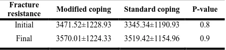
Table 2. Mean ± standard deviation (SD) of the load (N) at fracture (the fracture resistance)
Cercon sintering machine (DeguDent, Hanau,
Germany) at 1450°C for 5 hours. Upon completion,
the internal surfaces of the copings were cleaned with
a cotton pellet soaked in alcohol and also with a steam
cleaner. The adaptation of the coping with the die was
examined using a light body silicone paste (Fit
Checker, GC America Inc., Alsip, IL, USA). The
interfering points on the die were relieved using a
round diamond bur (D+Z, Frankfurt, Germany). The
A2 shade of the Cercon Ceram Kiss porcelain
(DeguDent, Hanau, Germany) was applied in a 1mm
uniform thickness and was fired in two steps. An
index of the first porcelain-veneered coping was made
using a transparent template, which was used for the
fabrication of other crowns.
After ensuring the complete seating of the copings,
the dies and crowns were separately cleaned in an
ultrasonic bath and were dried. In order to remove all
debris, the internal surfaces of the crowns were
cleaned using 37% phosphoric acid. The Panavia
F2.0 cement (Kuraray Dental, Tokyo, Japan) was
prepared according to the manufacturer’s instructions
and was applied to the crowns. The crowns were then
seated on the corresponding dies with a mild finger
pressure, and a constant pressure of 15N was applied
on the specimens using a 0.5kg weight via an acrylic
connector fabricated equal to the size of the crowns.
After 2 to 3 seconds of light-curing and removal
of excess cement, the crown margins were protected
using an air-inhibiting material (OxyGuard, Kuraray
Dental, Tokyo, Japan) until the cement was completely
Fracture
resistance Modified coping Standard coping P-value
Initial 3471.52±1228.93 3345.34±1190.93 0.8
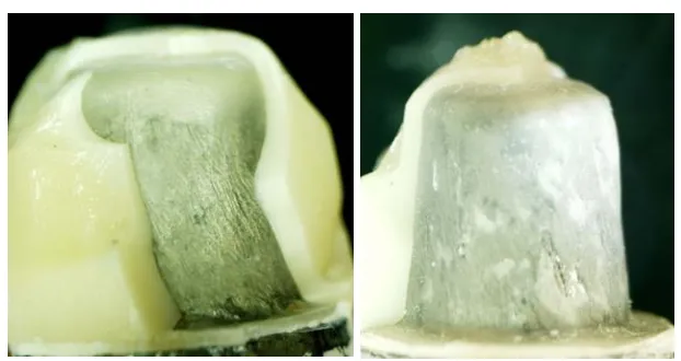
Fig. 2: External surface of a fractured specimen with a standard core (SC) design at ×10 magnification
set (3 minutes). The specimens were then subjected
to 5000 thermal cycles (Vafaei Industrial, Iran)
between 5-55°C (20 seconds in a hot bath, 10
seconds of dwell time, and 20 seconds in a cold
bath). Afterwards, the specimens were mounted in a
self-polymerizing acrylic resin.
The point of load application, at the center of the
occlusal surface, was marked on the specimens
which were placed in the universal testing machine
(Zwick/Roell, Germany). The load was applied at a
crosshead speed of 1mm/minute with a round tip of
4mm diameter.
The fracture load was recorded at two phases: the
initial fracture and final fracture. When the first
fracture occurred, it was recorded as the “initial
fracture” [1,3], and loading was continued until the
catastrophic fracture occurred [5,19,21,22]; thus, the
“final fracture” was measured. The mean fracture
load was calculated. Statistical analysis was performed
using independent samples T-test.
To ensure the accuracy, the specimens were
evaluated under a stereomicroscope (Nikon, Tokyo,
Japan) after the final fracture to determinate the
mode of failure (at the cement-core and core-veneer
interfaces). The modes of failure were divided into
three categories: Adhesive failure (failure at the
veneer-core or core-cement interfaces), cohesive
failure (failure within the cement layer, core layer or
veneer layer) and mixed failure (a combination of
both adhesive and cohesive failures in different
areas).
RESULTS
The means and standard deviations (SD) of the initial and
final fracture resistance in the MC design and SC design
are shown in Table 2. Independent samples T-test revealed
no statistically significant differences between the two
coping designs, neither in the initial (P=0.8) nor in the
final (P=0.9) fracture resistance. Table 3 shows the failure
modes of the specimens at ×63 magnification. Figures 2
and 3 show the specimens under the stereomicroscope.
DISCUSSION
The changes applied to the core design of the present
study were based on the trestle design suggested for
metal-ceramic crowns and were aimed to decrease the
risk of fracture in PFM restorations. Miller [18] stated
that the trestle design of the metal copings for single
crowns must include a proximal strut, a buttressing
shoulder, a reinforcing collar, shear resistant cusps, and
abutment seal. Since the trestle design is believed to be
optimal for metal-ceramic crowns, in the current study,
this design was used for zirconia copings to assess its
efficacy for all-ceramic zirconia restorations.
Fig. 3: External surface of a fractured specimen with a modified core (MC) design at ×10 magnification
in the SC group was lower than that at the initial fracture
in the MC group. Veneer fracture occurred more
frequently in the SC specimens (n=7) under lower levels
of stress. However, in the MC group, ceramic veneer
chipping occurred in only two specimens prior to core
fracture. This result may be explained by the greater
veneer support provided by the MC design. Finite
element analyses have also shown that compressive
stresses (instead of tensile stresses) are present at the
veneer-shoulder interface; thus, the support provided by
the shoulder finish line could reduce the risk of veneer
fracture [23, 24]. Moreover, the buttressing shoulder
with an increased height serves as a shock-absorber at
the areas near the crown margins; this area provides a
great support for the veneering porcelain under
functional loads [25].
In 2004, Sundh and Sjogren [22] concluded that the
adapted core framework design caused a greater
improvement in the fracture resistance compared to the
0.5mm-thick standard design. In 2011, Kokubo et al [3]
reported similar results and showed that the
uniform-thickness coping required the minimum amount of
fracture load. Also, the cuspal configuration of the
uniform-thickness coping design and the modified
coping margins increased the fracture resistance of the
veneering porcelain [3]. The modified design of the
coping margins adopted by Kokubo et al [3] was based
on a design suggested by Marchack et al [26] in 2008.
The authors recommended further modifications of the
margin design (i.e. increasing the height of the collar);
however, some studies reported that the modified
framework design did not improve the fatigue resistance
of the crowns [3,27]. Such controversy in the results
may be attributed to the differences in the type of the
materials, tests, and methods of load application.
Bonfante et al [19] and Silva et al [28] also assessed the
efficacy of an MC design and showed that the modified
margin design provided a greater support for the
porcelain. The details of the coping design, load
application status, and type of the materials used in the
two above-mentioned studies were different from those
of our study. The mode of failure in both groups of the
current study was mainly the mixed fracture. Adhesive
failure (at the core-veneer interface) was more frequent
in the MC group (n=6) compared to the SC samples
(n=2). In all the specimens, the fracture initiated at the
site of load application and extended laterally. In the MC
group, the fracture line extended toward the borders of
the proximal strut and did not involve the buttressing
shoulder or the proximal strut body. Conversely, in the
SC group, the fracture extended toward the margins;
these findings were in accordance with those of
Bonfante et al [29].
Table 3. Frequency and percentage of the failure modes at ×63 magnification
Mixed
N(%)
Cohesive N(%)
Adhesive N(%) Modified
coping
Die-Core 11(92) 0 1(8)
Core-Veneer 5(42) 1(8) 6(50)
Standard coping
Die-Core 9 (75) 0 3(25)
stress are concentrated at the margins [6,27]. Thus, the
SC design with a uniform thickness may serve as a
weak point in all-ceramic restorations. On the other
hand, the differences in the modes of failure among
various studies may be attributed to the different types
of ceramics, die materials, manufacturing techniques,
specimen designs, and various thicknesses of the walls
and luting agents [30]. It is suggested to predefine
the location of failure assessment in subsequent
studies to standardize the observation area under
the stereomicroscope.
Similar to previous studies [3,31], the static load was
used to assess the fracture resistance in this study.
Although the fracture strength test is highly important
for ceramic restorations, static load application has some
limitations and does not perfectly simulate the clinical
condition. It does not provide any information on the
long-term behavior of the materials or their properties
when exposed to cyclic fatigue in the oral environment.
Moreover, the complex oral environment and the role of
factors such as saliva and patient-related habits, etc.
cannot be ideally simulated by the current laboratory
techniques. These items are among the limitations of
this study. Therefore, the results of static tests must be
cautiously interpreted [30,32].
In the current study, similar to that of Beuer et al [20],
standard metallic dies were used for the fabrication of
specimens. According to Schererr and de Rijk [30], the
higher elastic modulus of the die resulted in a higher
fracture resistance. The elastic modulus of metallic dies
is much higher than that of dentin (200 GPa versus 18.3
GPa) [20]; therefore, these dies undergo a limited
transformation and as a result, low shear stresses are
created in the internal surfaces of the crowns; this is
considered an advantage [30]. Thus, the fracture
resistance values obtained by the application of these
dies may be much higher than those of the dentinal dies.
On the other hand, metallic dies have a significant role
in standardizing the preparations and obtaining crowns
with identical physical qualities. These are among the
resin-based cements is higher than that of the crowns
luted by conventional cements [33].
CONCLUSION
There were no statistically significant differences
in the fracture resistance between the modified
and standard coping designs, neither in the initial
nor in the final fracture strength. Most of the
specimens in both groups showed the mixed
failure mode. Based on the results, the modified
core design may not significantly improve the
fracture resistance of zirconia restorations.
REFERENCES
1- Larsson C, El Madhoun S, Wennerberg A, Vult von Steyern P. Fracture strength of yttria-stabilized tetragonal zirconia polycrystals crowns with different design: an in vitro study. Clin Oral Implants Res. 2012 Jul;23(7):820-6. 2- Della Bona A, Kelly JR. The clinical success of all-ceramic restorations. J Am Dent Assoc. 2008 Sep;139 Suppl:8S-13S.
3- Kokubo Y, Tsumita M, Kano T, Fukushima S. The influence of zirconia coping designs on the fracture load of all-ceramic molar crowns. Dent Mater J. 2011 May;30(3):281-5.
4- Rekow ED, Silva NR, Coelho PG, Zhang Y, Guess P, Thompson VP. Performance of dental ceramics: challenges for improvements. J Dent Res. 2011 Aug; 90(8):937-952.
5- Reich S, Petschelt A, Lohbauer U. The effect of finish line preparation and layer thickness on the failure load and fractography of ZrO2 copings. J Prosthet Dent. 2008 May;99(5):369-376.
6- Pogoncheff CM, Duff RE. Use of zirconia collar to prevent interproximal porcelain fracture: A clinical report. J Prosthet Dent. 2010 Aug;104(2):77-79. 7- Monaco C, Tucci A, Esposito L, Scotti R. Microstructural changes produced by abrading Y-TZP in presintered and sintered conditions. J Dent. 2013 Feb;41(2):121-6.
FPDs substructures. J Oral Rehabil. 2010 Apr;37(4):292-9. 9- Benetti P, Della Bona A, Kelly JR. Evaluation of thermal compatibility between core and veneer dental ceramics using shear bond strength test and contact angle measurement. Dent Mater. 2010 Aug;26(8):743-50. 10- Tholey MJ, Swain MV, Thiel N. Thermal gradients and residual stresses in veneered Y-TZP frameworks. Dent Mater. 2011 Nov;27(11):1102-10. 11- Nakamura K, Adolfsson E, Milleding P, Kanno T, Ortengren U. Influence of grain size and veneer firing process on the flexural strength of zirconia ceramics. Eur J Oral Sci. 2012 Jun;120(3):249-54. 12- Fischer J, Grohmann P, Stawarczyk B. Effect of zirconia surface treatments on the shear strength of zirconia/veneering ceramic composites. Dent Mater J. 2008 May;27(3):448-54.
13- Cattani Lorente M, Scherrer SS, Richard J, Demellayer R, Amez-Droz M, Wiskott HW. Surface roughness and EDS characterization of a Y-TZP dental ceramic treated with the CoJetTM Sand. Dent Mater. 2010 Nov;26(11):1035-42.
14- Salimi H, Mosharraf R, Savabi O. Effect of framework design on fracture resistance of zirconium oxide posterior fixed partial dentures. Dent Res J (Isfahan). 2012 Nov;9(6):764-9.
15- Son YH, Han CH, Kim S. Influence of internal-gap width and cement type on the retentive force of zirconia copings in pullout testing. J Dent. 2012 Oct;40(10):866-72.
16- Inokoshi M, Kameyama A, De Munck J, Minakuchi S, Van Meerbeek B. Durable bonding to mechanically and/or chemically pre-treated dental zirconia. J Dent. 2013 Feb;41(2):170-9.
17- Tang X, Tan Z, Nakamura T, Yatani H. Effects of aging on surface textures of veneering ceramics for zirconia frameworks. J Dent. 2012 Nov;40(11):913-20. 18 Miller LL. Framework design in ceramo -metal restorations. Dent Clin North Am. 1977 Oct;21(4):699-716.
19- Bonfante EA, da Silva NR, Coelho PG, Bayardo-Gonzalez DE, Thompson VP, Bonfante G. Effect of framework design on crown failure. Eur J Oral Sci. 2009 Apr;117(2):194-9.
20- Beuer F, Aggstaller H, Edelhoff D, Gernet W.
Effect of preparation design on the fracture resistance of zirconia crown copings. Dent Mater J. 2008 May;27(3):362-7.
21- Rosentritt M, Steiger D, Behr M, Handel G, Kolbeck C. Influence of substructure design and spacer settings on the in vitro performance of molar zirconia crowns. J Dent. 2009 Dec;37(12):978-83. 22- Sundh A, Sjogren G. A comparison of fracture strength of yttrium-oxide- partially-stabilized zirconia ceramic crowns with varying core thickness, shapes and veneer ceramics. J Oral Rehabil. 2004 Jul;31(7):682-8.
23- Dejak B, Mlotkowski A, Romanowicz M. Finite element analysis of stresses in molars during clenching and mastication. J Prosthet Dent. 2003 Dec;90(6):591-7. 24- Rafferty BT, Janal MN, Zavanelli RA, Silva NR, Rekow ED, Thompson VP, et al. Design features of a three-dimensional molar crown and related maximum principal stress. A finite element model study. Dent Mater. 2010 Feb;26(2):156-63.
25- Ha SR, Kim SH, Han JS, Yoo SH, Jeong SC, Lee JB, et al. The influence of various core designs on stress distribution in the veneered zirconia crown: a finite element analysis study. J Adv Prosthodont. 2013 May;5(2):187-97.
26- Marchack BW, Futatsuki Y, Marchack CB, White SN. Customization of milled zirconia copings for all-ceramic crowns: a clinical report. J Prosthet Dent. 2008 Mar;99(3):169-73.
27- Lorenzoni FC, Martins LM, Silva NR, Coelho PG, Guess PC, Bonfante EA, et al. Fatigue life and failure modes of crowns systems with a modified framework design. J Dent. 2010 Aug;38(8):626-34. 28- Silva NR, Bonfante EA, Rafferty BT, Zavanelli RA, Rekow ED, Thompson VP, et al. Modified Y-TZP core design improves all-ceramic crown reliability. J Dent Res. 2011 Jan;90(1):104-8. 29- Bonfante EA, Rafferty B, Zavanelli RA, Silva NR, Rekow ED, Thompson VP, et al. Thermal/mechanical simulation and laboratory fatigue testing of an alternative yttria tetragonal zirconia polycrystal core-veneer all-ceramic layered crown design. Eur J Oral Sci. 2010 Apr;118(2):202-9.