Available online on 15.11.2018 at http://jddtonline.info
Journal of Drug Delivery and Therapeutics
Open Access to Pharmaceutical and Medical Research© 2011-18, publisher and licensee JDDT, This is an Open Access article which permits unrestricted non-commercial use, provided the original work is properly cited
Open Access
Review Article
Technology Overview and Current Biomedical Application of Polymeric
Nanoparticles
Gurpreet Singh *, Abdul Faruk, Preet Mohinder Singh Bedi
Department of Pharmaceutical Sciences, Guru Nanak Dev University, Amritsar Punjab-143005, India.
ABSTRACT
Polymeric nanoparticles are of great importance in the treatment of various diseases, due to the flexibility in the modification of their structures. Recent advances in the field of nanotechnology facilitate the engineering of multifunctional polymeric nanoparticles. All the scientific efforts of the pharmaceuticals companies are mainly focusing on two basic aspects, one is to discover new molecules of potential therapeutic interest and second is to develop of a new drug delivery system. In the last few decades, research and development (R&D) scientists has directed their efforts toward formulating novel drug delivery systems that includes sustained and controlled release, modified release and targeted drug release dosage forms. Application of nanoscience and nanotechnology has opened several new possibilities in development of formulation This review compiles the different preparation methods of polymeric nanoparticles and then briefly explained their current potential applications.
Keywords: Polymeric nanoparticles, PLGA, Biomedical applications, Biodegradable, Dialysis method
Article Info:Received 27 Sep, 2018; Review Completed 25 Oct 2018; Accepted 27 Oct 2018; Available online 15 Nov 2018
Cite this article as:
Singh G, Faruk A, Bedi PMS, Technology Overview and Current Biomedical Application of Polymeric Nanoparticles , Journal of Drug Delivery and Therapeutics. 2018; 8(6): 285-295 DOI:http://dx.doi.org/10.22270/jddt.v8i6.2015
*Address for Correspondence:
Gurpreet Singh, Department of Pharmaceutical Sciences, Guru Nanak Dev University, Amritsar Punjab-143005, India.
INTRODUCTION
It has been well documented that drugs predicted from in vitro studies, fails to reach at target site in adequate quantities in vivo studies1. Correspondingly, a high amount of the administered drug is left to act on healthy tissues, often to the point of generating dose-limiting side effects2. Research related to delivery of drug is clearly moving from the micro to nano size range. Therefore, nanotechnology is evolving as a potential field in medicine that may provide significant therapeutic benefits of existing drugs. The most difficult tasks for pharmaceutical researcher is to develop an effective nano drug delivery system which is capable of carrying drugs, specifically to a desired site of action. Attempts have been made to reformulate the existing conventional formulations into nano delivery systems for better therapeutic use and positive scientific breakthroughs. These systems mainly include polymeric and solid lipid nanoparticles, liposomes and nanoemulsions. The ultimate objective of these nanodelivery systems is to markedly improve the efficacy and reduce the toxic effects of a drug3.
Recently, nanoparticles based delivery system has been proposed as promising colloidal drug carriers system for such purpose4. Nanoparticles (NPs) are a type of colloidal drug delivery system which is comprised of particles in size range from 10 to 1000 nm. Nanoparticles may or may not results size related properties that differ significantly from those observed properties in fine or macro particles5. Nowadays, nanoparticles are widely used as carrier system in variety of applications due to their ability to cross organ barriers such as cell membrane and blood brain barrier etc6. They are made up from biocompatible polymer and lipids that provide sustained and controlled release effect by either improved dissolution or diffusion mechanism7,8. Now a day’s nanoparticles are considered as very prolific device for drug delivery system. Dr. Gregory Gregoriad proposed the first liposome’s in 1974 as nanoparticulate drug delivery systems which resulted in several breakthrough discoveries by multidisciplinary approaches3.
nanotechnology as a multidisciplinary field. Currently, major research aimed at the development of biocompatible nanocarriers for drugs delivery, cell imaging and for other biomedical applications9.
POLYMERIC NANOPARTICLES
Polymeric nanoparticles (PNs) are submicron-sized colloidal particles in which therapeutic agent can be encapsulated within their polymeric matrix or adsorbed to the surface of nanoparticles. There are two types of
nanoparticles on basis of formulation method i.e., Nanospheres and Nanocapsules10,11 as depicted in Figure 1. Nanospheres have a matrix system in which drug is uniformly entrapped, dispersed or encapsulated within the particles or attached to their surfaces. Nanocapsules are the liquid-solid core system in which the drug is restricted to a polymer membrane of natural or synthetic polymers12. Active moiety is protective by coating and can be used for the controlled release and targeting of drugs13,14.
Figure 1: Schematic representation of nanospheres and nanocapsules Polymeric nanoparticles are prepared from a wide variety
of natural or synthetic biodegradable (e.g. albumin, chitosan, alginate, PLGA) and non-biodegradable polymers as describe in Table-1. By the virtue of their nano size, polymeric nanoparticles can also be targeted to specific cells and locations inside the body15. Depending on the nature of polymer properties, they are designed in such a way that they can be activated by change in the environmental conditions such as physiological pH, temperature, or chemical stimuli16. Macrophages are well recognized phagocytic cells of the reticuloendothelial system and responsible for the uptake and clearance of
administered drug loaded nanoparticles. Generally, when drug loaded nanoparticles are opsonized, phagocytosis/ endocytosis may take place and nanoparticles are degraded in a phagolysosome/endolysosome17. However, nanoparticles have the capability to escape from the endolysomal compartment which may allows the delivery of drug to the cytoplasm and finally to the nucleus. Thus, NPs are easily taken up by phagocytic cells and that’s makes them ideal for intracellular delivery of anti-retroviral drugs. Other applications of NPs include cytoplasmic release of plasmid vectors and therapeutic agents15,18,19.
Table 1: List of commonly used polymers Type of polymers Class of polymers Examples
Synthetic polymers Polyamide Polyamino acids, Polypeptides
Polyester Poly(glycolide), Poly(D,L-lactide), Poly(D,L-lactide-co-glycolide), Poly(ε-caprolactone), Poly(dioxanone) Poly(hydroxybutyrate)
Polyanhydride Poly[bis(p-carboxyphenoxy) propane-co-sebacic acid], Poly(fatty acid dimmer-co-sebacic acid), Poly(sebacic-co-recinolic acid)
Naturally occurring polymers
Polysaccharides Dextran, Chitosan, Alginate, Starch, Hyaluronic acid, Gellan
Proteins Collagen, Gelatin, Bovine serum albumin (BSA), Human serum albumin (HSA)
BIODEGRADABLE POLYMERS
Biodegradable polymers are polymer that are comprised of monomers linked to each other through a functional group and have unstable linkage in backbone that degrades within the body by enzymatic or chemical degradation as a result of natural biodegradable process15. Also when used as drug delivery system it gets eliminated from body and
The major biomedical applications of biodegradable polymers are 15, 21, 22
Constructing depot injections
Biodegradable sutures
Replacement of bone grafts
In tissue generation
For protein drug delivery
For gene drug delivery for sustain and controlled drug delivery systems
Common characteristics of all biodegradable polymers are23
Stability and compatibility with the drug molecule
Biocompatible and biodegradable in nature
Ease of production on a larger scale
Suitable for sterilization, and
Flexibility to prepare delivery system with multiple release profiles
The general advantages of nanoparticles includes24,25 1. Due to small and narrow particle size distribution of
nanoparticles, site specific drug delivery can be achieved
2. Provide a sustained and controlled release of active drug over a long period
3. Protection from chemical and enzymatic degradation of incorporated drug
4. Provide a predefine drug release profile
5. They can be lyophilized and spray dried to resolved stability issues
6. Surface modification can be easily done to achieve both active and passive drug targeting
7. They offer better therapeutic effectiveness response per unit dose as compare to conventional dosages form
8. Biodegradability in bloodcirculation
9. pH sensitive polymeric nanoparticles can enhanced the oral bioavailability of poorly water soluble drugs26 10. Site specific drug can be achieved by surface targeting ligand nanoparticles/magnetic nanoparticles for cancer therapy, vaccine and targeted antibodies27
Limitation of Nanoparticles
1. Particle aggregation during storage results in increase the particle size which is difficult in physical handling28
2. Limited drug loading and burst release29
3. Overall high production cost and scaling up problems30
4. Intracellular degradation of nanoparticles due to
phagocytosed by cells they may cause cytotoxic effects31
METHODS OF PREPARATION OF POLYMERIC NANOPARTICLES
The selection of materials and method of preparation is dependent on following factors:
Size of desired nanoparticles
Inherent and physicochemical properties of the drug, e.g., aqueous solubility and stability32
Surface charge properties and permeability33
Degree of biodegradability, biocompatibility, Antigenicity and toxicity34
Desired drug release rate profile35
Emulsion and diffusion method
This is the most widely used method of preparation for polymeric nanoparticles. In this method, the encapsulating polymer is dissolved in partially water-miscible organic phase (such as benzyl alcohol, propylene carbonate, ethyl acetate). The organic phase is emulsified by stirring with an aqueous solution of a suitable surfactant i.e., anionic sodium dodecyl sulfate (SDS), non-ionic polyvinyl alcohol (PVA) or cationic didodecyl dimethyl ammonium bromide (DMAB). The diffusion of the organic solvent and the counter diffusion of water into the emulsion droplets induce formation of nanospheres or nanocapsules, according to their oil-to-polymer ratio36.
This technique presents several advantages, such as high encapsulation efficiency, no need for homogenization, high batch-to-batch reproducibility, ease of scale-up, and narrow size distribution. However, disadvantages includes that are the high volumes of water is to be eliminated from the suspension and less suitable for water-soluble drugs due to leakage into the saturated-aqueous external phase during emulsification, reducing encapsulation efficiency37. While, parameters that influenced the size of nanoparticle are: polymer concentration, solvent nature, surfactant/polymer molecular mass, viscosity, stirring rate, nature of solvent, temperature and rate of water addition36.
Emulsification-solvent evaporation method
water-soluble drugs39,40. This technique has been successfully used for encapsulation of a hydrophobic drug, while in case of hydrophilic drug entrapment was less41. Further, modification of this method includes automated batch scale production and is known as high pressure emulsification solvent evaporation process42. This method involves preparation of an emulsion (O/W or W/O) which is then subjected to homogenization under high pressure followed by high speed stirring to remove any organic solvent residue43. The size of nanoparticles can be controlled by amount and type of polymer, surfactant, viscosity of organic and aqueous phases, temperature and rate of stirring44. Commonly used polymers are Polylactic acid, poly (lactic-co-glycolic acid), Ethyl cellulose15, and Polycaprolactone45.
Oil in water emulsion technique (single emulsion) This technique is based on the emulsification of an organic phase, which contains polymer and drug in an aqueous phase, followed by the removal of the organic solvent by evaporation method. A number of hydrophilic surfactants such as polyvinyl alcohol, or Pluronic F68 are generally used in an aqueous phase. For the formation of nanoemulsion, size reduction of emulsion droplet is done by sonication or microfluidization. The evaporation step is required to eliminate the organic solvent present in the organic phase. This leads to the precipitation of the polymer as nanoparticles with a diameter in the nanometric range46.
There are some considerable variables which effects the preparation of nanoparticles that includes molecular mass and concentration of polymer, co-polymer ratio and end groups, surfactant nature, phase ratio, solvent nature, rate of evaporation, drug entrapment, additives and sterilization47.
Double-emulsion (w/o/w) method
This method is a modification of emulsification solvent evaporation method and is useful for encapsulation of proteins and hydrophilic drugs. In this method, the primary emulsion (w/o) is prepared by a aqueous solution containing the hydrophilic drug and polymer in the organic phase containing a suitable surfactant, having low HLB value such as Span 80, Pluronic-F68. The primary emulsion is produced by strong shear stress and re-emulsified in an external aqueous phase of a surfactant for formation of water in-oil-in water (w/o/w) emulsion. Further, droplet size reduction of emulsion is achieved with the help of sonication or homogenization. Finally, organic phase residue removed with the help of evaporation under vacuum and nanoparticles can be collected by centrifugation at high speed48. The major drawback of this method is the formulation of large sized nanoparticles and the leakage of hydrophilic drugs. Overall production of nanoparticles depends upon polymer/surfactant ratio, polymer concentration, type and nature of surfactant, viscosity, energy input, evaporation and phase ratios49 .
Solvent displacement/Nanoprecipitation
This technique was first described by Fessi et al. 1989 and is also called as solvent displacement method50. This method is commonly used to incorporate lipophilic drugs and involves the precipitation of polymer from an organic phase. In this method, drug, polymer and lipophilic surfactant (e.g., phospholipids) are added in a semi-polar water-miscible solvent (i.e., acetone or ethanol). Under magnetic stirring, this solution is added drop wise into an
aqueous phase solution containing a stabilizer. Rapid diffusion of the solvent, facilitate instant formation of colloidal nano suspension. Then organic solvent is removed under reduced pressure from the suspension51. This technique is suitable for lipophilic drugs which allow the formation of nanocapsules with high drug loading efficiencies. This method is not suitable for water miscible solvents, in which the diffusion rate is high to produce spontaneous emulsification and may leads to instability when added in water. In some cases, the entrapment efficiency of drugs can be increase by use of acetone/dichloromethane which increases the mean particle size. The particles size and drug entrapment efficiency depend upon the rates of addition of the organic phase into the aqueous phase52. This technique has been used for various polymers such as poly(lactic-co-glycolic acid), Poly(lactic acid) and Polycaprolactone etc 41,53.
Dialysis method
This method is a simple and relatively effective method for preparation of polymeric nanoparticles with small and narrow size distribution. Briefly, drug and polymer is dissolved in an organic solvent and are put in the dialysis tube (semi-permeable membranes having a appropriate molecular weight cut off) which is kept in a aqueous phase. The organic phase diffuses out through the pore of dialysis membrane into the aqueous phase which resulted in decrease in interfacial tension between two phase. Subsequently, displacement of organic solvent results in loss of solubility of polymer and may form homogenous suspension of nanoparticles54. The mechanism of formation of nanoparticles by dialysis method is similar to that of nanoprecipitation suggested by the Fessi et al50.
Emulsion Polymerization Method
This is in situ polymerization of monomers in an aqueous solution containing surfactant which results in formation of nanoparticles. Drug is added during polymerization process or is adsorbed on nanoparticles after completed polymerization. The nanoparticle can be purified and recovered after removal of residues of stabilizers and surfactants by centrifugation and reconstituted in an isotonic medium55, 56. One another alternative method is mini-emulsion polymerization method, in which co-stabilizer and high-shear energy (sonication, ultrasound, etc) is used with mixture of monomer, surfactant and a initiator57. This method has been reported for the preparation of nanoparticles from organic and inorganic materials and also for hybrid nanoparticles58.
Salting out method
nanospheres. In final step, salting out agent is removed by centrifugation60. The salting out method is suitable for heat sensitive substances. Important variables to be considered includes polymer concentration, nature and concentration of surfactant, molecular mass of polymer, stirring speed and time, and solvent used and cryoprotectants61,53 .
Coacervation or Ionic Gelation method
Nowadays, much attention of research has been focused on production of biodegradable nanoparticles using polymers such as chitosan, gelatin and sodium alginate having features like biocompatibility and low toxicity. Calvo et al introduced a method for preparation of hydrophilic chitosan nanoparticles by ionic gelation62. In this method, one aqueous phases consisted of solution of chitosan polymer and a di-block-co-polymer ethylene oxide or propylene oxide (PEO-PPO) and the other phase is solution of sodium tripolyphosphate. Nano size coacervates are formed due to an interaction of cationic group of chitosan and anionic tripolyphosphate. Coacervates are produced due to electrostatic interaction between both aqueous phases whereas conversion of material from liquid to gel takes place due to ionic interaction at room temperature. Cross-linking agent such as glutaraldehyde solution (25 %) can be used for hardening of coacervates63. Desired property of the nanoparticles can be achieved by concentration of cross linker, temperature, pH, addition rate and agitation speed64.
Spray drying method
Spray drying has been considered as widely used method for the production of nano/micron-sized particles of heat-sensitive materials. In this method, solution droplet is converted into a dry particle by evaporation of the solvent in a single-step process. Thermo-labile compounds such as proteins and enzymes intended for therapeutic and diagnostic purposes have been successfully developed as spray-dried product. This method is widely used to improve the morphology and particle size of dried powder by varying the process variables and the formulation factors. It is also suitable for protein drugs which are administered as spray dried powders by pulmonary and nasal routes for therapeutic purposes65. It has been reported that dried particles of water soluble or water insoluble drugs can be prepared by use of various
polymers which resolved the issue of drug leakage66 and thus, the drug amount in particles can be accurately determined67. Innovative advancement in spray drying technology is impact on development of new spray dryers for pharmaceutical industry, which resulted in high product yield with nano size range68.
Supercritical fluid technology method
In conventional methods, organic solvents used are unsuitable for environment as well as to physiological and biological systems. Therefore, the supercritical fluid technology has been introduced as environmentally safe method for preparation of nanoparticles69. Supercritical fluids remains as a single phase without impact of pressure and having intermediate properties of a liquid and a gas. They are commonly used in the production of inorganic and hybrid nanomaterials. Supercritical carbon dioxide (SCCO2) is the most extensively used fluid because of its safe and non-flammable nature. They are commonly employed in supercritical anti-solvent (SAS) and rapid expansion of critical solution (RESS) methods. SAS method, a solid sample is dissolved in an organic or inorganic liquid solvent and which injected into a supercritical fluid under the high pressure. Under these conditions, miscibility of solid solute is reduced in the supercritical fluid which results in the precipitation of the solute and the subsequently formation of nanoparticles70. While in RESS method, the solid sample is saturated in a supercritical solvent and is passed through a very fine nozzle at high speed that resulted in precipitation of the solute due to expansion/decompression effect of the system. The overall morphology and size distribution of solute particles are dependent on expansion conditions. Other factors that affect overall production process include nature of solute, supercritical solvent, operating pressure and temperature conditions. Supercritical fluid technology is appropriate for bulk production but costly design of equipment is major limitation71 .
APPLICATIONS OF POLYMERIC NANOPARTICLES The extensive advancement in the area of science results in innovative ideas which lead to novel drug delivery systems. Recently reported outcomes of polymeric nanoparticles are summarized in table 2.
Table 2: The research outcomes of polymeric nanoparticle S.
No Carrier system Drug /Molecule Recent applications Year Ref.
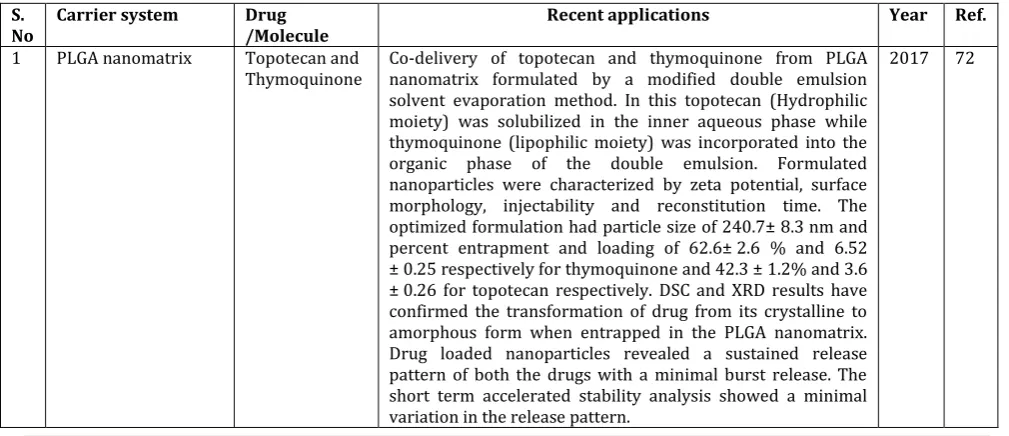
1 PLGA nanomatrix Topotecan and
Thymoquinone Co-delivery of topotecan and thymoquinone from PLGA nanomatrix formulated by a modified double emulsion solvent evaporation method. In this topotecan (Hydrophilic moiety) was solubilized in the inner aqueous phase while thymoquinone (lipophilic moiety) was incorporated into the organic phase of the double emulsion. Formulated nanoparticles were characterized by zeta potential, surface morphology, injectability and reconstitution time. The optimized formulation had particle size of 240.7± 8.3 nm and percent entrapment and loading of 62.6± 2.6 % and 6.52 ± 0.25 respectively for thymoquinone and 42.3 ± 1.2% and 3.6 ± 0.26 for topotecan respectively. DSC and XRD results have confirmed the transformation of drug from its crystalline to amorphous form when entrapped in the PLGA nanomatrix. Drug loaded nanoparticles revealed a sustained release pattern of both the drugs with a minimal burst release. The short term accelerated stability analysis showed a minimal variation in the release pattern.
2 Biodegradable poly(d,l) lactic acid nanoparticles
Tamoxifen
citrate Biodegradable poly (d,l) lactic acid nanoparticles were prepared by modified spontaneous emulsification solvent diffusion method. In vitro studies for cytotoxicity revealed that MCF-7 and MDA-MB-231 cells lines were more sensitive to tamoxifen loaded nanoparticles than tamoxifen citrate alone. DNA ladder and the expression of Bax to Bcl-2 ratio were higher in tamoxifen loaded nanoparticles than that in alone tamoxifen citrate.
2016 73
3 Monomethoxy polyethylene glycol amine-polylactide-co-glycolide (mPEG-PLGA) co-polymer
Gemcitabine Gemcitabine, a nucleoside analog, has a short half-life in systemic circulation due to its enzymatic degradation.To overcome this problem, monomethoxy polyethylene glycol amine-polylactide-co-glycolide (mPEG-PLGA) co-polymer was synthesized. Gemcitabine loaded mPEG-PLGA nanoparticles (NPs) exhibited sustained drug release profile, compatible with blood and enhanced cellular uptake. The cell cytotoxicity of mPEG-PLGA NPs were observed in MiaPaCa-2 and MCF-7 cells. The half-life of gemcitabine loaded nanoparticles was remarkably enhanced (19 folds) as compared to pure gemcitabine which improved anticancer efficacy in Ehrlich ascites bearing Balb-c mice.
2016 74
4 Poly(d,l-lactide-co-glycolide) (PLGA) nanoparticles nanoprecipitation method
Rapamycin and
Piperine Rapamycin (RPM) with a chemosensitizer (piperine) loaded Poly(d,l-lactide-co-glycolide) (PLGA) nanoparticles were prepared by nanoprecipitation method for improve oral bioavailability and efficacy. Prepared nanoparticles showed sustained in vitro drug release and it has been found that the uptake of the Rapamycin (P-gp substrate) increased in the presence of piperine in an everted gut sac method. Pharmacokinetic studies showed improved bioavailability of 4.8 folds in combination with piperine. An in vitro cell line studies indicates better efficacy of rapamycin nanoparticles compared to free drug solution which suggests that the use of a combination of rapamycin with piperine nanoparticles would be a effective approach in the treatment of breast cancer.
2016 75
5 PLGA nanoparticles, Emulsified
nanoprecipitation technique
Tamoxifen Tamoxifen loaded PLGA nanoparticles (Tmx-PLGA) were prepared by emulsified nanoprecipitation technique. Tmx-PLGA has been evaluated for its better DNA cleavage potential, cytotoxicity using Dalton's lymphoma ascite cells and MDA-MB231 breast cancer cells. In vitro cytotoxicity studies indicate that Tmx-PLGA showed excellent DNA cleavage potential as compared to pure Tmx. Fluorescence imaging of nuclear fragmentation and condensation exhibiting significant increase of apoptosis (70%) in PLGA-Tmx while pure drug (58%). Enhanced DNA cleavage potential, nuclear fragmentation and condensation in apoptotic cells confirm greater bioavailability of PLGA-Tmx as compared to pure Tmx. Tamoxifen loaded PLGA nanoparticles may act as a novel vehicle for the treatment of cancer.
2016 76
6 Biotin-F127-PLA or F127-PLA polymeric nanoparticles
Camptothecin Camptothecin incorporated into biotin-PLA or F127-PLA polymeric nanoparticles (NPs) prepared by a dialysis method. Results indicate that the targeted CPT NPs exhibited regular spherical shape (mean diameter 180 nm). In vitro release exhibited an initial burst (40%) within 12 h, followed by a slow release of camptothecin. The in vitro antitumor effect of the Camptothecin -loaded nanoparticles was determined against H22 cells using an MTT assay which exerted significant antitumor effects as compared to free Camptothecin. The targeted Camptothecin NPs showed increased in vivo tumor inhibition.
2016 77
7 PLGA/Solutol HS15
nanoparticles Docetaxel Docetaxel loaded PLGA/Solutol HS15 nanoparticles (NPs) were fabricated by a modified emulsification solvent evaporation method. in vitro release studies, indicates that emulsifying property of Solutol HS15 seemed to contribute to the enhanced drug release of Docetaxel from NPs at physiological pH. These NP can be a promising local anticancer drug delivery system for cancer therapy.
2016 78
8 biotinylated
lactide-co-glycolide)
nanoparticles were prepared for tumor targeted and prolonged delivery system for Epirubicin (EPB). The results revealed encapsulation efficiency (84.1 ± 3.4%), spherical shaped, higher positive zeta potential compared to the unmodfied EPB-loaded PLGA NPs. The in vitro drug release studies showed constant drug release kinetics during the first 48 h and the drug burst release significantly decreased in comparison to the unmodified PLGA NPs. The results of MTS assays and the flow cytometry and the confocal microscope showed that Bio-CS-PLGA NPs markedly increased the cytotoxicity and extent of cellular uptake of EPB. In MCF-7 tumorbearing nude mice, EPBloaded biotinylated chitosan -PLGA NPs were efficiently accumulated in the tumors which displayed greater potential for application as the carriers of anti-cancer drugs.
9 Eudragit E 100 nanoparticles, Emulsification diffusion evaporation method
Curcumin Curcumin loaded Eudragit E 100 nanoparticles were prepared by emulsification diffusion evaporation method to enhance the bioavailability and anti-cancer efficacy. The in vitro cytotoxicity sulphorhodamine B assay, showed 19-fold reduction in IC50 when treated with curcumin loaded nanoparticles as compared to pure curcumin, Pharmacokinetic studies revealed ~91-fold increase in Cmax and ~95-fold increase in AUC0. The in vivo anti-cancer activity showed a significant increase in efficacy compared with pure curcumin, as observed by tumor volume, body weight and survival rate. Its offer a great potential to improve bioavailability and efficacy of hydrophobic chemotherapeutic anticancer drug.
2016 80
10 Lipid polymer hybrid
nanoparticles Docetaxel Docetaxel (DTX) lipid polymer hybrid nanoparticles consist of a pH-responsive PEG layer that gets detached prior to its cellular uptake. Docetaxel was added into the lipid core of the nanoparticles, which was then protected with the pH-responsive block co-polymer polyethylene glycol-b-polyaspartic acid using a modified-emulsion method. Drug release from docetaxel loaded nanoparticles was pH-sensitive, which is helpful in tumor targeting. The negative surface charge and PEG shell of vehicle enhanced the blood circulation time and improved physiological activity of docetaxel loaded nanoparticles as compared to free docetaxel. In vivo anticancer effect of Docetaxel lipid polymer hybrid nanoparticles was further confirmed by the elevated levels of poly ADP ribose polymerase and caspase-3 found in the tumors after treatment. Thus, the results indicate that Docetaxel lipid polymer hybrid nanoparticles system could be an effective new treatment for cancer.
2015 81
11 methoxy poly(ethylene glycol)-poly(lactide) polymeric
nanoparticles
5-Flurouracil 5-FU loaded methoxy poly (ethylene glycol)-poly (lactide) based polymeric nanoparticles were prepared by nanoprecipitation method in order to increase the efficacy against breast cancer. The prepared nanoparticles were evaluated by DLS, TEM, in vitro release kinetics, and in vivo parameters. The average particle diameter of 5-FU loaded NP was ~110 nm. The NPs exhibited a pH-dependent drug release pattern and in vitro cytotoxicity assay showed the enhanced cytotoxic effect of drug loaded NPs in comparison to free drug. Flow cytometer analysis showed that nanoparticle system remarkably arrested the G2/M phase of cell cycle with significant amount of apoptosis cells in late and early phase. Nanoparticle formulation significantly decreased the tumor burden of mice with minimal signs of adverse effect. The favorable results obtained from this study makes 5-FU loaded methoxy poly(ethylene glycol)-poly(lactide) based polymeric nanoparticles one of the possible alternative for the successful breast cancer therapy.
2015 82
12 poly(lactic-co-glycolic acid)coating on Mg-Al layered double hydroxide (LDH) nanoparticles
Methotrexate It has been reported that development of methotrexate loaded poly(lactic-co-glycolic acid)coating on Mg-Al layered double hydroxide (LDH) nanoparticles prepared by double and single emulsion-solvent evaporation technique. The optimized nanoparticles were assessed for in vitro drug release kinetics,
time and dose dependent in vitro cell viability assay and in vitro MTX uptake study using MG-63 cell line (human osteosarcoma). The results of in vivo pharmacokinetic study revealed the much higher therapeutic efficacy of the optimized PLGA-LDH-MTX and PLGA-MTX nanoparticles in terms of the enhanced half life of the MTX and the slow clearance rate compared to those of the pure drug.
13 pectin nanoparticles 5-Fluorouracil 5-FU-loaded pectin nanoparticles (5-FU-NPs) with an average diameter of 300 nm are found to posses greater potency in killing cancer cells in HepG2 and A549 cell lines compared to that of the free drug. Pharmacokinetics study using Sprague Dawley rats further confirmed that the 5-FU-loaded nanoparticles showed a longer half life in the circulation fluids than the free 5-fluorouracil.
2014 84
14 poly(d, l-lactide-co-glycolide)
nanoparticles
Carboplatin Carboplatin-loaded poly(d, l-lactide-co-glycolide) nanoparticles were formulated by double emulsion-solvent evaporation technique. Nanoparticles showed sustained release of carboplatin over 7 days. Cellular uptake of carboplatin encapsulated in nanoparticles was several fold higher than that with free carboplatin in A549 (lung) and MA148 (ovarian) tumor cells and reduction in the IC50 of carboplatin in several cell lines. Confocal microscopic analysis revealed the existence of carboplatin nanoparticles in lysosomes, cytoplasm, and the nucleus of cells. These results revealed the enhanced Cellular uptake, therapeutic efficacy and reduced toxicity may be achieved with this approach.
2014 85
CONCLUSION
The advantageous effects of polymeric nanoparticles depend on their physicochemical properties such as size, shape, and surface properties. The potential advantages of nanoparticles are improved bioavailability, increased aqueous solubility, increased bio-distribution of drug in the body and targeting the drug to specific location within the body. Emerging technologies and method of preparation
play a critical for development of safe and effective drug delivery system. Polymeric nanoparticles based drug delivery system has a promising future in the areas of diagnosis, imaging, and therapeutics
Conflicts of interest: Nil
Acknowledgment: Nil
REFERENCES
1. Alfarouk KO, Stock CM, Taylor S, Walsh M, Muddathir AK, Verduzco D, et al. Resistance to cancer chemotherapy: failure in drug response from ADME to P-gp. Cancer cell international. 2015; 15:71. PubMed PMID: 26180516. Pubmed Central PMCID: 4502609.
2. Jabir NR, Tabrez S, Ashraf GM, Shakil S, Damanhouri GA, Kamal MA. Nanotechnology-based approaches in anticancer research. International journal of nanomedicine. 2012; 7:4391-408. PubMed PMID: 22927757. Pubmed Central PMCID: 3420598. 3. Yashwant P, Deepak T. Drug delivery nanoparticles
formulation and characterization, Drugs and Pharmaceutical Science Series. Informa healthcare USA, 2009; 191:1-30. 4. Manallack DT, Prankerd RJ, Yuriev E, Oprea TI, Chalmers DK.
The significance of acid/base properties in drug discovery. Chemical Society reviews. 2013; 42(2):485-96. PubMed PMID: 23099561. Pubmed Central PMCID: 3641858
5. Buzea C, Pacheco, II, Robbie K. Nanomaterials and nanoparticles: sources and toxicity. Biointerphases. 2007; 2(4):MR17-71. PubMed PMID: 20419892.
6. Abhilash M.. Potential applications of Nanoparticles. 1. International Journal of Pharma and Bio Sciences. 2010; 1(1):1–12.
7. Chan JM, Valencia PM, Zhang L, Langer R, Farokhzad OC. Polymeric nanoparticles for drug delivery. Methods in molecular biology. 2010; 624:163-75. PubMed PMID: 20217595.
8. Muller RH, Mader K, Gohla S. Solid lipid nanoparticles (SLN) for controlled drug delivery - a review of the state of the art. European journal of pharmaceutics and biopharmaceutics : official journal of Arbeitsgemeinschaft fur Pharmazeutische
Verfahrenstechnik eV. 2000; 50(1):161-77. PubMed PMID: 10840199.
9. Cheng CM, Wu KC. Nanomaterials and nanofabrication for biomedical applications. Science and technology of advanced materials. 2013; 14(4):040301. PubMed PMID: 27877583. Pubmed Central PMCID: 5090309.
10. Sahoo SK, Labhasetwar V. Nanotech approaches to drug delivery and imaging. Drug discovery today. 2003; 8(24):1112-20. PubMed PMID: 14678737.
11. Parveen S, Sahoo SK. Polymeric nanoparticles for cancer therapy. Journal of drug targeting. 2008; 16(2):108-23. PubMed PMID: 18274932.
12. Letchford K, Burt H. A review of the formation and classification of amphiphilic block copolymer nanoparticulate structures: micelles, nanospheres, nanocapsules and polymersomes. European journal of pharmaceutics and biopharmaceutics : official journal of Arbeitsgemeinschaft fur Pharmazeutische Verfahrenstechnik eV. 2007; 65(3):259-69. PubMed PMID: 17196803.
13. Diaspro A, Krol S, Cavalleri O, Silvano D, Gliozzi A. Microscopical characterization of nanocapsules templated on ionic crystals and biological cells toward biomedical applications. IEEE transactions on nanobioscience. 2002; 1(3):110-5. PubMed PMID: 16696300.
14. Kothamasu P, Kanumur H, Ravur N, Maddu C, Parasuramrajam R, Thangavel S. Nanocapsules: the weapons for novel drug delivery systems. BioImpacts : BI. 2012; 2(2):71-81. PubMed PMID: 23678444.
International Journal of Pharmacology and Pharmaceutical Sciences. 2014; 1:30-42.
16. Jeong B, Gutowska A. Lessons from nature: stimuli-responsive polymers and their biomedical applications. Trends in biotechnology. 2002; 20(7):305-11. PubMed PMID: 12062976. 17. Gustafson HH, Holt-Casper D, Grainger DW, Ghandehari H. Nanoparticle Uptake: The Phagocyte Problem. Nano today. 2015; 10(4):487-510. PubMed PMID: 26640510. Pubmed Central PMCID: 4666556.
18. Gunaseelan S, Gunaseelan K, Deshmukh M, Zhang X, Sinko PJ. Surface modifications of nanocarriers for effective intracellular delivery of anti-HIV drugs. Advanced drug delivery reviews. 2010; 62(4-5):518-31. PubMed PMID: 19941919. Pubmed Central PMCID: 2841563.
19. Dizaj SM, Jafari S, Khosroushahi AY. A sight on the current nanoparticle-based gene delivery vectors. Nanoscale research letters. 2014; 9(1):252. PubMed PMID: 24936161. Pubmed Central PMCID: 4046008.
20. Ulery BD, Nair LS, Laurencin CT. Biomedical Applications of Biodegradable Polymers. Journal of polymer science Part B, Polymer physics. 2011; 49(12):832-64. PubMed PMID: 21769165. Pubmed Central PMCID: 3136871.
21. Vilar G, Tulla-Puche J, Albericio F. Polymers and drug delivery systems. Current drug delivery. 2012; 9(4):367-94. PubMed PMID: 22640038.
22. Gross RA, Kalra B. Biodegradable polymers for the environment. Science. 2002; 297(5582):803-7. PubMed PMID: 12161646.
23. Li X, Jasti B. Design of Controlled Release Drug Delivery Systems. New York: McGrawHill, 2006. P. 271-304.
24. Mudshinge SR, Deore AB, Patil S, Bhalgat CM. Nanoparticles: Emerging carriers for drug delivery. Saudi pharmaceutical journal : SPJ : the official publication of the Saudi Pharmaceutical Society. 2011; 19(3):129-41. PubMed PMID: 23960751. Pubmed Central PMCID: 3744999.
25. El-Say KM, El-Sawy HS. Polymeric nanoparticles: Promising platform for drug delivery. International journal of pharmaceutics. 2017; 528(1-2):675-91. PubMed PMID: 28629982.
26. Wang XQ, Zhang Q. pH-sensitive polymeric nanoparticles to improve oral bioavailability of peptide/protein drugs and poorly water-soluble drugs. European journal of pharmaceutics and biopharmaceutics : official journal of Arbeitsgemeinschaft fur Pharmazeutische Verfahrenstechnik eV. 2012; 82(2):219-29. PubMed PMID: 22885229.
27. Yang HW, Hua MY, Liu HL, Huang CY, Wei KC. Potential of magnetic nanoparticles for targeted drug delivery. Nanotechnology, science and applications. 2012; 5:73-86. PubMed PMID: 24198498. Pubmed Central PMCID: 3781723. 28. Hotze EM, Phenrat T, Lowry GV. Nanoparticle aggregation:
challenges to understanding transport and reactivity in the environment. Journal of environmental quality. 2010; 39(6):1909-24. PubMed PMID: 21284288
29. De Jong WH, Borm PJ. Drug delivery and nanoparticles:applications and hazards. International journal of nanomedicine. 2008; 3(2):133-49. PubMed PMID: 18686775. Pubmed Central PMCID: 2527668.
30. Paliwal R, Babu RJ, Palakurthi S. Nanomedicine scale-up technologies: feasibilities and challenges. AAPS PharmSciTech. 201; 15(6):1527-34. PubMed PMID: 25047256. Pubmed Central PMCID: 4245446.
31. Barthel AK, Dass M, Droge M, Cramer JM, Baumann D, Urban M, et al. Imaging the intracellular degradation of biodegradable polymer nanoparticles. Beilstein journal of nanotechnology. 2014; 5:1905-17. PubMed PMID: 25383302. Pubmed Central PMCID: 4222285.
32. Wu L, Zhang J, Watanabe W. Physical and chemical stability of drug nanoparticles. Advanced drug delivery reviews. 2011; 63(6):456-69. PubMed PMID: 21315781.
33. Frohlich E. The role of surface charge in cellular uptake and cytotoxicity of medical nanoparticles. International journal of nanomedicine. 2012; 7:5577-91. PubMed PMID: 23144561. Pubmed Central PMCID: 3493258.
34. Li RY, Liu ZG, Liu HQ, Chen L, Liu JF, Pan YH. Evaluation of biocompatibility and toxicity of biodegradable poly (DL-lactic acid) films. American journal of translational research. 2015;
7(8):1357-70. PubMed PMID: 26396667. Pubmed Central PMCID: 4568792.
35. Wallace SJ, Li J, Nation RL, Boyd BJ. Drug release from nanomedicines: Selection of appropriate encapsulation and release methodology. Drug delivery and translational research. 2012; 2(4):284-92. PubMed PMID: 23110256. Pubmed Central PMCID: 3482165.
36. Quintanar-Guerrero D, Allemann E, Fessi H, Doelker E. Preparation techniques and mechanisms of formation of biodegradable nanoparticles from preformed polymers. Drug development and industrial pharmacy. 1998; 24(12):1113-28. PubMed PMID: 9876569.
37. Takeuchi H, Yamamoto H, Kawashima Y. Mucoadhesive nanoparticulate systems for peptide drug delivery. Advanced drug delivery reviews. 2001; 47(1):39-54. PubMed PMID: 11251244.
38. Song CX, Labhasetwar V, Murphy H, Qu X, Humphrey WR, Shebuski RJ, et al. Formulation and characterization of biodegradable nanoparticles for intravascular local drug delivery. Journal of Controlled Release. 1997; 43(2):197-212. 39. Alex R, Bodmeier R. Encapsulation of water-soluble drugs by a
modified solvent evaporation method. I. Effect of process and formulation variables on drug entrapment. Journal of microencapsulation. 1990; 7(3):347-55. PubMed PMID: 2384837.
40. Obeidat WM, Price JC. Viscosity of polymer solution phase and other factors controlling the dissolution of theophylline microspheres prepared by the emulsion solvent evaporation method. Journal of microencapsulation. 2003; 20(1):57-65. PubMed PMID: 12519702.
41. Reis CP, Neufeld RJ, Ribeiro AJ, Veiga F. Nanoencapsulation I. Methods for preparation of drug-loaded polymeric nanoparticles. Nanomedicine. 2006; 2(1):8-21. PubMed PMID: 17292111
42. Jaiswal J, Gupta SK, Kreuter J. Preparation of biodegradable cyclosporine nanoparticles by high-pressure emulsification-solvent evaporation process. Journal of controlled release : official journal of the Controlled Release Society. 2004; 96(1):169-78. PubMed PMID: 15063039.
43. Soppimath, K. S., Aminabhavi, T. M., Kulkarni, A. R., & Rudzinski, W. E. Biodegradable polymeric nanoparticles as drug delivery devices. J Control Release, 2001; 70(1-2):1-20. 44. Tice TR, Gilley RM. Preparation of injectable controlled-release
microcapsules by a solvent-evaporation process. Journal of Controlled Release. 1985; 2:343-52.
45. Lemarchand C, Gref R, Passirani C, Garcion E, Petri B, Muller R, et al. Influence of polysaccharide coating on the interactions of nanoparticles with biological systems. Biomaterials. 2006; 27(1):108-18. PubMed PMID: 16118015.
46. Sjostrom B, Kaplun A, Talmon Y, Cabane B. Structures of nanoparticles prepared from oil-in-water emulsions. Pharmaceutical research. 1995; 12(1):39-48. PubMed PMID: 7724486.
47. Shunmugaperumal T, Sudalaimuthu Ramachandran S, Raj B, Thenrajan RS. Manufacturing techniques and excipients used during the formulation of oil-in-water type nanosized emulsions for medical applications. Journal of Excipients and Food Chemicals. 2010; 1(1):11-29.
48. Ficheux MF, Bonakdar L, Leal-Calderon F, Bibette J. Some Stability Criteria for Double Emulsions. Langmuir : the ACS journal of surfaces and colloids. 1998; 14(10):2702-6. 49. McCall RL, Sirianni RW. PLGA nanoparticles formed by single-
or double-emulsion with vitamin E-TPGS. Journal of visualized experiments : JoVE. 2013; 27(82):51015. PubMed PMID: 24429733. Pubmed Central PMCID: 4106449.
50. Fessi H, Puisieux F, Devissaguet JP, Ammoury N, Benita S. Nanocapsule formation by interfacial polymer deposition following solvent displacement. International journal of pharmaceutics. 1989; 55(1):R1-R4.
51. Barichello JM, Morishita M, Takayama K, Nagai T. Encapsulation of hydrophilic and lipophilic drugs in PLGA nanoparticles by the nanoprecipitation method. Drug development and industrial pharmacy. 1999; 25(4):471-6. PubMed PMID: 10194602.
release kinetics. Journal of controlled release : official journal of the Controlled Release Society. 2002; 83(3):389-400. PubMed PMID: 12387947.
53. Galindo-Rodriguez S, Allemann E, Fessi H, Doelker E. Physicochemical parameters associated with nanoparticle formation in the salting-out, emulsification-diffusion, and nanoprecipitation methods. Pharmaceutical research. 2004; 21(8):1428-39. PubMed PMID: 15359578.
54. Nah JW, Paek YW, Jeong YI, Kim DW, Cho CS, Kim SH, et al. Clonazepam release from poly(DL-lactide-co-glycolide) nanoparticles prepared by dialysis method. Archives of pharmacal research. 1998; 21(4):418-22. PubMed PMID: 9875469
55. Ekman B, Sjöholm I. Improved Stability of Proteins Immobilized in Microparticles Prepared by a Modified Emulsion Polymerization Technique. Journal of pharmaceutical sciences. 1978; 67(5):693-6.
56. Lowe PJ, Temple CS. Calcitonin and insulin in isobutylcyanoacrylate nanocapsules: protection against proteases and effect on intestinal absorption in rats. The Journal of pharmacy and pharmacology. 1994; 46(7):547-52. PubMed PMID: 7996380.
57. Crespy D, Landfester K. Miniemulsion polymerization as a versatile tool for the synthesis of functionalized polymers. Beilstein journal of organic chemistry. 2010; 6:1132-48. PubMed PMID: 21160567. Pubmed Central PMCID: 3002022. 58. Landfester K. Miniemulsion polymerization and the structure
of polymer and hybrid nanoparticles. Angewandte Chemie. 2009; 48(25):4488-507. PubMed PMID: 19455531.
59. Ibrahim H, Bindschaedler C, Doelker E, Buri P, Gurny R. Aqueous nanodispersions prepared by a salting-out process. International journal of pharmaceutics. 1992; 87(1):239-46. 60. Rao JP, Geckeler KE. Polymer nanoparticles: Preparation
techniques and size-control parameters. Progress in Polymer Science. 2011; 36(7):887-913.
61. Allemann E, Leroux JC, Gurny R, Doelker E. In vitro extended-release properties of drug-loaded poly(DL-lactic acid) nanoparticles produced by a salting-out procedure. Pharmaceutical research. 1993; 10(12):1732-7. PubMed PMID: 7905625.
62. Calvo P, Remuñán-López C, Vila-Jato JL, Alonso MJ. Novel hydrophilic chitosan-polyethylene oxide nanoparticles as protein carriers. Journal of Applied Polymer Science. 1997; 63(1):125-32.
63. Grenha A. Chitosan nanoparticles: a survey of preparation methods. Journal of drug targeting. 2012; 20(4):291-300. PubMed PMID: 22296336.
64. Nasti A, Zaki NM, de Leonardis P, Ungphaiboon S, Sansongsak P, Rimoli MG, et al. Chitosan/TPP and chitosan/TPP-hyaluronic acid nanoparticles: systematic optimisation of the preparative process and preliminary biological evaluation. Pharmaceutical research. 2009; 26(8):1918-30. PubMed PMID: 19507009.
65. Patel BB, Patel JK, Chakraborty S. Review of patents and application of spray drying in pharmaceutical, food and flavor industry. Recent patents on drug delivery & formulation. 2014; 8(1):63-78. PubMed PMID: 24720661.
66. Esposito E, Cervellati F, Menegatti E, Nastruzzi C, Cortesi R. Spray dried Eudragit microparticles as encapsulation devices for vitamin C. International journal of pharmaceutics. 2002; 242(1-2):329-34. PubMed PMID: 12176273.
67. Sinsuebpol C, Chatchawalsaisin J, Kulvanich P. Preparation and in vivo absorption evaluation of spray dried powders containing salmon calcitonin loaded chitosan nanoparticles for pulmonary delivery. Drug design, development and therapy. 2013; 7:861-73. PubMed PMID: 24039397. Pubmed Central PMCID: 3770519.
68. Gallo L, Bucala V. A Review on Influence of Spray Drying Process Parameters on the Production of Medicinal Plant Powders. Current drug discovery technologies. 2018. E-pub PubMed PMID: 30068280.
69. Deshpande PB, Kumar GA, Kumar AR, Shavi GV, Karthik A, Reddy MS, et al. Supercritical fluid technology: concepts and pharmaceutical applications. PDA journal of pharmaceutical
science and technology. 2011; 65(3):333-44. PubMed PMID: 22293238.
70. Kalani M, Yunus R. Application of supercritical antisolvent method in drug encapsulation: a review. International journal of nanomedicine. 2011; 6:1429-42. PubMed PMID: 21796245. Pubmed Central PMCID: 3141870.
71. Debenedetti PG, Tom JW, Kwauk X, Yeo SD. Rapid expansion of supercritical solutions (ress ): fundamentals and applications. Fluid Phase Equilibria. 1993; 82:311-21.
72. Verma D, Thakur PS, Padhi S, Khuroo T, Talegaonkar S, Iqbal Z. Design expert assisted nanoformulation design for co-delivery of topotecan and thymoquinone: Optimization, in vitro characterization and stability assessment. Journal of Molecular Liquids. 2017; 242(Supplement C):382-94.
73. Ravikumara NR, Bharadwaj M, Madhusudhan B. Tamoxifen citrate-loaded poly(d,l) lactic acid nanoparticles: Evaluation for their anticancer activity in vitro and in vivo. Journal of biomaterials applications. 2016; 31(5):755-72. PubMed PMID: 27664187. Epub 2016/10/30. eng.
74. Khare V, Singh A, Mahajan G, Alam N, Kour S, Gupta M, et al. Long-circulatory nanoparticles for gemcitabine delivery: Development and investigation of pharmacokinetics and in-vivo anticancer efficacy. European journal of pharmaceutical sciences : official journal of the European Federation for Pharmaceutical Sciences. 2016; 92:183-93. PubMed PMID: 27404580. Epub 2016/07/13. eng.
75. Katiyar SS, Muntimadugu E, Rafeeqi TA, Domb AJ, Khan W. Co-delivery of rapamycin- and piperine-loaded polymeric nanoparticles for breast cancer treatment. Drug delivery. 2016; 23(7):2608-16. PubMed PMID: 26036652. Epub 2016/10/18. eng.
76. Pandey SK, Patel DK, Maurya AK, Thakur R, Mishra DP, Vinayak M, et al. Controlled release of drug and better bioavailability using poly(lactic acid-co-glycolic acid) nanoparticles. International journal of biological macromolecules. 2016; 89:99-110.
77. Yang A, Liu Z, Yan B, Zhou M, Xiong X. Preparation of camptothecin-loaded targeting nanoparticles and their antitumor effects on hepatocellular carcinoma cell line H22. Drug delivery. 2016; 23(5):1699-706. PubMed PMID: 25148540.
78. Cho HJ, Park JH, Kim DD, Yoon IS. Poly(lactic-co-glycolic) Acid/Solutol HS15-Based Nanoparticles for Docetaxel Delivery. Journal of nanoscience and nanotechnology. 2016; 16(2):1433-6. PubMed PMID: 27433600.
79. Chen H, Xie LQ, Qin J, Jia Y, Cai X, Nan W, et al. Surface modification of PLGA nanoparticles with biotinylated chitosan for the sustained in vitro release and the enhanced cytotoxicity of epirubicin. Colloids and surfaces B, Biointerfaces. 2016; 138:1-9. PubMed PMID: 26638176. 80. Chaurasia S, Chaubey P, Patel RR, Kumar N, Mishra B.
Curcumin-polymeric nanoparticles against colon-26 tumor-bearing mice: cytotoxicity, pharmacokinetic and anticancer efficacy studies. Drug development and industrial pharmacy. 2016; 42(5):694-700. PubMed PMID: 26165247.
81. Yuan Z, Qu X, Wang Y, Zhang DY, Luo JC, Jia N, et al. RETRACTED: Enhanced antitumor efficacy of 5-fluorouracil loaded methoxy poly(ethylene glycol)-poly(lactide) nanoparticles for efficient therapy against breast cancer. Colloids and surfaces B, Biointerfaces. 2015; 128:489-97. PubMed PMID: 25779606.
82. Ray S, Mishra A, Mandal TK, Sa B, Chakraborty J. Optimization of the process parameters for the fabrication of a polymer coated layered double hydroxide-methotrexate nanohybrid for the possible treatment of osteosarcoma. RSC Advances. 2015; 5(124):102574-92.
83. Yu CY, Wang YM, Li NM, Liu GS, Yang S, Tang GT, et al. In vitro and in vivo evaluation of pectin-based nanoparticles for hepatocellular carcinoma drug chemotherapy. Molecular pharmaceutics. 2014; 11(2):638-44. PubMed PMID: 24383625.
About the Authors:
Gurpreet Singh
Department of Pharmaceutical Sciences, Guru Nanak Dev University, Amritsar Punjab-143005, India. ORCID ID: http://orcid.org/0000-0001-5436-2697,
Researcher ID: D-9909-2014, Scopus Author ID: 56166600300 E-mail: [email protected], Mobile: +91-9814085601
Dr. Abdul Faruk
Presently, Head, Department of Pharmaceutical Sciences, HNB Garhwal University (A Central University) Chauras Campus, P.O. Kilkeleshwar, Via Kirtinagar Distt. Tehri Garhwal-249161 Uttrakhand E-mail: [email protected]
Email: [email protected] Mobile: +91-9412079188, +91-9456348123
Prof. Preet Mohinder Singh Bedi
Head, Department of Pharmaceutical Sciences, Guru Nanak Dev University, Amritsar, Punjab 143005, India E-mail: [email protected]