Differentiating giant cell tumor of bone from
patellofemoral syndrome: a case study
Jason Bonar, BScKin, DC
1Shannon Clutton Carr, BKin, MPT, MCPA
2Diana De Carvalho, DC, PhD
2, 3Jay S. Wunder, MD, FRCSC
41 Department of Graduate Studies, Canadian Memorial Chiropractic College, Toronto, Ontario, Canada 2 Health and Performance Centre, University of Guelph, Guelph, Ontario, Canada
3 Division of Epidemiology/Biomechanics, Discipline of Medicine, Faculty of Medicine, Memorial University of Newfoundland, St. John’s, NL,
Canada
4 University Musculoskeletal Oncology Unit, Mount Sinai Hospital and Department of Surgery, University of Toronto, Toronto, Ontario, Canada
Corresponding Author: Dr. Jason Bonar
Canadian Memorial Chiropractic College, 6100 Leslie Street, Toronto, ON M2H 3J1 Tel: (416) 482-2340
Email: [email protected] © JCCA 2015
*Informed consent was obtained from the patient for the preparation and publication of this manuscript.
Balancing the assessment of musculoskeletal dysfunctions with a high level of suspicion for non-mechanical origins can be a challenge for the clinician examining a sports injury. Without timely diagnosis, non-mechanical complaints could result in surgery or loss of limb. This case describes the discovery of a Giant Cell Tumor of Bone (GCTB) following the re-evaluation of an athlete who had undergone five years of conservative management for patellofemoral pain syndrome (PFPS). Knee injuries account for 32.6% of sports injuries with PFPS being the most common and most likely diagnosis for anterior knee pain. GCTB is a benign aggressive bone tumor with a predilection for the juxta-articular region of the knee, comprising up to 23% of all benign bone tumors, and commonly occurs in the second to fourth decades. This case report illustrates the difficulty in accurately diagnosing healthy athletes, reviews common differentials for knee complaints and explores helpful diagnostic procedures.
Trouver le bon équilibre entre l’évaluation des
(JCCA. 2016;60(1):57-65)
k e y w o r d s: chiropractic, patellofemoral pain
syndrome, bone neoplasms, sports medicine, diagnostic errors
de cas montre la difficulté du diagnostic précis des athlètes en bonne santé, examine les écarts communs pour les plaintes liées au genou et explore les procédures utiles de diagnostic.
(JCCA. 2016;60(1):57-65)
m o t s c l é s : chiropratique, syndrome fémoro-rotulien
douloureux, tumeurs des os, médecine sportive, erreurs de diagnostic
Introduction
Differentiating between mechanical and non-mechanic-al pain is one of the most important steps in the assess-ment of a patient; although this can be challenging with athletes. Mechanism of injury, associated symptoms, red flags and risk factors picked up in the medical history can lead clinicians toward potential non-mechanical origins of a complaint. However, aspects of the history can also distract clinicians initially. Once management begins, poor compliance and re-aggravation can also skew prog-nosis for the working diagprog-nosis. We present the case of a recreational soccer player who was originally diagnosed and treated for mechanical knee pain. Re-evaluation of the case resulted in a potentially limb sparing discovery of a locally aggressive benign bone tumor. This case stresses the importance of maintaining a high level of suspicion, even when faced with seemingly healthy athletes. The following report will highlight how athletic injuries may mask pathology, while discussing common sources of an-terior knee pain and detailing tumors of the knee.
Case Presentation
An otherwise healthy 30-year-old female presented to a chiropractor with a complaint of right knee pain. She re-ported that this condition began approximately five years ago and attributes it to playing soccer. Past impressions have included an irritated meniscus and patellofemoral pain syndrome. The symptoms have recently become pro-gressive, although she did get relief from icing.
Examination of the patellofemoral joint and muscles of the knee were unremarkable. There was positive med-ial joint line tenderness on palpation. Orthopedic tests for
ligamentous stability were negative for excessive mo-tion; however, Slocum test, anterior-posterior glide with external rotation of the shin reproduced the knee pain. Functional examination found single leg standing and squat aggravated the chief complaint and McMurray’s test produced pain, without click. Duck walk was found to be non-painful at the hip, but reproduced medial joint line tenderness of the right knee. The patient was referred to her family doctor for a second opinion and imaging. A plain film series and MRI scan of the knee were then requisitioned to rule out meniscal injury and the patient was referred to physiotherapy for assessment and treat-ment.
Thessely’s test was negative, while McMurray’s test was painful without click. Manual muscle testing was rated using the Oxford scale with left gluteus medius rated 4, 4- on the right, hamstrings 4+ bilaterally, and gluteus max-imus 4 bilaterally. She was diagnosed with patellofemoral pain syndrome (PFPS) with a differential diagnosis of right medial meniscal injury.
Plain radiographs were taken and demonstrated a mult-iseptated “soap bubbly” lytic lesion in the medial femoral condyle (Figure 1). Differentials suggested by the radiol-ogist included giant cell tumor (GCTB), aneurysmal bone cyst (ABC), osteoblastoma, or chronic osteomyelitis and advanced imaging was recommended. The MRI scan pro-vided a more detailed description of the nature and size of the lesion and helped rule out malignancy (Figures 2
Figure 2.
Right knee magnetic resonance imaging (T2 weighted fat-saturated) axial view. This pre-surgical image demonstrates a lesion in the medial femoral condyle
projecting anterior-posterior 4.5 x 2.9 cm with the visualization of several fluid-fluid levels.
Figure 1.
Right knee, anterior-posterior view plain film radiograph. This pre-surgical image demonstrates a multiseptated “soap bubbly” lytic lesion in the medial femoral condyle. Differentials suggested by the radiologist included giant cell tumor (GCTB), aneurysmal bone cyst (ABC), osteoblastoma, or chronic osteomyelitis.
and 3). A well-circumscribed multi-septated lesion with a sclerotic border measuring 4.5 x 2.9 cm with several fluid-fluid levels was visualized. There was no cortical disruption, periosteal reaction or expansion of the medial femoral condyle; also no soft tissue mass was visualized and there was no bone marrow edema.
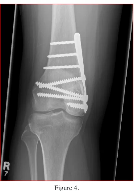
cells in a mononuclear stroma, while the mononuclear cells showed moderate atypia and extensive hemosiderin deposition, suggestive of GCTB. Based on this benign diagnoisis, aggressive curettage and high speed burring were performed to remove the tumor. During surgery both solid tumor and blood filled cystic areas were identified. The final pathology revealed a GCTB with a secondary ABC. A portion of the distal femoral medial condyle was removed until only normal appearing bone remained. A periarticular plate and screws were used to support the morcellized cancellous allograft bone (Figure 4 and 5).
Discussion
This case highlights an example where an underlying pathological condition went clinically unnoticed for a number of years. Considering the good health of the pa-tient and the fact that the only symptom was knee pain, this should not be a surprise. In fact, we are trained
know-ing that serious pathology comprises a very small per-centage of complaints presenting to a musculoskeletal practitioner. Therefore, the point of this case presenta-tion is to emphasize the importance of the re-evaluapresenta-tion, a thorough work up and a second opinion in cases with persistent symptoms. Further, it is an excellent starting point for a review of the differential diagnosis of persis-tent knee pain (Table 1).
In retrospect, the patient could have been asked more pointed questions regarding red flags (in this case the re-evaluation history only included general questions about health status and whether or not there were any changes in the health history). Red flag symptoms includ-ing night sweats, weight loss, malaise etc. could be present in tumours of metastatic origin, but none were present in this case aside from history of melanoma and unremit-ting pain. The reproduction of pain during the orthopaedic tests could have been due to compressive forces on the
Figure 3.
Figure 4.
Right knee, anterior-posterior view plain film radiograph. This post-surgical image demonstrates the hardware (A
periarticular plate and screws) utilized post-curettage to cover and support the site of morcellized cancellous
allograft bone used for reconstruction.
Figure 5.
Right knee, lateral view plain film radiograph. This post-surgical image demonstrates the side view of the periarticular plate and screws used to cover and support
the allograft reconstruction post-curettage.
bone itself. Regardless of the exact mechanism of pain, the decision to refer the patient for a second opinion and imaging was largely based on a lack of specific findings from the physical examination and the history of un-resolved symptoms with no past imaging.
Differential Diagnosis of Knee Pain
Roughly 33% of all sports injuries involve the knee (Table 1).1 PFPS is the most commonly diagnosed clinical con-dition in athletes with non-traumatic anterior knee pain.1-2 In a military population, with comparable incidence rates, females were found to suffer from PFPS 2.23 times more frequently than males.3-4 At a specialty center dealing with musculoskeletal trauma, meniscal injury was the most
the proximal links in the lower extremity are more sig-nificantly associated with the dysfunction noted in PFPS.8 Positive risk factors for the development of PFPS identi-fied in the literature include: muscular weakness around the knee and/or hip; single leg stance strength deficits; de-creased trunk proprioception; tight illiotibial band; gen-eral ligament laxity; large Q-angle; patellar compression or tilting.9-10 Abnormal vastus medialis oblique/vastus lateralis reflex timing has also been considered; however this is proving to be less significant than first theorized
ac-cording to recent systematic reviews.11 The female athlete is in a high risk category due to relatively larger Q-an-gles, potential ligamentous laxity, differences in muscular girth, and even effects of hormone fluctuations through-out the menstrual cycle.3,12-13 One weakness to the patellar tracking theory is the poor correlation with expected lat-eral tilt or displacement of the patella on radiographs and symptomatology.14 More recent observational trials have demonstrated significantly lower cross-sectional girth and diameter of the quadriceps musculature as measured Table 1.
Common sources of knee pain.
Common Pathologies Leading to Anterior Knee Pain (AKP) Articular Cartilage Injury
Bone Tumors
Chondromalacia Patellae Hoffa’s Disease
Iliotibial Band Syndrome Loose Bodies
Neuromas
Osgood-Schlatter Disease Osteochondritis Dissecans Patellar Instability/Subluxation Patellar Stress Fracture
Patellar Tendinopathy Patellofemoral Arthritis Patellofemoral Pain Syndrome Pes Anserine Bursitis
Plica Synovialis
Prepatellar Bursitis Previous Surgery Quadriceps Tendinopathy
Referred from L/S or Hip Joint Pathology Saphenous Neuritis
Sinding-Larsen-Johansson Syndrome Symptomatic Bipartite Patella
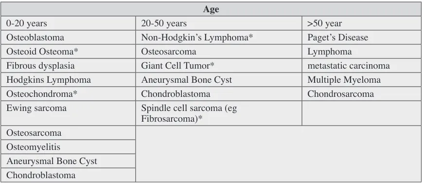
Table 2.
Common bone tumors and conditions by age of incidence. Asterisks (*) indicate tumors commonly affecting the femur or tibia around the knee.
Age
0-20 years 20-50 years >50 year
Osteoblastoma Non-Hodgkin’s Lymphoma* Paget’s Disease
Osteoid Osteoma* Osteosarcoma Lymphoma
Fibrous dysplasia Giant Cell Tumor* metastatic carcinoma
Hodgkins Lymphoma Aneurysmal Bone Cyst Multiple Myeloma
Osteochondroma* Chondroblastoma Chondrosarcoma
Ewing sarcoma Spindle cell sarcoma (eg Fibrosarcoma)*
Osteosarcoma Osteomyelitis
on MRI; however a significant difference in the ratio of vastus lateralis and vastus medialis was not demonstrat-ed.14 Further research using MRI paired with kinematic analysis revealed that what is more important is abnormal femur motion and not that of the patella.15 This abnor-mal femur motion in PFPS is suggested to be the result of reduced hip torque into abduction and external rotation noted on a step down task.16 Similarly, decreased hip ab-duction force and associated increased hip adab-duction an-gle at the end of a run of variable distances was measured in PFPS subjects.17
Investigations on the natural history of PFPS have been poor, making it difficult to know when to consider alterna-tive differential diagnoses. Two studies have attempted long-term follow-up of patients with PFPS. The group found that 27% of athletes recovered within an average of 8 months, while the remaining patients continued to have pain at 5 years.4 Of the unrecovered population, half re-ported being able to cope with the pain, and only 20% of athletes were forced to completely cease sport participa-tion, and 6% reporting time off work.4 Variables strongly associated with poorer prognosis include: female gender, pain severity on visual analog scale, Anterior Knee Pain score, patellar hypermobility, and a sedentary lifestyle.4, 18 Misdiagnosis in Athletes
Misdiagnosis, specifically tumors about the knee in ath-letes is an important issue looked at in the literature.19 One report from a large orthopedic hospital helped illustrate the incidence of misdiagnosed knee pathology in athletic populations.20 The investigators reviewed 667 cases and found 25 tumor patients which were originally misdiag-nosed as an athletic injury, and resulted in inappropriate invasive procedures.20 The authors concluded that 15 pa-tients had suffered significant detrimental effects to their final clinical outcomes due to misdiagnosis, 3 of which resulted in limb amputation.20 While the incidence of these cases was found to be relatively low, the results are potentially devastating to the patient. The most common cause for misdiagnosis reported by the authors was poor quality radiographs and refusal to consider alternative diagnoses in the absence of clinical improvement.20 Tumors Around the Knee
Although GCTB is an uncommon cause of knee pain, it has been reported to accounts for 13.7% (8–23%) of all
benign primary bone tumors.21 GCTB is considered to be “quasi-malignant” or a “borderline” malignancy making up a 5-8% of all primary malignant bone tumors.21-22 It is associated with a very low risk of lung metastasis, even in the absence of histologic malignancy, although it is treated locally as a benign tumor.21 The vast majority of patients with GCTB are between 20–50 years of age.22-23 GCTB has a predilection for juxta-articular locations (i.e. metaphysis and epiphysis) and is located most commonly around the knee.23
ABC is also an uncommon cause of knee pain, 6% of benign bone tumors, which occurs typically in the first two decades of life.22 ABC frequently causes bone de-struction and cortical expansion prior to their discovery.23 In adults ABC can also form secondary to an underlying GCTB or other benign bone tumor. 21, 25-25 In the case pre-sented above, pathological analysis of the excised tissue found evidence of both GCTB and secondary ABC. There are other bone tumors that fit this case presentation based on location and demographic information (Table 2). Patients with benign aggressive bone tumors usual-ly present with rapid onset of symptoms and functional disability, unlike the patient in this case report who had persistent, but slowly worsening symptoms over a course of 5 years. The typical management of benign aggressive lesions including GCTB and ABC is tumor resection by aggressive curettage and high speed burring followed by reconstruction with either bone graft or cement, or less commonly by en bloc resection.26-27 A retrospective re-view of 621 patients at a Chinese hospital specializing in musculoskeletal oncology reported a local recurrence rate of 8.6% after extensive curettage and burring.20
Imaging
avoid-ing inadvertent tumor rupture, spread of lesion and some-times amputation.20
Summary
We have presented a case of underlying knee pathology that had remained undiagnosed for a number of years. The important feature to note during the evolution of this case was the re-evaluating clinician’s willingness to en-gage in collaborative care when faced with progressive or unresolving symptoms. On a subsequent evaluation, the patient’s presentation still appeared mechanical on physical exam; however, symptoms were increasing in severity making the clinical progression appear more ur-gent, warranting imaging. While MRI remains a sensitive imaging modality for early diagnosis, radiographs may be a logical first step.
One challenge with this case is the self-limiting na-ture of PFPS in the absence of ligamentous instability or intra-articular injury. This allows patient to continue to access pain management over a period of time, often seek-ing out various health care providers due to frustration, while allowing serious pathology to go unsuspected. This case illustrates the importance of a thorough re-evalua-tion, consideration of differentials and follow-up for per-sistent self-limiting complaints. Maintaining a high level of suspicion in athletic or active populations should be exercised early so as to avoid delayed diagnosis and hast-en recovery.
References:
1. Steinbrück K. Epidemiology of sports injuries—25-year analysis of sports orthopedic-traumatologie ambulatory care. Sportverletz Sportschaden. 1999;13(2): 38 – 52. 2. Kransdorf M. Malignant soft-tissue tumors in a large
referral population: distribution of diagnoses by age, sex, and location. Am J Roentgen. 1995; 164(1): 129-134. 3. Boling M, Padua D, Marshall S, Guskiewicz K, Pyne S, Beutler A. Gender differences in the incidence and prevalence of patellofemoral pain syndrome. Scand J Med Sci Sports. 2010; 20(5): 725–730.
4. Blønd L, Hansen L. Patellofemoral pain syndrome in athletes: a 5-7-year retrospective follow-up study of 250 athletes. Acta Orthop Belg. 1998; 64(4): 393–400. 5. Clayton R, Brown C. The epidemiology of
musculoskeletal tendinous and ligamentous injuries. Injury. 2008; 39(12): 1338-1344.
6. Snoeker B, Bakker E, Kegel C, Lucas C. Risk factors for meniscal tears: a systematic review including meta-analysis. J Orthop Sports Phys Ther. 2013; 43(6): 352-367.
7. Englund M, Guermazi A, Gale D, Hunter D, Aliabadi P, Clancy M, Felson D. Incidental meniscal findings on knee MRI in middle-aged and elderly persons. N Engl J Med. 2008; 359: 1108-1115.
8. Powers C. The influence of abnormal hip mechanics on knee injury: a biomechanical perspective. J Orthop Sports Phys Ther. 2010;40(2):42-51.
9. Boling MC, Padua DA, Marshall SW, Guskiewicz K, Pyne S, Beutler A. A prospective investigation of biomechanical risk factors for patellofemoral pain syndrome: the Joint Undertaking to Monitor and Prevent ACL Injury (JUMP-ACL) cohort. Am J Sports Med. 2009; 37: 2108–2116. 10. Witvrouw E, Lysens R, Bellemans J, Cambier D,
Vanderstraeten G. Intrinsic risk factors for the development of anterior knee pain in an athletic population. a two-year prospective study. Am J Sports Med. 2000; 28(4): 480-489.
11. Chester R, Smith TO, Sweeting D, Dixon J, Wood S, Song F. The relative timing of VMO and VL in the aetiology of anterior knee pain: a systematic review and meta-analysis. BMC Musculoskelet Disord. 2008; 9: 64.
12. Wojtys, Edward M et al. Association between the menstrual cycle and anterior cruciate ligament injuries in female athletes. Am J Sports Med. 1998; 614-619. 13. Slauterbeck, James R et al. The menstrual cycle, sex
hormones, and anterior cruciate ligament injury. J Athl Train. 2002: 275.
14. Giles, Lachlan S et al. Does quadriceps atrophy exist in individuals with patellofemoral pain? A systematic literature review with meta-analysis. J Orthop Sports Phys Ther. 2013: 766-776.
15. Souza R, Draper C, Fredericson M, Powers C. Femur rotation and patellofemoral joint kinematics: a weight-bearing magnetic resonance imaging analysis. J Orthop Sports Phys Ther. 2010; 40(5): 277-285.
16. Bolgla, L, Malone T, Umberger B,Uhl T. Hip strength and hip and knee kinematics during stair descent in females with and without patellofemoral pain syndrome. J Orthop Sports Phys Ther. 2008;38(1):12–18.
17. Dierks T, Manal K , Hamil J, Davis I. Proximal and distal influences on hip and knee kinematics in runners with patellofemoral pain during a prolonged run. Orthop Sports Phys Ther. 2008;38(8):448–456.
18. Kujala UM, Jaakkola LH, Koskinen SK, et al. Scoring of patellofemoral disorders. Arthroscopy. 1993; 9: 159–163. 19. Muscolo DL, Ayerza MA, Makino A, Costa-Paz M,
Aponte-Tinao LA. Tumors about the knee diagnosed as athletic injuries. J Bone Joint Surg. 2003; 85(7): 1209– 1214.
20. Niu X, Zhang Q, Hao L, Ding Y, Li Y, Xu H, Liu W. Giant cell tumor of the extremity: retrospective analysis of 621 Chinese patients from one institution. J Bone Joint Surg Am. 2012; 94(5): 461-467.
Of Bone Tumors And Tumor-Like Lesions Medical Radiology. Springer Berlin Heidelberg; 2009. p. 321-336. 22. Yochum TR, Rowe LJ. Essentials of Skeletal Radiology.
3rd. ed. Philadelphia: Lippincott Williams & Wilkins; 2005.
23. Taylor JAM, Hughes TH, Resnick DL. Skeletal Imaging: Atlas of the spine and extremities. Elsevier Health Sciences; 2009.
24. Chakarun CJ, Forrester DM, Gottsegen CJ, Patel DB, White EA, Matcuk GR Jr. Giant cell tumor of bone: review, mimics, and new developments in treatment. Radiographics. 2013; 33(1): 197-211.
25. Low SF, Hanafiah M, Nurismah MI, Suraya A. Challenges in imaging and histopathological assessment of a giant cell tumour with secondary aneurysmal cyst in the patella. BMJ Case Rep. 2013; 20.
26. Turcotte RE, Wunder JS, Isler MH, Bell RS, Schachar N, Masri BA, Moreau G, Davis AM; Canadian Sarcoma Group. Giant cell tumor of long bone: a Canadian Sarcoma Group study. Clin Orthop Relat Res. 2002; 397: 248-258. 27. Blackley HR, Wunder JS, Davis AM, White LM, Kandel R, Bell RS. Treatment of giant-cell tumors of long bones with curettage and bone-grafting. J Bone Joint Surg Am. 1999; 81(6): 811-820.