Malathion induced testicular toxicity and oxidative damage in male mice: the protective effect of curcumin
Full text
Figure


Related documents
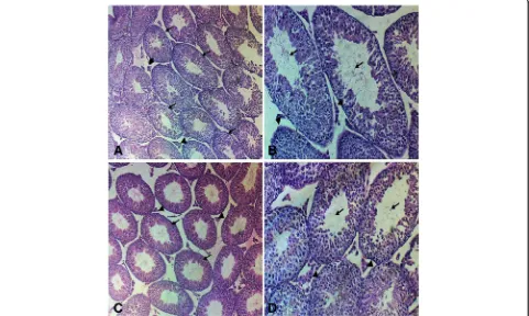
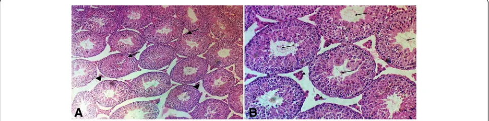
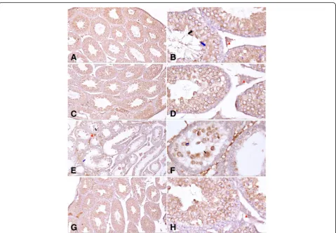
significant reduction in testicular weight, epididymal sperm count, motility, serum level of.. FSH, LH and testosterone with damaged histoarcihtecture of testes
Obtained results in groups receiving malathion show that LH hormone levels in different groups have no significant change compared with the control group (p<0.05), however
were in line with the results of the present study in that administration of Malathion significantly reduced catalase and serum total antioxidant level,
Results: Significant changes in the normal range of testosterone, FSH, LH serum levels and seminiferous tubule apoptotic cells were detected in CVS group compared to
In noise exposed rats the serum testosterone level was reduced which is similar as in immobilization test where the stress did not alter plasma luteizing hormone (LH) levels,
Therefore, the pathological changes in biochemical markers of reproductive function such as significant decrease in testosterone, LH and FSH concentrations and the
The results demonstrated a signi ficant improvement of sex hormones serum levels (FSH, LH, total testosterone, SHBG, calcu- lated free testosterone, and estradiol), semen volume
We studied the serum gonadotrophins which are follicle stimulating hormone (FSH), luteinizing hormone (LH) and testosterone, relative reproductive organs weight,
