O R I G I N A L A R T I C L E
Open Access
Suppression of
OsVPE3
Enhances Salt
Tolerance by Attenuating Vacuole Rupture
during Programmed Cell Death and Affects
Stomata Development in Rice
Wenyun Lu
1†, Minjuan Deng
2†, Fu Guo
1, Mingqiang Wang
1, Zhanghui Zeng
1, Ning Han
1, Yinong Yang
1,3,
Muyuan Zhu
1and Hongwu Bian
1*Abstract
Background:Vacuolar processing enzymes (VPEs) are cysteine proteinases that act as crucial mediators of programmed cell death (PCD) in plants. In rice, however, the role of VPEs in abiotic stress-induced PCD remains largely unknown. In this study, we generatedOsVPE3overexpression and suppression transgenic lines to elucidate the function of this gene in rice.
Results:Survival rate and chlorophyll retention analyses showed that suppression ofOsVPE3clearly enhanced salt stress tolerance in transgenic rice compared with wild type. Furthermore, fragmentation of genomic DNA was inhibited in plants with down-regulatedOsVPE3. Vital staining studies indicated that vacuole rupture occurred prior to plasma membrane collapse during salt-induced PCD. Notably, overexpression ofOsVPE3promoted vacuole rupture, whereas suppression ofOsVPE3attenuated or delayed the disintegration of vacuolar membranes. Moreover, we found that suppression ofOsVPE3caused decreased leaf width and guard cell length in rice.
Conclusions:Taken together, these results indicated that suppression ofOsVPE3enhances salt tolerance by attenuating vacuole rupture during PCD. Therefore, we concluded thatOsVPE3plays a crucial role in vacuole-mediated PCD and in stomatal development in rice.
Keywords:OsVPE3, Programmed cell death, Rice, Salt stress, Stomata, Vacuolar processing enzyme
Background
Rice (Oryza sativa L.) is one of the most important cereal crops for more than half of the world’s population. According to estimates, the world will need to produce 25% more rice by 2030 to meet the challenges of feeding increasing populations. However, the increasing soil salinization of limited farmland is becoming a serious global threat to sustained rice production (Khatun and Flowers 1995; Sahi et al. 2006; Gao et al. 2007). There-fore, enhancing salt-tolerance is a serious concern for crop breeding programs.
Programmed cell death (PCD) is a highly conserved and genetically controlled process in multicellular organ-isms. PCD is involved in maintaining cellular homeostasis, development and senescence(Azeez et al. 2007; Williams and Dickman 2008), and this process is triggered by a var-iety of abiotic and biotic stresses (Huh et al. 2002; Lam 2004; Gechev et al. 2006). Plant and animal cells share many hallmarks of PCD, including cytoplasm shrinkage, chromatin condensation, DNA cleavage, mitochondrial swelling, organelle disruption and plasma membrane col-lapse (Mittler et al. 1997; van Doorn 2011; De Pinto et al. 2012). However, plants also exhibit unique features of PCD due to the presence of chloroplasts and vacuoles (Samuilov et al. 2003; Hatsugai et al. 2006; Kim et al. 2012; Wituszynska et al. 2015). Vacuoles are storage organelles that function as reservoirs for both hydrolytic enzymes * Correspondence:[email protected]
†Equal contributors
1Institute of Genetics and Regenerative Biology, Key Laboratory for Cell and
Gene Engineering of Zhejiang Province, College of Life Sciences, Zhejiang University, Hangzhou, China
Full list of author information is available at the end of the article
© The Author(s). 2016Open AccessThis article is distributed under the terms of the Creative Commons Attribution 4.0 International License (http://creativecommons.org/licenses/by/4.0/), which permits unrestricted use, distribution, and reproduction in any medium, provided you give appropriate credit to the original author(s) and the source, provide a link to the Creative Commons license, and indicate if changes were made.
Luet al. Rice (2016) 9:65
and defence proteins, and vacuoles play several roles in stress response, development and pathogen defence. Re-cent studies have suggested that vacuole-mediated cell death is a response to various stresses in plants (Hatsugai et al. 2006; Hatsugai et al. 2015). Under salt stress, early events include the production of reactive oxygen species (ROS) and increased cytoplasmic calcium concentrations (Dionisio-Sese and Tobita 1998; Menezes-Benavente et al. 2004; Kudla et al. 2010) followed by PCD. In addition, vacuole rupture is a trigger for nuclear degradation during PCD (Obara et al. 2001). However, the genes and regula-tory networks involved in vacuole-mediated cell death re-main unidentified.
Vacuolar processing enzymes (VPEs) are cysteine pro-teinases involved in the processing of vacuolar proteins and the maturation of seed storage proteins in plants (Haranishimura et al. 1991; Hiraiwa et al. 1993; Rojo et al. 2003; Wang et al. 2009). VPEs are expressed in senescent tissues, and their expression patterns have been linked to PCD (Hara-Nishimura et al. 1998; Kinoshita et al. 1999). For example, it has been shown that VPEs regulate sys-tematic cell death induced by viral, aluminium and heat stress, which is mediated by caspase-1-like activity during PCD (Hatsugai et al. 2004b; Li et al. 2012; Kariya et al. 2013). VPEs and caspase-1 share conserved structural properties, particularly the Asp pocket of caspase-1, which includes three crucial amino acids (Arg-179, Arg-341 and Ser-347) (Hatsugai et al. 2006). Although VPEs and caspase-1 share many similarities, the subcellular localiza-tions of these two proteases are completely different. Namely, VPEs localize to vacuoles, whereas animal caspases localize to the cytosol. As initiators of plant PCD, VPEs trigger cell death through vacuolar collapse (Hatsugai et al. 2004a). To date, several types of VPEs have been reported to be involved in PCD as follows: NbVPE1a and NbVPE1b in Nicotiana; and AtγVPE in Arabidopsis. In rice, however, the role of VPEs in the vacuole-mediated cell death remains unknown.
The rice genome contains fourVPEhomologous genes as follows:OsVPE1(Os04g45470),OsVPE2(Os01g37910),
OsVPE3(Os02g43010) and OsVPE4(Os05g51570) (Deng
et al. 2011). Phylogenetically, OsVPE1 and OsVPE3 are more similar to Arabidopsis AtβVPE, whereas OsVPE2
and OsVPE4 are more similar to AtαVPE and AtγVPE.
OsVPE1, a homolog ofAtβVPE, plays a crucial role in the maturation of glutelins in seeds (Wang et al. 2009). Previ-ous work by our group has shown that the transcription of
OsVPE2 and OsVPE3, but not of OsVPE1 and OsVPE4,
can significantly enhance salt-induced PCD (Deng et al. 2011; Kim et al. 2014). In this study, we aimed to elucidate the role ofOsVPE3in the context of salt stress. To deter-mine the function ofOsVPE3in rice under salt stress, we generated transgenic lines to either overexpress and suppressOsVPE3, and we found thatOsVPE3regulates
the collapse of vacuolar membranes during PCD. In addition, we found thatOsVPE3plays a role in leaf and stomata development in rice.
Results
Expression pattern and localization of OsVPE3
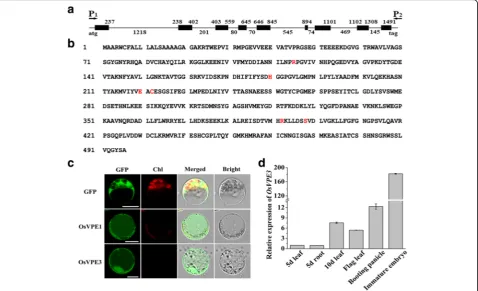
A comparison between genomic DNA and cDNA revealed thatOsVPE3(Os02g43010) contains nine exons and eight introns (Fig. 1a), and OsVPE3 encodes a peptide of 496 amino acids. The red letters in Fig. 1 indicate essential amino acids in the catalytic site and substrate pocket that mediate caspase-like activity (Fig. 1b) (Nicholson 1999; Hara-Nishimura et al. 2005; Hatsugai et al. 2015).
To determine the subcellular localization of OsVPE3, the full-length cDNA was fused to green fluorescent protein (GFP) under control of the 35S promoter. An
OsVPE1-GFP fusion gene driven by the 35S promoter
was used as a control. OsVPE1, a seed-type vacuolar processing enzyme, has been shown to localize to vacuoles in onion cells (Wang et al. 2009). These constructs were transiently transformed into rice protoplasts. We searched for GFP signals in vacuoles for the 35S:OsVPE1-GFP and
35S:OsVPE3-GFP fusion constructs, and no GFP signal
was observed in vacuoles for the 35S: GFP construct (Fig. 1c). It was reported that GFP was unstable in the acidic vacuoles of plants under the light condition, be-cause light would be-cause the rapid degradation of 27-kDa GFP by pre-existing proteinase in vacuoles at pH5.5 (Tamura et al., 2003). In protoplast transient assay, iso-lated protoplasts were kept in the dark condition to protect from light damage, thus the GFP fluorescence can be observed in the vacuoles..Consequently, this result suggested that OsVPE3 is a vacuole-targeting protein simi-lar to OsVPE1.
To investigate the expression pattern of OsVPE3 in rice, transcription levels were examined in various tissues by quantitative RT-PCR. Expression analysis revealed that OsVPE3 is expressed in tissues of the leaf, root, booting panicle and immature embryo (Fig. 1d). OsVPE3 expres-sion increased rapidly in the leaves of 10-day-old seedlings compared with 5-day-old seedlings. The highest transcrip-tional expression level ofOsVPE3was detected in imma-ture embryos. These results demonstrated thatOsVPE3is actively transcribed in the leaves and developing embryos of rice.
Generation of Transgenic Rice Lines
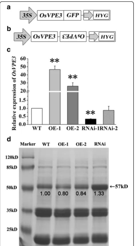
In our previous work, we found that the expression of OsVPE3 is dramatically induced by salt stress (Deng et al. 2011; Kim et al. 2014). However, the role of the OsVPE3gene in rice remains largely unknown. To inves-tigate if OsVPE3 is involved in the process of PCD,
OsVPE3was overexpressed and suppressed using
were used for further investigation. Quantitative RT-PCR analysis confirmed that the expression levels of OsVPE3 were strongly increased in the overexpression lines (OE-1 and OE-2) and decreased significantly in the RNA interference transgenic lines (RNAi-1) compared with WT (Fig. 2c).
Previous studies showed that OsVPE1 plays a crucial role in the maturation of glutelins in seeds (Wang et al. 2009). To examine the role ofOsVPE3on seed proteins, we performed SDS-PAGE analysis to compare the pro-teins in WT and transgenic lines. Our data showed that the major storage proteins yielded three bands of ap-proximately 20, 40 and 57 kDa in rice (Fig. 2d). The 57-kDa protein is a glutelin precursor (Yamagata et al. 1982; Krishnan and Okita 1986). Compared with WT, 57 kDa protein levels was increased to 1.33 fold in the OsVPE3-RNAi line, whereas reduced to approximately 0.80-0.84 in the overexpression lines.(Fig. 2d). Accord-ingly, the grain width and 1000-grian weight of the RNAi line decreased significantly compared with WT and overexpression lines (Additional file 1: Figure S1). These results indicated that OsVPE3 is involved in the processing of seed storage proteins in rice.
Suppression of OsVPE3 Enhances Salt Stress Tolerance in Rice
Based on our previous studies, we hypothesized that
OsVPE3 might act as a trigger in salt-induced PCD. To
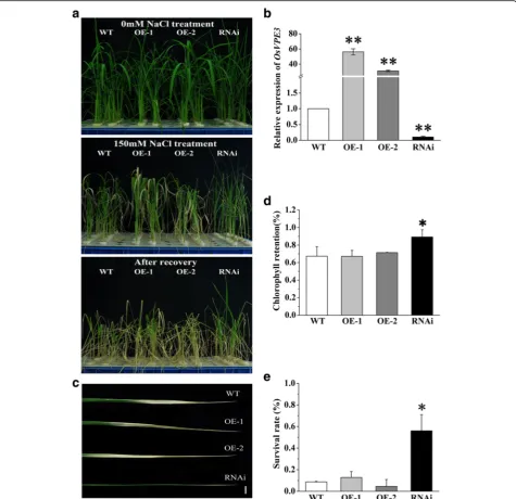
determine if OsVPE3 is involved in salt stress tolerance in rice, 4-week-old WT and transgenic plants were treated with 150 mM NaCl for 3 days. Before NaCl treatment, the expression levels of OsVPE3were measured in the fourth leaf of each line, confirming that the transcript levels of OsVPE3were increased in overexpression lines (OE-1 and OE-2) and significantly suppressed in the RNAi transgenic lines (Fig. 3a, b). Under salt stress, chlorosis is a common symptom in rice leaves. Chlorophyll contents were mea-sured after 3 days of NaCl treatment, and then survival rates were calculated after 7 days of recovery culture under normal growth conditions. When the plants were exposed to 150 mM NaCl for 3 days, the leaves of WT and overex-pression lines turned yellow. In contrast, the chlorosis phenotype was significantly weakened in the RNAi line (Fig. 3c, d). Approximately 56.1 ± 15.0% of RNAi plants remained alive after recovery culture, whereas the survival rates of WT and overexpression transgenic lines (OE-1, OE-2) were significantly lower than that of RNAi line
Fig. 1Sequence analysis, subcellular localization and expression pattern of OsVPE3.aTheOsVPE3gene structure based on the genomic sequence and full-length cDNA clone (BAC41387). Black boxes represent exons. Arrows indicate the primers used to isolateOsVPE3cDNA.bOsVPE3 amino acid sequence. Amino acids necessary for caspase-like activity are indicated by coloured letters.cThe subcellular localization of OsVPE1 and OsVPE3 by confocal fluorescent microscopy. Scale bar = 10μm.dqRT-PCR analysis ofOsVPE3expression in various rice tissues. Total RNA was extracted from leaf, root, booting panicle and immature embryo samples.OsUBQ5was used as an internal control. Values are means, and error bars represent the SD from three independent experiments
(Fig. 3a, e). Remarkably, the RNAi line had a significantly higher chlorophyll content and relative survival rate com-pared with the WT and overexpression lines (Fig. 3d, e). These results demonstrated that suppression of OsVPE3 inhibits the development of chlorosis and improves salt tol-erance in rice.
Suppression of OsVPE3 Inhibits the Formation of Dna Ladders in Salt-Induced PCD
To further investigate the effect ofOsVPE3on rice toler-ance to salt stress,3-day-old WT and transgenic seedlings were treated with high concentration NaCl (100 mM, 150 mM) for 3 days. The results showed that RNAi seed-lings had the highest root relative elongation rate and overexpression seedlings were the most sensitive to salt stress (Fig. 4a). At the same time, Evans Blue staining con-firmed that the root tips of the RNAi line maintained highest cell viability after exposure to 150 mM NaCl for 3 days compared to other lines (Fig. 4b). The frag-mentation of genomic DNA is a typical biochemical and morphological feature of PCD. To determine if the suppression of OsVPE3 can inhibit salt-induced PCD, DNA fragmentation was measured in transgenic lines and WT plants treated with 300 mM NaCl for 8 h. As shown in Fig. 4c, clear and visible DNA laddering was observed in the OsVPE3-overexpression lines, whereas DNA laddering was significantly inhibited in the RNAi line. This result suggested that suppression ofOsVPE3 inhibits the formation of DNA ladders during salt-induced PCD.
BecauseOsVPE3has high homology withOsVPE1, we performed qRT-PCR experiment to detect the expres-sion levels of OsVPE homologous genes in the roots of transgenic lines (Fig. 4d). The results showed that the expression level of OsVPE3 was decreased dramatically in RNAi line; meanwhile, OsVPE1 expression was also reduced compared with the WT. The expression level of
OsVPE3was much more decreased thanOsVPE1in the
RNAi line. Our previous work has shown that the tran-scription of OsVPE2 and OsVPE3, but not of OsVPE1 andOsVPE4, can significantly increase in the salt-induced PCD (Deng et al. 2011; Kim et al. 2014), suggesting that the transcription ofOsVPE1 was not responsive to salt stress in rice roots (Deng et al. 2011). Therefore, we conclude thatOsVPE3plays a crucial role in salt stress-mediated PCD.
Suppression of OsVPE3 Helps Maintain the Integrity of Vacuolar Membranes During Salt-Induced Pcd
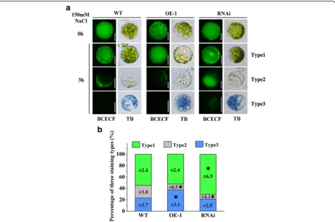
Unlike in animal cells, the vacuole is a unique organelle used to store a variety of hydrolytic enzymes and defence proteins in plant cells (Neuhaus et al. 1991; Yamada et al. 2001). The rupture of vacuolar membranes releases these components and leads to cell death (Mino et al. 2006). To elucidate the role of OsVPE3 in the integrity of vacuolar membranes during PCD, rice protoplasts were stained with Trypan Blue and the BCECF-AM fluorescent probe. Trypan Blue staining is a reporter of cell death based on disintegrated cellular membranes, and BCECF-AM labels the vacuole lumen as green fluorescence to reveal intact vacuoles (Swanson and Jones 1996). BCECF-staining
revealed that protoplasts prior to NaCl treatment accu-mulated strong fluorescence signal in vacuoles in both WT and transgenic lines (Fig. 5a, the first horizontal low). Protoplasts subjected to 100 mM NaCl treatment for 3 h can be sorted into three types as follows (Fig. 5a, b): Type 1, BCECF-positive and Trypan Blue-negative, indicating living cells with intact cellular membranes
and vacuoles; Type 2, BCECF-negative and Trypan Blue-negative, indicating living cells with disintegrated vacuolar membranes (as shown by distribution of BCECF fluorescence signal outside of the vacuoles); and Type 3, BCECF-negative and Trypan Blue-positive, indicating dead cells with collapsed vacuoles (as shown by no BCECF fluorescence signal). After 100 mM NaCl treatment, the
Fig. 3Suppression ofOsVPE3improved survival rate and inhibited chlorosis under salt stress.aPlants were grown for 4 weeks, treated with 150 mMNaCl for 3 days and then allowed to recover for 7 days.bExpression levels ofOsVPE3in WT and transgenic lines as determined by qRT-PCR. Total RNA was extracted from the fourth leaves.OsUBQ5was used as an internal control. Values are means, and error bars represent the SD from three independent experiments. Asterisks indicate a significant difference between WT and transgenic lines (t-test; **,P< 0.01).cThe fourth leaves of NaCl-treated plants. Bar = 1 cm.dChlorophyll retention in the fourth leaves of NaCl-treated plants prior to recovery. Values are means, and error bars represent the SD from three independent experiments. Asterisks indicate a significant difference between WT and transgenic lines (t-test; *,P< 0.05).eSurvival rates of NaCl-treated plants 7 days after recovery. Values are means, and error bars represent the SD (n> 20) of three independent experiments. Asterisks indicate a significant difference between WT and transgenic lines (t-test; *,P< 0.05)
survival protoplasts (type 1) decreased to 54.6 ± 2.4 and 52.7 ± 2.4% of the levels in the WT and OE-1 lines, re-spectively. In contrast, the survival rate of protoplasts (Type 1) from the RNAi line was 70.3 ± 6.5%, which was significantly higher than that of the WT and OE-1 lines. The results showing that many of the protoplasts (Type 2) with degraded vacuolar membranes were alive suggested that cell death was preceded by vacuolar collapse. Only 8.4 ± 4.1% of the RNAi cells belonged to Type 2, which in-volved vacuole rupture before cell death. Interestingly, 37.5 ± 3.1% of OsVPE3-overexpressing cells belonged to Type 3, which was significantly higher than in the WT and RNAi lines. Notably, certain Type 3 protoplasts from the RNAi line still showed weak fluorescence in the vacuolar region, thereby suggesting thatOsVPE3-RNAi attenuated or delayed the disintegration of vacuolar membranes even after cell death. Taken together, these results demonstrated
that suppression ofOsVPE3prevents vacuole rupture dur-ing salt-induced PCD (Fig. 5a).
Suppression of OsVPE3 Results in Decreased Leaf Width and Stomatal Size
Compared with WT, there were no obviously morpho-logical changes in the transgenic plants at the early seed-ling stage. However, after four weeks of growth, the RNAi transgenic line exhibited narrower leaves than WT (Fig. 6a). Blade width of the fourth leaf was approximately 6.00 ± 0.54 cm in the WT plants, whereas the blade width was approximately 5.50 ± 0.33 cm in the RNAi line (Fig. 6b). Further observation revealed stomata size in the RNAi line was smaller than in WT plants (Fig. 6c). Statis-tical analysis showed that only 25% of guard cells were longer than 20μm in the RNAi line whereas 64% in the WT plants. The majority of guard cells were between 15
and 20μm in the RNAi line (Fig. 6d). There were no sig-nificant differences in leaf width and guard cell length be-tween the OE-1 line and WT plants. The expression pattern ofOsVPE3 showed thatOsVPE3 is actively tran-scribed in the leaves and developing embryos of rice (Fig. 1d). We speculated that the expression levels of
OsVPE3were abundant enough for the leaf development
in WT, thus the OsVPE3-overexpression lines exhibited weak phenotype compared with the WT.
In addition, we tested the percentage of water loss of detached leaves from WT and transgenic lines. The data indicated that the percentage of water loss of RNAi leaves was significantly lower than other lines (Additional file 1: Fig. S2), suggesting suppression ofOsVPE3enhanced the dehydration tolerance. The above results demonstrated that suppression of OsVPE3 caused decreased leaf width and guard cell length, promoting a dehydration tolerance in rice leaves.
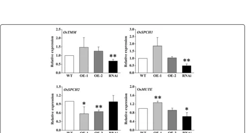
Furthermore, we analysed the expression of genes re-lated to stomatal development by qRT-PCR. According
to known reports, there are five critical genes related to the development of stomata in rice, namely OsTMM
(TOO MANY MOUTHS), OsSPCH1 (SPEECHLESS),
OsSPCH2, OsMUTE and OsFAMA (Liu et al. 2009).
OsTMMis a receptor for extracellular ligand, character-ized as a set of key regulators in stomatal production and patterning (Balcerowicz and Hoecker 2014).OsSPCH1,
OsSPCH2,OsMUTEandOsFAMAare essential
transcrip-tion factors in the stomata patterning and development (Liu et al. 2009). We monitored the expressions of these genes (the data ofOsFAMAis not shown because of its low expression level in rice leaves). As shown in Fig. 7, the ex-pression levels of OsTMM, OsSPCH1 andOsMUTE were significantly down-regulated in the RNAi line compared with WT. Consistently, these three genes showed up-regulated tendencies in overexpression lines, while only the expression ofOsSPCH2decreased significantly. It revealed that OsVPE3 might mainly affect the level expression of
OsTMM, OsSPCH1andOsMUTEin stomata
developmen-tal pathway to change the guard cell size.
Fig. 5Suppression ofOsVPE3alleviates vacuole rupture under salt stress.aProtoplasts from WT and transgenic lines were stained with the BCECF-AM vital dye (final concentration = 10μM) for 2 h and then treated with 100 mM NaCl for 3 h. Before the BCECF fluorescent images were inspected, the protoplasts were stained with Trypan Blue (TB). Type 1 protoplasts were living cells with intact vacuoles. Type 2 protoplasts were living cells with disintegrated vacuolar membranes. Type 3 protoplasts were dead.bStatistical analysis of three types of protoplasts after treatment with 100 mM NaCl. Data represent the means ± SD (n> 100) for three independent replicates. Asterisks indicate a significant difference between the WT and transgenic lines (t-test; *,P< 0.05)
Fig. 6Fourth leaf width and guard cell size in WT and transgenic lines.aImages of fourth leaves of WT and transgenic lines. Bars = 1 cm.
bMeasurements of the maximum fourth leaf width in WT and transgenic lines. Values indicate the mean, and error bars represent the SD (n>10). An asterisk indicates a significant difference between the WT and transgenic lines (t-test;P< 0.05).cImages of stomata in WT and transgenic lines. Bars = 5μm.dPercentage of stomata with various guard cell lengths in WT and transgenic lines (n> 500)
Discussion
The main purpose of this study was to understand the role of OsVPE3in vacuole-mediated PCD following salt stress in rice. OsVPE3 overexpression and suppression transgenic lines were created to elucidate the function of
OsVPE3in rice. Our results demonstrated that OsVPE3
plays a crucial role in the salt stress response by regulat-ing the collapse of vacuolar membranes durregulat-ing PCD. In addition, OsVPE3 affected leaf and stomata guard cell development in rice.
High salinity is one of the most important abiotic stresses during crop breeding. High salinity activates the salt-overly-sensitive (SOS) system in plants, which leads to sodium exclusion from the cytosol (Zhu 2003). Under high salt conditions, excess Na+accumulates in the plant, thus increasing influx of Na+and efflux of K+(Serrano and Rodriguez-Navarro 2001; Horie et al. 2012). It has been proposed that these changes can decrease the cytosolic K/Na ratio, thus elevating concentrations of the intra-cellular second messenger Ca2+ (Kudla et al. 2010) and causing ROS bursts (Zhu 2001). As a consequence of increased K/Na ratios and salinity-induced ROS, pro-grammed cell death would be finally triggered (Huh et al. 2002; Lin et al. 2006; Shabala 2009). A recent study in rice by our group revealed that overexpression of BCL-2, an anti-apoptotic protein, significantly reduces NaCl-induced K+ efflux and represses the expression of VPEs, thereby alleviating PCD symptoms(Deng et al. 2011; Kim et al. 2014).
Salt-induced PCD in plants and animals shares many consequences, including DNA fragmentation, nuclear condensation, nuclear deformation, mitochondrial in-volvement and endonuclease activity (Li et al. 2007; Jiang et al. 2008). Analysis of DNA laddering indicated that PCD occurs in rice seedlings under salt treatment (Fig. 4c). Compared with WT, overexpression of OsVPE3 strongly enhanced genomic DNA fragmentation, whereasOsVPE3 interference strongly repressed DNA fragmentation during PCD. This finding suggested that OsVPE3 likely plays a crucial role in the salt-induced PCD in rice.
The VPE family was originally identified as a group of processing enzymes responsible for the maturation of seed storage proteins in protein storage vacuoles (Haranishimura et al. 1991; Haranishimura et al. 1993). It was originally reported that NtVPEs have caspase-1 activ-ity and are essential for the virus-induced hypersensitive response involving PCD, which led to the proposition of a new cell death mechanism mediated by VPE and cellular vacuoles (Hatsugai et al. 2004b). Subsequently, a similar function was proposed for AtγVPE in mycotoxin-induced cell death (Kuroyanagi et al. 2005). Recently, further stud-ies have shown that VPEs are also involved in cell death under abiotic stresses (Zhang et al. 2013b). For example, AtγVPE, which is mediated by MPK6, affects
heat-shock-induced PCD in Arabidopsis (Li et al. 2012). Our study found thatOsVPE3regulated salt-induced PCD in rice.
Vacuoles are essential organelles in plants, which have multiple functions including storage of a wide variety of ions, proteins and other metabolites, and maintain cyto-solic ion homeostasis (Boller and Wiemken 1986; Rea and Sanders 1987). Plant vacuoles also play critical roles in stress responses, development and pathogen defence. There have been many reports concerning the relation-ship between vacuoles and cell death (Hara-Nishimura and Hatsugai 2011; Higaki et al. 2011). Vacuole rupture triggers nuclear degradation during PCD (Obara et al. 2001). The discovery of VPE functions in vacuoles could further explain the molecular mechanism of vacuole-me-diated PCD. Similar to characterized OsVPE1, OsVPE3 lo-calized to vacuoles (Fig. 1c). We found that vacuole integrity was impaired prior to cell death under salt treat-ment in WT plants. However, suppression of OsVPE3 markedly enhanced the maintenance of vacuolar mem-branes even in the context of cell death. In contrast, overex-pression of OsVPE3 accelerated the rupture of vacuole membranes (Fig. 5). Our results strongly supported the hy-pothesis thatOsVPE3plays a crucial role in vacuole rupture during PCD. To date, the mechanism of VPE-mediated vacuole rupture remains unclear, due to a lack of evidence that the VPEs interact other vacuolar proteins. Based on the characteristics of VPEs, we propose that VPEs expres-sion is upregulated by disturbances in ion homeostasis in response to abiotic stresses and that VPE precursors are self-activated to process vacuolar hydrolases and proteases, leading to vacuole rupture and cell death.
In addition to vacuole rupture, there is another inter-esting finding concerning the role ofOsVPE3in the de-velopment of stomata in rice. The stoma is known to be an important structure for controlling gaseous exchange and water release by transpiration (Assmann 1993), and function under stress conditions (Zhang et al. 2013a). At the same time, stomata movement may be affected by VPEs in Arabidopsis (Albertini et al. 2014). We found that leaves in the RNAi line were more curled than in wild type and that this phenomenon was more apparent under salt stress (Fig. 3). Further research showed that stomata size in the RNAi line was smaller than in WT plants (Fig. 6c, d). Small stomata may alter transpiration to improve resistance in plants. Transcriptome analysis has revealed strong expression ofAtγVPEin Arabidopsis guard cells (Albertini et al. 2014). qRT-PCR analysis has confirmed that γVPE expression in guard cells is higher than in whole leaves, thus suggesting that this gene plays a critical role in guard cells (Albertini et al. 2014). More-over, Arabidopsis γVPE knockout mutants reduced sto-mata opening and increased resistance to desiccation (Albertini et al. 2014). Our data showed that suppression
of OsVPE3 down-regulated the expression levels of
OsTMM, OsSPCH1 and OsMUTE in the stomata de-velopmental pathway, leading to affect guard cell size in the OsVPE3-RNAi line. This finding suggests that OsVPE3might play a role in stomata development in rice.
Conclusions
Our results demonstrated that OsVPE3 plays a crucial role in the salt stress-induced PCD by regulating the col-lapse of vacuolar membranes. In addition, OsVPE3 af-fected leaf and stomata guard cell development in rice. These findings are relevant for enhancing salt-tolerance via genetic engineering in crop breeding programs.
Methods
Growth Conditions
Seeds were surface sterilized with 10% sodium hypochlorite (v/v) for 30 min, rinsed 5 times in deionized water and soaked in deionized water at 30 °C for 2 days in the dark. After germination, the seeds were transferred to a nutrient solution culture (Yoshida et al. 1976) at pH 5.0–6.0 in a greenhouse at 28 °C under a 16:8 h light: dark cycle. The 3-day-old seedlings were treated with 300 mM NaCl for DNA ladder detection. For the expression pattern analyses, tissues were collected at different stages fol-lowing seedling growth under these conditions. After 4 weeks of growth, the plants were treated with various concentrations of NaCl.
Constructs and Plant transformation
All wild-type and mutant transgenic lines were generated in the Oryza sativa L. ssp. japonica cv. Nipponbare rice background.
For the overexpression constructs, the cDNA sequence
ofOsVPE3 (approximately 1488 bp) was amplified from
the cDNA library of Nipponbare using gene-specific pri-mer pairs. The fragments were then cloned into the pENTR⁄D-TOPO vector (Invitrogen, Carlsbad, CA, USA) and then into the destination vector (pH7FWG2.0) by LR clonase reactions.
For the RNA interference (RNAi) constructs, a 745-bp fragment was amplified from OsVPE3, inserted into the pENTR⁄D-TOPO vector (Invitrogen) and then cloned into pH7GWIWG2 (I) by LR clonase reactions.
Rice transformation was performed using the Agrobacter-ium tumefaciens-mediated co-cultivation method. Trans-formed calli were selected on hygromycin medium. T0 plants were self-pollinated over two generations to obtain homozygous T2 transgenic seeds. Homozygous T3 seeds were used in this study.
Primers used in this work are listed in Additional file 1: Table S1. The gene constructs used for rice transformation were verified by sequencing.
Total RNA extraction and qRT-PCR Assay For Osvpe3 Expression
Total RNA was isolated using the TRIzol RNA extrac-tion kit (Invitrogen), and first-strand cDNA was synthe-sized using the ReverTra Ace qPCR RT Master Mix with gDNA Remover for qRT-PCR (TOYOBO). Quantitative RT-PCR was performed using the Master cyclerep real-plex system (Eppendorf, Hamburg, Germany) and the SYBR PrimeScript RT-PCR kit (Perfect Real Time; TaKaRa). OsUBQ5 was amplified as a control for the template. All primers used in this work are listed in Additional file 1: Table S1.
Protein extraction from rice grains and SDS-PAGE assays
Mature seeds harvested from T3 homozygous plants were used for protein extraction. The protein extraction was performed on the described methods (Takemoto et al. 2002), and the proteins were analysed by SDS-PAGE. The density of the protein bands was measured by using the Image J software.
Subcellular Localization
The OsVPE1:GFP and OsVPE3:GFP fusions were created using Gateway cloning (Invitrogen). The full-length cDNAs
of OsVPE1 and OsVPE3 (lacking stop codons) were
amplified and inserted into the pENTR/D-TOPO vec-tor (Invitrogen), and they were subsequently cloned into pUGW5 using LR clonase reactions. The fusion constructs were transformed into rice protoplasts using PEG-mediated transfection according to our previous study (Bai et al. 2014). Following transformation, rice protoplasts were incubated at 25 ± 2 °C in the dark for 12–16 h prior to observation with a Zeiss LSM710 NLO two-photon microscopy (Germany).
Chlorophyll Content Assay
Four-week-old plants were treated with 150 mM NaCl for 3 days prior to measuring chlorophyll content. Chlorophyll was extracted from the tip of the fourth leaf. Briefly, each sample (0.1 g) was placed into a 10-mL tube containing 4 mL of extraction solution (1: 1 ethanol: acet-one) and incubated in the dark for 24 h at 25 °C. Absorb-ance of the extracts at 663 and 645 nm was measured using a Spekol spectrocolorimeter (Carl Zeiss GmbH, Jena, Germany). Total chlorophyll content was calculated using the following formulas (Yu et al. 2006):
Chl a
½ ¼ 12:7A663−2:69 A645 V=ð1000 WÞ
Chl b
½ ¼ 22:9 A645−4:68 A663 V=ð1000 WÞ
Chl aþ b
½ ¼ 20:2 A645 þ 8:02 A663 V=ð1000 WÞ
Root Elongation Assay
After germination, sterilized seeds were sown on filter paper moistened with 0.1 mM CaCl2. Before treatment with NaCl, the primary root lengths of 3-day-old seed-lings were measured and recorded as LC0and LT0. After treatment with NaCl (0, 100, 150 mM) for 3 days, the primary root lengths were recorded as LC3and LT3. Root relative elongation rates were calculated using the for-mula previously reported by Panet al. (2004):
RERð Þ ¼% ðLT3−LT0Þ=ðLC3–LC0Þ 100%
Evans Blue Staining
The cell viability of primary root was evaluated using Ev-ans Blue staining. After treatment with 0 or 150 mM NaCl for 3 days, 6-day-old seedlings were stained with 2.5% Evans Blue for 10 min and then washed twice with deionized water. Prior to observation, the seedlings were soaked in transparent agent for 12 h.
DNA Ladder Analysis
After 8 h of treatment with 300 mMNaCl, the roots tips of the rice seedlings were collected and ground in liquid nitrogen. DNA was isolated using the CTAB method and then digested with 100 g/mL DNase-free RNase for 1 h at 37 °C to eliminate RNA contamination. For each sample, an aliquot of DNA (20 μg) was separated using a 2% (w/v) agarose gel, stained with 0.1μg/mL ethidium bromide in TE buffer (10 mMTris-HCl, pH 8.0; 0.5 mM EDTA) and washed once with TE buffer. The fragmented DNA was observed under UV light using a photostation (UVI, Cambridge, UK).
BCECF and Trypan Blue Staining
Protoplasts were incubated with 10 μM BCECF-AM (Molecular Probes, USA) for 2 h at 25 °C in the dark and then treated with 100 mM NaCl for 3 h. Prior to obser-vation, protoplasts were washed twice with W5 solution and stained with 0.04% Trypan Blue for 3 min. BCECF sig-nal was visualized with excitation at 465–495 nm and emis-sion at 515–555 nm using a band pass filter based on a previous method (Tang et al. 2012).
Leaf Water Loss Assay
Plants germinated under normal growth conditions for 4 weeks. The leaves were detached from various lines with same age and position, and weighed immediately as the initial fresh weight. They were then placed in clean filter papers, and incubated at 25 °C. The decreases in fresh weight were recorded at every 20 min for 5 h. Water loss was presented as percentage of fresh weight loss versus the initial fresh weight (Zhang et al. 2012).
Stomata Observation
The dental resin impression method was used with nail polish as an impression material (Kagan et al. 1992; Geisler et al. 2000). Impressions were observed on glass slides using a Zeiss LSM710 NLO two-photon microscope (Mannheim, Germany).
Additional files
Additional file 1:Supporting information.Table S1.List of primers used in this study (F, forward primer; R, reverse primer; q, quantitative real-time PCR).Figure S1.The grain width and 1000-grian weight of the WT, over-expression lines and RNAi line.Values are means, and error bars represent the SD from three independent experiments. Asterisks indicate a significant difference between WT and transgenic lines (t-test; **, P< 0.01).Figure S2.The percentages of water loss of detached leaves from WT and transgenic lines.Values are means, and error bars represent the SD from three independent experiments. (DOCX 825 kb)
Abbreviations
BCECF-AM:2’,7’ -bis-(2-carboxyethyl)-5-(and-6)-carboxyfluorescein-acetoxymethyl; BCL-2: B-cell lymphoma-2; GFP: Green fluorescent protein; MPK6: Mitogen-activated protein kinase 6; PCD: Programmed cell death; qRT-PCR: Real time quantitative polymerase chain reaction; ROS: Reactive oxygen species; SOS: Salt-overly-sensitive; VPE: Vacuolar processing enzymes
Acknowledgements
This study was supported by the National Natural Science Foundation of China (Grant number 31171615) and the Natural Science Foundation of Zhejiang Province (Y15C130004).
Competing interests
The authors declare that they have no competing interests.
Authors’contributions
LW, DM, ZM and BH contributed to the experimental design. LW, DM, GF, WM, ZZ and BH contributed to experiment performance, data analysis and drafted the manuscript. HN and YY contributed to good advice of designing experiment. All authors read and approved the final manuscript.
Author details 1
Institute of Genetics and Regenerative Biology, Key Laboratory for Cell and Gene Engineering of Zhejiang Province, College of Life Sciences, Zhejiang University, Hangzhou, China.2State Key Laboratory Breeding Base for Zhejiang Sustainable Pest and Disease Control, Institute of Virology and Biotechnology, Zhejiang Academy of Agricultural Sciences, Hangzhou, China. 3Department of Plant Pathology and Huck Institute of Life Sciences,
Pennsylvania State University, University Park, PA 16802, USA.
Received: 27 July 2016 Accepted: 21 November 2016
References
Albertini A, Simeoni F, Galbiati M, Bauer H, Tonelli C, Cominelli E (2014) Involvement of the vacuolar processing enzyme gamma VPE in response of Arabidopsis thaliana to water stress. Biol Plantarum 58:531–538
Assmann SM (1993) Signal-Transduction in Guard-Cells. Annu Rev Cell Biol 9:345–375 Azeez A, Sane AP, Bhatnagar D, Nath P (2007) Enhanced expression of serine
proteases during floral senescence in Gladiolus. Phytochemistry 68:1352–1357 Bai Y, Han N, Wu J, Yang Y, Wang J, Zhu M, Bian H (2014) A transient gene
expression system using barley protoplasts to evaluate microRNAs for post-transcriptional regulation of their target genes. Plant Cell Tissue Organ Cult 119:211–219
Balcerowicz M, Hoecker U (2014) Auxin - a novel regulator of stomata differentiation. Trends Plant Sci 19:747–749
Boller T, Wiemken A (1986) Dynamics of Vacuolar Compartmentation. Annu Rev Plant Phys 37:137–164
De Pinto MC, Locato V, De Gara L (2012) Redox regulation in plant programmed cell death. Plant, Cell Environ 35:234–244
Deng MJ, Bian HW, Xie YK, Kim Y, Wang WZ, Lin EP, Zeng ZH, Guo F, Pan JW, Han N, Wang JH, Qian Q, Zhu MY (2011) Bcl-2 suppresses hydrogen peroxide-induced programmed cell death via OsVPE2 and OsVPE3, but not via OsVPE1 and OsVPE4, in rice. Febs Journal 278:4797–4810
Dionisio-Sese ML, Tobita S (1998) Antioxidant responses of rice seedlings to salinity stress. Plant Sci 135:1–9
Gao JP, Chao DY, Lin HX (2007) Understanding abiotic stress tolerance mechanisms: Recent studies on stress response in rice. J Integr Plant Biol 49:742–750 Gechev TS, Van Breusegem F, Stone JM, Denev I, Laloi C (2006) Reactive oxygen
species as signals that modulate plant stress responses and programmed cell death. Bioessays 28:1091–1101
Geisler M, Nadeau J, Sack FD (2000) Oriented asymmetric divisions that generate the stomatal spacing pattern in Arabidopsis are disrupted by the too many mouths mutation. Plant Cell 12:2075–2086
Hara-Nishimura I, Hatsugai N (2011) The role of vacuole in plant cell death. Cell Death Differ 18:1298–1304
Haranishimura I, Inoue K, Nishimura M (1991) A Unique Vacuolar Processing Enzyme Responsible for Conversion of Several Proprotein Precursors into the Mature Forms. FEBS Lett 294:89–93
Haranishimura I, Takeuchi Y, Nishimura M (1993) Molecular Characterization of a Vacuolar Processing Enzyme Related to a Putative Cysteine Proteinase of Schistosoma-Mansoni. Plant Cell 5:1651–1659
Hara-Nishimura I, Kinoshita T, Hiraiwa N, Nishimura M (1998) Vacuolar processing enzymes in protein-storage vacuoles and lytic vacuoles. J Plant Physiol 152:668–674
Hara-Nishimura I, Hatsugai N, Nakaune S, Kuroyanagi M, Nishimura M (2005) Vacuolar processing enzyme: an executor of plant cell death. Curr Opin Plant Biol 8:404–408
Hatsugai N, Kuroyanagi M, Yamada K, Meshi T, Hara-Nishimura I, Nishimura M (2004a) Vacuolar processing enzyme exhibiting caspase-1-like activity is involved in TMV-induced hypersensitive cell death in tobacco. Plant and Cell Physiology 45, S143-S143
Hatsugai N, Kuroyanagi M, Yamada K, Meshi T, Tsuda S, Kondo M, Nishimura M, Hara-Nishimura I (2004b) A plant vacuolar protease, VPE, mediates virus-induced hypersensitive cell death. Science 305, 855–858
Hatsugai N, Kuroyanagi M, Nishimura M, Hara-Nishimura I (2006) A cellular suicide strategy of plants: vacuole-mediated cell death. Apoptosis 11:905–911 Hatsugai N, Yamada K, Yamada S, Hara-Nishimura I (2015) Vacuolar processing
enzyme in plant programmed cell death. Front Plant Sci 6:234 Higaki T, Kurusu T, Hasezawa S, Kuchitsu K (2011) Dynamic intracellular
reorganization of cytoskeletons and the vacuole in defense responses and hypersensitive cell death in plants. J Plant Res 124:315–324
Hiraiwa N, Takeuchi Y, Nishimura M, Haranishimura I (1993) A Vacuolar Processing Enzyme in Maturing and Germinating-Seeds - Its Distribution and Associated Changes during Development. Plant Cell Physiol 34:1197–1204
Horie T, Karahara I, Katsuhara M (2012) Salinity tolerance mechanisms in glycophytes: An overview with the central focus on rice plant. Rice 5:11 Huh GH, Damsz B, Matsumoto TK, Reddy MP, Rus AM, Ibeas JI, Narasimhan ML,
Bressan RA, Hasegawa PM (2002) Salt causes ion disequilibrium-induced programmed cell death in yeast and plants. Plant J 29:649–659 Jiang AL, Cheng YW, Li JY, Zhang W (2008) A zinc-dependent nuclear
endonuclease is responsible for DNA laddering during salt-induced programmed cell death in root tip cells of rice. J Plant Physiol 165:1134–1141 Kagan ML, Novoplansky N, Sachs T (1992) Variable Cell Lineages Form the
Functional Pea Epidermis. Ann Bot 69:303–312
Kariya K, Demiral T, Sasaki T, Tsuchiya Y, Turkan I, Sano T, Hasezawa S, Yamamoto Y (2013) A novel mechanism of aluminium-induced cell death involving vacuolar processing enzyme and vacuolar collapse in tobacco cell line BY-2. J Inorg Biochem 128:196–201
Khatun S, Flowers TJ (1995) Effects of Salinity on Seed Set in Rice. Plant, Cell Environ 18:61–67
Kim C, Meskauskiene R, Zhang S, Lee KP, Lakshmanan Ashok M, Blajecka K, Herrfurth C, Feussner I, Apel K (2012) Chloroplasts of Arabidopsis are the source and a primary target of a plant-specific programmed cell death signaling pathway. Plant Cell 24:3026–3039
Kim YH, Wang MQ, Bai Y, Zeng ZH, Guo F, Han N, Bian HW, Wang JH, Pan JW, Zhu MY (2014) Bcl-2 suppresses activation of VPEs by inhibiting cytosolic Ca2+ level with elevated K+ efflux in NaCl-induced PCD in rice. Plant Physiol Bioch 80:168–175
Kinoshita T, Yamada K, Hiraiwa N, Kondo M, Nishimura M, Hara-Nishimura I (1999) Vacuolar processing enzyme is up-regulated in the lytic vacuoles of vegetative tissues during senescence and under various stressed conditions. Plant J 19:43–53
Krishnan HB, Okita TW (1986) Structural Relationship among the Rice Glutelin Polypeptides. Plant Physiol 81:748–753
Kudla J, Batistic O, Hashimoto K (2010) Calcium Signals: The Lead Currency of Plant Information Processing. Plant Cell 22:541–563
Kuroyanagi M, Yamada K, Hatsugai N, Kondo M, Nishimura M, Hara-Nishimura I (2005) Vacuolar processing enzyme is essential for mycotoxin-induced cell death in Arabidopsis thaliana. J Biol Chem 280:32914–32920
Lam E (2004) Controlled cell death, plant survival and development. Nat Rev Mol Cell Bio 5:305–315
Li JY, Jiang AL, Zhang W (2007) Salt stress-induced programmed cell death in rice root tip cells. J Integr Plant Biol 49:481–486
Li Z, Yue HY, Xing D (2012) MAP Kinase 6-mediated activation of vacuolar processing enzyme modulates heat shock-induced programmed cell death in Arabidopsis. New Phytol 195:85–96
Lin JS, Wang Y, Wang GX (2006) Salt stress-induced programmed cell death in tobacco protoplasts is mediated by reactive oxygen species and
mitochondrial permeability transition pore status. J Plant Physiol 163:731–739 Liu T, Ohashi-Ito K, Bergmann DC (2009) Orthologs of Arabidopsis thaliana
stomatal bHLH genes and regulation of stomatal development in grasses. Development 136:2265–2276
Menezes-Benavente L, Teixeira FK, Kamei CLA, Margis-Pinheiro M (2004) Salt stress induces altered expression of genes encoding antioxidant enzymes in seedlings of a Brazilian indica rice (Oryza sativa L.). Plant Sci 166:323–331 Mino M, Murata N, Date S, Inoue M (2006) Collapse of vacuole is a direct factor
of cell death in the seedling of interspecific hybrid of Nicotiana gossei x N-tabacum. Plant Cell Physiol 47:S182–S182
Mittler R, Simon L, Lam E (1997) Pathogen-induced programmed cell death in tobacco. J Cell Sci 110:1333–1344
Neuhaus JM, Sticher L, Meins F, Boller T (1991) A Short C-Terminal Sequence Is Necessary and Sufficient for the Targeting of Chitinases to the Plant Vacuole. Proc Natl Acad Sci U S A 88:10362–10366
Nicholson DW (1999) Caspase structure, proteolytic substrates, and functionduring apoptotic cell death. Cell Death Differ 6:1028–1042 Obara K, Kuriyama H, Fukuda H (2001) Direct evidence of active and rapid
nuclear degradation triggered by vacuole rupture during programmed cell death in Zinnia. Plant Physiol 125:615–626
Pan JW, Ye D, Wang LL, Hua J, Zhao GF, Pan WH, Han N, Zhu MY (2004) Root border cell development is a temperature-insensitive and Al-sensitive process in barley. Plant Cell Physiol 45, 751–760.
Rea PA, Sanders D (1987) Tonoplast Energization - 2 H+ Pumps, One Membrane. Physiol Plantarum 71:131–141
Rojo E, Zouhar J, Carter C, Kovaleva V, Raikhel NV (2003) A unique mechanism for protein processing and degradation in Arabidopsis thaliana. Proc Natl Acad Sci U S A 100:7389–7394
Sahi C, Singh A, Kumar K, Blumwald E, Grover A (2006) Salt stress response in rice: genetics, molecular biology, and comparative genomics. Funct Integr Genomics 6:263–284
Samuilov VD, Lagunova EM, Gostimsky SA, Timofeev KN, Gusev MV (2003) Role of chloroplast photosystems II and I in appoptosis of pea guard cells. Biochemistry-Moscow 68:912–917
Serrano R, Rodriguez-Navarro A (2001) Ion homeostasis during salt stress in plants. Curr Opin Cell Biol 13:399–404
Shabala S (2009) Salinity and programmed cell death: unravelling mechanisms for ion specific signalling. J Exp Bot 60:709–711
Swanson SJ, Jones RL (1996) Gibberellic acid induces vacuolar acidification in barley aleurone. Plant Cell 8:2211–2221
Takemoto Y, Coughlan SJ, Okita TW, Satoh H, Ogawa M, Kumamaru T (2002) The Rice Mutant esp2 Greatly Accumulates the Glutelin Precursor and Deletes the Protein Disulfide Isomerase. Plant Physiology 128(4):1212–1222.
Tamura K, Shimada T, Ono E, Tanaka Y, Nagatani A, Higashi S, Watanabe M, Nishimura M, Hara-Nishimura I (2003) Why green fluorescent fusion proteins have not been observed in the vacuoles of higher plants. Plant J 35:545–555
van Doorn WG (2011) Classes of programmed cell death in plants, compared to those in animals. J Exp Bot 62:4749–4761
Wang YH, Zhu SS, Liu SJ, Jiang L, Chen LM, Ren YL, Han XH, Liu F, Ji SL, Liu X, Wan JM (2009) The vacuolar processing enzyme OsVPE1 is required for efficient glutelin processing in rice. Plant J 58:606–617
Williams B, Dickman M (2008) Plant programmed cell death: can't live with it; can't live without it. Mol Plant Pathol 9:531–544
Wituszynska W, Szechynska-Hebda M, Sobczak M, Rusaczonek A, Kozlowska-Makulska A, Witon D, Karpinski S (2015) LESION SIMULATING DISEASE 1 and ENHANCED DISEASE SUSCEPTIBILITY 1 differentially regulate UV-C-induced photooxidative stress signalling and programmed cell death in Arabidopsis thaliana. Plant, Cell Environ 38:315–330
Yamada K, Matsushima R, Nishimura M, Hara-Nishimura I (2001) A slow maturation of a cysteine protease with a granulin domain in the vacuoles of senescing arabidopsis leaves. Plant Physiol 127:1626–1634
Yamagata H, Sugimoto T, Tanaka K, Kasai Z (1982) Biosynthesis of Storage Proteins in Developing Rice Seeds. Plant Physiol 70:1094–1100
Yoshida S, Forno D, Cock J, Gomez K (1976) Laboratory Manual for Physiological Studies of Rice, 3rd edn. International Rice Research Institute, Manila, Philippines Yu XJ, Li GX, Xu D, Dong XL, Qi XX, Deng YQ (2006) An improvement of cucumber
cotyledon greening bioassay for cytokinins. Acta Physiol Plant 28:9–11 Zhang KW, Xia XY, Zhang YY, Gan SS (2012) An ABA-regulated and Golgi-localized
protein phosphatase controls water loss during leaf senescence in Arabidopsis. Plant J 69:667–678
Zhang HY, Niu XL, Liu J, Xiao FM, Cao SQ, Liu YS (2013a) RNAi-Directed Downregulation of Vacuolar H+−ATPase Subunit A Results in Enhanced Stomatal Aperture and Density in Rice. PloS one 8 (7) e69046 Zhang J, Li QF, Huang WW, Xu XY, Zhang XL, Hui MX, Zhang MK, Zhang LG
(2013b) A Vacuolar Processing Enzyme RsVPE1 Gene of Radish Is Involved in Floral Bud Abortion under Heat Stress. International journal of molecular sciences 14, 13346–13359
Zhu JK (2001) Plant salt tolerance. Trends Plant Sci 6:66–71
Zhu JK (2003) Regulation of ion homeostasis under salt stress. Curr Opin Plant Biol 6:441–445
Submit your manuscript to a
journal and benefi t from:
7 Convenient online submission 7 Rigorous peer review
7 Immediate publication on acceptance 7 Open access: articles freely available online 7 High visibility within the fi eld
7 Retaining the copyright to your article
Submit your next manuscript at 7 springeropen.com