R E S E A R C H A R T I C L E
Open Access
Volar locking plate versus external fixation
with optional additional K-wire for
treatment of AO type C2/C3 fractures: a
retrospective comparative study
Xiaofei Yu
1,2, Yadong Yu
1,2*, Xinzhong Shao
1,2, Yanbin Bai
1,2and Tong Zhou
1,2Abstract
Purpose:This study aimed to compare the radiographic and functional results of Arbeitsgemeinschaftfür Osteosynthesefragen (AO) type C2/C3 fracture of distal radius between volar locking plate (VLP) and external fixation (EF).
Methods: It was a retrospective comparative study. Between January 2015 and March 2018, a total of 62/117 patients who underwent EF (23) or VLP fixation (39) for AO type C2/C3 distal radius fractures were assessed. The follow-up period was at least 12 months. Gartland–Werley scale and the disabilities of the arm, shoulder, and hand (DASH) scale were used to evaluate the overall functional outcomes; wrist range of motion and grip strength were measured. The radiographic parameters included radial inclination, volar tilt, radial length, ulnar variance, and articular step-off. All of the comparisons were performed using SPSS 21.0.
Results: The mean follow-up time was 17.1 months. At final visit, VLP performed better in wrist flexion (69.7° vs 62.3°, p< 0.001), forearm pronation (73.1° vs 64.8°,p= 0.027) and supination (70.6° vs 63.1°,p= 0.033) than EF, but not different with regard to other kinematic parameters. No significant difference was found between two groups, in term of Gartland-Werley or DASH score (p> 0.05). The ulnar variance and articular step-off was significantly more improved in VLP than EF group, being 0.6 vs 1.6 mm (p= 0.002) and 0.5 vs 1.2 mm (p= 0.007). The overall rate of complications did not differ in both groups (28.2% vs 34.5%) (p= 0.587).
Conclusions:Compared to EF, VLP fixation showed better performance in wrist mobility, correction of ulnar variance, and improving articular congruence, but with the comparable overall functional outcomes and complication rate.
Keywords:Unstable intra-articular fracture, Distal radius, Clinical outcome, Volar plate fixation, External fixation
Introduction
Distal radius fracture is the most common fracture type in the department of orthopedics or emergency, and over 40% of them involved the articular surface [1].
Arbeitsgemeinschaftfür Osteosynthesefragen (AO) type C2/C3 distal radius fracture is an unstable completely intra-articular fracture with metaphyseal simple or multifragmentary, which is typically indicated for
surgical treatment. Evidences have shown that articular step-off more than 2 mm could increase risk of trau-matic arthritis by over four times [2,3]; radial shortening caused the increased pressure in the distal radioulnar joint [4], and a dorsal angulation over 20° beyond the original position resulted in a transfer of loading across the radioscaphoid and the ulnocarpal joints [5,6].
During the past 10 years, volar locking plate (VLP) has gained the most popularity in the treatment of distal radius fractures, due to its superior biomechanical property [7,8]. By contrast, external fixation (EF) is not so extensively used, but was preferred by a fair number of surgeons due to its easy application, improved
© The Author(s). 2019Open AccessThis article is distributed under the terms of the Creative Commons Attribution 4.0 International License (http://creativecommons.org/licenses/by/4.0/), which permits unrestricted use, distribution, and reproduction in any medium, provided you give appropriate credit to the original author(s) and the source, provide a link to the Creative Commons license, and indicate if changes were made. The Creative Commons Public Domain Dedication waiver (http://creativecommons.org/publicdomain/zero/1.0/) applies to the data made available in this article, unless otherwise stated. * Correspondence:dryyd1965@126.com
1
Department of Hand Surgery, The Third Hospital of Hebei Medical University, NO.139 Ziqiang Road, Shijiazhuang 050051, Hebei, People’s Republic of China
reduction by ligamentotaxis, no need of secondary pro-cedure, and the acceptable results. However, the higher complication rate should be a concern, including pin-tract infection, loss of reduction, the radial sensory nerve injury, and complex regional pain syndrome [9–11].
Randomized controlled trials (RCTs) or cohort studies have demonstrated the advantages of VLP over EF for treatment of overall types of distal fractures, especially at early postoperative period [12–14]. As for AO type C2/C3 fractures, the reported results were varied and even contradictory, either treated by VLP or EF alone [3, 15,
16], or combined [5,17]. However, as far as we know, data on the direct comparison of clinical or radiographic out-comes for treatment of such fractures were scarce [18,
19]. The study aimed to compare the EF and the VLP fix-ation for treatment of AO type C2/C3 distal radius frac-tures, in terms of radiological outcomes, functional outcomes, and complications.
Methods
This was a retrospective study, approved by the ethics committee board of The Third Hospital of Hebei Medical University. Inclusion criteria were as follows: age of 18 years or older, definite diagnosis of AO type
C2 or C3 fracture, fresh fracture (< 14 days from fracture occurrence), no prior surgery at the injured wrist, unilat-eral fracture, no concomitant fracture at the injured limb, treatment by VLP or EF, and complete follow-up data available. Exclusion criteria were old fracture, systematic skeletal diseases (e.g., hyperparathyroidism) or local disorder (e.g., tumors, Paget disease, or rheuma-toid arthritis), treatment other than VLP or EF, patients lost to follow-up, or incomplete data.
Surgical technique External fixation
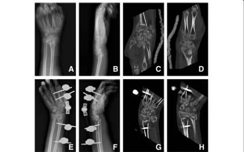
After the initial reduction maneuver, continuous slight traction was applied to maintain the reduction and align-ment. The external fixator (Zengli Medical Instrument Corporation, Hengshui, China) was fixed on the radius with 4-mm Schanz pins and the second or third metacar-pal bone with 3-mm pins (Fig.1). For fractures that were unsatisfactorily reduced or with the significant articular surface collapse or significant displacement of the larger fragment, a small incision was made at the volar side of the distal radius and the periosteum elevator was introduced to elevate the collapsed fragments under direct vision. In cases of bone detect or seriously impacted
fragments, autogenous bone graft or allograft was applied. In most cases, additional average of three K-wires, gener-ally two from the radial side and one from the ulnar side, were used for additional stability.
In the early postoperative period (1–3 days), functional exercises of shoulder, elbow, and finger joints were started to prevent joint stiffness. At 6 weeks after surgery, K-wires and the external fixator were removed and wrist exercises were started. Routine X-ray was taken at the postoperative 3 weeks, 6 weeks, and 3 months to evaluate the status of bone union.
VLP fixation
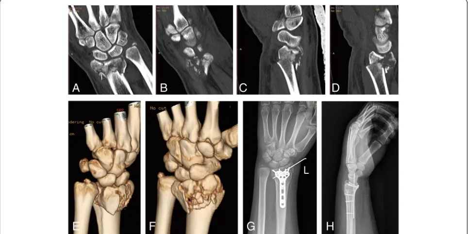
Under local or general anesthesia and control of tourni-quet in supine position, modified Henry approach was used to make a 10–12-cm longitudinal incision along the course of the flexor carpi radialis (FCR). FCR tendon, the flexor pollicis longus tendon, and radial nerve were retracted ulnarly, and brachioradialis and radial blood vessels were retracted radially. Then, pronator quadratus muscle was elevated from its radial origin and retracted ulnarly to expose the fracture fragments. Every fragment was reduced and re-confirmed under the fluoroscopic guidance. As for impacted fragments into the articular surface or metaphysis, periosteum elevator is introduced to elevate the fragments. Autogenous bone graft or allograft was applied to fill the bone detect, if necessary. Temporary fixation with K-wires was used to stabilize the reduced fragments. 2.4 mm or 3.5 mm T-shape
locking plate (Synthes™, Shanghai, China; Wego™, Shan-dong, China; Trauson Medical Instrument Corporation, Jiangsu, China) and screws were placed, with additional K-wires for auxiliary fixation when necessary (Fig. 2). Postoperatively, cast immobilization was applied for 4 weeks. Early motion of the finger, elbow, and shoulder was started on the first postoperative day. At the 15th day, dressings and sutures were removed. At the 29th day, cast and auxiliary K-wires were removed; active and passive wrist rehabilitation begun and gradually strengthened.
Follow-up and postoperative evaluation
The minimum follow-up period was 12 months. The ob-jective functional outcomes were wrist motion range and grip strength. A goniometer was used to measure the wrist flexion, extension, supination, and pronation; a Jamar dynamometer (Jamar, Preston, USA) was used to measure the grip strength. All of these measurements were assessed in comparison with the contralateral uninjured wrist, using percentage as the indicator. The patient-reported DASH score system [20] and the phys-ician-based Gartland–Werley scale [21] were used to indicate the overall functional outcome. DASH question-naire was used to evaluate patients’ ability to perform the daily activities, with a score range from 0, represent-ing no disability, to 100 points representrepresent-ing maximum disability (the higher the score, the more severe the dis-ability). Gartland–Werley scale is a validated
based scoring system, which combines residual deform-ity, subjective findings, the ROM, the postoperative com-plications, and poor finger function. The scale ranges from 0 to 52 points, with a higher score representing poorer outcome.
Standard posteroanterior and lateral radiographs were used to measure volar tilt, radial inclination, radial length, ulnar variance, and articular step-off. The Jupiter criteria were used to judge the presence of arthritic changes on the final radiographs [22]. At each visit (postoperative 2 weeks, 6 weeks, 3 months, 6 months, and 12 months), any potential complication was evalu-ated and documented, either from patients’ self-reports or surgeons’ check-up, including infection, plate/screw loosening, neuropathy or nerve injury, tendon-related issues, loss of reduction, chronic regional pain syn-drome, malunion, nonunion, re-fracture, and others.
Statistical analysis
For comparison of continuous variables (age, surgical duration, volar tilt, radial inclination, radial length, ulnar variance, and articular step-off), Student’s t test or Mann–Whitney U test was used, based on their data distribution status. As for categorical variables (gender, handedness, injury mechanism, complications), Pearson chi-square test or Fisher’s exact test was used, if appro-priate. A p value < 0.05 was considered as statistically significant. All the analyses were performed using the SPSS 21.0 software (IBM, Armonk, NY, USA).
Results
One hundred and seventeen AO type C2/C3 distal frac-tures were treated surgically in 95 patients, from January 2015 to March 2018. Fifty-one patients with 53 fractures were excluded according to our criteria, with 62 frac-tures included for data analysis. The injury mechanism was as follows: fall from standing height (23), motor vehicle collision (14), sports-related injury (7), fall from greater height (6), industrial machinery injury (4), and others (8). Thirty-seven patients had involvement of the right wrist and 25 patients had involvement of the left wrist. Eight (12.9%) patient had concomitant ulnar styloid fracture.
In the VLP group, there were 23 men and 16 women and their average age was 52.0 years. In the EF group, there were 13 men and 10 women, with an average age of 50.5 years. There was no significant difference between both groups with respect to age, gender, side of injured wrist, handedness, injury mechanism, time to operation, bone graft, and duration of follow-up (p> 0.05). The significantly more operative time (105 min vs 92 min,p= 0.023) and greater proportion of fractures requiring K-wire auxiliary fixation (73.9% vs 33.3%) were found in the VLP group, compared to the EF group.(Table1).
Satisfactory reduction (defined as dorsal tilt < 10°, volar tilt < 20°, radial inclination > 10°, radial shortening < 2 mm, and articular step-off < 1 mm at the immediate postoperative X-rays) was achieved in all fractures, viewed from the postoperative immediate X-rays.
The mean follow-up was 17.1 months (range, 12 to 47 months). The results showed that VLP performed better in wrist flexion (69.7° vs 62.3°,p< 0.001), forearm prona-tion (73.1° vs 66.8°, p= 0.027), and supination (70.6° vs 63.1°,p= 0.033) than EF. With respect to other parame-ters (extension, ulnar deviation, and radial deviation), no significant difference was found (p> 0.05) (Table2).
The mean grip strength of the operated wrist was 96% that of the contralateral uninjured wrist in the VLP group, in comparison with 94% in the EF group (p= 0.893) (Table2).
Regarding radiographic parameters, there is no signifi-cant difference for comparison of volar tilt, radial inclin-ation, or radial length (p> 0.05). The ulnar variance at the final radiographs in the VLP group was 0.6 ± 1.3 mm, and in the EF group was 1.6 ± 1.8 mm, indicating a significant difference (p= 0.002). The radial-carpal articular step-off in the VLP group was significantly less than that in the EF group (0.5 ± 1.1 mm vs 1.2 ± 1.4 mm,
p= 0.007) (Table3).
VLP group exhibited a better Gartland–Werley score (2.5 ± 2.7 vs 3.7 ± .2.4) than EF group, although the dif-ference did not approach to statistical level (p= 0.086). As for DASH, both groups exhibited similar excellent performances, scoring 16 ± 12 in the EF group vs 12 ± 15 points in the VLP group (p= 0.162) (Table3).
In the EF group, 9 complications occurred in 8 pa-tients, indicating the incidence of 34.5% (8/23); the pin infection was the most common complication (3, 13.0%), followed by complex regional pain syndrome (2, 8.7%), fixation failure requiring second operation (1, 4.3%), sen-sory branch of radial nerve injury (1, 4.3%), reduce loss requiring re-adjustment (1, 4.3%), and traumatic radial-carpal arthritis (grade 1, by Knirk and Jupiter criteria). In the VLP group, 13 complications in 11 patients were noted, including 4 cases of plate/screw issues (plate prominence, screw too long or penetration), 3 cases of carpal tunnel syndrome, 2 cases of tendon rupture or tendonitis, 2 case of scar hypertrophy, 1 case of complex regional pain syndrome type I (CRPS) which required long-term physiotherapy, and 1 case of superficial wound infection which resolved by antibiotic therapy. The overall rate of complications did not differ between both groups (p= 0.587), although the difference seemed great (34.8% vs 28.2%) (Table4).
Discussion
but no one exhibited overwhelming superiority to the others [3,19, 23–25]. The optimal treatment method of choice is still a controversial issue. In the study, we com-pared the EF and VLP for treatment of type C2/C3 distal radius fractures and demonstrated the superiority of VLP in maintaining joint stability and articular congru-ence and improving joint mobility. However, at the final
follow-up, the overall complications and the functional score based on DASH or Gartland–Werley scale did not differ significantly (p> 0.05).
During the past decade, the use of VLP has gained the most popularity in the treatment of unstable distal radius fractures, due to its advantages. On the one hand, the open manner via volar approach allowed good
Table 1Descriptive comparison of perioperative variables between the EF and VLP group
Variable External fixation Volar locking plate p
n, % or mean ± SD n, % or mean ± SD
Age 50.5 ± 15.2 52.0 ± 14.6 0.372
Gender 0.850
Men 13 56.5 23 59.0
Women 10 43.5 16 41.0
Wrist fractured 0.690
Right 13 56.5 20 51.3
Left 10 43.5 19 48.7
Handedness 0.495
Dominant 15 65.2 22 56.4
Non-dominant 8 34.8 17 43.6
Mechanism 0.380
Fall from standing height 8 34.8 15 38.5
Motor vehicle collision 7 30.4 7 17.9
Sports injury 2 8.7 5 12.8
Fall from height 1 4.3 5 12.8
Industrial machinery injury 3 13.0 1 2.6
Others 2 8.7 6 15.4
Type 0.857
C2 17 73.9 28 71.8
C3 6 26.1 11 28.2
Time to operation (days) 4.5 ± 3.3 5.8 ± 4.0 0.114
Surgical duration (min) 92 ± 36 105 ± 45 0.023
K-wire auxiliary fixation 17 73.9 13 33.3 0.002
Bone graft 7 30.4 11 28.2 0.852
Duration of follow-up 19.0 ± 6.0 16.0 ± 6.4 0.518
Table 2Comparison of wrist ROM and grip strength at the last visit
External fixation VLP fixation
Mean (sd) % of value on contralat. side Mean (sd) % of value on contralat. side
Flexion (deg) 62.3 (7.7) 89.6 69.7 (9.6) 95.6 < 0.001
Extension (deg) 60.2 (11.8) 92.7 61.1 (10.9) 93.7 0.657
Pronation (deg) 66.8 (9.6) 88.3 73.1 (8.7) 94.6 0.027
Supination (deg) 63.6 (8.9) 87.2 70.6 (10.8) 95.2 0.033
Radial deviation (deg) 19.4 (7.7) 90.4 19.7 (6.8) 91.3 0.798
Ulnar deviation (deg) 29.5 (4.6) 91.8 31.0 (5.7) 94.2 0.276
exposure of fracture fragments for easy manipulation, which was more useful in reducing the compressed or impacted fragments. The fixed-angle and locking screw/ hole allowed securing the small fragments and provided better support. By comparison, traction alone in EF might not be effective because ligamentotaxis primarily functioned via strong volar links. This could be used to largely explain the difference of final articular step-off (0.5 mm vs 1.2 mm) and ulnar variance (0.6 mm vs 1.6 mm) between both methods. This result was consistent with the previous reports, where EF or VLP was applied for treatment of AO type C unstable distal radius frac-tures [26, 27]. In these studies, researchers observed a difference value of 0.8 mm (2.2 mm vs 1.4 mm,−0.4 mm vs −1.2 mm) in ulnar variance at the final visit (> 52 weeks). But in the other two randomized studies, no difference was observed, either for articular step-off or ulnar variance [18,28].
We did not observe a significant difference of volar tilt value at the final follow-up, although VLP was superior to EF in correcting volar angulation and afforded enough support via subchondral distal locking screws for a certainly long term (e.g., 12 months). By comparison, loss in volar angulation would continue after removal of the external fixator, from 0.9° at immediate surgery to
4.2° at 6-month follow-up [29]. It should be noted that the small sample size in both groups (23 and 39) should be a concern because of their inadequate ability to detect the true difference in volar tilt, which likely leads to a type II statistical error. If adequate sample size was provided, the advantages of VLP in correction and main-tenance of radiographic parameters would be more statistically prominent.
Most previous studies have demonstrated the advantages of VLP over EF in functional recovery at the early postoper-ative period (< 3 months) [19, 26, 30]. But as for the mid-or long-term period results, controversies exist. In a retrospective cohort of 115 patients with AO type C2/C3 fractures, Richard et al. [10] demonstrated the better DASH score and more improved pronation/supination arc in VLP group at postoperative 12 months. Williksen et al. [26] conducted a RCT study of 104 AO type C fractures and did not observe the significant difference of DASH score and other functional parameters between the two groups, but only the better wrist supination (90° vs 76°) in the VLP group. In a study of 69 AO type C fractures, wrist and pronation range was significantly more improved in the VLP group, while Gartland–Werley or patient-rated wrist evaluation (PRWE) score was non-significantly different [19]. In this study, VLP showed a significantly better per-formance in wrist flexion, pronation, and supination (p< 0.05). This might be attributed to the facts that VLP fix-ation allowed an earlier wrist mobilizfix-ation and maintained improved anatomic parameter until fracture union. We suggested the former one (improved anatomic parameters) should be a more important contributing factor, because in this study, VLP fixation did not maximize the benefit of allowing early-period wrist mobilization (approximately at 29th day) and furthermore prolonged casting after plate fixation did not decrease wrist motion [31].
Despite the advantages, VLP could not be applied in some fracture types, for example, the comminuted very distal fractures or osteoporotic fractures that do not allow
Table 3Comparison of radiographic parameters, functional
outcomes, and complications between the two groups at the last visit (> 12 months)
Variable External fixation VLP p
Volar tilt (deg) 4.9 (5.3) 5.5 (6.1) 0.317
Radial inclination (deg) 20.8 (3.5) 22.2 (4.1) 0.538
Radial height (mm) 10.8 (1.7) 10.4 (1.6) 0.693
Ulnar variance (mm) 1.6 (1.8) 0.6 (1.3) 0.002
Articular step-off (mm) 1.2 (1.4) 0.5 (1.1) 0.007
DASH (points) 16 (12) 12 (15) 0.162
Gartland–Werley score (points) 3.7 (2.4) 2.5 (2.7) 0.086
Table 4Complications in both groups
External fixation,n(%) VLP,n(%) p
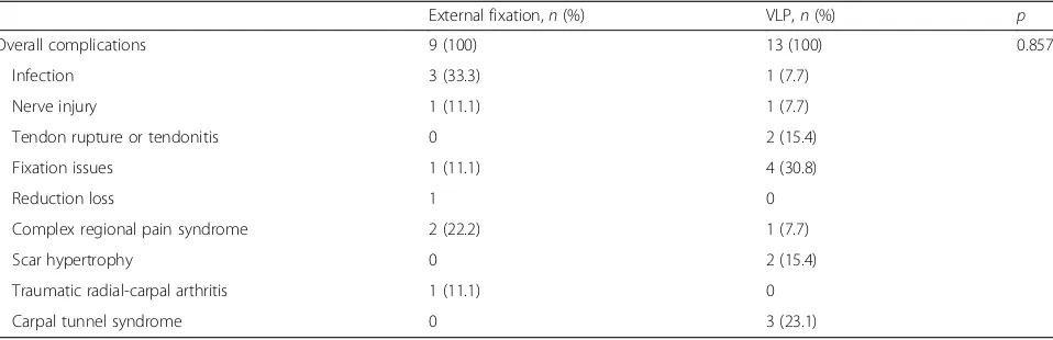
Overall complications 9 (100) 13 (100) 0.857
Infection 3 (33.3) 1 (7.7)
Nerve injury 1 (11.1) 1 (7.7)
Tendon rupture or tendonitis 0 2 (15.4)
Fixation issues 1 (11.1) 4 (30.8)
Reduction loss 1 0
Complex regional pain syndrome 2 (22.2) 1 (7.7)
Scar hypertrophy 0 2 (15.4)
Traumatic radial-carpal arthritis 1 (11.1) 0
screw insertion [32]. In such fractures, K-wireaugmented EF could be a better choice, which more likely yields successful results. The continuous distraction of external fixator, together with additional K-wires to secure the comminuted fragments and bone graft to fill the defect, could provide greater stability. In this study, over 70% of EF was supplemented by K-wires, and 30% of cases required bone graft to fill the bone defect, and indeed, these combinations demonstrated the excellent or good functional and radiographic outcomes [27,33].
With respect to complications, we did not find a sig-nificant difference in the incidence rate, 34.5% in the EF group and 28.2% in the VLP group, both of which was in range of the reported figures [10,18,19, 26, 33]. Cao et al. [33] retrospectively reviewed 226 type C3 distal radius fractures treated by external fixator in the elderly patients, and reported a rate of 18.6% (42/226) for over-all complications, 10% for loss of reduction, 6.2% for joint stiffness, 2.2% for traumatic arthritis, and 0.5% for pin-tract infection. Richard et al. [10] reported a signifi-cantly higher overall rate of complications in the EF group (52.5%, 31/59) than that in the VLP group (25%, 14/56). In a meta-analysis of 9 RCTs, Esposito et al. [34] concluded the significantly higher incidence of overall complications or infection in EF over VLP, but non-significant for re-operation, osteoarthritis, malu-nion, nerve deficit, complex regional pain syndrome, painful retained hardware requiring removal, carpal tunnel syndrome, stiffness, tendon rupture, or tendon-itis. It is often difficult to compare these reported varied figures due to differences in study design, patient characteristics, data collection, and follow-up period. On the other hand, complications from physician re-ports and patient rere-ports are different, with the former emphasizing check-up related complications while the latter often take symptoms as the major concern. McKay et al. [35] suggested not all suboptimal results should be considered as complications, unless it is at-tributable to a specifically diagnosed complication; the authors proposed a complication checklist to improve prospective data collection.
There were several limitations to this study. Firstly, the retrospective design was its inherent limitation in accuracy of data collection. Secondly, the procedures in this study were performed by 14 surgeons and the sur-gical choice was mainly dependent on surgeons’ prefer-ence, both of which might affect the outcomes. Thirdly, the patient’s pre-injury baseline radiographic parameters or functional state, potential factors influ-encing the comparative results and the recovery process, could not be examined. Fourthly, the sample size was small due to the scarcity of this complex in-jury, which might lead to a type II statistical error when evaluating some variables.
Conclusion
In summary, VLP fixation demonstrated its better per-formance in wrist mobility (wrist flexion, pronation, and supination), correction of ulnar variance, and improving articular congruence. As for DASH or Gartland–Werley score, other radiographic or functional parameters, and complications, both fixation methods showed similar results. Future research with better design and large sample is needed to verify our results and explore poten-tial contributors that influence the adverse results or complications.
Abbreviations
AO:Arbeitsgemeinschaftfür Osteosynthesefragen; CRPS: Complex regional pain syndrome; CT: Computed tomography; DASH: Disabilities of the arm, shoulder, and hand scale; EF: External fixation; FCR: Flexor carpi radialis; RCT: Randomized controlled trail; VLP: Volar locking plate
Acknowledgements
We are grateful to Y Lin and B Zhang of the Department of Orthopedics and to Q Zhan of the Department of Statistics and Applications for their kind assistance.
Authors’contributions
YY designed the study. YB and TZ followed up the patients and documented the data. XS analyzed and interpreted the data. XY wrote the manuscript, and YY approved the final version of the manuscript. All authors read and approved the final manuscript.
Funding
No organization sponsored the research.
Availability of data and materials
All the data will be available upon motivated request to the corresponding author of the present paper.
Ethics approval and consent to participate
This study was approved by the ethics committee of The Third Hospital of Hebei Medical University. Informed consent was obtained from all the participants.
Consent for publication
Written informed consent was obtained from each patient to authorize the publication of their data.
Competing interests
The authors declare that they have no competing interests.
Received: 1 July 2019 Accepted: 7 August 2019
References
1. Zhang Y. Clinical epidemiology of orthopaedic trauma. Stuttgart: Thieme; 2016. 2. Pruitt DL, et al. Computed tomography scanning with image reconstruction
in evaluation of distal radius fractures. J Hand Surg Am. 1994;19(5):720–7. 3. Rein S, et al. Results of dorsal or volar plate fixation of AO type C3 distal
radius fractures: a retrospective study. J Hand Surg Am. 2007;32(7):954–61. 4. Crisco JJ, et al. Effects of distal radius malunion on distal author proof
radioulnar joint mechanics—an in vivo study. J Orthop Res. 2010;25(4):547–55. 5. Deng Y, Zhang Q, Wang Q, et al. Combination of volar buttress plate with
external fixatior for the distal radius fractures of type C3 caused by high-energy injuries. China J Orthop Trauma. 2009;22(7):543–6.
6. Md MAOM, et al. Anatomic and x-ray film studies of the distal articular surface of the radius. J Hand Surg. 1996;21(4):567.
7. Bentohami A, et al. Complications following volar locking plate fixation for distal radial fractures: a systematic review. J Hand Surg Eur Vol. 2014;39(7):745–54. 8. Disseldorp DJ, et al. Dorsal or volar plate fixation of the distal radius: does
9. Kreder HJ, et al. Indirect reduction and percutaneous fixation versus open reduction and internal fixation for displaced intra-articular fractures of the distal radius: a randomised, controlled trial. J Bone Joint Surg Br. 2005;87(6):829–36.
10. Richard MJ, et al. Analysis of the complications of palmar plating versus external fixation for fractures of the distal radius. J Hand Surg Am. 2011; 36(10):1614–20.
11. Reynolds PR, Beredjiklian PK. External fixation of distal radius fractures: do benefits outweigh complications? Curr Opin Orthop. 2001;12(4):286–9. 12. Fu Q, et al. Volar locking plate versus external fixation for distal radius
fractures: a meta-analysis of randomized controlled trials. Indian J Orthop. 2018;52(6):602–10.
13. Gouk CJC, et al. Volar locking plate fixation versus external fixation of distal radius fractures: a meta-analysis. J Hand Surg Eur Vol. 2018;43(9):954–60. 14. Walenkamp MM, et al. Functional outcome in patients with unstable distal
radius fractures, volar locking plate versus external fixation: a meta-analysis. Strategies Trauma Limb Reconstr. 2013;8(2):67–75.
15. Chou YC, et al. Dorsal and volar 2.4-mm titanium locking plate fixation for AO type C3 dorsally comminuted distal radius fractures. J Hand Surg Am. 2011;36(6):974–81.
16. Earp BE, Foster B, Blazar PE. The use of a single volar locking plate for AO C3-type distal radius fractures. Hand. 2015;10(4):649–53.
17. Cao J, Shen G, Lu J, et al. Volar locking compression plate combined with external fixation for treatment of AO type C3 distal radius fractures. Clin Med China. 2013;29:118–20.
18. Jeudy J, et al. Treatment of complex fractures of the distal radius: a prospective randomised comparison of external fixation‘versus’locked volar plating. Injury. 2012;43(2):174–9.
19. Kumbaraci M, et al. Retrospective comparison of external fixation versus volar locking plate in the treatment of unstable intra-articular distal radius fractures. Eur J Orthop Surg Traumatol. 2014;24(2):173–8.
20. Hudak PL, et al. Development of an upper extremity outcome measure: the DASH (disabilities of the arm, shoulder, and head). Am J Ind Med. 1996; 29(6):602-608.
21. Von RJ, et al. Characteristics of two different locking compression plates in the volar fixation of complex articular distal radius fractures. Bone Joint Res. 2012;1(6):111–7.
22. Shin EK, Jupiter JB. Radioscapholunate arthrodesis for advanced degenerative radiocarpal osteoarthritis. Tech Hand Up Extrem Surg. 2007;11(3):180. 23. Disseldorp DJG, et al. Dorsal or volar plate fixation of the distal radius: does
the complication rate help us to choose? Jnl Wrist Surg. 2016;05(03):202–10. 24. Hull P, et al. Volar locking plates versus K-wire fixation of dorsally displaced
distal radius fractures--a functional outcome study. J Trauma Acute Care Surg. 2011;70(6):125–8.
25. Karantana A, et al. Surgical treatment of distal radial fractures with a volar locking plate versus conventional percutaneous methods: a randomized controlled trial. J Bone Joint Surg A. 2013;95(19):1737–44.
26. Williksen JH, et al. Volar locking plates versus external fixation and adjuvant pin fixation in unstable distal radius fractures: a randomized, controlled study. J Hand Surg Am. 2013;38(8):1469–76.
27. Gereli A, Nalbantoglu U, Kocaoglu B, Turkmen M. Comparison of palmar locking plate and K-wire augmented external fixation for intra-articular and comminuted distal radius fractures. Acta Orthop Traumatol Turc. 2010;44(3):212–9.
28. Roh YH, et al. A randomized comparison of volar plate and external fixation for intra-articular distal radius fractures. J Hand Surg Am. 2015;40(1):34–41. 29. Dicpinigaitis P, Wolinsky P, Hiebert R, Egol K, Koval K, Tejwani N. Can
external fixation maintain reduction after distal radius fractures? J Trauma. 2004;57(4):845–50.
30. Wilcke MK, Abbaszadegan H, Adolphson PY. Wrist function recovers more rapidly after volar locked plating than after external fixation but the outcomes are similar after 1 year. Acta Orthop. 2011;82(1):76–81. 31. Lozano-Calderon SA, et al. Wrist mobilization following volar plate fixation
of fractures of the distal part of the radius. J Bone Joint Surg Am. 2008;90(6): 1297–304.
32. Fu YC, et al. Use of an external fixation combined with the buttress-maintain pinning method in treating comminuted distal radius fractures in osteoporotic patients. J Trauma. 2006;60(2):330–3.
33. Cao F, Zhou W, Xia T, Liu M, Liu Y, Liu J, Liu G, et al. Diagnosis and treatment of complications following trans-articular external fixation for elderly patients with distal radius fracture of type C3. Chin J Orthop Trauma. 2018;20(11):960–3.
34. Esposito J, et al. External fixation versus open reduction with plate fixation for distal radius fractures: a meta-analysis of randomised controlled trials. Injury. 2013;44(4):409–16.
35. Mckay SD, et al. Assessment of complications of distal radius fractures and development of a complication checklist. J Hand Surg. 2001;26(5):916–22.
Publisher’s Note