Volume 2011, Article ID 748194,4pages doi:10.1155/2011/748194
Case Report
Primitive Neuroectodermal Tumor of the Liver: A Case Report
Eduardo Cambruzzi,
1Enilde Eloena Guerra,
2Hamilton Cardoso Hilgert,
2Herbert Jorge Schmitz,
2Vin´ıcius Lopes Silva,
2Daniel Marini Milani,
2Ricardo Pedrini Cruz,
2and Raul Pruinelli
21Department of Pathology, Nossa Senhora da Conceic¸˜ao Hospital, Porto Alegre, Rio Grande do Sul, Brazil 2Department of Surgery, Nossa Senhora da Conceic¸˜ao Hospital, Porto Alegre, Rio Grande do Sul, Brazil
Correspondence should be addressed to Eduardo Cambruzzi,[email protected]
Received 5 September 2011; Revised 26 September 2011; Accepted 29 September 2011
Academic Editor: Estella M. Matutes
Copyright © 2011 Eduardo Cambruzzi et al. This is an open access article distributed under the Creative Commons Attribution License, which permits unrestricted use, distribution, and reproduction in any medium, provided the original work is properly cited.
Primary liver sarcomas represent a rare group of neoplasias, with angiosarcoma being the most common histological type. Primi-tive neuroectodermal tumor (PNET) represents a high malignant neoplasia that usually affects the central nervous system and soft tissues. An 18-year-old male patient was admitted with clinical complains of pain in the right upper abdominal quadrant. The clin-ical evaluation revealed a solid mass in the right hepatic lobe. On the gross examination of the resected liver specimen, the right lobe of the liver was replaced by a yellow-red solid mass measuring 21 cm in its largest dimension. On the histopathology, a tu-mor composed of small round blue cells with little cytoplasm and round nuclei was identified. The lesion revealed positive immu-noexpression for vimentin and CD99 and negative immunostaining for desmin, CD45, cytokeratin, and neuroblastoma protein, suggesting, then, the diagnosis of PNET. Although it is an unusual tumor, it should be considered in the differential diagnosis of liv-er masses, especially in young patients.
1. Introduction
Liver neoplastic lesions include many different histological
types of primary benign and malignant masses and high rates of metastatic processes. Primary tumors can be solid or cystic and can arise from hepatocyte, bile duct epithelium, neu-roendocrine cells, mesenchymal cells, and, rarely, from
heter-otopic tissues [1,2]. Hepatoblastoma and mesenchymal
ha-martoma are usually found in the pediatric population. He-patocellular carcinoma represent the single most common histologic type of malignant epithelial tumors of the liver (about 85–90%), being frequently associated to cirrhosis and chronic viral hepatitis. Primary hepatic sarcomas are exceed-ingly unusual, accounting for only 1% to 2% of all malignant tumors arising in the liver, with angiosarcoma and undiffer-entiated sarcoma being the most common histologic types
[1,2].
PNET represents a family of tumors which shows varying
degrees of neuronal differentiation with an Ewing’s sarcoma
gene rearrangement, most often as a consequence of a t (11; 22) (q24; q12) chromosomal translocation. PNET is a
high-ly malignant neoplasm most commonhigh-ly involving the central nervous and skeletal system, and it is composed of small, round, uniform cells. Because of the undifferentiated appear-ance of the tumor cells, it looks as if the original cell might be an undifferentiated mesenchymal cell [3–5]. Herein, the au-thors report a case of PNET arising in the liver and review the morphologic and immunohistochemical findings of this tu-mor in a rare topography.
2. Case Report
2 Case Reports in Medicine
(a) (b)
Figure 1: Preoperative CT scan showing a large, solid mass in the liver.
Figure 2: PNET arising in the liver: diffuse sheet of uniform small
round cells in a lobulated growth pattern, HE, 200x.
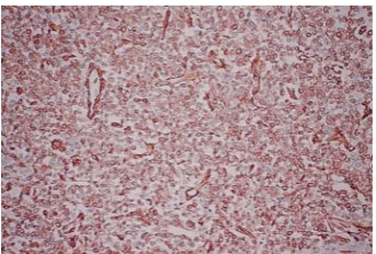
Figure 3: PNET arising in the liver: positive immunoexpression for
vimentin, streptavidin-biotin, 200x.
In follow-up CT, areas of partial necrosis and no tumor size regression were found. The patient underwent exploratory laparotomy. The right lobe of the liver was partially replaced by a yellow-purple-red solid tumor, which was in continuity with the right kidney and hepatic colon flexure. There were no signs of peritoneal disease and no evidence of metastasis in liver segments 1 to 3. A right extended hepatectomy with en bloc resection of the right kidney, gallbladder, and partial
Figure 4: PNET arising in the liver: the tumor cells show
immunoreactivity for CD99, streptavidin-biotin, 200x.
colectomy (hepatic colon flexure) with primary anastomosis was performed.
The surgical specimen consisted of a portion of the liv-er, of the gallbladdliv-er, a segment of the colon, and of the right kidney, previously fixed in formalin, measuring 30 cm
×25 cm×13.5 cm, and weighing 4313 g. On cut surface, the
thrombosis extending from the iliac vessels to the right atri-um and multiple metastatic implants in the lungs were iden-tified in thoracic CT. The patient had a sudden death due to massive pulmonary thromboembolism. At this time, it was not possible to perform genetic studies to establish the line-age of the neoplasm.
3. Discussion
PNETs are small round cell tumors that belong to the Ewing’s sarcoma family of tumors. These tumors are divided in two main categories: central (derived from the neural tube) and peripheral (outside the central nervous system) [6–10].
PNETs were first described by Stout in 1918 [7, 10, 11]
and represent less than 1% of all sarcomas [7,12]. These
sarcomas predominantly affect bones and deep soft tissue and rarely affect visceral organs. PNET can be found in the thorax (44%), abdomen and pelvis (44%), extremities
(20%), and 6% in the head and neck areas [9,12]. Visceral
primary commitment has been described with increasing frequency in the pancreas, vagina, stomach, small bowel, ovaries, esophagus, and kidney [5–10].
The gross appearance of the tumor varies. In general, it is multilobulated, soft, and friable; it rarely exceeds 10 cm in its largest dimension. Its cut surface has a gray-yellow or gray-tan appearance, often with large areas of necrosis, cyst formation, or hemorrhage. Despite the extensive necrosis,
calcification is rare [4,5,13].
At microscopy, the typical PNET shows a predomi-nantly lobular growth pattern, with little or no stroma. It is composed of poorly differentiated small round cells containing darkly staining, round or oval nuclei. The pale eosinophilic cytoplasm is indistinct except in areas where the cells are more mature and the elongated hair-like cytoplasmic extensions coalesce to form rosettes. Most of the rosettes are similar to those seen in neuroblastomas and contain a central solid core of neurofibrillary material. Rarely do the rosettes contain a central lumen or vesicle. Some tumors are composed of cords or trabeculae of small round cells that can
be mistaken by a carcinoid tumor or a small-cell undiff
eren-tiated carcinoma. The presence of areas of necrosis is a usual finding. There are uncommon tumors with glial, ependymal,
cartilaginous, or epithelial differentiation. Some areas can
resemble a fibrosarcoma or a malignant schwannoma [4,
5,13,14]. The ultrastructural features include the presence
of elongated cell processes that interdigitate with each other and contain small dense-core granules (neurosecretory granules) that measure 50–100 nm and occasionally contain
microtubules [14,15]. On immunostains, many PNETs are
usually positive for CD99, neuron-specific enolase, Leu-7, S-100, and synaptophysin, and negative for desmin and Myogenin/MyoD1. Some cases are positive for cytokeratin
[3–5,14–16]. PNET characteristically exhibits a neural
phe-notype, expressing the MIC2-protein (CD99), and display the t (11; 22) (q24; q12) chromosomal translocation in about
85%–95% of the cases [3,7,8,17–22].
The differential diagnosis includes neuroblastoma (pos-itive for NB84), desmoplastic small round cell tumor, mes-enchymal chondrosarcoma, alveolar rhabdomyosarcoma,
non-Hodgkin’s lymphoma, metastatic pulmonary small-cell carcinoma, cutaneous neuroendocrine carcinoma, small-cell osteosarcoma, and poorly differentiated synovial sarcoma
[3–5,13,18–20,22].
Treatment with surgery alone for extraskeletal PNET
is considered insufficient, and multimodal treatment with
chemotherapy and radiotherapy is frequently performed. Despite the multimodal treatment, most patients show a rapid dissemination of the disease. Key prognostic factors that adversely influence the outcome are the presence of me-tastatic disease at the time of the initial diagnosis, large tu-mor size, extensive necrosis, central axis tutu-mors, and poor re-sponse to initial chemotherapy. Patients with type 1 EWS/FLI fusion transcripts appear to have longer disease-free survival
than those with other fusion transcript types [3,5,6,19–21].
4. Conclusion
PNETs are usually aggressive small blue cell tumors arising in the central nervous or skeletal system. To the best of our knowledge, this is the third case report of this tumor arising in liver parenchyma. Although it is an unusual liver tumor associated with poor prognosis, PNET should be considered in the differential diagnosis of liver masses, especially in young patients.
References
[1] R. N. M. MacSween, A. D. Burt, B. C. Portmann, K. G. Ishak, P. J. Scheuer, and P. P. Anthony, Pathology of the Liver, Churchill Livingstone, London, UK, 4th edition, 2002.
[2] K. G. Ishak, P. P. Anthony, and L. H. Sobin, Histological
Typ-ing of Tumors of the Liver. World Health Organization Inter-national Histological Classification of Tumors, Springer, Berlin,
Germany, 2nd edition, 1994.
[3] K. K. Unni, C. Y. Inwards, J. A. Bridge, L. G. Kindblom, and L. E. Wold, “Tumors of the bones and joints,” in AFIP Atlas of
Tu-mor Pathology, vol. 2, pp. 209–222, ARP Press, Silver Spring,
Md, USA, 4th edition, 2005.
[4] F. Vakar-L ´opez, A. G. Ayala, A. K. Raymond, and B. Czerniak, “Epithelial phenotype in Ewing’s sarcoma/primitive neuroec-todermal tumor,” International Journal of Surgical Pathology, vol. 8, no. 1, pp. 59–65, 2000.
[5] S. Movahedi-Lankarani, R. H. Hruban, W. H. Westra, and D. S. Klimstra, “Primitive neuroectodermal tumors of the pan-creas,” The American Journal of Surgical Pathology, vol. 26, no. 8, pp. 1040–1047, 2002.
[6] R. B. Colovic, N. M. Grubor, M. T. Micev, S. V. Matic, H. D. E. Atkinson, and S. M. Latincic, “Perigastric extraskeletal Ewing’s sarcoma: a case report,” World Journal of Gastroenterology, vol. 15, no. 2, pp. 245–247, 2009.
[7] T. Welsch, G. Mechtersheimer, S. Aulmann et al., “Huge primi-tive neuroectodermal tumor of the pancreas: report of a case and review of the literature,” World Journal of Gastroenterology, vol. 12, no. 37, pp. 6070–6073, 2006.
[8] H. Doi, S. Ichikawa, A. Hiraoka et al., “Primitive neuroecto-dermal tumor of the pancreas,” Internal Medicine, vol. 48, no. 5, pp. 329–333, 2009.
4 Case Reports in Medicine
neuroectodermal tumors: a clinicopathologic and molecular study,” Human Pathology, vol. 32, no. 10, pp. 1109–1115, 2001. [10] J. Y. Park, S. Lee, H. J. Kang, H. S. Kim, and S. Y. Park, “Prima-ry Ewing’s sarcoma-primitive neuroectodermal tumor of the uterus: a case report and literature review,” Gynecologic
Onco-logy, vol. 106, no. 2, pp. 427–432, 2007.
[11] A. P. Stout, “A tumor of the ulnar nerve,” Proceedings of New
York Pathological Society, vol. 12, pp. 2–12, 1918.
[12] A. Ousadden, K. Mazaz, A. Amraoui, F. Kettani, M. C. Chef-chaouni, and K. Ait Taleb, “Primary hepatic localization of the PPNET (primitive peripheral neuroectodermal tumors). Case report,” Annales de Chirurgie, vol. 130, no. 4, pp. 254– 256, 2005.
[13] L. P. Dehner, “Primitive neuroectodermal tumor and Ewing’s sarcoma,” The American Journal of Surgical Pathology, vol. 17, no. 1, pp. 1–13, 1993.
[14] Y. Hachitanda, M. Tsuneyoshi, M. Enjoji, A. Nakagawara, and K. Ikeda, “Congenital primitive neuroectodermal tumor with epithelial and glial differentiation: an ultrastructural and immunohistochemical study,” Archives of Pathology and
Laboratory Medicine, vol. 114, no. 1, pp. 101–105, 1990.
[15] S. Ushigome, T. Shimoda, K. Takaki et al., “Immunocyto-chemical and ultrastructural studies of the histogenesis of Ewing’s sarcoma and putatively related tumors,” Cancer, vol. 64, no. 1, pp. 52–62, 1989.
[16] A. Llombart-Bosch, M. J. Terrier-Lacombe, A. Peydro-Olaya, and G. Contesso, “Peripheral neuroectodermal sarcoma of soft tissue (peripheral neuroepithelioma): a pathologic study of ten cases with differential diagnosis regarding other small, round-cell sarcomas,” Human Pathology, vol. 20, no. 3, pp. 273–280, 1989.
[17] E. de Alava and W. L. Gerald, “Molecular biology of the Ew-ing’s sarcoma/primitive neuroectodermal tumor family,”
Jour-nal of Clinical Oncology, vol. 18, no. 1, pp. 204–213, 2000.
[18] B. H. Kushner, S. I. Hajdu, S. C. Gulati, R. A. Erlandson, P. R. Exelby, and P. H. Lieberman, “Extracranial primitive neuroec-todermal tumors: the memorial sloan-kettering cancer center experience,” Cancer, vol. 67, no. 7, pp. 1825–1829, 1991. [19] M. C. Le Deley, O. Delattre, K. L. Schaefer et al., “Impact of
EWS-ETS fusion type on disease progression in Ewing’s sar-coma/peripheral primitive neuroectodermal tumor: prospec-tive results from the cooperaprospec-tive Euro-E.W.I.N.G. 99 trial,”
Journal of Clinical Oncology, vol. 28, no. 12, pp. 1982–1988,
2010.
[20] J. A. van Doorninck, L. Ji, B. Schaub et al., “Current treatment protocols have eliminated the prognostic advantage of type 1 fusions in Ewing sarcoma: a report from the Children’s Oncology Group,” Journal of Clinical Oncology, vol. 28, no. 12, pp. 1989–1994, 2010.
[21] E. De Alava, C. Antonescu, A. Panizo et al., “Why do Ewing’s sarcomas with EWS/FLI-1 type 1 transcripts behave better?”
Modern Pathology, vol. 11, article 981A, 1998.
[22] S. Mani, D. Dutta, and K. de Binay, “Primitive neuroecto-dermal tumor of the liver: a case report,” Japanese Journal of
Submit your manuscripts at
http://www.hindawi.com
Stem Cells
International
Hindawi Publishing Corporationhttp://www.hindawi.com Volume 2014
Hindawi Publishing Corporation
http://www.hindawi.com Volume 2014
INFLAMMATION
Hindawi Publishing Corporation
http://www.hindawi.com Volume 2014
Behavioural
Neurology
Endocrinology
International Journal ofHindawi Publishing Corporation
http://www.hindawi.com Volume 2014
Hindawi Publishing Corporation
http://www.hindawi.com Volume 2014
Disease Markers
Hindawi Publishing Corporation
http://www.hindawi.com Volume 2014
BioMed
Research International
Oncology
Journal of Hindawi Publishing Corporationhttp://www.hindawi.com Volume 2014
Hindawi Publishing Corporation
http://www.hindawi.com Volume 2014
Oxidative Medicine and Cellular Longevity
Hindawi Publishing Corporation
http://www.hindawi.com Volume 2014
PPAR Research
The Scientific
World Journal
Hindawi Publishing Corporationhttp://www.hindawi.com Volume 2014
Immunology Research
Hindawi Publishing Corporation
http://www.hindawi.com Volume 2014
Journal of
Obesity
Journal ofHindawi Publishing Corporation
http://www.hindawi.com Volume 2014
Hindawi Publishing Corporation
http://www.hindawi.com Volume 2014
Computational and Mathematical Methods in Medicine
Ophthalmology
Journal ofHindawi Publishing Corporation
http://www.hindawi.com Volume 2014
Diabetes Research
Journal ofHindawi Publishing Corporation
http://www.hindawi.com Volume 2014
Hindawi Publishing Corporation
http://www.hindawi.com Volume 2014
Research and Treatment
AIDS
Hindawi Publishing Corporation
http://www.hindawi.com Volume 2014
Gastroenterology Research and Practice
Hindawi Publishing Corporation
http://www.hindawi.com Volume 2014
Parkinson’s
Disease
Evidence-Based Complementary and Alternative Medicine
Volume 2014