Thoracic Chondrosarcoma
Kalliopi Alpantaki,
1George Datsis,
1Odysseas Zoras,
2Alkisti Kampouroglou,
1Ioannis Drositis,
3George Halkiadakis,
2, 3and Pavlos Katonis
11Departments of Orthopaedics and Traumatology, University Hospital of Heraklion, Voutes, Heraklion, 71003 Crete, Greece 2Department of Surgery, University Hospital of Heraklion, Voutes, Heraklion, 71003 Crete, Greece
3Department of Thoracic Surgery, University Hospital of Heraklion, Voutes, Heraklion, 71003 Crete, Greece
Correspondence should be addressed to Pavlos Katonis,katonis@hol.gr
Received 11 December 2010; Accepted 10 March 2011
Academic Editor: Shaji Kumar
Copyright © 2011 Kalliopi Alpantaki et al. This is an open access article distributed under the Creative Commons Attribution License, which permits unrestricted use, distribution, and reproduction in any medium, provided the original work is properly cited.
Chondrosarcomas of the spine are rare and difficult to treat. In this paper a case of thoracic chondrosarcoma is presented. Chondrosarcomas of the spine are generally smaller, more difficult to excise and are followed by higher local recurrence compared with chondrosarcomas of the peripheral skeleton. The tumor is radio- and chemoresistant, making the surgical treatment of utmost importance. The most important prognostic factor for local control is wide or marginal tumor resection. Our patient was treated in two stages, with total excision of the tumor, using cryosurgery. Liquid nitrogen was used to freeze the damaged tissue at a cellular level and made the excision more efficient.
1. Introduction
Chondrosarcoma is the most frequent primary malignant bone tumour. It constitutes a family of malignant tumors in which the cells differentiate into cartilage and may originate from healthy bones (primary) or from chondromas or osteo-chondromas with sarcomatous degeneration (secondary). Chondrosarcomas are classified into three categories: central, peripheral, and periostal as well as two rare entities: mes-enchymal chondrosarcoma and clear-cell chondrosarcoma. Primary chondrosarcoma of the spine is an extremely rare tumor. Only six to ten percent of chondrosarcomas arise from the mobile spine. The maximum incidence of chondrosarcoma is found in adult, male patients with a tumor diameter being generally greater than 5 cm.
Radiographically it is identified as an area of bone destruction and a calcified soft tissue mass. The soft tissue mass may be absent in lesions originating from a vertebral body. Magnetic resonance imaging (MRI) can best demon-strate the extent of the lesion and of the cord compression. Usually chondrosarcoma does not manifest signs of cord compression at early onset [1].
Chondrosarcoma of the spine is challenging to manage. The clinical course of chondrosarcomas originating from the spine is usually long because of the slow growth of the tumour [1]. Local recurrence and metastases, resistant to most protocols of radiation therapy and chemotherapy, may occur in more than 10 years after surgical removal. Surgical excision has been advocated as the primary mode of therapy for treating chondrosarcoma. For best results (longer survival, lower rate of recurrence) a more aggressive surgical approach is preferred [1–7].
We report a case of thoracic chondrosarcoma, which was treated with total resection, using cryosurgrery. Cryosurgery is found to be effective against a localized disease and solid tumors larger than 1 cm. Liquid nitrogen was used to freeze the damaged tissue at a cellular level and form ice crystals inside the cells of the tumor making the excision more efficient and with lower rates of side effects. More damage to the tumor occurs when blood vessels supplying the diseased tissue freeze.
Figure 1: The posteroanterior chest radiograph shows a
well-defined radiopaque lesion.
chondrosarcoma of the spine using cryosurgery [10–14]. It is a field that should be reported and studied in depth.
2. Case Presentation
A 3year-old male consulted our department with a 7-month history of mild thoracic spine pain after a fall; in addition to the thoracic spine pain he noticed a painful, slowly growing mass. He had no past medical history, and physical examination was unremarkable except for the presence of a hard and tender thoracic spine mass. His father died of hepatic cancer. Clinical blood examination did not show any abnormality, including alkaline phosphatase.
Initial posteroanterior chest radiograph revealed the presence of a left-sided soft tissue mass located at the level of the aortic arch (Figure 1). The lesion’s margins and the absence of a positive “silhouette” sign with the mediastinal structures indicated an extrapulmonary and posterior position, a finding that was verified in the lateral chest radiograph. A CT examination was subsequently per-formed upon request, which showed a predominantly soft tissue tumour with internal areas of mineralisation. The mineralised elements were amorphous, scattered, ring-like, and arcuate-shaped. Rib destruction and infiltration was also noted. Furthermore, widening of the adjacent neural foramina was depicted and an MRI examination was done to clarify this finding. MRI verified neural foramina invasion as well as significant spinal cord compression and contralateral displacement. Despite that fact, the patient remained com-pletely asymptomatic, which was attributed to an indolent and slow growth of the tumour. The mass showed low signal intensity in T1-w images and heterogeneous but predomi-nantly high signal intensity in T2-w images. An associated superficial component of the tumour, which corresponded to the palpable finding, was also better demonstrated on MRI
(Figure 2). Mineralised areas that were depicted in the CT
examination demonstrated low signal intensity in all pulse sequences. Gadolinium enhanced T1-w fat saturated images showed intense, heterogeneous tumour enhancement. The mass extended from the anterior to the left of the vertebral body at T3–T6 measuring 7.5×6×5.5 cm and in the left
A H
P F
(a)
A H
P F
(b)
A
R L
P
(c)
Figure 2: The sagittal T1-w (a) MR image shows a hypointense
lobulated lesion. The mass demonstrates heterogeneous but pre-dominantly high signal intensity in the sagittal T2-w. (b) MR image. Note the superficial palpable component of the tumor. (c) The T2 axial view shows how the tumor is positioned next to the ventral structures and its relationship to the spinal cord.
(a) (b)
Figure 3: The postoperative plain X-ray.
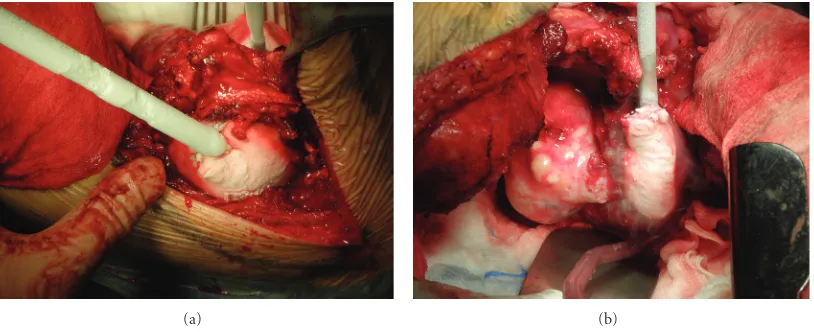
(a) (b)
Figure 4: The second stage of the procedure. The tumour had 3 lobes and was treated with liquid nitrogen (−17◦C for 15 min/lobe). The
equipment used was Candela CS5 (Spembly Medical).
The surgical procedure was conducted in two stages. Posterior then anterior approaches were planned. For the first stage, the patient was placed prone and an incision was made in the midline extending from C5 to T7. The spinous process and bilateral inferior facets of T3 were removed. The posterior bony elements of T4 and T5 were removed. Instrumentation was placed from C5 to C7 and T3, T6, T7 and fixed with pedicle screws. A combination of Vertex and CD Horizon system (Metronic) of posterolateral fixation was used. Fusion was then accomplished by decorticating the transverse processes and an autograft with bone chips was applied after the exposed dura had been protected (Figure 3). The second stage of the procedure was performed 3 months after the first stage. The patient was placed in a lateral position with his right side down; surgical excision of the 5th allowed a large thoracotomy. The tumour had 3 lobes and was treated with liquid nitrogen (−17◦C for 15 min/lobe). The equipment used was Candela CS5 (Spembly Medical) with up to 5 probes, which can be sterilized and reused. We used 3.5 and 10 mm rod-shaped and a plate probe (for small superficial tumors) with liquid N2 (55 psi) which can
reach –196◦C (tissue temperature is usually 30 degrees higher
(Figure 4). Subsequently, the tumour with a part of the 6th
rib was resected. Macroscopically, the tumour removal was at least marginal. The wound was copiously irrigated and hemostasis was attained. The wound was closed in multiple layers.
Histopathology of the resected specimens was grade 1 and grade 2 chondrosarcoma. According to the histopatho-logic examination of the resected specimen the tumour was marginally resected during the posterior approach and en bloc resected with disease-free margins during the anterior operation. Postoperative MRI and CT scan 6 months following the operation confirm the en bloc resection of the tumour (Figure 5).
3. Discussion
(a) (b)
(c) (d)
(e)
Figure 5: Postoperative MRI (a, b, c) and CT scan (d, e) show no evidence of recurrence six months after the operation.
clearly described [15]. Spine chondrosarcoma is generally a low-grade, slowly growing tumor that tends to recur if inadequately managed [2,5]. Although sarcomas are gener-ally chemosensitive and radiosensitive, the chondrosarcoma subtype is resistant to the known protocols of chemotherapy and also radioresistant, consequently the role of surgical
Since the role of the extent of surgical resection appears to have such an impact on recurrence rate and time of tumor recurrence, the best surgical technique should be used, to offer optimal results [19]. Our results suggest that cryosurgery reduces bleeding. At the end of the first stage of the surgical procedure when no cryosurgery was applied bleeding was estimated at 1.51, in comparison to 75 mL at the second approach with the use of cryosurgery. Our results suggest that the use of cryotherapy improves the surgical technique and offers the best chance for a marginal resection of the tumor. Liquid nitrogen was used to freeze the damaged tissue at a cellular level and form ice crystals inside the cells of the tumor, making the excision more efficient. Careful preoperative planning for surgical tumor removal is mandatory. In addition, literature suggests that cryosurgery reduces the risk of scattering of malignant cells and tissue during the surgical procedure [13,14].
Although cryosurgery is found to be effective for use against localized disease and solid tumors larger than 1 cm, little is mentioned in the literature concerning the use of liquid nitrogen in the treatment of spine chondrosarcoma [10–14]. It is a field that should be reported and studied in depth.
References
[1] S. Boriani, F. De Lure, S. Bandiera et al., “Chondrosarcoma of the mobile spine: report on 22 cases,” Spine, vol. 25, no. 7, pp. 804–812, 2000.
[2] J. E. York, R. H. Berk, G. N. Fuller et al., “Chondrosarcoma of the spine: 1954 to 1997,” Journal of Neurosurgery, vol. 90, no. 1, pp. 73–78, 1999.
[3] Y. Matsuda, K. Sakayama, Y. Sugawara et al., “Mesenchymal chondrosarcoma treated with total en bloc spondylectomy for 2 consecutive lumbar vertebrae resulted in continuous disease-free survival for more than 5 years: case report,” Spine, vol. 31, no. 8, pp. E231–E236, 2006.
[4] E. Tessitore, K. Burkhardt, and M. Payer, “Primary clear-cell chondrosarcoma of the cervical spine: case illustration,”
Journal of Neurosurgery: Spine, vol. 4, no. 5, p. 424, 2006.
[5] M. Quiriny and M. Gebhart, “Chondrosarcoma of the spine:a report of three cases and literature review,” Acta Orthopaedica
Belgica, vol. 74, no. 6, pp. 885–890, 2008.
[6] G. Rao, D. Suki, I. Chakrabarti et al., “Surgical management of primary and metastatic sarcoma of the mobile spine,” Journal
of Neurosurgery: Spine, vol. 9, no. 2, pp. 120–128, 2008.
[7] N. Kawahara, K. Tomita, H. Murakami, S. Demura, K. Yoshioka, and T. Miyazaki, “Total excision of a recurrent chondrosarcoma of the thoracic spine: a case report of a seven-year-old boy with fifteen years follow-up,” Spine, vol. 35, no. 11, pp. E481–E487, 2010.
371–375, 2004.
[11] E. R. Ahlmann, L. R. Menendez, A. N. Fedenko, and T. Learch, “Influence of cryosurgery on treatment outcome of low-grade chondrosarcoma,” Clinical Orthopaedics and Related Research, no. 451, pp. 201–207, 2006.
[12] I. C. M. Van Der Geest, M. H. De Valk, J. W. J. De Rooy, M. Pruszczynski, R. P. H. Veth, and H. W. B. Schreuder, “Oncological and functional results of cryosurgical therapy of enchondromas and chondrosarcomas grade,” Journal of
Surgical Oncology, vol. 98, no. 6, pp. 421–426, 2008.
[13] D. G. Mohler, R. Chiu, D. A. McCall, and R. S. Avedian, “Curettage and cryosurgery for low-grade cartilage tumors is associated with low recurrence and high function,” Clinical
Orthopaedics and Related Research, vol. 468, no. 10, pp. 2765–
2773, 2010.
[14] B. S. Souna, N. Belot, H. Duval, F. Langlais, and H. Thomazeau, “No recurrences in selected patients after curet-tage with cryotherapy for grade I chondrosarcomas,” Clinical
orthopaedics and related research, vol. 468, no. 7, pp. 1956–
1962, 2010.
[15] S. P. Kelley, R. U. Ashford, A. S. Rao, and R. A. Dickson, “Primary bone tumours of the spine: a 42-year survey from the Leeds Regional Bone Tumour Registry,” European Spine
Journal, vol. 16, no. 3, pp. 405–409, 2007.
[16] H. D. Suit, M. Goitein, and J. Munzenrider, “Definitive radiation therapy for chordoma and chondrosarcoma of base of skull and cervical spine,” Journal of Neurosurgery, vol. 56, no. 3, pp. 377–385, 1982.
[17] A. J. Fenoy, J. D. W. Greenlee, A. H. Menezes et al., “Primary bone tumors of the spine in children,” Journal of Neurosurgery, vol. 105, no. 4, pp. 252–260, 2006.
[18] Y. Hu, Q. Xia, J. Ji, and J. Miao, “One-stage combined posterior and anterior approaches for excising thoracolumbar and lumbar tumors: surgical and oncological outcomes,” Spine, vol. 35, no. 5, pp. 590–595, 2010.
Submit your manuscripts at
http://www.hindawi.com
Stem Cells
International
Hindawi Publishing Corporation
http://www.hindawi.com Volume 2014
Hindawi Publishing Corporation
http://www.hindawi.com Volume 2014
Hindawi Publishing Corporation
http://www.hindawi.com Volume 2014
Behavioural
Neurology
Endocrinology
International Journal of Hindawi Publishing Corporationhttp://www.hindawi.com Volume 2014
Hindawi Publishing Corporation
http://www.hindawi.com Volume 2014
Disease Markers
Hindawi Publishing Corporation
http://www.hindawi.com Volume 2014
BioMed
Research International
Oncology
Journal of Hindawi Publishing Corporationhttp://www.hindawi.com Volume 2014
Hindawi Publishing Corporation
http://www.hindawi.com Volume 2014 Oxidative Medicine and Cellular Longevity Hindawi Publishing Corporation
http://www.hindawi.com Volume 2014
PPAR Research
The Scientific
World Journal
Hindawi Publishing Corporationhttp://www.hindawi.com Volume 2014
Immunology Research
Hindawi Publishing Corporation
http://www.hindawi.com Volume 2014
Journal of
Obesity
Journal ofHindawi Publishing Corporation
http://www.hindawi.com Volume 2014
Hindawi Publishing Corporation
http://www.hindawi.com Volume 2014 Computational and Mathematical Methods in Medicine
Ophthalmology
Journal ofHindawi Publishing Corporation
http://www.hindawi.com Volume 2014
Diabetes Research
Journal ofHindawi Publishing Corporation
http://www.hindawi.com Volume 2014
Hindawi Publishing Corporation
http://www.hindawi.com Volume 2014
Research and Treatment
AIDS
Hindawi Publishing Corporation
http://www.hindawi.com Volume 2014
Gastroenterology Research and Practice
Hindawi Publishing Corporation
http://www.hindawi.com Volume 2014
Parkinson’s
Disease
Evidence-Based Complementary and Alternative Medicine
Volume 2014