University of Pennsylvania
ScholarlyCommons
Publicly Accessible Penn Dissertations
2017
Dissecting The Functions Of Atr In Replication
Fork Stability
Theonie Anastassiadis
University of Pennsylvania, [email protected]
Follow this and additional works at:
https://repository.upenn.edu/edissertations
Part of the
Cell Biology Commons
,
Genetics Commons
, and the
Molecular Biology Commons
This paper is posted at ScholarlyCommons.https://repository.upenn.edu/edissertations/3070
For more information, please [email protected].
Recommended Citation
Anastassiadis, Theonie, "Dissecting The Functions Of Atr In Replication Fork Stability" (2017).Publicly Accessible Penn Dissertations. 3070.
Dissecting The Functions Of Atr In Replication Fork Stability
Abstract
Genome maintenance is required for cellular viability, and failure to preserve genomic integrity is associated with an increased risk of diseases, such as cancer. To ensure genomic stability, cells have checkpoints that control cell cycle progression in the event of DNA damage or incomplete DNA replication. The DNA replication checkpoint is regulated by the ATR-CHK1 pathway that stabilizes stalled replication forks and prevents their collapse into DNA double-strand breaks (DSBs). Two distinct models have been proposed to explain how ATR stabilizes stalled forks: 1) through local modulation of fork remodelers, such as
SMARCAL1 inhibition, and 2) through inhibition of CDK-dependent pathways, such as inhibition of the AURKA-PLK1 pathway, which prevent cell cycle progression. However, it remains unclear which stabilization function is essential for fork stability and whether specific sites in the genome depend on one function over the other.
In an effort to test if an essential part of fork stabilization is mediated through inhibiting CDK-dependent pathways, such as inhibiting premature activation of the AURKA-PLK1 pathway, we established a system to hyper-activate the AURKA-PLK1 pathway to determine if it is sufficient to cause fork collapse. We found that fork collapse was not achievable solely through Aurora A overexpression nor with overexpression of its co-activators, TPX2 and BORA, but rather that CDK1 activation was also required. To test if CDK1-activation is sufficient to promote fork collapse, we inhibited WEE1, which short-circuits the cell cycle checkpoint function of ATR without inhibiting its fork-proximal activity. Using flow cytometry based fork collapse assays and genome-wide detection of RPA accumulation using RPA ChIP-Seq, we show that WEE1 and ATR inhibition cause similar levels of fork collapse at overlapping genomic locations in a CDK1-depdendent manner under conditions of partial replication inhibition (low dose aphidicolin). Notably, treatment with WEE1 inhibitor (WEE1i) alone was also sufficient to cause replication fork collapse, and did so more rapidly and to a higher degree than treatment with ATR inhibitor (ATRi) alone. Interestingly, clear differences in site specificity were observed when WEE1i was combined with ATRi, suggesting that particular sites in the genome may be slightly more dependent on the local functions of ATR than others. Thus, cell cycle
checkpoint abrogation by WEE1i is sufficient to cause replication fork collapse in a manner similar to ATRi; however, site-specific roles for ATR remain. Together our findings indicate that the cell cycle checkpoint of ATR is key in stabilizing replication forks at a majority of sites in the genome. These findings could be leveraged to develop cancer treatments that exploit combinations of oncogenic genomic breakage signatures with that of WEE1 or ATR inhibitors.
Degree Type Dissertation
Degree Name
Doctor of Philosophy (PhD)
Graduate Group
Cell & Molecular Biology
First Advisor Eric J. Brown
Keywords
ATR, Replication fork, WEE1
Subject Categories
Cell Biology | Genetics | Molecular Biology
DISSECTING THE FUNCTIONS OF ATR IN REPLICATION FORK STABILITY Theonie Anastassiadis
A DISSERTATION in
Cell and Molecular Biology
Presented to the Faculties of the University of Pennsylvania in
Partial Fulfillment of the Requirements for the Degree of Doctor of Philosophy
2018
Supervisor of Dissertation
_________________________
Eric J. Brown, Ph.D.
Associate Professor of Cancer Biology
Graduate Group Chairperson
_________________________
Daniel S. Kessler, Ph.D.
Associate Professor of Cell and Developmental Biology
Dissertation Committee
Luca Busino, Ph.D., Assistant Professor of Cancer Biology
Brad Johnson, MD, Ph.D., Associate Professor of Pathology and Laboratory Medicine Mark Lemmon, Ph.D., Professor of Pharmacology Yale University
ii
DEDICATION PAGE
This doctoral thesis is dedicated to my family.
To my dad who has inspired me to work hard, think smart, be prepared and have fun while doing it. Your courage, strength and dedication is something I admire every day. Thank you for your guidance.
To my mom whose love, thoughtfulness and boundless care has always lifted me through hard times. Your independence, curiosity and love for life have served as a model after which I mold my life. Merci pour tous les mots encourageants et les mille caresses.
To my sister for all those times we laughed so hard that our legs gave out. And of course, for being the person I enjoy being totally weird with! You always know how to make me smile. Thank you for your support.
iii
ACKNOWLEDGMENT
Craig Bassing, Luca Busino, Brad Johnson, Daniel Kessler, Mark Lemmon, Kathy O’Connor-Cooley, Matt Weitzman, Rugang Zhang: Thank you for your support, guidance and words of wisdom that steered me scientifically through the PhD. Your input and advice were greatly appreciated.
Eric Brown, Laura Butler, Veena Jagannathan, Yancy Lo, Dillon Malloney, Laura Murillo, Ryan Ragland, Nishita Shastri, Jessica Erli Tang, Yu-Chen Tsai: Thank you for your scientific feedback on my projects, for consoling me when experiments (or projects!) did not work and for cheering with me when they did. Most of all thank you for supporting me through this journey. Eric, thank you particularly for supporting my scientific independence and for immersing me in the field of DNA damage by having me attend so many conferences.
Karlene Reilly: Thank you for taking a chance on me when no one else would. I am thankful for the time you spent training me and later providing me with guidance when I felt lost. You and Jay have provided me with many opportunities for growth, for which I am eternally grateful. Thank you for all that you’ve done in helping me thrive.
Jeff Peterson: Thank you for making me feel scientifically valued and for encouraging my love for research. I will forever be thankful for the scientific training and opportunities you provided me. Jacob Blanton, Laura Blanton, Kate Byrne, Hannah Dada, Cassie Gafford, Jessica Grindheim, Eunsun Kim, Jessica Kim, Piotr Kopinski, Rena Loughlin, Naomi Philips, Mary Welsh, Bihui Xu, Marla Yee: For the last 6-12 years, you have filled my life with love, support, kindness and laughter. Thank you for making the everyday feel so special. You hold a special place in my heart and I am so grateful to have you in my life.
Kristy Shine and Ian Kowalczyk: Thank you for keeping me active and reminding me that there is more to life than lab. I cherish our adventures in the great outdoors conquering mountains, scaling walls, admiring nature and breathing fresh air while discovering charming towns in addition to ourselves. These will be some of my favorite detours taken in graduate school. Thank you for those amazing memories. Forever your pickle.
Mark Kozlowski: Thank you for your kindness over the last couple of years. Your support means so much to me. I love that you taught me to see life through a different lens.
Aurore Anastassiadis, Daniele Anastassiadis, Panos Anastassiadis and Maya: Thank you for supporting me emotionally throughout my life, and of course through my PhD. Words cannot express how thankful I am to have you in my life and to have had you cheering me on and guiding me since the very beginning. I love you.
iv ABSTRACT
DISSECTING THE FUNCTIONS OF ATR IN REPLICATION FORK STABILITY Theonie Anastassiadis
Eric J. Brown
Genome maintenance is required for cellular viability, and failure to preserve genomic integrity is associated with an increased risk of diseases, such as cancer. To
ensure genomic stability, cells have checkpoints that control cell cycle progression in the event of DNA damage or incomplete DNA replication. The DNA replication checkpoint is regulated by the ATR-CHK1 pathway that stabilizes stalled replication forks and prevents their collapse into DNA double-strand breaks (DSBs). Two distinct models have been proposed to explain how ATR stabilizes stalled forks: 1) through local modulation of fork remodelers, such as SMARCAL1 inhibition, and 2) through inhibition of CDK-dependent pathways, such as inhibition of the AURKA-PLK1 pathway, which prevent cell cycle progression. However, it remains unclear which stabilization function is essential for fork stability and whether specific sites in the genome depend on one function over the other.
v
vi
TABLE OF CONTENTS
DEDICATION PAGE ... II
ACKNOWLEDGMENT ... III
ABSTRACT ... IV
LIST OF ILLUSTRATIONS ... VIII
CHAPTER 1: INTRODUCTION: THE ROLE OF DNA REPLICATION CHECKPOINTS
IN REPLICATION FORK DYNAMICS ... 1
Introduction ... 2
DNA Replication ... 3
Replication stress ... 7
DNA Replication Checkpoint ... 13
The functions of ATR in DNA replication and repair ... 16
Repair of collapsed replication forks ... 22
G2-M cell cycle kinases ... 24
Genomic instability in cancer and therapeutic implications ... 30
Summary ... 32
Figure Legends ... 34
Figures ... 35
References ... 37
CHAPTER 2: PREMATURE ACTIVATION OF THE AURKA-PLK1 PATHWAY IS
INSUFFICIENT TO INDUCE REPLICATION FORK COLLAPSE ... 64
Summary ... 65
vii
Results ... 68
Discussion ... 77
Material and Methods ... 81
Figure Legends ... 84
Figures ... 88
References ... 101
CHAPTER 3: CELL CYCLE CHECKPOINT FUNCTION OF ATR IS REQUIRED FOR
REPLICATION FORK STABILITY. ... 109
Summary ... 110
Introduction ... 111
Results ... 113
Discussion ... 123
Material and Methods ... 127
Figure Legends ... 132
Figures ... 137
Supplemental Figures ... 151
References ... 154
CHAPTER 4: DISCUSSION ... 159
The role of the Aurora A-PLK1 pathway in replication fork collapse ... 160
ATR stabilizes stalled replication forks through its cell cycle function ... 163
Therapeutic implications and combinatorial treatments ... 169
Future Directions ... 171
Figure Legends ... 176
Figures ... 178
viii
LIST OF ILLUSTRATIONS
Figure 1.1. Model figure for Chapter 2. Figure 1.2. Model figure for Chapter 3.
Figure 2.1. MCF10A cells can inducibly overexpress AURKA.
Figure 2.2. Aurora A overexpression is not sufficient to activate PLK1 or induce DSBs. Figure 2.3. Induction and activation of chromatin-bound AURKAS51D-ERT2 after 4-OHT treatment.
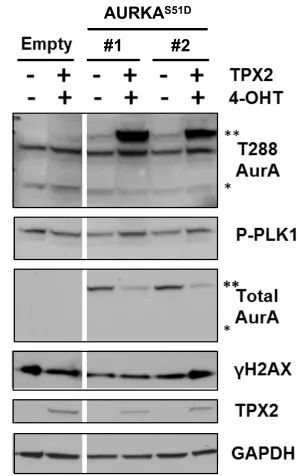
Figure 2.4. Transient overexpression of TPX2 in stably expressing AURKAS51D-ERT2 cells slightly increased phospho-PLK1 levels.
Figure 2.5. Transient co-overexpression of AURKAS51D-ERT2 with TPX2 increases levels of AURKA autophosphorylation.
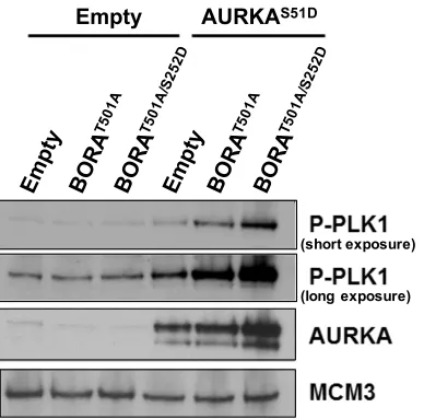
Figure 2.6. Transient co-overexpression of AURKAS51D-ERT2 with BORA activates PLK1.
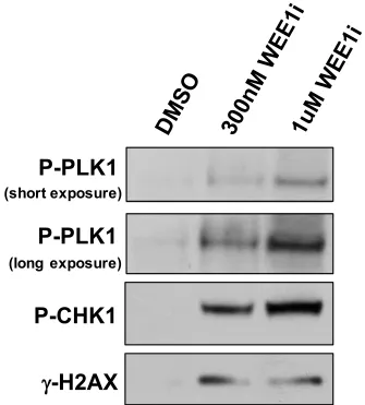
Figure 2.7. WEE1 inhibition is sufficient to activate the AURKA-PLK1 pathway and induce fork collapse.
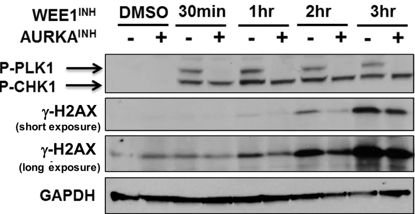
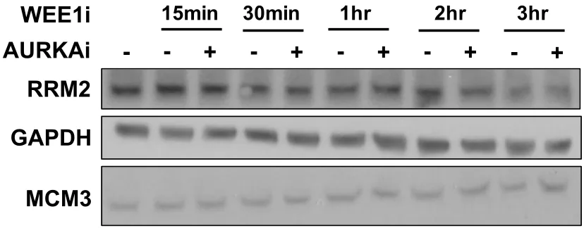
Figure 2.8. WEE1 inhibition-induced DSB formation depends on AURKA.
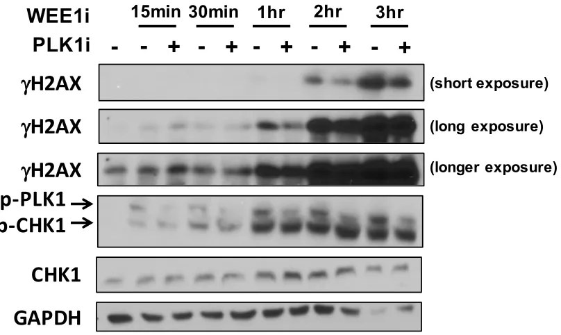
Figure 2.9. WEE1 inhibition-induced DSB formation does not affect RRM2 levels. Figure 2.10. WEE1 inhibition-induced DSB formation depends on PLK1.
Figure 2.11. AURKA and PLK1 inhibition prevent WEE1 inhibition-induced replication
fork collapse.
Figure 2.12. Overexpression of stabilized AURKA increases reliance on ATR but not on CHK1 for genome stability.
Figure 3.1. Model figure
ix
Figure 3.3. WEE1 inhibition activates CDK1 and ATR signaling and leads to RRM2 depletion.
Figure 3.4. WEE1 and ATR inhibition results in similar levels of DNA damage under conditions of replication stress.
Figure 3.5. WEE1 inhibition prevents DNA replication to a greater extent than ATR inhibition.
Figure 3.6 CDK1 inhibition decreases gH2AX accumulation and rescues DNA replication
inhibition induced by WEE1 and ATR inhibition.
Figure 3.7. RPA ChIP-Seq peaks overlap between WEE1- and ATR-inhibited cells with
or without aphidicolin.
Supplemental Figure 3.1. Determining the IC50 for WEE1 and ATR inhibitor in MEF cells.
Supplemental Figure 3.2. WEE1 and ATR inhibition results in similar levels of DNA damage under conditions of replication stress.
Supplemental Figure 3.3. RPA ChIP-Seq peaks have similar signal intensity between WEE1- and ATR-inhibited cells with or without aphidicolin.
Figure 4.1. Model figure: ATR stabilizes forks by preventing the premature activation of the CDK1-AURKA-PLK1 pathway.
Figure 4.2. Model figure the fork stabilizing functions of ATR.
Figure 4.3. Signaling cascades activated by WEE1 inhibition that result in fork collapse. Figure 4.4. Signaling cascades downstream of CDK1 that result in fork stalling and collapse.
x
1
CHAPTER 1: Introduction: The role of DNA replication checkpoints in replication
fork dynamics
Theonie Anastassiadis
Department of Cancer Biology, University of Pennsylvania, Philadelphia, PA,
2
Introduction
The survival of an organism depends on its ability to faithfully and efficiently transmit genetic material from mother to daughter cell. During each cell cycle, human cells must accurately replicate over three billion base pairs of DNA in a short period of time and protect the genome from spontaneous or induced DNA damage that could result in alterations or loss of genetic information, ultimately contributing to cancer or age-related pathologies. Indeed, DNA is constantly under assault and can be damaged in a multitude of ways, including from replication-induced mismatches, chemically-induced crosslinks and adducts, base damage from ultraviolet light, and single- and double-strand breaks resulting from ionizing radiation or chemical reactions (Ciccia and Elledge, 2010). These DNA lesions affect the stability of the genome as well as impact its ability to be accurately duplicated. In response to these lesions, cells have evolved protective pathways that detect and repair damaged DNA, and pause cell cycle progression to give cells time to resolve the problematic lesion (Blackford and Jackson, 2017). Defects in these pathways lead to the accumulation of DNA damage over time, giving rise to an increased mutation burden that often results in age-related pathologies, such as cancer (Tubbs and Nussenzweig, 2017). Understanding the essential mechanisms at play during replication fork stabilization, collapse and repair in the context of the genome, of the proteome and of the molecular signaling cascades, will
3
DNA Replication
DNA replication is the process by which a doubled stranded DNA (dsDNA) molecule is duplicated to produce an identical copy. This process is divided into three phases: initiation, elongation and termination. Initiation occurs when the replicative DNA helicase unwinds the origin of replication; during elongation the replication fork copies the chromosome using semi-conservative DNA synthesis; and finally, termination results when converging replication forks converge (Siddiqui, On and Diffley, 2013). Specific regulation of the replication phases is crucial to assure accurate duplication of the genome to prevent loss or amplification of genetic information. Indeed, replication initiation is tightly controlled to ensure that the genome is only replicated once per cell cycle (Diffley, 1996; Blow and Dutta, 2005). In this section, we will elaborate on the sequence of events and factors involved in replication regulation.
4
Though these findings are still in their infancy, many advances have been made in our understanding of how the genetic sequence, chromatin milieu and nuclear architecture influence the location, timing and activation of origins.
Initiation of DNA replication is comprised of two major steps: origin licensing and origin firing. Origins are licensed in late mitosis and early G1 phases by loading the pre-replicative complex (pre-RC) onto chromatin (Bell and Stillman, 1992; Remus et al., 2009; Riera, Tognetti and Speck, 2014). The pre-RC complex is comprised of the origin recognition complex (ORC) and the double hexameric minichromosome maintenance 2-7 (MCM2-2-7) helicase complex, that is recruited by CDC6 and the licensing factor CTD1 (Cvetic and Walter, 2006; Randell et al., 2006; You and Masai, 2008). A greater number of origins are licensed than fired (Woodward et al., 2006). The unfired origins are termed dormant origins and are only used to complete replication when the progression of a nearby replication fork is compromised as a result of fork slowing, stalling or collapse (Ge and Blow, 2010).
Origin firing occurs when the MCM2-7 helicases are activated in S phase to begin the elongation phase of replication. These events are controlled through a cell
cycle mediated kinase signaling cascade, primarily regulated by DBF4-dependent kinase (DDK) and cyclin-dependent kinase (CDK) (Heller et al., 2011; Tanaka and Araki, 2013).
5
components of the replisome, such as TIMELESS, TIPIN and Claspin, will be recruited (Gotter, Suppa and Emanuel, 2007; Yoshizawa-Sugata and Masai, 2007; Masai et al., 2010). The ORC complex is removed from chromatin and degraded through ubiquitin-mediated proteolysis to prevent another MCM helicase from being loaded onto the ORC, which would lead to re-replication of that genomic loci (Méndez et al., 2002).
Once origins have fired and DNA synthesis is initiated, replication forks move in opposite directions away from the origin. The helicase unwinds the duplex DNA, separating the parental strands, each of which will be used as a template for DNA synthesis (Masai et al., 2010). Replicative polymerases are proteins that synthesize DNA in a 3’ to 5’ direction (Loeb and Monnat, 2008). Polymerase alpha initiates strand synthesis, polymerase epsilon synthesizes DNA on the leading strand, and polymerase delta synthesizes small stretches of DNA (Okazaki fragments) in between RNA primers laid by primases on the lagging strand (Lujan, Williams and Kunkel, 2016). These primers are eventually removed and Ligase 1 ligates Okazaki fragments to one another. There are many other factors involved in DNA replication that contribute to faithful genome replication and are adversely affected under conditions of replication stress. In addition, it is hypothesized that the replisome composition is altered or modified under different conditions of replication stress (Yoo et al., 2004; Mailand et al., 2006; Mamely
6
Finally, replication termination occurs when two converging replication forks meet or when a fork meets a chromatin end. The field has predominantly focused on elucidating the mechanisms involved in replication initiation and elongation, leaving much to still be understood about replication termination. During the process of termination, the following steps occur in most organisms: replication fork convergence, completion of DNA synthesis, replisome disassembly and decatenation (Dewar and Walter, 2017). When replication forks encounter each other, their replisomes are disassembled. Though this process has been shown to be mediated through ubiquitin signaling and to involve the ubiquitin selective protein segregase p97, it remains to be determined if the replisome is actively disassembled or not (Meerang et al., 2011). The mechanism by which DNA synthesis is completed is also under debate. Some groups argue that the single-stranded gap remaining between the leading stand and the last Okazaki fragment is filled by the same mechanism utilized during elongation, whereas other groups argue this process requires replisome disassembly (Dewar and Walter, 2017). Finally, catenated DNA is created by replicating the last turn of the parental duplex DNA, which must be resolved before chromosome segregation. However, the topological stress needs to be managed until chromosome segregation takes place, leaving the field with important questions regarding the factors and pathways that are involved in decatenation and relief of topological stress (Postow et al., 2001). If all phases are successfully executed, the cell will have achieved complete and accurate genome duplication, which it can pass on to the daughter cell.
7
checkpoints, that work to prevent these DNA replication perturbations from inducing disease-causing genomic instability.
Replication stress
Replication stress is a problem that cells often face during DNA replication that can have significant consequences on the stability of the genome, and ultimately, on cell survival. DNA replication renders cells particularly susceptible to DNA lesions that can block replication fork progression. Stalled forks can collapse into DSBs or be improperly repaired, leading to a loss of genetic information that gives rise to many diseases, such as cancer (Mcgowan, 2003; Paulsen and Cimprich, 2007). There is no uniform description of replication stress nor unique cellular markers that specifically define this phenomenon. As a result, the definition of replication stress is constantly evolving, though some broad characteristics are agreed upon by the field. Generally, replication stress is defined as the slowing or stalling of replication fork progression (Zeman and Cimprich, 2014a). Though physical structures change depending on the lesion, all replication stress-induced structures share a common feature: a tract of single-stranded DNA (ssDNA). This tract is generated either from a functional uncoupling of the DNA replicative helicase from the replicative polymerase, or from endonucleolytic processing of DNA that resects the DNA strand where a nick or gap was left unrepaired, forming a stretch of ssDNA. Prolonged exposure of ssDNA triggers the activation of the DNA replication checkpoint pathway, the details of which will be discussed in the following section (Byun et al., 2005; MacDougall et al., 2007).
8
from metabolic and chemical reactions. Most exogenous sources of replication stress arise from chemical mutagens and ultraviolet (UV) light that generate DNA lesions that either block the helicase (e.g. DNA-topoisomerase adducts, intra-strand crosslinks, and bulky DNA adducts), or that block polymerase progression, (e.g. base pair adducts, polymerase inhibitors [such as aphidicolin], and chemicals [such as hydroxyurea] that deplete deoxyribonucleotides [dNTP] pools) (Ciccia and Elledge, 2010). Limiting exposure to these agents when it is possible should be a priority to prevent genomic instability that could drive malignant transformation of cells.
There also exists a plethora of endogenous sources of replication stress that cannot be mitigated by limiting exposure; instead, DNA damage repair pathways have naturally evolved to resolve these inevitable lesions. Ironically, nicks and gaps are naturally-occurring intermediates in many DNA repair pathways and if encountered by replication machinery, will stall the forks, eventually causing them to collapse. Though it remains controversial whether or not nicks and gaps behind the fork can cause fork stalling, nicks and gaps ahead of the replication fork can collapse into DSBs through duplex unwinding. Indeed, a nick or gap on the leading strand will cause the helicase to slide off the DNA, leading to fork collapse, and a nick or gap on the lagging strand could result in fork progression problems that cause fork stalling. Many unrepaired DNA lesions, such as inter-strand crosslinks and protein-DNA crosslinks that can be caused by metabolic pathway products (e.g. reactive aldehydes generated during alcohol metabolism) can also be barriers to replication fork progression (Mirkin and Mirkin, 2007; Dalgaard JZ, 2011).
9
polymerases faithfully match base pairs but are less specific at discriminating between dNTPs and ribonucleotides, which they surprisingly often incorporate erroneously (Dalgaard, 2012). This replication defect is recognized and corrected through the ribonucleotide excision repair pathway (Reijns et al., 2012; Sparks et al., 2012). If not removed, the incorporated ribonucleotide will stall the polymerase and the DNA damage tolerance pathway will be required to bypass the lesion (Anglana et al., 2003; Nick McElhinny et al., 2010; Lazzaro et al., 2012).
DNA replication requires many different components to be successfully completed, so when any are limiting, it can lead to polymerase slowing and eventual fork stalling (Poli et al., 2012; Sørensen and Syljuåsen, 2012). Some well characterized insufficiencies are those of nucleotides and replisome factors. Depletion of these factors often result from aberrant regulation of replication initiation leading to an excess of origin firing that overwhelm the existing pools of factors (Beck et al., 2012). It has been well documented that overexpression or constitutive activation of oncogenes, such as RAS, MYC or cyclin E, lead to increased origin firing, resulting in insufficient nucleotide levels or increased collision with transcriptional machinery (Halazonetis, Gorgoulis and Bartek, 2008; Bester et al., 2011; Jones et al., 2012; Burrell et al., 2013).
Because replication and transcription machinery are both active during S phase
10
techniques, including DNA combing and electron microscopy, have established that encounters between the transcription and replication complexes cause replication stress and eventual DNA damage (Paulsen et al., 2009; Wahba et al., 2011; Stirling et al., 2012). DNA damage induced by replication-transcription interference is a consequence of either R loop formation or resulting topological stress (García-Muse and Aguilera, 2016). R loops occur when RNA polymerases pause due to secondary DNA structures or collide with the replication fork (Aguilera and García-Muse, 2012). Topological stress occurs when a replication fork approaches a transcribed region that cannot rotate freely, such as when it is tethered to the nuclear periphery (Bermejo et al., 2011). If the region is not detached from the nuclear pore, accumulation of positive supercoiling in front of the fork will result in fork collapse and fork reversal (Bermejo et al., 2011).
11
and transcription machinery (Helmrich et al., 2013). However, because CFS fragility does not correlate with the expression of these genes in some cell lines and the rate of the replication fork is unperturbed, this hypothesis is unlikely (Le Tallec et al., 2013). Another explanation of CFS fragility is their paucity of replication origins, preventing the ability of stalled forks to be rescued by replication initiation at other origins (Letessier et al., 2011). The fact that CFS replicate in late S phase exacerbates this problem as dormant origins in areas surrounding CFS have a limited amount of time to initiate and complete replication. As these incompletely replicated genomic loci enter mitosis, the resulting aberrant replication intermediates are processed by nucleases, such as Mus81-Eme1 or ERCC1, resulting in DSBs and promoting disease-inducing genomic instability (Naim et al., 2013; Ying et al., 2013). Many deletions and chromosomal rearrangements found in cancers occur at these CFS, supporting their contribution to cancer-causing genome instability (Bignell et al., 2010).
12
genetic instability and are associated and contribute to the pathology of diseases such as Fragile X syndrome, Friedreich’s ataxia and Myotonic dystrophy (Castel, Cleary and Pearson, 2010; Mirkin and Mirkin, 2014; Jones, Houlden and Tabrizi, 2017).
Finally, secondary structure-forming DNA are known to cause replication stress. Some of these sequences include the aforementioned tandem trinucleotide repeats, which can form hairpins or triplexes that can block fork progression. Recently, our laboratory has discovered additional hard-to-replicate tandem repeats that can cause secondary structures and that were shown to stall or collapse replication forks (Shastri, Tsai et al., under revision). G-quadruplexes are secondary structures that form from GC-rich DNA sequences (Técher et al., 2017). Structural stabilization of G-quadruplexes by chemical means or loss of the helicases required to unwind them increase DSB formation and cause a loss of genetic information at those sites (Bochman, Paeschke and Zakian, 2012; Paeschke et al., 2013).
As discussed, there are many endogenous and exogenous sources of replication stress and more are still being discovered. In addition, levels of replication stress, as well as the source of stress, appear to elicit varying cellular responses as well as different
patterns of genomic lesions and instability. Exposure to replication stress will activate the DNA replication checkpoint response to overcome these stresses and protect genomic
13 DNA Replication Checkpoint
To ensure genomic stability, cells have evolved detection and repair mechanisms that monitor DNA lesions and coordinate repair with cell-cycle progression (Blackford and Jackson, 2017). Indeed, cell cycle checkpoints control cell cycle progression and allow cells to pause the cell cycle to provide time for DNA damage to be repaired before cell division resumes (Zhou et al., 2000; Kastan and Bartek, 2004a). If the damage cannot be resolved, checkpoint activation can trigger permanent cell cycle arrest (senescence) or cause cells to undergo programmed cell death (apoptosis) to prevent propagation of the mutated cell (Branzei and Foiani, 2009; Sperka, Wang and Rudolph, 2012). Once DNA damage is recognized by the cell, cellular signaling cascades are triggered to promote DNA repair. Different types of DNA damage activate cell cycle checkpoint pathways that regulate distinct repair mechanisms. More specifically, physical DNA lesions, such as DSBs, will trigger the ataxia telangiectasia mutated (ATM) and the DNA-dependent protein kinase (DNA-PK) kinase-regulated DNA damage checkpoint, whereas DNA replication stress will activate the ataxia telangiectasia and Rad3-related protein (ATR)-regulated DNA replication checkpoint (Maréchal and Zou, 2013; Blackford and Jackson, 2017). These signaling cascades will activate many downstream effector proteins as well as other cellular responses to mediate repair, ultimately resolving the DNA damage and resuming cell cycle progression.
14
through screens for S and G2 checkpoint defects and screens for hypersensitivity to hydroxyurea and methyl methanesulfonate (Allen et al., 1994; Kato and Ogawa, 1994; Weinert, Kiser and Hartwell, 1994). The human gene was later cloned revealing its PIKK domain, adding it to the PIKK protein family along with ATM and DNA-PK (Bentley et al., 1996; Cimprich et al., 1996). However, unlike ATM and DNA-PK, ATR is essential for embryonic development and crucial for genomic integrity (E. J. Brown & Baltimore, 2003; E J Brown & Baltimore, 2000).
Many types of DNA replication fork obstructions can cause replication stress and activate ATR but they all share a similar resulting DNA structure at the fork, which consists of single-stranded DNA (ssDNA) (MacDougall et al., 2007). The unwinding minichromosome maintenance (MCM) helicase, but not the DNA replicative polymerase, can overcome most DNA lesions leading to their uncoupling. This results in an accumulation of ssDNA between the helicase and the polymerase, which is rapidly coated by the ssDNA-binding protein complex, replication protein A (RPA) (Byun et al., 2005). The tract of ssDNA can further be expanded by exonucleases. In addition to ssDNA tracts being formed from uncoupling of the helicase and polymerase, ssDNA tract can be generated by endonucleolytic processing of other forms of DNA lesions (Raderschall, Golub and Haaf, 1999). The RPA-ssDNA complex then serves as a platform to recruit the ATR-interacting protein (ATRIP), which is required for the interaction of ATR with the stalled replication fork but not sufficient for ATR activation at the fork (Cortez et al., 2001; Zou and Elledge, 2003; Ball, Myers and Cortez, 2005; Kim
et al., 2005; Namiki and Zou, 2006).
15
(ETAA1). TOPBP1 is recruited to the to the ATR-ATRIP complex by a ssDNA-double-stranded DNA (dsDNA) junction (Michael et al., 2000; Ellison and Stillman, 2003; Bomgarden et al., 2004; Kumagai et al., 2006; MacDougall et al., 2007; Mordes et al., 2008). This junction is also necessary for the loading of the RAD9-RAD1-HUS1 (9-1-1) checkpoint clamp complex by the RAD17/RFC2-5 clamp loader complex (Bermudez et al., 2003; Delacroix et al., 2007; Lee, Kumagai and Dunphy, 2007; Navadgi-Patil and Burgers, 2009). Once the 9-1-1 clamp is loaded on the ssDNA-dsDNA junction, it recruits TOPBP1 to the ATR-ATRIP-RPA coated ssDNA in conjunction with the MRE11-RAD50-NBS1 (MRN) complex and RAD9-RAD1-HUS1-interacting nuclear orphan (RHINO), although the mechanisms by which the MRN complex and RHINO contribute to TOPBP1 recruitment remain elusive (Cotta-Ramusino et al., 2011; Duursma et al., 2013; Lee and Dunphy, 2013; Lindsey-Boltz et al., 2015). Once TOPBP1 is recruited, it interacts with the ATR-ATRIP complex through its ATR activation domain, which interacts with ATR to activate it. The second, and recently identified, ATR activator is ETAA1, which like TOPBP1 contains an ATR-activation domain but unlike TOPBP1 interacts with the RPA-ssDNA platform through direct binding of RPA (Bass et al., 2016; Feng et al., 2016; Haahr et al., 2016). It remains to be determined if these activators recognize different ssDNA structures that require distinct repair signaling pathways to be resolved, possibly dictated through a specificity in downstream substrates of ATR. Once
16
The functions of ATR in DNA replication and repair
Once activated, ATR stimulates multiple signaling cascades that protect genomic integrity by halting cell cycle progression, limiting firing of novel origins of replication, stabilizing the replication fork, and promoting replication fork repair and restart (Saldivar, Cortez and Cimprich, 2017).
ATR prevents cell cycle progression
By coordinating cell cycle arrest with replication, ATR prevents cells from entering prematurely into mitosis before replication defects are resolved and before replication is complete. ATR exerts its checkpoint function on the cell cycle primarily through its most prominent and well-characterized downstream effector, the checkpoint kinase 1 (CHK1) (Guo et al., 2000; Hekmat-Nejad et al., 2000; Liu et al., 2000; Zhao and Piwnica-Worms, 2001). ATR activates CHK1 by phosphorylating its serine 317 and 345 sites. The interaction between ATR and CHK1 is facilitated by Claspin, an adaptor protein localized at the fork (Kumagai and Dunphy, 2000; Liu et al., 2000; Zhao and Piwnica-Worms, 2001; Liu, Song and Zou, 2012). It has also been suggested that the timeless (TIM) and timeless-interacting protein (Tipin) complex might be involved in ATR
activation of CHK1 through its recruitment to stalled forks. Indeed, TIM and Tipin loss lead to a decrease in the ability of ATR to activate CHK1 (Chou and Elledge, 2006;
17
CHK1 controls cell cycle progression by phosphorylating the three CDC25 phosphatases, ultimately leading to their degradation or sequestration (Löffler et al., 2006). The CDC25 phosphatases remove the inhibitory phosphorylation of the cyclin-dependent kinases (CDKs). Therefore, CHK1-cyclin-dependent inactivation of the CDC25 proteins prevents CDK activation and premature entry into mitosis (Piwnica-Worms et al., 1998). CHK1 phosphorylation of CDC25A triggers its ubiquitin-mediated degradation, facilitated by the SCFβ-TRCP E3 ubiquitin ligase complex (Busino et al., 2003; Jin et al., 2003). CDC25A degradation prevents CDK1 activation to inhibit entry into mitosis and prevents CDK2 activation to prevent origin firing (Chen, Ryan and Piwnica-Worms, 2003a). CHK1 also phosphorylates CDC25C, resulting in 14-3-3 binding and sequestration to the cytoplasm, which prevents CDK1 activation (Kasahara et al., 2010). These events provide cells with sufficient time to respond to replication perturbations and finish replication before entry into mitosis.
ATR inhibits novel origin firing
The ATR-CHK1 axis also inhibits global origin firing under conditions of replication stress to prevent additional fork stalling until the stress is resolved (Diffley and
Santocanale, 1998; Costanzo et al., 2003; Karnani and Dutta, 2011; Yekezare, Gomez-Gonzalez and Diffley, 2013). ATR limits origin firing by preventing replication initiation by
18
activation and origin firing. The CHK1 yeast homologue was shown to phosphorylate Dbf4 to suppress DDK activity and ATR was shown to phosphorylate DDK in human cells, though this site was not shown to affect DDK activity. Further studies are required to determine if the ATR-CHK1 checkpoint can inhibit DDK-dependent CDC45 loading and origin firing (Heffernan et al., 2007; Zegerman and Diffley, 2010). In addition, CHK1 prevents origin firing through CDC25A degradation resulting in CDK2 inhibition thus preventing CDC45 loading (Mailand et al., 2000; Zhao, Watkins and Piwnica-Worms, 2002). Finally, CDC45 loading and origin firing was shown to be suppressed by CHK1-mediated phosphorylation of Treslin, which could be another mechanism by which the ATR-CHK1 axis controls origin firing (Guo et al., 2015). Of note, the ATR-CHK1 checkpoint prevents origin firing globally but interestingly allows local origin firing (Diffley, 1996; Ge and Blow, 2010). These functions that initially appear at odds with one another actually promote genomic stability by allowing local dormant origins to fire to ensure complete replication in the region surrounding the stall site but prevent global and late-replicating origins from firing to avoid stalling of additional replication forks until the replication stress-inducing event is resolved. The mechanism by which local origins evade checkpoint suppression of origin firing remains to be elucidated; however, some evidence suggests that CDC45 might already be loaded onto local dormant origins in the replication problematic area thereby bypassing the step inhibited by the checkpoint
(Thomson, Gillespie and Blow, 2010).
ATR controls dNTP levels
19
levels of nucleotides was ATR’s primary genome-protecting function from studies in budding yeast, where lethality resulting from ATR homologue loss could be rescued by overexpressing ribonucleotide reductase (Rnr), the enzyme responsible for dNTP production (Huang, Zhou and Elledge, 1998; Zhao, Muller and Rothstein, 1998). In human, ATR is required for ribonucleoside-diphosphate reductase subunit M2 (RRM2) expression and to prevent its CDK-cyclin F mediated degradation (Zhang et al., 2009; D’Angiolella et al., 2012). Finally, mice carrying a hypomorphic mutation of ATR exhibited prolonged survival and reduced levels of genomic instability when crossed to mice expressing supra-physiological levels of RRM2 (Lopez-Contreras et al., 2015). Though sometimes overlooked, ATR’s function in maintaining replication-favorable levels of dNTPs is key in maintaining genomic stability.
ATR stabilizes stalled replication forks
In addition to its regulatory roles in cell cycle, origin firing and nucleotide levels, ATR is best known for its replication fork stabilizing functions. By stabilizing replication forks, ATR prevents stalled forks from collapsing into DSBs and allows them to reinitiate replication once the stalling agent has been eliminated (Lopes et al., 2001; Tercero and
Diffley, 2001). Indeed, ATR loss has been shown to increase levels of genomic instability as measured by elevated levels of DSBs and fork-associated recombination structures,
such as reversed forks (E J Brown & Baltimore, 2000; Eric J Brown & Baltimore, 2003; Myung, Datta, & Kolodner, 2001). The mechanism by which this occurs has been under fierce debate in the field and many models have been proposed.
20
phosphorylates many replisome components under condition of replication stress, including DNA polymerases, the MCM2-7 helicase, the clamp loader RFC1-5 and the Claspin-timeless-tipin-AND1 complex (Block, Yu and Lees-Miller, 2004; Cortez, Glick and Elledge, 2004; Olson et al., 2006; Göhler et al., 2011). In addition, many replisome factors, such as FANCM and Claspin, are targeted for degradation following extended fork stalling (Yoo et al., 2004; Mailand et al., 2006; Mamely et al., 2006; Peschiaroli et al., 2006; Kee et al., 2009). Finally, our laboratory has observed decreased levels of replisome components under conditions of replication stress in ATR-depleted cells and showed that suppression of RNF4, PLK1 or AURKA, which are involved in degradation pathways, rescues the replication restart defect seen in ATR-depleted cells (Ragland et al., 2013). Together these data suggest that removal of components required for replication prevent the replication of restart once forks stall. Though there are studies supporting this model, recent genomic and proteomic data have contested this model (De Piccoli et al., 2012; Dungrawala et al., 2015). More studies are needed to better define in which context (cell cycle phase, organism, replication stress, etc) this model might be applicable.
structure-21
specific endonuclease subunit SLX4 in complex with several structure-specific nucleases, such as SLX1, MUS81-EME1 and XPF-ERCC1 (Cotta-Ramusino et al., 2005; Hanada et al., 2007; Forment et al., 2011; Wyatt et al., 2013a, 2017; Duda et al., 2016). In support of this model are studies that show that loss of SLX4 decreases DSB formation in ATR-inhibited cells (Couch et al., 2013a; Ragland et al., 2013). Of note, ATR’s role in protecting forks from nuclease-dependent fork collapse is also reinforced by its indirect inhibition of CDK as some of the structure-specific endonuclease are activated by CDK (Domínguez-Kelly et al., 2011). Indeed, in the absence of ATR, CDK levels are not regulated under conditions of replication stress, and nucleases are prematurely activated causing cleavage-induced fork collapse.
In line with this second model, it is also possible that DSB formation from fork collapse might actually be an intermediate generated during a process of homologous recombination (HR)-mediated replication restart. It was recently shown that the S phase specific endonuclease MUS81-EME2 is responsible for fork cleavage of stalled replication forks to allow for replication restart (Alessandra Pepe and West, 2014a). Therefore, it is possible that nuclease-dependent stalled fork cleavage is an attempt at replication restart in ATR-depleted cells.
A third model is that ATR stabilizes forks by regulating pathways that promote
replication fork restart, such as template switching, DNA damage tolerance pathways, and HR-mediated fork restart. Indeed, ATR phosphorylates translesion polymerases,
reversionless 1 (REV1) and Pol h, proposing that ATR allows DNA lesion bypass
RAD51-22
dependent restart-promoting pathways (Vassin et al., 2009; Murphy et al., 2014; Ahlskog
et al., 2016; Buisson et al., 2017). Finally, ATR could protect forks by promoting replication restart by processing repair intermediates through the regulation of recombination repair factors, such as the BLM and WRN helicases, which are ATR substrates and involved in fork restart to prevent collapse (Davies et al., 2004; Ammazzalorso et al., 2010).
A final model has proposed that ATR stabilizes forks indirectly by preventing the exhaustion of RPA. Under conditions of replication stress and ATR inhibition, an excess of ssDNA is generated exceeding the amount of RPA available to coat it, thereby rendering the uncovered forks susceptible to nuclease cleavage (Toledo et al., 2013; Toledo, Neelsen and Lukas, 2017). More studies are needed to build a stronger case for this model though it is not difficult to imagine that an insufficiency of protective factors could play an important role in triggering increased fork collapse after fork stalling.
Every model is backed by convincing experimental data and no model excludes the other so it is possible that the mechanism of fork stabilization by ATR might be context dependent or a combination of the different models. Additional studies will help
further clarify the mechanism(s) by which ATR stabilizes replication forks and which of its functions are crucial for its fork stabilizing role (Chapter 3).
Repair of collapsed replication forks
23
substrates of structure-specific nucleases that process them in DSBs (Fekairi et al., 2009; Couch et al., 2013a; Ragland et al., 2013; Szakal and Branzei, 2013a; Wyatt et al., 2013a; Sarbajna, Davies and West, 2014). Indeed, MUS81-EME2 is a nuclease complex that is only active in S phase and that is required for fork restart by cleaving the fork to induce DSB formation (Alessandra Pepe and West, 2014a). In the absence of the ATR-regulated checkpoint, aberrant CDK signaling promotes premature activation of G2/M nucleases, such as SLX4-SLX1 and MUS81-EME1, which process the unprotected fork into a DSB (Couch et al., 2013a; Ragland et al., 2013).
Once DSBs are formed, the ATM-CHK2-regulated DNA damage checkpoint pathway is activated. DSB repair occurs by homologous recombination, which uses the sister chromatid as a DNA template to prevent loss of genetic information (West, 2003). The DSB is resected by the MRN complex at the 5’ end, recruiting ATM, and generating RPA-coated ssDNA overhangs (Lamarche, Orazio and Weitzman, 2010). Following its activation via autophosphorylation, ATM phosphorylates its substrates, among which are the H2AX histone variant and the CHK2 kinase (Rogakou et al., 1998; Matsuoka et al.,
2000). Phosphorylated H2AX, gH2AX, regulates the repair process and CHK2 arrests
the cell cycle (Paull et al., 2000; Kuo and Yang, 2008; Chanoux et al., 2009). Of note, under conditions of replication stress, ATR will also phosphorylate H2AX (Ward and
Chen, 2001). gH2AX acts as a surrogate marker of DSB and recruits repair factors at the
lesion. gH2AX spreads along the chromatin up to several hundreds of kilobases on either
end of the break, either as a means to remodel the chromatin to enhance repair protein recruitment and accessibility to the site, or to prevent transcription-replication collision
and formation of R loops because gH2AX-marked chromatin is transcriptionally silenced
24
with the MDC1 adaptor protein, gH2AX recruits the RNF8 and RNF168 E3 ubiquitin
ligases, which promote the ubiquitin-mediated recruitment of the BRCA1-RAP80 complex in a CtIP-dependent manner (Stucki et al., 2005; Shao et al., 2009; Ohta, Sato and Wu, 2011; Strauss and Goldberg, 2011). Following extended resection by MRN in coordination with BLM helicase and EXO1 or DNA2 nucleases, BRCA1 will then recruit BRCA2 via PALB2, which will mediate RAD51 loading onto the resected ssDNA overhangs, displacing RPA to form RAD51 filaments (Sy, Huen and Chen, 2009;
Peterson et al., 2011). RAD51 orchestrates strand exchange via D-loop formation and homologous recombination ensues to complete the repair (Liu et al., 2010). Once the
DSB is repaired, replication can continue and be completed by novel origin firing or by a nearby actively replicating fork.
G2-M cell cycle kinases
CDK-cyclin control of cell cycle
25
(Giacinti and Giordano, 2006). Progression through S phase is regulated by CDK2-cyclin E as well as CDK2-cyclin A in S phase and cyclin A in G2. Finally, the CDK1-cyclin B complex drives cells into mitosis followed by cell division (Norbury and Nurse, 1992; Weinberg, 1995; Murray, 2004). We will focus on the players involved in entry into G2-M phase and their role in cancer.
Regulation of CDK1
CDK1 is the only CDK that is essential for cell cycle progression as it can compensate for the loss of all other cell cycle CDKs (CDK2, CDK4 and CDK6) (Santamaria and Ortega, 2006). As mentioned above, CDK1 is required for entry into mitosis and needs to be inactivated for cells to exit mitosis. In late mitosis, CDK1 is inactivated through proteasome-mediated degradation of cyclin B by the anaphase-promoting complex/cyclosome (APC/C) complex to promote mitotic exit (Gavet and Pines, 2010). Once CDK1 is inactive, cells can separate their chromosomes and undergo cytokinesis.
Cyclin levels are not the only regulators of CDKs. CDKs are also regulated by their phosphorylation levels. CDK1 is inactivated when phosphorylated at its Thr14 and Tyr15 sites by membrane associated tyrosine- and threonine-specific cdc2-inhibitory kinase (MYT) and WEE1, respectively, and it is activated by removal of these phosphates by the CDC25 phosphatases (Parker and Piwnica-Worms, 1992; Mueller et
al., 1995).
Regulation of the Aurora A- PLK1 pathway
26
chromosomal segregation, but our focus will be on their role and regulation in the context of cell cycle progression and entry into mitosis (Asteriti, De Mattia and Guarguaglini, 2015).
Aurora A is a serine-threonine kinase that is first expressed in late S phase (Walter et al., 2000; Seki, Coppinger and Jang, 2008). Aurora A kinase promotes mitotic entry through phosphorylation and activation of the serine-threonine kinase PLK1 (Macůrek et al., 2008). Indeed, PLK1 activates the CDK1-cyclin B complex by activating the CDC25C phosphatase which activates CDK1, as well as by inducing the degradation of WEE1, the negative regulator of CDK1 (Nigg, 2001; Seki, Coppinger and Jang, 2008; S. M. a Lens, Voest and Medema, 2010). The CDK1-AURKA-PLK1 mitotic entry pathway is regarded as a switch, which once activated is difficult to inhibit. Once the CDK1, Aurora A and PLK1 kinases are activated, they enter into a positive feedback loop that ultimately irreversibly drives mitotic entry (Seki, Coppinger and Jang, 2008; Seki, Coppinger, Du, et al., 2008; Feine et al., 2014).
Aurora A has an autophosphorylation site on its activation loop at conserved residue Thr288, which was thought to be a mark of activation until a recent study
showed that it was possible for Aurora A to be autophosphorylated and not be active (Bischoff et al., 1998; Walter et al., 2000; Littlepage et al., 2002; Bayliss et al., 2003a;
27
Aurora A autophosphorylation, (2) by shielding the autophosphorylation site from phosphatases through conformational change, and (3) by inhibiting Aurora A recognition by the ubiquitin E3 ligase APC/C with its co-activator Cdh1 (APC/CCdh1). Two well-characterized Aurora A binding proteins are Bora and TPX2 (Kufer et al., 2002a; Eyers
et al., 2003; Eyers and Maller, 2004; Hutterer, Berdnik, Wirtz-Peitz, Zigman, et al., 2006; Seki, Coppinger, Jang, et al., 2008; Bruinsma et al., 2014a). Bora localizes Aurora A in the nucleoplasm and plays an essential role in driving the G2/M transition by promoting Aurora A-mediated activation of PLK1 (Seki, Coppinger and Jang, 2008). TPX2 localizes Aurora A to mitotic spindles after nuclear envelope break down and protects Aurora A dephosphorylation at its Thr288 site by inducing a conformational change in its activation loop (Kufer et al., 2002a; Bayliss et al., 2003a; Eyers et al., 2003; Li, Cao and Zheng, 2003; Tsai et al., 2003a; Eyers and Maller, 2004; Dodson and Bayliss, 2012b). Aurora A is known to interact with several other binding proteins, including HEF1, PAK1, Arpc1b, and PUM2, which aid Aurora A with its other functions (Elena N. Pugacheva and Golemis, 2005; Zhao et al., 2005; Molli et al., 2010; Huang et al., 2011).
28
partners (Chan et al., 2008b). Indeed, PLK1 regulates Aurora A binding partner
preference, which in turn regulates its localization and function, through b
-TRCP-dependent Bora degradation.
The AURKA-PLK1 pathway is important for recovery after DNA damage. Indeed, PLK1 is required for mitotic entry following G2 cell cycle arrest induced by DNA damage (van Vugt, Brás and Medema, 2004; Peng, 2013; Hyun et al., 2014). PLK1 also regulates the activation of MUS81-EME1 and stimulates its association with the SLX4 endonuclease complex, promoting replication restart through DSB generation at stalled
replication forks (Matos et al., 2011; Gallo-Fernández et al., 2012; Muñoz-Galván et al., 2012; Szakal and Branzei, 2013a). Due to its prominent and impactful role in the cell
cycle and in the checkpoint pathways, it is not surprising that the AURKA-PLK1 pathway is tightly regulated in various ways, such as by stabilization, degradation, localization, phosphorylation and partner protein interaction.
Cell cycle: a target of DDR
Under conditions of DNA damage, cell cycle checkpoints are activated to block cell cycle progression and allow time for DNA repair. As we previously discussed, the DNA damage checkpoints achieve cell cycle control through modulation of CDK activity. CHK1 phosphorylation inactivates the CDC25 phosphatases and activates WEE1 kinase to inhibit CDK1 and CDK2, forcing cells to arrest in G2 phase. In addition, the AURKA-PLK1 pathway is similarly inhibited during the checkpoint to prevent premature entry into mitosis (Parrilla et al., 2016; Bruinsma et al., 2017)
29
Cancer can be defined as uncontrolled cell proliferation that results from unregulated cell cycle activity, caused either by mutations in signaling pathways or in genes coding for cell cycle proteins. CDKs are frequently dysregulated in cancers, making them a therapeutic target (Otto and Sicinski, 2017). Interestingly, CDK1 activity is rarely deregulated in cancer, though studies have shown that it contributes to tumorigenesis (Otto and Sicinski, 2017). Indeed, CDK1 knockdown in liver was shown to prevent NRAS-driven liver tumor formation and CDK1 inhibition prevented KRAS-driven colorectal cancer xenograft growth in mice (Diril et al., 2012; Costa-Cabral et al., 2016).
WEE1 kinase is overexpressed in many cancers, such as melanoma and glioblastoma. Interestingly, heterozygous deletion of WEE1 in mammary tissue increased the incidence of mammary tumor formation, whereas homozygous deletion did not, suggesting that complete loss of WEE1 might oppose tumor formation (Mir et al., 2010; Vassilopoulos et al., 2015). Despite this finding, WEE1 is still considered an oncogene and is being actively pursued as a cancer therapeutic. A promising WEE1 inhibitor, AZD1775, in currently performing well alone and in combinatorial therapies in over twenty clinical trials, with WEE1 inhibition proving to be synthetic lethal with other compounds, such as PARP and HDAC inhibitors, in several cancers (Rajeshkumar et al., 2011; Karnak et al., 2014; Mueller et al., 2014).
30
an oncogene (Dutertre, Descamps and Prigent, 2002; Marumoto et al., 2002; Meraldi, Honda and Nigg, 2002a; S. M. a Lens, Voest and Medema, 2010). PLK1 is often overexpressed in tumors and correlates with poor prognosis (S. M. a Lens, Voest and Medema, 2010). The mechanism by which PLK1 contributes to tumorigenesis remains unclear though it is speculated that it compromises the cell cycle checkpoints, resulting in genomic instability through premature progression through the cell cycle (Osada and Simizu, 2000; Kanaji et al., 2006). Interestingly, PLK1 loss in mice increases the incidence of tumors, suggesting PLK1 might play a tumor suppression role in certain contexts (L.-Y. Lu et al., 2008). Both Aurora A and PLK1 inhibitors are being pursued in clinical trials and appear to be promising therapeutics (Otto and Sicinski, 2017).
Genomic instability in cancer and therapeutic implications
31
bypass prevent cells from undergoing senescence and apoptosis and facilitate their clonal outgrowth and possible dissemination.
In support of genetic instability driving tumorigenesis, many studies have established that early tumorigenesis is associated with activation of a DNA damage response (DDR), which protects against development into malignancy (DiTullio et al., 2002; Bartkova et al., 2005; Karakaidos et al., 2005). Furthermore, it was shown that oncogene expression results in DNA replication stress, which if unrepairable will trigger oncogene-induced senescence (Bartkova et al., 2006; Di Micco et al., 2006; Fikaris et al., 2006; Bartek, Bartkova and Lukas, 2007; Burhans and Weinberger, 2007). Finally, models in which repair pathway proteins, such as ATR and ATM, are lost display increased tumor incidence, suggesting their pivotal role in cancer prevention (Harper and Elledge, 2007; Jackson and Bartek, 2009; Ciccia and Elledge, 2010; Negrini, Gorgoulis and Halazonetis, 2010). Together, these data highlight the importance of the DNA replication and damage checkpoints in preventing genomic instability and disease.
The pattern of mutations found in specific cancers can be utilized to subtype them. Studies have shown that cancers that share similar genome alterations respond
similarly to certain therapies and offer comparable prognostics. Therefore, increasing our understanding of how these patterns arise, whether it be from sites in the genome being
32
pathway suppresses the growth of a broad spectrum of cancers and synergistically increases DSBs and cell death (Gilad et al., 2010a; Murga et al., 2011a; Toledo et al., 2011; Ma et al., 2012; Prevo et al., 2012; Schoppy et al., 2012a).
In addition, by further understanding how the genome is stabilized and by characterizing the features that render parts of the genome more sensitive to certain stabilization pathways will enhance our comprehension of tumor pathogenesis and our ability to develop novel and more targeted therapeutics. Using genome-wide and proteomic techniques to probe the genome landscape and the replication dynamics observed under conditions of replication stress in combination with compromised genome stabilization mechanisms will improve our understanding of the key pathways involved in driving tumor progression and inform our patient treatment courses, from discovering exploitable synthetic lethal interactions to identifying responsive patient cohorts.
Summary
Previous work from our laboratory has shown that fork collapse in ATR-deficient cells is mediated through the activation of the AURKA-PLK1 pathway, suggesting that a
key part of ATR-mediated replication fork stabilization is a consequence of inhibiting premature activation of the AURKA-PLK1 pathway (Ragland et al., 2013) (Figure 1.1).
33
collapse. However, when WEE1 kinase, a negative regulator of CDK1, was inhibited, fork collapse occurred, suggesting that one major fork stabilizing function of ATR is to prevent premature activation of the mitotic CDK1-AURKA-PLK1 pathway to avoid premature entry into mitosis.
To further investigate the mechanisms essential in replication fork stabilization, we used genome-wide techniques and proteomics to determine whether ATR maintains fork integrity through its cell cycle checkpoint role or its local fork protection role, whether some sites of the genome are more dependent on one role over the other for genome stability and whether some replisome components or recruited factors play important roles in stabilization (Figure 1.2). In Chapter 3, we show that in the context of partial replication inhibition, WEE1 and ATR inhibition cause similar levels of fork collapse at overlapping genomic locations and that fork collapse at these sites is dependent on CDK1 and Aurora A kinase. Interestingly, while WEE1 inhibition is sufficient to cause replication fork collapse in a manner similar to ATR inhibition, WEE1 inhibition further promotes fork collapse through mechanisms distinct from ATR inhibition because WEE1 inhibition produces a distinct subset of sites compared to ATR inhibition. This could result from WEE1 inhibition leading to a depletion nucleotide levels, ultimately causing an additional source of replication stress. We show that the cell cycle checkpoint function of ATR is the essential mechanism by which ATR maintains fork integrity under conditions of replication stress.
34 Figure Legends
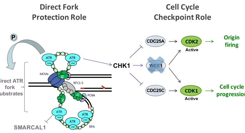
Figure 1.1. Model figure for Chapter 2: ATR stabilizes forks by preventing the premature activation of the CDK1-AURKA-PLK1 pathway. When ATR is absent or inhibited, the AURKA-PLK1 pathway is prematurely activated and results in premature entry into mitosis as well as in the degradation or removal of replisome components, leading to fork stalling and inability to restart replication.
35 Figures
Figure 1.1
36
Figure 1.2
WEE1
MCMs
RFC1-5
PCNA
RPA e
ATR
ATRIP
ATR
ATRIP
ATR
ATRIP
ATR
ATRIP
ATR
ATRIP
Cell cycle progression
Origin firing
Cell Cycle
Checkpoint Role
Direct Fork
Protection Role
Direct ATR fork substrates
CHK1
SMARCAL1
CDC25C CDC25A
37 References
Aguilera, A., & García-Muse, T. (2012). R loops: from transcription byproducts to threats to genome stability. Molecular Cell, 46(2), 115–24. https://doi.org/10.1016/j.molcel.2012.04.009
Ahlskog, J. K., Larsen, B. D., Achanta, K., & Sørensen, C. S. (2016). ATM/ATR-mediated phosphorylation of PALB2 promotes RAD51 function. EMBO Reports, 17(5), 671–681. https://doi.org/10.15252/embr.201541455
Allen, J. B., Zhou, Z., Siede, W., Friedberg, E. C., & Elledge, S. J. (1994). The SAD1/RAD53 protein kinase controls multiple checkpoints and DNA damage-induced transcription in yeast. Genes & Development, 8(20), 2401–15. Retrieved from http://www.ncbi.nlm.nih.gov/pubmed/7958905
Ammazzalorso, F., Pirzio, L. M., Bignami, M., Franchitto, A., & Pichierri, P. (2010). ATR and ATM differently regulate WRN to prevent DSBs at stalled replication forks and promote replication fork recovery. The EMBO Journal, 29(18), 3156–3169. https://doi.org/10.1038/emboj.2010.205
Anglana, M., Apiou, F., Bensimon, A., & Debatisse, M. (2003). Dynamics of DNA replication in mammalian somatic cells: nucleotide pool modulates origin choice and interorigin spacing. Cell, 114(3), 385–94. https://doi.org/10.1016/S0092-8674(03)00569-5
Aparicio, T., Guillou, E., Coloma, J., Montoya, G., & Méndez, J. (2009). The human GINS complex associates with Cdc45 and MCM and is essential for DNA replication. Nucleic Acids Research, 37(7), 2087–2095. https://doi.org/10.1093/nar/gkp065
Asteriti, I. A., De Mattia, F., & Guarguaglini, G. (2015). Cross-Talk between AURKA and Plk1 in Mitotic Entry and Spindle Assembly. Frontiers in Oncology, 5, 283. https://doi.org/10.3389/fonc.2015.00283
Ball, H. L., Myers, J. S., & Cortez, D. (2005). ATRIP Binding to Replication Protein A-Single-stranded DNA Promotes ATR-ATRIP Localization but Is Dispensable for Chk1 Phosphorylation. Molecular Biology of the Cell, 16(5), 2372–2381. https://doi.org/10.1091/mbc.E04-11-1006
Barlow, J. H., Faryabi, R. B., Callén, E., Wong, N., Malhowski, A., Chen, H. T., … Nussenzweig, A. (2013). Identification of early replicating fragile sites that contribute to genome instability. Cell, 152(3), 620–32. https://doi.org/10.1016/j.cell.2013.01.006
Barr, F. (2004). Polo-like kinases and the orchestration of cell division. Nature Reviews Molecular Cell Biology, 5(6), 429–441. https://doi.org/10.1038/nrm1401