Matz, M (2017) Factors influencing ovarian cancer survival world-wide. PhD thesis, London School of Hygiene & Tropical Medicine. DOI: https://doi.org/10.17037/PUBS.03482691
Downloaded from: http://researchonline.lshtm.ac.uk/3482691/
DOI:10.17037/PUBS.03482691
Usage Guidelines
Please refer to usage guidelines at http://researchonline.lshtm.ac.uk/policies.html or alterna-tively contactresearchonline@lshtm.ac.uk.
1
Factors influencing ovarian cancer survival worldwide
Lisa Melissa Matz
Thesis submitted in accordance with the requirements for the degree
of
Doctor of Philosophy
University of London
January 2017
Department of Non-Communicable Disease Epidemiology
Faculty of Epidemiology and Population Health
LONDON SCHOOL OF HYGIENE & TROPICAL MEDICINE
No funding received
2
Declaration
I, Lisa Melissa Matz, confirm that the work presented in this thesis is my own.
Where information has been derived from other sources, I confirm that this has
been indicated in the thesis.
Signed:
3
Dedication
This thesis is dedicated to my mom, who taught me the true meanings of courage
and strength
4
Acknowledgements
First and foremost, I would like to thank sincerely my supervisors, Professor Michel Coleman and Dr. Claudia Allemani, for their continued guidance and encouragement. It has been an honour and inspiration to work with them throughout these past three years. Additionally, I would like to thank Professor Martin Gore, who bravely stepped in at the last minute to be my clinical advisor. Our discussions on ovarian cancer were insightful and an invaluable contribution to this thesis.
This thesis would not have been possible without the hard work of the CONCORD Central Analytic Team in cleaning the massive data set from the cancer registries that was used in the analyses. I cannot thank them enough for preparing these data; they all deserve medals for their incredible efforts.
I would also like to thank the various members of the Cancer Survival Group at the London School of Hygiene and Tropical Medicine who welcomed me warmly to the group over three years ago, and have been a constant source of support and encouragement throughout my PhD. I would especially like to thank Yuki, Natalia and Lisa for their help with scheduling meetings with my supervisors – it was quite a difficult task at times! My family, though thousands of miles away, have supported me throughout these past few years. I could not have done it without their unwavering support and belief that I could do anything (even if I kept telling them I could not, in fact, be two places at once). I would also like to thank endlessly my friends who have encouraged me when I doubted myself, laughed with me when things were rough, distracted me when things were stressful and listened to me talk relentlessly about work while at the pub– I could not have done this without any of you.
5
I would especially like to thank Anjali, Eileen, Gui, Joy and Lily: you ladies have been a consistent source of support, despite all of us being scattered across several time zones and continents. I have always felt I could run to you when I needed to. Friends do not get any better than you.
To all of my officemates – thank you for understanding how stressful and intense working on a PhD can be. I wish those of you still working on your degrees the best of luck. Finally, I would like to thank my mom, Linda, to whom this thesis is dedicated. She taught me that courage is facing your greatest fears head on, without hesitation; and that strength is facing those fears day after day. I am the person I am today, and doing something I enjoy immensely, because of her endless strength and courage. And for that, I will never be able to thank her enough.
6
Abstract
Ovarian cancer survival varies widely worldwide. This variation may be explained by several factors, including international variation in the histological subtypes of ovarian cancer, stage at diagnosis and race/ethnicity.
Data used for this thesis were extracted from the CONCORD-2 study. The CONCORD-2 study collected data for 793,098 adult women (aged 15-99 years) in 61 countries who were diagnosed during the 15-year period 1995-2009 with a cancer of the ovary. Ovarian cancer was defined broadly to include tumours of the fallopian tube, uterine ligaments and adnexa, other specified and unspecified female genital organs, peritoneum or retroperitoneum. Age-standardised net survival was the main outcome for each analysis. The worldwide distribution of and international variation in histological groups of ovarian cancer was examined, as an approach to understanding international differences in overall ovarian cancer survival. International comparisons of ovarian cancer survival have traditionally analysed ovarian cancer as a single homogenous group. However, ovarian cancer comprises several histologically distinct subtypes, which have very different survival outcomes. Survival from the most common histology, type II epithelial, was much lower than that for other histological groups in most countries.
International differences in stage-specific net survival were also explored, where adequate data were available, in order to understand the impact of stage at diagnosis on survival. Survival from localised tumours was much higher overall, and for each histological group, than for advanced-stage disease in all countries.
Net survival by race was estimated for Israel, New Zealand and the United States. Survival was consistently higher for the majority racial group than for the minority group.
7
The results presented in this thesis provide a valuable contribution to the understanding of variations in ovarian cancer survival, which may thus be used to inform health care policies and plans to reduce disparities in survival.
8
Table of Contents
DECLARATION ... 2 DEDICATION ... 3 ACKNOWLEDGEMENTS ... 4 ABSTRACT ... 6 LIST OF TABLES ... 11 LIST OF FIGURES ... 13 CHAPTER 1: INTRODUCTION ... 141.1 ANATOMY AND BIOLOGY OF THE OVARY AND FALLOPIAN TUBES ... 14
1.1.1 OVARIES ... 14
1.1.2 FALLOPIAN TUBES ... 16
1.1.3 THE MENSTRUAL CYCLE ... 16
1.2 OVARIAN CANCER ... 18
1.2.1 EPITHELIAL OVARIAN, FALLOPIAN TUBE AND PRIMARY PERITONEAL CANCER ... 18
1.2.2 GERM CELL... 28
1.2.3 SEX CORD-STROMAL ... 30
1.3 DEFINITION OF OVARIAN CANCER ... 31
1.4 CLASSIFICATION SYSTEMS FOR STAGE OF DISEASE AT DIAGNOSIS ... 32
1.4.1 FÉDÉRATION INTERNATIONALE DE GYNÉCOLOGIE ET D'OBSTÉTRIQUE (FIGO) ... 33
1.4.2 TUMOUR NODE METASTASIS (TNM) ... 33
1.4.3 SURVEILLANCE,EPIDEMIOLOGY, AND END RESULTS (SEER)SUMMARY STAGE 2000 ... 33
1.5 SCREENING FOR OVARIAN CANCER ... 33
1.6 AIMS AND OBJECTIVES ... 41
CHAPTER 2: LITERATURE REVIEW ... 42
2.1 METHODS ... 42
2.2 SURVIVAL AND HISTOLOGY ... 42
2.3 SURVIVAL AND STAGE AT DIAGNOSIS ... 45
2.4 SURVIVAL AND RACE/ETHNICITY ... 47
2.4.1 UNITED STATES ... 47
2.4.2 NEW ZEALAND ... 50
2.5 SURVIVAL AND PLACE OF RESIDENCE ... 51
9
2.5.2 INTERNATIONAL DIFFERENCES ... 51
2.6 SURVIVAL AND SOCIOECONOMIC STATUS ... 52
2.7 SURVIVAL AND TREATMENT ... 56
2.8 OTHER INFLUENCES ON OVARIAN CANCER SURVIVAL ... 59
CHAPTER 3: MATERIAL AND METHODS ... 61
3.1 METHODS TO ACHIEVE THE AIM AND OBJECTIVES OF THE PHD ... 61
3.2 THE CONCORD PROGRAMME ... 61
3.2.1 POPULATION-BASED CANCER REGISTRY DATA ... 62
3.3 INCIDENCE, PREVALENCE AND MORTALITY ... 74
3.3.1 INCIDENCE ... 74
3.3.2 PREVALENCE ... 75
3.3.3 MORTALITY ... 75
3.4 NET SURVIVAL ... 76
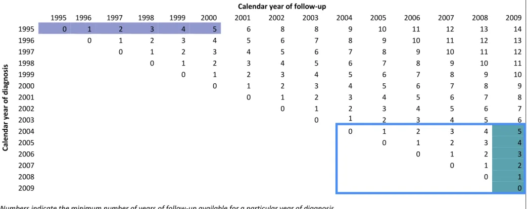
3.5 COHORT, PERIOD AND COMPLETE APPROACHES ... 79
3.6 AGE-STANDARDISATION ... 82
3.7 STATISTICAL ANALYSES... 83
CHAPTER 4: HISTOLOGICAL GROUPS OF OVARIAN CANCER: WORLDWIDE DISTRIBUTION ... 85
4.1 INTRODUCTION ... 85
4.2 MATERIAL AND METHODS ... 86
4.3 RESULTS ... 91
4.3.1 TOPOGRAPHICAL SUB-SITE ... 91
4.3.2 OVARIAN CANCER HISTOLOGY ... 93
4.4 DISCUSSION ... 136
CHAPTER 5: OVARIAN CANCER SURVIVAL BY HISTOLOGICAL GROUP ... 144
5.1 INTRODUCTION ... 144
5.2 MATERIAL AND METHODS ... 144
5.3 RESULTS ... 151
5.3.1 HISTOLOGICAL GROUP BY COUNTRY AND CALENDAR PERIOD ... 151
5.3.2 HISTOLOGICAL GROUP BY SUB-SITE ... 199
5.3.3 SURVIVAL FOR HISTOLOGICAL SUBTYPES ... 203
5.4 DISCUSSION ... 205
CHAPTER 6: OVARIAN CANCER SURVIVAL BY STAGE AT DIAGNOSIS ... 211
6.1 INTRODUCTION ... 211
6.2 MATERIAL AND METHODS ... 211
10
6.3.1 STAGE AT DIAGNOSIS ... 216
6.3.2 STAGE AT DIAGNOSIS AND HISTOLOGY ... 232
6.4 DISCUSSION ... 237
CHAPTER 7: OVARIAN CANCER SURVIVAL BY RACE/ETHNICITY ... 241
7.1 INTRODUCTION ... 241
7.2 MATERIAL AND METHODS ... 241
7.3 RESULTS ... 244
7.4 DISCUSSION ... 250
CHAPTER 8: STRENGTHS AND LIMITATIONS ... 252
8.1 STRENGTHS ... 252
8.2 LIMITATIONS ... 252
CHAPTER 9: CONCLUSION ... 256
9.1 DISTRIBUTION OF HISTOLOGY ... 256
9.2 SURVIVAL BY HISTOLOGICAL GROUP ... 257
9.3 SURVIVAL BY STAGE AT DIAGNOSIS ... 258
9.4 SURVIVAL BY RACE/ETHNICITY ... 258
9.5 FUTURE DIRECTIONS ... 259
9.6 CONCLUSION ... 260
REFERENCES ... 264
APPENDIX A: CONCORD-2 DATA SPECIFICATION ... 275
APPENDIX B: ETHICAL APPROVALS ... 325
11
List of tables
TABLE 1.1 COMPATIBILITY OF FIGO AND TNM STAGING SYSTEMS ... 34
TABLE 1.2 SEER SUMMARY STAGE 2000 CLASSIFICATION ... 35
TABLE 2.1 KEY TOPICS AND SEARCH TERMS FOR LITERATURE REVIEW ... 43
TABLE 3.1 MAIN VARIABLES FOR ANALYSIS ... 72
TABLE 4.1 OVARIAN CANCER HISTOLOGICAL GROUPS AND SUBTYPES ... 87
TABLE 4.2 DISTRIBUTION (%) OF TOPOGRAPHY (SUB-SITE) BY CONTINENT AND CALENDAR PERIOD OF DIAGNOSIS, 1995-2009, 51 COUNTRIES ... 92
TABLE 4.3 DISTRIBUTION (%) OF TOPOGRAPHY (SUB-SITE) BY OVARIAN CANCER HISTOLOGICAL GROUP, CALENDAR PERIOD AND CONTINENT, 1995-2009, 51 COUNTRIES ... 95
TABLE 4.4 WORLDWIDE DISTRIBUTION (%) OF OVARIAN CANCER BY HISTOLOGICAL GROUP AND CALENDAR PERIOD, 1995-2009, 51 COUNTRIES ... 100
TABLE 4.5 DISTRIBUTION (%) OF HISTOLOGICAL GROUPS BY CONTINENT AND CALENDAR PERIOD OF DIAGNOSIS, 1995-2009 ... 104
TABLE 4.6 DISTRIBUTION (%) OF HISTOLOGICAL GROUPS BY COUNTRY AND CALENDAR PERIOD OF DIAGNOSIS, 1995-2009... 105
TABLE 4.7 EPITHELIAL OVARIAN CANCER SUBTYPES: WORLDWIDE DISTRIBUTION (%) BY CALENDAR PERIOD, 1995-2009, 51 COUNTRIES ... 121
TABLE 4.8 EPITHELIAL OVARIAN CANCER SUBTYPES: DISTRIBUTION (%) BY CONTINENT AND CALENDAR PERIOD, 1995-2009... 123
TABLE 4.9 EPITHELIAL OVARIAN CANCER SUBTYPES: DISTRIBUTION (%) BY COUNTRY AND CALENDAR PERIOD, 1995-2009... 125
TABLE 5.1 OVARIAN CANCER HISTOLOGICAL GROUPS AND SUBTYPES ... 148
TABLE 5.2 WORLDWIDE DISTRIBUTION (%) OF OVARIAN CANCER HISTOLOGY AND MEAN AGE AT DIAGNOSIS, 1995-2009, 60 COUNTRIES ... 152
TABLE 5.3 FIVE-YEAR AGE-STANDARDISED NET SURVIVAL (NS, %) (95% CI) BY HISTOLOGICAL GROUP (EPITHELIAL TUMOURS), COUNTRY AND CALENDAR PERIOD, 1995-2009, 60 COUNTRIES ... 156
TABLE 5.4 FIVE-YEAR AGE-STANDARDISED NET SURVIVAL (NS, %) (95% CI) BY HISTOLOGICAL GROUP (NON-EPITHELIAL TUMOURS), COUNTRY AND CALENDAR PERIOD, 1995-2009, 60 COUNTRIES ... 168
TABLE 5.5 FIVE-YEAR AGE-STANDARDISED NET SURVIVAL (NS, %) (95% CI) BY COUNTRY AND CALENDAR PERIOD FOR ALL TUMOURS, TUMOURS OF SPECIFIC MORPHOLOGY, TUMOURS OF NON-SPECIFIC MORPHOLOGY AND TUMOURS WITH MISSING MORPHOLOGY, 1995-2009, 60 COUNTRIES ... 184
TABLE 5.6 FIVE-YEAR AGE-STANDARDISED NET SURVIVAL (NS, %) (95% CI) BY SUB-SITE, HISTOLOGICAL GROUP, COUNTRY AND CALENDAR PERIOD, UNITED KINGDOM AND UNITED STATES, 1995-2009 ... 200
TABLE 5.7 FIVE-YEAR AGE-STANDARDISED NET SURVIVAL (NS, %) (95% CI) BY HISTOLOGICAL SUBTYPE, COUNTRY AND CALENDAR PERIOD, UNITED KINGDOM AND UNITED STATES, 1995-2009 ... 204
TABLE 6.1 WORLDWIDE DISTRIBUTION (%) OF STAGE AT DIAGNOSIS AND MEAN AGE AT DIAGNOSIS, 2001-2009, 25 COUNTRIES ... 217
TABLE 6.2 FIVE-YEAR AGE-STANDARDISED NET SURVIVAL (NS, %) (95% CI) BY COUNTRY, REGISTRY, CALENDAR PERIOD AND STAGE AT DIAGNOSIS, 1995-2009, 25 COUNTRIES ... 222
TABLE 6.3 DISTRIBUTION (%) OF STAGE AT DIAGNOSIS BY HISTOLOGICAL GROUP AND CALENDAR PERIOD, UNITED STATES, 2001-2009 ... 234
TABLE 6.4 FIVE-YEAR AGE-STANDARDISED NET SURVIVAL (NS, %) BY HISTOLOGICAL GROUP, STAGE AT DIAGNOSIS AND CALENDAR PERIOD, UNITED STATES, 2001-2009 ... 235
TABLE 7.1 OVARIAN CANCER HISTOLOGICAL GROUPS AND SUBTYPES ... 243
TABLE 7.2 FIVE-YEAR AGE-STANDARDISED NET SURVIVAL (NS, %) BY RACE, COUNTRY AND CALENDAR PERIOD, 1995-2009... 246
12
TABLE 7.3 FIVE-YEAR AGE-STANDARDISED NET SURVIVAL (NS, %) BY RACE, HISTOLOGICAL GROUP AND PERIOD OF DIAGNOSIS, UNITED STATES, 1995-2009 ... 247 TABLE 7.4 FIVE-YEAR AGE-STANDARDISED NET SURVIVAL (NS, %) FOR EPITHELIAL OVARIAN CANCER BY RACE AND
13
List of figures
FIGURE 1.1 THE FEMALE REPRODUCTIVE SYSTEM ... 15
FIGURE 1.2 THE MENSTRUAL CYCLE... 17
FIGURE 1.3 THE HISTOLOGICAL SUBTYPES OF OVARIAN CANCER, INCLUDING FALLOPIAN TUBE AND PRIMARY PERITONEAL TUMOURS ... 19
FIGURE 1.4 WILSON AND JUNGNER CLASSIC SCREENING CRITERIA ... 36
FIGURE 1.5 UPDATES TO THE CLASSIC SCREENING CRITERIA ... 38
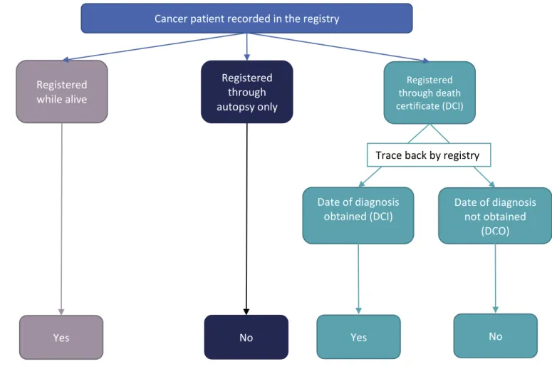
FIGURE 3.1 METHODS OF CANCER PATIENT REGISTRATION AND CRITERIA FOR INCLUSION IN SURVIVAL ANALYSES .... 67
FIGURE 4.1 DATA EXCLUSION FLOW CHART FOR THE WORLDWIDE DISTRIBUTION OF OVARIAN CANCER HISTOLOGY, 1995-2009 ... 88
FIGURE 4.2 ANATOMIC SUB-SITE DISTRIBUTION WITHIN HISTOLOGICAL GROUP BY CONTINENT, 1995-2009 ... 94
FIGURE 4.3 WORLDWIDE DISTRIBUTION (%) OF OVARIAN CANCER HISTOLOGY: 51 COUNTRIES, 1995-2009 ... 101
FIGURE 4.4 DISTRIBUTION (%) OF OVARIAN CANCER HISTOLOGICAL GROUPS BY CONTINENT, 2005-2009 ... 103
FIGURE 4.5 DISTRIBUTION (%) OF OVARIAN CANCER HISTOLOGICAL GROUPS BY COUNTRY (CENTRAL AND SOUTH AMERICA AND NORTH AMERICA), 2005-2009 ... 116
FIGURE 4.6 DISTRIBUTION (%) OF OVARIAN CANCER HISTOLOGICAL GROUPS BY COUNTRY (ASIA AND OCEANIA), 2005-2009 ... 117
FIGURE 4.7 DISTRIBUTION (%) OF OVARIAN CANCER HISTOLOGICAL GROUPS BY COUNTRY (EUROPE), 2005-2009 ... 118
FIGURE 5.1 DATA EXCLUSION FLOW CHART FOR NET SURVIVAL ANALYSIS BY OVARIAN CANCER HISTOLOGICAL GROUP, 1995-2009 ... 146
FIGURE 5.2 FIVE-YEAR AGE-STANDARDISED NET SURVIVAL (%) FOR TYPE I EPITHELIAL TUMOURS, 1995-1999 .... 153
FIGURE 5.3 FIVE-YEAR AGE-STANDARDISED NET SURVIVAL (%) FOR TYPE I EPITHELIAL TUMOURS, 2000-2004 .... 154
FIGURE 5.4 FIVE-YEAR AGE-STANDARDISED NET SURVIVAL (%) FOR TYPE I EPITHELIAL TUMOURS, 2005-2009 .... 155
FIGURE 5.5 FIVE-YEAR AGE-STANDARDISED NET SURVIVAL (%) FOR TYPE II EPITHELIAL TUMOURS, 1995-1999 ... 164
FIGURE 5.6 FIVE-YEAR AGE-STANDARDISED NET SURVIVAL (%) FROM TYPE II EPITHELIAL TUMOURS, 2000-2004 165 FIGURE 5.7 FIVE-YEAR AGE-STANDARDISED NET SURVIVAL (%) FROM TYPE II EPITHELIAL TUMOURS, 2005-2009 166 FIGURE 6.1 DATA EXCLUSION FLOW CHART FOR NET SURVIVAL ANALYSIS BY STAGE AT DIAGNOSIS, 2001-2009 ... 213
FIGURE 6.2 FIVE-YEAR AGE-STANDARDISED NET SURVIVAL (%) FOR LOCALISED TUMOURS, 2004-2009 ... 219
FIGURE 6.3 FIVE-YEAR AGE-STANDARDISED NET SURVIVAL (%) FOR ADVANCED TUMOURS, 2004-2009 ... 220
FIGURE 6.4 FIVE-YEAR AGE-STANDARDISED NET SURVIVAL (%) FOR TUMOURS WITH MISSING STAGE, 2004-2009221 FIGURE 6.5 FIVE-YEAR AGE-STANDARDISED NET SURVIVAL (%) FOR OVARIAN CANCER BY STAGE AT DIAGNOSIS AND HISTOLOGICAL GROUP, UNITED STATES, 2004-2009 ... 236
14
Chapter 1: Introduction
Ovarian cancer ranks 7th in both incidence and mortality among women worldwide. In 2012, an estimated 238,000 women were diagnosed with and 151,000 women died from ovarian cancer, representing 4% of all new cancer diagnoses and 4% of cancer deaths among women1. Early symptoms, such as persistent abdominal pain, bloating or decreased appetite, are vague2, and most women present with disease at an advanced stage3.
1.1
Anatomy and biology of the ovary and fallopian tubes
1.1.1
Ovaries
The ovary is the primary endocrine gland of the female reproductive system. It has two main functions: to produce the eggs and to secrete the female sex hormones, oestrogen and progesterone4,5. Suspended within the peritoneal cavity by the broad ligament, the ovaries are paired organs that are attached to either side of the body of uterus by the ovarian ligaments [Figure 1.1]. The ovarian ligaments extend from the posterior side of the uterus as the round ligament. The tubal extremity of the ovary is attached to the broad ligament by the suspensory ligament of the ovary. The tubal extremity is the area of the ovary closest to the fimbriated end of the fallopian tube5. Recent evidence suggests that the fimbriated end of the fallopian tube may be the primary site of origin for most pelvic high-grade serous tumours among women6.
The ovary has three main components: surface, cortex and medulla. The surface is made up of epithelial cells, one of the three main types of cell found in the ovaries. The cortex is located just below the ovarian surface epithelium and contains the outer supporting stroma and the follicles, which produce the eggs. Stromal cells form the supporting structural tissue of the cortex and produce oestrogen and progesterone, and germ cells are found in the follicles. The medulla is the central part of the ovary: it contains the inner
15
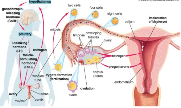
Figure 1.1 The female reproductive system
Source: Editors of Encyclopædia Britannica. Fallopian tube: anatomy - uterus
[illustration]. 2012. [cited 12 September 2016]. Available from:
16
supporting stroma and a rich neurovascular network providing blood to the ovary from the ovarian arteries7.
1.1.2
Fallopian tubes
The fallopian tubes comprise the fimbriae, infundibulum, ampulla and isthmus [Figure 1.1]. The infundibulum is the end of the tube closest to the ovary into which the finger-like fimbriae help gather the ovulated egg(s) into the fallopian tube. The ampulla is the longest part of the tube and the most common site of fertilisation of the egg by a sperm. The fallopian tube narrows at the uterine end to form the isthmus which enters into the body of the uterus5.
1.1.3
The menstrual cycle
The ovaries are involved in the regulation of the menstrual cycle as part of the endocrine system, through a complex feedback loop [Figure 1.2]. The cycle begins with the follicular phase when the hypothalamus recognises low levels of oestrogen and progesterone in the bloodstream and secretes gonadotropin-releasing hormone (GnRH). The pituitary gland responds to the release of GnRH by producing and releasing luteinising hormone (LH) and follicle-stimulating hormone (FSH). LH and FSH signal the ovaries to produce oestrogen and progesterone, which stimulate growth of the follicles and prepare the uterus for pregnancy. During the follicular phase, one dominant egg-producing follicle develops from a primordial follicle to a matured egg. Once the follicle has completely matured, ovulation occurs. During ovulation, the ovarian surface epithelium ruptures to release the egg into the fimbriated end of the fallopian tube where it then travels to the uterus4.
The second half of the menstrual cycle is called the luteal phase and occurs immediately after ovulation. During the luteal phase, the lining of the dominant follicle grows to form a corpus luteum [Figure 1.2]. The corpus luteum temporarily secretes oestrogen and
17
Figure 1.2 The menstrual cycle
Source: Editors of Encyclopædia Britannica. Fallopian tube: anatomy - pituitary gland: secretion and function of gonadotropins [illustration]. 2013. [cited 12 September 2016]. Available from: https://www.britannica.com/science/fallopian-tube/images-videos/The-hypothalamus-and-pituitary-gland-control-the-secretion-of-gonadotropins/102076.
18
progesterone to thicken the lining of the uterus for pregnancy and prevent further ovulation. If pregnancy does not occur, the corpus luteum stops producing hormones and degenerates. This reduction in oestrogen and progesterone will be recognised by the hypothalamus and a new cycle will begin4.
1.2
Ovarian cancer
Since the early part of the 20th century, it has been recognised that ovarian cancer is not a single disease, but comprised of various histologically different tumour types8. Ovarian cancers have generally been divided into epithelial and non-epithelial groups for many years. Epithelial, germ cell and sex cord-stromal tumours are the commonest types of ovarian cancer. They can be further subdivided into distinct histological subtypes [Figure 1.3]. The developmental pathway and clinical prognosis for a particular ovarian tumour depends upon the histological subtype2.
1.2.1
Epithelial ovarian, fallopian tube and primary peritoneal cancer
Epithelial ovarian cancer is the most common type of ovarian tumour, making up 90% of all primary malignant ovarian cancers9. Histological subtypes of epithelial tumours primarily include: clear cell, endometrioid, mucinous, serous, squamous, transitional cell (Brenner) and undifferentiated carcinoma2,10. Recent work has enabled finer subdivision of epithelial ovarian cancers into different groups according to a combination of morphological, molecular and clinical characteristics10-14. Each histological subtype has distinct molecular pathways that influence chemosensitivity, the pattern of metastasis and the probability of survival11,15.
Under one proposed classification scheme, “type I” epithelial tumours include low-grade serous, endometrioid, clear cell, mucinous, squamous and transitional cell (Brenner) carcinomas. They often present at an early stage, may arise from borderline ovarian tumours or endometriosis, and typically have a good prognosis10-12,16.
19
Malignant ovarian tumours
Germ cell
Dysgerminomas
Immature teratoma
Mixed germ cell
Yolk sac
Embryonal carcinoma
Non-gestational
choriosarcoma
Epithelial
Type I
Low-grade serous Endometrioid Clear cell Mucinous SquamousTransitional cell (Brenner)
Type II
High-grade serous Carcinosarcoma Undifferentiated carcinomaSex cord-stromal
Adult granulosa cell
Sertoli-Leydig cell
Fibrosarcoma
Steroid cell
Other
miscellaneous
20
Using the same classification scheme, “type II” epithelial tumours comprise high-grade serous carcinoma, undifferentiated carcinomas and malignant mixed mesodermal tumours (carcinosarcoma). They account for around 75% of epithelial ovarian cancers, typically present at an advanced stage and have a poor prognosis10-12,16.
Fallopian tube and primary peritoneal carcinoma arise outside the anatomical ovary, but most tumours at these sites are now considered to be part of the spectrum of ovarian malignancies.
Primary fallopian tube carcinoma is a rare cancer that presents clinically in a similar manner to epithelial ovarian cancer and is treated clinically in the same way17. Malignant subtypes of fallopian tube carcinoma include clear cell, endometrioid, mucinous, serous, transitional cell and undifferentiated epithelial carcinomas2. Non-epithelial fallopian tube tumours are extremely rare, accounting for only around 7% of malignant fallopian tube cancers, and can include leiomyosarcoma and germ cell tumours2,18.
Primary peritoneal carcinoma in women is also extremely rare. Macroscopically, primary peritoneal carcinoma may look like an epithelial ovarian carcinoma that has spread to the abdomen, and microscopically, the cells often resemble those of epithelial ovarian carcinoma19. Primary peritoneal carcinomas are also managed in the same way as advanced-stage epithelial ovarian cancer10,17. Non-epithelial types of primary peritoneal cancer do occur, such as malignant mesothelioma, desmoplastic small round cell tumours and solitary fibrous tumours, but these types account for only one-third of primary peritoneal cancers2,18.
Due to the anatomical location of the three sites, primary fallopian tube and peritoneal carcinomas may be diagnosed as primary epithelial ovarian cancer12,17. Around 15% of tumours diagnosed as primary ovarian have been found to actually be primary peritoneal tumours2. During 1995-2004, age-adjusted annual incidence of primary ovarian
21
carcinoma in the US was much higher (119.9 per million) than for primary peritoneal (6.78 per million) or fallopian tube (3.72 per million) carcinomas18. The incidence of ovarian carcinoma fell over the 33-year period from 1973 to 2005, and the incidence of peritoneal and fallopian tube carcinomas increased. The decrease in incidence in ovarian cancer over time may be partially artificial and attributable to the establishment of guidelines in 1993 to define primary peritoneal carcinoma18,20.
The guidelines for diagnosis of primary fallopian tube cancer are more restrictive than those for primary ovarian cancer. In order for a tumour to be considered a primary fallopian tube carcinoma, the majority of the tumour has to be within the fallopian tube rather than the ovary, and there must be evidence of an intraepithelial tubal carcinoma. Additionally, there must be a clear transition from benign to malignant epithelium21.
A diagnosis of primary peritoneal cancer is rare because the guidelines for diagnosis are also very restrictive. Regardless of whether there is extensive tumour involvement of the peritoneum or other abdominal organs, if the tumour within the ovary is greater than 5mm, the cancer is, nevertheless, considered to be primary ovarian rather than primary peritoneal carcinoma12,20.
Subtypes of epithelial ovarian, fallopian tube and primary peritoneal cancer
Serous
Serous carcinoma is the most common histological subtype at all three primary sites2,18. Around 40-50% of ovarian cancers and around 66-90% of fallopian tube carcinomas are serous tumours2,22. Non-serous peritoneal carcinoma is extremely uncommon2.
Serous tumours of ovarian, tubal and peritoneal origin are divided into low-grade and high-grade serous carcinoma depending upon the degree of differentiation2. Low-grade serous carcinoma (LGSC) is distinct from high-grade serous carcinoma (HGSC); progression from low-grade to high-grade only occurs rarely10. Low-grade serous ovarian carcinomas
22
are relatively rare, accounting for only 5% of serous ovarian carcinomas. Low-grade serous fallopian tube carcinoma is also very rare2.
High-grade serous ovarian carcinoma is the most common subtype of epithelial ovarian cancer, and women with high-grade serous ovarian tumours typically present at older ages than women with low-grade tumours. Most serous peritoneal tumours are high-grade and resemble high-high-grade serous ovarian tumours2.
Endometrioid carcinoma
Endometrioid tumours are the second most common type of ovarian and fallopian tube carcinoma18. Around 20-33% of women diagnosed with endometrioid tumours also have primary endometrial cancer or hyperplasia9. Most endometrioid tumours are early-stage and are confined to the ovary at diagnosis, with only 17% of women diagnosed with bilateral tumours. Endometrioid tumours are generally well-differentiated and low-grade, and women with low-grade tumours have higher survival than women diagnosed with high-grade tumours2.
Clear cell carcinoma
Clear cell tumours are generally large (around 15 cm in diameter), but unilateral2. Around 85% of clear cell tumours are stage I or II at diagnosis9. Survival from early-stage disease is high, but advanced-stage disease does not respond well to chemotherapy and thus, survival is lower2.
Mucinous carcinoma
Mucinous tumours account for around 3-4% of all ovarian tumours2. Mucinous ovarian tumours are usually stage I at diagnosis and are well-differentiated or moderately-differentiated. Survival from early-stage mucinous tumours is generally high, but advanced-stage disease, though rare, is often aggressive and does not respond well to chemotherapy. Metastatic tumours from other sites, such as the gastrointestinal tract, often mimic a primary ovarian mucinous tumour. Thus, identifying a mucinous tumour as
23
a primary ovarian tumour is often difficult. Primary ovarian tumours are more frequently unilateral and larger than mucinous tumours at other primary sites23.
Transitional cell (Brenner) carcinoma
Transitional cell tumours are large (16-20 cm in diameter) and usually confined to the ovary at diagnosis. Around 80% of these tumours are stage I at diagnosis and only 12% are bilateral. Women diagnosed with stage I disease have high 5-year survival, while women diagnosed with tumours that have spread outside the ovaries have similar survival to other advanced epithelial tumours2.
Squamous carcinoma
Squamous carcinomas are an extremely rare form of malignant epithelial ovarian tumour comprised of squamous cells that do not originate from germ cells. Squamous tumours are primarily high-grade and survival from these tumours is poor19. Around 34% of women diagnosed with squamous carcinoma have stage I disease, while 21% and 25% have stage III and stage IV, respectively. Survival varies by stage and can be as high as 86% for early stage tumours, but as low as 3% for stage IV tumours24.
Carcinosarcoma
Carcinosarcomas can also be referred to as malignant mixed mesodermal or malignant mixed Müllerian tumours. These tumours are rare, accounting for only 2% of ovarian tumours. Most women are diagnosed at advanced stages with high-grade large tumours (14 cm)2.
Undifferentiated carcinoma
An undifferentiated carcinoma is an epithelial tumour without differentiation, thus identifying a specific cell type is impossible. Most women diagnosed with undifferentiated carcinoma have advanced-stage disease and poor survival2.
24
Epidemiology of epithelial tumours
A strong family history of either breast or ovarian cancer in a first degree relative at an early age is a primary risk factor for epithelial ovarian cancer17. Older age of the woman is another important risk factor, and the median age at diagnosis for epithelial ovarian cancer is 60 years25. In a recent study from the US, peritoneal carcinomas were generally diagnosed at older ages (mean age at diagnosis of 67 years) than fallopian tube (mean age at diagnosis of 64 years) and ovarian (mean age at diagnosis of 63 years) carcinomas18.
While only 10% of epithelial ovarian cancers are due to genetic abnormalities, women with BRCA1 mutations have a 35-50% increased risk of disease, and women with BRCA2 mutations have a 10-30% increased risk26. Lynch syndrome can also increase a woman’s risk of ovarian cancer by 3-33%27. Women with BRCA mutations are more likely to develop type II tumours12. BRCA mutations are also more common in women with fallopian tube and peritoneal cancer than women in the general population2. Additional risk factors include endometriosis, nulliparity, early menarche, late menopause and lack of oral contraceptive use9,26.
Biological mechanisms of epithelial tumours
The pathogenesis of ovarian cancer is not fully understood. There are two conventional theories regarding the development of epithelial ovarian cancer. The first theory refers to incessant ovulation, or the repeated wounding and repair of the surface epithelium of the ovary during ovulation28. This repetition increases epithelial proliferation for repair and, therefore, the frequency of DNA mutations and the formation of ovarian cortical inclusion cysts25. Ovarian inclusion cysts form through invagination of the ovarian surface epithelium into the surrounding ovarian stroma12. The second theory involves the increase of gonadotropic hormones (LH and FSH) during ovulation, which also increase proliferation, potentially leading to malignant transformation of the epithelium of ovarian inclusion cysts25,26,29. Most risk factors traditionally associated with epithelial ovarian
25
cancer, such as older age, nulliparity, early menarche, late menopause and lack of oral contraceptive use, can be used to support both theories of the development of ovarian cancer because they are all associated with an increased lifetime number of ovulations. Additionally, BRCA mutations result in a decreased ability to repair genetic damage, which increases risk of disease.
The primary critique of the theory of incessant ovulation is that the ovarian surface epithelium does not resemble the main histologic types of epithelial ovarian tumours (serous, endometrioid, mucinous, clear cell or transitional cell). The second theory, involving gonadotropic hormones, attempts to mitigate this weakness by assuming that, prior to malignant transformation, ovarian inclusion cysts undergo metaplastic change. The cysts, which comprise the mesothelium (the cell type of the ovarian surface epithelium), undergo metaplasia and are converted to cell types representing the primary histological subtypes of epithelial ovarian cancer12,25.
Epithelial ovarian cancer has conventionally been defined as cancer that begins in the ovaries and this idea is central to both traditional theories of epithelial ovarian cancer development. However, while some epithelial ovarian tumours may start in the ovaries, given the lack of evidence of a precursor lesion arising from or in the ovary, it is possible that some epithelial ovarian tumours may originate outside the ovaries and only involve the ovaries secondarily12.
LGSC is thought to develop in a step-wise manner from a serous cystadenoma or adenofibroma to an atypical proliferative serous tumour (APST). APSTs are serous borderline tumours which then progress to non-invasive micropapillary serous borderline tumours (MPSC). Once an MPSC becomes invasive, it is considered an LGSC30.
The pathogenesis of HGSC is less clear than that of LGSC30. Most high-grade serous ovarian tumours are now thought to originate in the fallopian tubes rather than the ovary31-33.
26
Recent studies examining the fallopian tubes of high-risk women with BRCA mutations have found that early-stage invasive tubal carcinomas are present in up to 70% of women with high-grade serous ovarian carcinoma, and serous tubal intraepithelial carcinoma (STIC) is now considered to be a precursor lesion for this particular histological subtype of epithelial ovarian cancer. Malignant cells from a STIC may shed and implant on the ovarian surface during ovulation when the fimbriated end of the fallopian tube is in close contact with the ovary. Additionally, normal, non-malignant tubal epithelial cells may shed from the fimbria and implant on the ovary to form an inclusion cyst. Once implanted, the cyst may then undergo malignant transformation12.
The aetiology of fallopian tube cancer is unknown, but some studies show a protective effect of oral contraceptive use and parity, similar to the pattern observed for serous ovarian carcinoma22. Similarly, the aetiology for primary peritoneal carcinoma is unknown and difficult to establish because this type of cancer is so rare. There is some evidence that primary peritoneal carcinomas develop along the same pathway as ovarian carcinoma, particularly high-grade serous ovarian carcinoma18. However, while contraceptive use may decrease the risk of primary peritoneal carcinoma, increasing parity may not be as protective as for ovarian cancer22.
Endometrioid and clear cell tumours are known to develop from endometriotic cysts, which are said to be the result of endometrial tissue implanted on the ovary26 or passing through the fallopian tube12 due to endometriosis. Endometriosis is a disease that primarily affects women of reproductive age, and around 10% of women of reproductive age have endometriosis. Endometriosis is when cells similar to the ones lining the uterus are found outside the womb; these cells are linked to the menstrual cycle, growing and bleeding along with the cells lining the uterus. However, unlike the cells lining the uterus, the cells are not shed from the body during menstruation. This can cause inflammation,
27
pain and the formation of scar tissue34. Endometriosis is an established risk factor for ovarian cancer26.
The molecular development of transitional cell and mucinous tumours is not well established, but these tumours may develop from transitional cell nests at the tubal-mesothelial junction. Transitional cell nests are clusters of benign epithelial cells, usually located in the connective tissue of the fallopian tubes. Transitional cell and mucinous tumours may develop from these cell nests located in the transitional epithelium between the fallopian tubes and peritoneum. Further, it is believed that mucinous and transitional cell tumours develop along the same molecular pathway and may develop simultaneously, although transitional cell tumours tend to be small and slow-growing, while mucinous tumours tend to be large and fast-growing12.
Treatment guidelines for epithelial tumours
Staging of ovarian cancer requires a surgical procedure to examine the spread of disease. During an exploratory laparotomy, samples of tissue from the ovary, fallopian tube, pelvic lymph nodes, omentum and diaphragm are biopsied and examined microscopically for malignant cells. The stage of disease is then based on the size, extent and location of the tumour (see section 1.4). Treatment for ovarian cancer depends on the stage of disease, therefore accurate staging of disease is critical for receipt appropriate treatment17.
Standard treatment for early-stage epithelial ovarian cancer includes surgery, usually consisting of hysterectomy, bilateral salpingo-oophorectomy and omentectomy, and platinum-based chemotherapy. Early-stage tumours are generally confined to the ovary and are well-differentiated. For those tumours, salpingo-oophorectomy (unilateral or bilateral) may be adequate on its own. Women with early-stage and well-differentiated tumours may receive unilateral salpingo-oophorectomy to preserve fertility. Adjuvant
28
chemotherapy with cisplatin, carboplatin and paclitaxel is recommended for all early-stage patients, except those with early-stage IA well-differentiated tumours17.
Treatment for advanced-stage tumours includes total abdominal hysterectomy, bilateral salpingo-oophorectomy, omentectomy, and other procedures to remove the tumour, such as debulking, followed by combination chemotherapy17. Optimal cytoreduction – where the residual tumour is less than 1cm – has been shown to improve survival significantly. Systemic chemotherapy is recommended for women who have residual disease of 1cm or more after surgery. Around 80% of advanced-stage tumours will relapse and should then be treated with either platinum-based treatment if the tumour was platinum-sensitive (i.e. the disease relapsed six months or more after cessation of initial treatment), or alternative options for platinum-resistant disease (i.e. the disease recurred within six months of stopping initial treatment or progressed during induction therapy)17.
Advanced-stage epithelial tumours may also be treated with intraperitoneal (IP) chemotherapy after surgery. Clinical trials have shown favourable outcomes for IP chemotherapy for women with platinum-sensitive, small residual tumours. Hyperthermic peritoneal chemotherapy (HIPEC) is another treatment option that has been only recently used to treat ovarian cancer. Exploratory trials are in progress to examine the most effective drug combination and time at target temperatures for drug delivery, in addition to defining which women will benefit the most from HIPEC17.
Treatment for fallopian tube and peritoneal carcinomas is the same as for epithelial ovarian tumours17,18.
1.2.2
Germ cell
Germ cell tumours are responsible for 3% of invasive ovarian tumours worldwide, though they can account for up to 20% of ovarian tumours in some East Asian countries19. Germ cell tumours include several histologically distinct tumour subtypes2. The majority (95%)
29
of germ cell tumours are benign mature cystic teratomas19,35. Dysgerminomas are the most frequent subtype of malignant germ cell tumour, but only make up 1-2% of all malignant ovarian tumours. Immature teratomas are the second most common subtype of malignant germ cell tumours. Mixed germ cell tumours represent about 8% of malignant germ cell tumours and are a mixture of two or more malignant germ cell tumours. The most common mixed germ cell tumour is a mixed dysgerminoma and yolk sac tumour. Pure yolk sac tumours, non-gestational choriocarcinomas and embryonal carcinomas are extremely rare subtypes of malignant germ cell tumours2.
Epidemiology of germ cell tumours
Germ cell tumours represent 60% of all malignant ovarian tumours among women aged 21 years or younger19. The average age at diagnosis for dysgerminoma is 22 years, immature teratomas are most common among women aged 30 or younger and the mean age at diagnosis is 16 years for mixed germ cell tumours2. While most risk factors for germ cell tumours are unknown, congenital malformations of the genital tract, Turner’s syndrome and gonadoblastomas are possible risk factors for dysgerminoma9.
The majority (60-70%) of malignant germ cell tumours are stage I or II at diagnosis, while only 20-30% are stage III. Stage IV tumours are extremely rare36.
Biological mechanisms of germ cell tumours
Germ cell tumours develop from benign germ cells, which are the egg-producing cells within the ovary. However, the pathway to malignant transformation of these cells is not clearly understood19,37. The duration of symptoms prior to diagnosis is generally only two to four weeks; therefore, germ cell tumours are thought to develop rapidly36.
Treatment guidelines for germ cell tumours
As germ cell tumours are primarily unilateral, standard surgery may be more conservative than for epithelial tumours38. Standard treatment typically involves unilateral salpingo-oophorectomy or total abdominal hysterectomy and bilateral salpingo-salpingo-oophorectomy,
30
plus platinum-based adjuvant combination chemotherapy. If the cancer is early-stage, unilateral salpingo-oophorectomy may be performed to preserve fertility in young women, and chemotherapy may not be required. For advanced-stage disease, unilateral salpingo-oophorectomy if followed by chemotherapy may be performed instead of bilateral salpingo-oophorectomy and hysterectomy, in order to preserve fertility39. Germ cell tumours generally respond well to chemotherapy38.
1.2.3
Sex cord-stromal
Sex cord-stromal tumours are a diverse group of rare ovarian tumours that can involve a variety of different cell types40. Subtypes of malignant sex cord-stromal tumours primarily include fibrosarcoma, steroid cell, adult granulosa cell and Sertoli-Leydig cell tumours2,19. The diversity of cell types and the fact that the tumours may be composed of one or more cell type leads to difficulty in correctly identifying the tumour subtype40. Adult granulosa cell tumours, the most common subtype, only comprise 1% of all malignant ovarian tumours and steroid cell tumours only account for 0.1% of ovarian tumours2.
Adult granulosa cell tumours are generally low-grade, unilateral and confined to the ovary at diagnosis2. Though adult granulosa cell tumours are slow-growing, these tumours have been known to recur up to 20 years after the initial diagnosis2. Sertoli-Leydig cell tumours are usually confined to the ovaries at diagnosis, and are well-differentiated40. A recent study found that 86% of Sertoli-Leydig cell tumours were stage I at diagnosis41.
Epidemiology of sex cord-stromal tumours
While the incidence of sex cord-stromal tumours is highest among women in their fifties26, these tumours can occur throughout the reproductive years and after menopause19,42. The average age of adult granulosa cell patients is 53 years, but the mean age for Sertoli-Leydig cell tumours is 25 years2. Risk factors for sex cord-stromal tumours are not well known, but may include race/ethnicity, obesity, family history of breast or ovarian cancer,
31
lack of oral contraceptive use and nulliparity, particularly for granulosa cell tumours43. BRCA mutations do not increase the risk of granulosa cell tumours44.
Biological mechanisms of sex cord-stromal tumours
The aetiology of sex cord-stromal tumours is unknown; however, the development of granulosa cell tumours may be associated with infertility and the use of ovulation-stimulating drugs19,44,45. There are two proposed pathways for development of granulosa cell tumours due to ovulation-stimulating drugs: the granulosa cell tumour may already exist within the ovary and the hormonal drugs trigger growth, or increased amounts of follicle-stimulating hormone may be carcinogenic to granulosa cells44,45. Granulosa cell tumours may also be associated with endometrial hyperplasia due to stimulation of the endometrium in response to the increase in oestrogen, which is secreted by granulosa cell tumours. Around 13% of women diagnosed with granulosa cell tumours also develop well-differentiated endometrial adenocarcinoma44.
Treatment guidelines for sex cord stromal tumours
The treatment for sex cord-stromal tumours is similar to that for epithelial ovarian tumours. Early-stage disease may be treated with conservative surgery, consisting of only unilateral salpingo-oophorectomy44. Adjuvant chemotherapy may be used to treat early-stage disease, but the benefits of such treatment are not yet confirmed42,46,47. For advanced disease, total abdominal hysterectomy, bilateral salpingo-oophorectomy and debulking surgery may be performed along with administration of platinum-based combination chemotherapy42,44,47. Accurate staging of the tumour is of particular importance because of the higher tendency for advanced-stage disease to relapse many years later. All patients should be observed indefinitely after initial treatment42.
1.3
Definition of ovarian cancer
Given the newly proposed theories of development for serous ovarian carcinoma involving the fallopian tubes, and the extra-ovarian nature of the development of
32
endometrioid, clear cell, mucinous and transitional cell tumours, it seems likely that the majority of epithelial ovarian tumours may actually originate outside the ovary10,12. Additionally, the restrictive guidelines for primary fallopian tube and peritoneal carcinoma lead to a bias in diagnosing pelvic tumours as “ovarian” carcinoma. Since 2000, fallopian tube and primary peritoneal carcinomas have been included in ovarian cancer trials17. Therefore, for the purposes of this thesis, the definition of ovarian cancer will include primary fallopian tube and peritoneal cancer as well as tumours of the uterine ligaments and adnexa, and other specified and unspecified female genital organs (International Classification of Diseases for Oncology, 3rd edition (ICD-O-3) topography codes C48.0-C48.2, C56.9, C57.0-C57.4 and C57.7-C57.9)48.
1.4
Classification systems for stage of disease at diagnosis
The stage of disease is important for accurate treatment of ovarian, tubal and peritoneal cancer. When examining cancer survival, stage at diagnosis is key, and the staging system should not allow for changes in stage after biopsy or initial treatment. The stage of the disease describes the extent of the spread of disease and is based on location of the primary tumour, tumour size, lymph node involvement and metastasis at diagnosis. Unless the disease is advanced at diagnosis, ovarian cancer is generally staged through surgery and pathological analysis of tissue samples of the tumour49. Three main staging systems for ovarian cancer are used: the Fédération Internationale de Gynécologie et d'Obstétrique (FIGO) system, the Union for International Cancer Control’s (UICC) Tumour Node Metastasis (TNM) system and the Surveillance, Epidemiology and End Results (SEER) programme’s Summary Stage 2000. Agreement between FIGO, UICC and the American Joint Committee for Cancer ensures that the different staging systems for ovarian cancer are compatible and comparable between countries.33
1.4.1
Fédération Internationale de Gynécologie et d'Obstétrique (FIGO)
The FIGO staging classification system for is unique to gynaecological tumours [Table 1.1]49. The staging guidelines are applicable to all histological subtypes of ovarian cancer, as well as primary fallopian tube and primary peritoneal carcinoma.
1.4.2
Tumour node metastasis (TNM)
Ovarian cancer can also be staged using the TNM system50. Tumours are classified on the basis of the size and extent of the primary tumour (T), involvement of regional lymph nodes (N) and the presence or absence of metastasis (M). Individual TNM values can be combined to create a grouped variable representing stages I-IV, which are compatible with FIGO stages I-IV. T, N and M may be determined through pathological or clinical examination, or both. Clinical examination for staging can include physical examination or imaging, while pathological examination involves microscopic examination of the tumour.
1.4.3
Surveillance, Epidemiology, and End Results (SEER) Summary
Stage 2000
SEER Summary Stage 2000 is primarily used by the North American Association of Central Cancer Registries51. It was developed by the US National Cancer Institute’s SEER programme. SEER Summary Stage 2000 is compatible with TNM and the stages and definitions of each stage are listed in Table 1.2. Previous work has shown that for ovarian cancer there is very little misclassification when converting SEER Summary Stage 2000 to TNM52.
1.5
Screening for ovarian cancer
Successful screening techniques for ovarian cancer have been difficult to develop. Screening tests should follow the classic guidelines proposed by Wilson and Jungner in 1968 [Figure 1.4]53. These guidelines have been updated over the past few decades, and new criteria build upon the guidelines originally proposed by Wilson and Jungner
34
Table 1.1 Compatibility of FIGO and TNM staging systems
FIGO T Na Mb Definition
I T1 N0 M0 Tumour limited to the ovaries or fallopian tubes
IA T1a N0 M0
Tumour limited to one ovary, capsule intact, or fallopian tube, no tumour on surface, no malignant cells in ascites or peritoneal washings
IB T1b N0 M0
Tumour limited to both ovaries, capsules intact, or fallopian tubes, no tumour on surface, no malignant cells in ascites or peritoneal washings
IC T1c1-3 N0 M0
Tumour limited to one or both ovaries or fallopian tubes with any of the following: surgical spill, capsule ruptured, tumour on ovarian surface, malignant cells in ascites or peritoneal washings
II T2 N0 M0
Tumour involves one or both ovaries or fallopian tubes with pelvic extension or primary peritoneal cancer
IIA T2a N0 M0 Extension and/or implants on the uterus and/or tubes and/or ovaries
IIB T2b N0 M0 Extension to other pelvic tissues
III T1/T2 N1 M0
Tumour involves one or both ovaries or fallopian tubes, or primary peritoneal cancer, with
microscopically confirmed peritoneal spread outside the pelvis and/or metastasis to the retroperitoneal lymph nodes
IIIA T3a2 N0/N1 M0 Positive retroperitoneal lymph nodes only or
microscopic peritoneal metastasis beyond the pelvis IIIB T3b N0/N1 M0 Macroscopic peritoneal metastasis beyond the pelvis
< 2cm
IIIC Any T N0/N1 M0 Macroscopic peritoneal metastasis beyond the pelvis > 2cm
IV Any T Any N M1 Distant metastasis excluding peritoneal metastases IVA Any T Any N M1 Pleural effusion with positive cytology
IVB Any T Any N M1 Parenchymal metastases and metastases to extra-abdominal organs
a N0 indicates no regional lymph node involvement and N1 indicates regional lymph node involvement. b M0 indicates no metastasis and MI indicates metastasis.
35
Table 1.2 SEER Summary Stage 2000 Classification
Stage Definition 0 In situ
1 Localised only
2 Regional spread by direct extension only 3 Regional lymph nodes involved only
4 Regional spread by both direct extension and lymph node involvement 5 Regional, NOS (not otherwise specified)
7 Distant site(s) or lymph node(s) are involved 9 Unknown if there is extension or metastasis
36
Wilson and Jungner classic criteria for screening
1. The condition sought should be an important health problem. 2. There should be an accepted treatment for patients with recognised
disease.
3. Facilities for diagnosis and treatment should be available.
4. There should be a recognisable latent or early symptomatic stage. 5. There should be a suitable test or examination.
6. The test should be acceptable to the population.
7. The natural history of the condition, including development from latent to declared disease, should be adequately understood.
8. There should be an agreed policy on whom to treat as patients. 9. The cost of case-finding (including diagnosis and treatment of patients
diagnosed) should be economically balanced in relation to possible expenditure on medical care as a whole.
10. Case-finding should be a continuing process and not a “once and for all” project.
37
[Figure 1.5]54. As ovarian cancer is a leading cause of death from gynaecological malignancy worldwide, it meets the first criterion for screening. Standard treatment guidelines for most types of ovarian cancer exist and access to treatment is generally available, which satisfy the second and third criteria.
Screening tests will only be useful in reducing mortality if a precursor lesion can be detected and the rate of tumour growth is slow enough to allow for early disease detection. Mathematical modelling using data from risk-reducing salpingo-oophorectomies among women with BRCA1 mutations suggests that serous tumours may be in situ, stage I or stage II for more than four years, and stage III or stage IV for an additional year, before they present clinically55. While it appears there may be a lengthy latent period for serous ovarian cancer, the latent period is not easily recognisable and there is no early symptomatic stage for most ovarian tumours, which is required for the fourth criterion.
Serous tumours tend to be small and slow-growing during this occult phase of development, remaining only 1 cm in diameter for the majority of the time before increasing to only 3 cm as the tumour progresses to stage III or IV55. Once the disease is stage III or IV, tumours grow rapidly, doubling in size every 2-3 months56. This evidence suggests that in order for a screening test to achieve 50% sensitivity, an annual screening test would need to be able to detect tumours as small as 1.3 cm in diameter. For 80% sensitivity, the screening test would need to detect tumours less than 0.4 cm in diameter, and for a 50% reduction in mortality from serous tumours, the annual screening test would need to detect tumours 0.5 cm in diameter55. Previous work has shown that in order to achieve a positive predictive value of 10% for a screening test for epithelial ovarian cancer (meaning that 10% of women who screen positive for epithelial ovarian
38
Updates to the classic screening criteria
1. The screening programme should respond to a recognised need. 2. The objectives of screening should be defined at the outset. 3. There should be a defined target population.
4. There should be scientific evidence of screening programme effectiveness. 5. The programme should integrate education, testing, clinical services and
programme management.
6. There should be quality assurance, with mechanisms to minimize potential risks of screening.
7. The programme should ensure informed choice, confidentiality and respect for autonomy.
8. The programme should promote equity and access to screening for the entire target population.
9. Programme evaluation should be planned from the outset. 10. The overall benefits of screening should outweigh the harm.
39
cancer actually have the disease), the screening test should have a sensitivity of greater than 75% and a specificity of at least 99.6%25.
Screening methods previously assessed in trials may not be able to detect such small tumours required to achieve a mortality benefit from annual screening. Lead-time bias will occur if a screening test detects disease earlier than it would have been if it had been diagnosed without screening, but does not result in a delay of death. With lead-time bias, the perceived survival time is longer with screening but this “improvement” in survival is due only to the earlier detection of disease through screening rather than an impact of the screening test on mortality. Screening techniques for ovarian cancer include pelvic examination, measurement of serum cancer antigen 125 (CA125) and transvaginal sonography, and used separately these tests have not been successful in reducing ovarian cancer mortality57. However, there is some evidence that when used in combination, ovarian cancer may be detected at an earlier stage. Recent results from the UK Collaborative Trial of Ovarian Cancer Screening (UKCTOCS) show that multimodal screening consisting of transvaginal ultrasound and CA125 assessment using the risk of ovarian cancer algorithm is more likely to lead to earlier diagnosis of ovarian or primary peritoneal cancer than no screening. The UKCTOCS also showed that multimodal screening could prevent up to 20% of ovarian cancer deaths. Women eligible for the trial were post-menopausal and did not have an increased risk of ovarian cancer57. Results from the Prostate, Lung, Colorectal and Ovarian (PLCO) Cancer Screening Trial in the US showed no evidence of a mortality benefit from screening58 and more research is needed to confirm the results from the UKCTOCS.
Further, given the histologically distinct subtypes of ovarian cancer, one screening test is unlikely to be sufficient for detecting all ovarian tumours. Therefore, screening techniques such as the multimodal screening method used in UKCTOCS would be difficult to
40
implement at the population level25. For type I tumours that grow slowly and tend to reach a large size while still confined to the ovary, pelvic examination and transvaginal ultrasound may be effective tools for early diagnosis. However, type I tumours are much less common and less aggressive than type II tumours, thus a mass screening programme for these tumours may not be appropriate. Requiring that screening tests for ovarian cancer focus on detecting tumours while still confined to the ovary is unlikely to be effective for type II tumours, which appear to develop outside the ovary. Effective screening tools for type II tumours might focus on detection while the tumour is small, rather than early stage, and would need to include examination of the fallopian tubes and transvaginal ultrasound12.
The target population for ovarian cancer screening tests may change based on the specific subtype of ovarian tumour for which the test is aiming to detect. Additionally, screening techniques may need to be tailored further for different risk groups because the effect of annual screening on ovarian cancer mortality may vary between low-risk women and high-risk women. High-risk women, particularly those with Lynch syndrome or BRCA mutations, may benefit from more frequent screening, such as 4-monthly CA125 assessment as implemented in Phase II of the UK Familial Ovarian Cancer Screening Study27,56. While risk stratification for screening for ovarian cancer may be necessary, it must also be cost-effective.
While there is some evidence of a mortality benefit from screening as shown in the UKCTOCS, more research is needed to confirm this result in the general population, and to achieve similar results in high-risk groups. Additionally, screening tests for other ovarian cancer subtypes need to be explored. Thus, the majority of the Wilson and Jungner screening criteria have yet to be satisfied for ovarian cancer.
41
1.6
Aims and objectives
The aim of this thesis is to examine various factors that may help to explain how and why ovarian cancer survival differs between and within countries.
Objective 1: Does the distribution of histology vary by country or geographic region, or over time?
Objective 2: Does survival vary between histological groups? Objective 3: Does survival vary by stage at diagnosis? Objective 4: Does survival vary by race/ethnicity?
42
Chapter 2:
Literature review
2.1
Methods
The aim of this literature review is to synthesise current knowledge on the factors that influence ovarian cancer survival. These factors include histology, stage at diagnosis, place of residence, race/ethnicity, treatment, socioeconomic status and health insurance status.
The Medline, EMBASE and Global Health databases were searched using the keywords detailed in Table 2.1 for articles published between January 1970 and August 2016. The review was limited to articles in English.
Additional articles for the literature review were found by hand-searching the references of articles included in the review.
2.2
Survival and histology
The different histological subtypes of ovarian cancer differ in aetiology and developmental pathways, risk factors, prognosis and survival outcome10,12. The majority of women are diagnosed with epithelial ovarian cancer, which confers the lowest survival of the three main types of ovarian cancer. In particular, survival for women with high-grade serous carcinoma is much lower than for other types of ovarian cancer. For women diagnosed between 1988 and 2001 with epithelial ovarian cancer in the US, 5-year disease-specific survival was highest for those with endometrioid tumours (71.5%) and lowest for those with serous tumours (38.6%). Women with mucinous (67.1%) and clear cell (64.6%) carcinoma also had relatively high survival compared to those with serous tumours59.
43
Table 2.1 Key topics and search terms for literature review
Key topics Search terms
Disease of interest (ovar* OR gynecol* or gynaecol*) AND (neoplasm* OR oncol* OR cancer* OR tumour* OR tumor*)
Factor of interest
histology morphology OR histology OR type* OR subtype* stage at diagnosis stage at diagnosis OR (cancer staging AND diagnosis) race/ethnicity rac* OR ethnicit*
place of residence geography OR region* OR residence
socioeconomic status socioeconomic status OR SES OR social class OR socioeconomic factor* OR socioeconomic difference* OR socioeconomic inequal* OR socioeconomic inequit* OR disparit* OR income OR education OR employment OR occupation* OR poverty OR deprivation
treatment treatment OR surgery OR chemotherapy
insurance status insurance OR insurance coverage OR insurance status
44
There is some evidence, however, that histology may not impact risk of death from ovarian cancer. Among women receiving treatment for ovarian cancer, particularly primary debulking surgery, at a tertiary care centre in Germany from 2000 to 2010, histological subtype did not influence mortality for women diagnosed with advanced-stage disease60. The results from this analysis suggest that differences in survival between histological subtypes may be due to differences in the distribution of stage at diagnosis within each subtype.
Survival from borderline ovarian tumours is extremely high. In a study of long-term survival from borderline tumours among women diagnosed from 2000 to 2007 in Sweden, 5-year relative survival from borderline tumours was 97%. Survival for women diagnosed from 1980 to 1989 was as high as 94% 10 years after diagnosis and 91% 15 years after diagnosis61.
The incidence of the various ovarian cancer histological subtypes varies with age62, and they may respond differently to standard treatment. In a prospective study conducted in India, younger women were more likely to be diagnosed with germ cell tumours while sex cord-stromal tumours were more common among older women, and epithelial tumours were diagnosed in women of all ages. The peak age of incidence ranged from 21 to 30 years for germ cell tumours, 51 to 60 years for sex cord-stromal tumours and 21 to 50 years for epithelial tumours63. A study in Iran in 2004 found that young women aged 20-29 and older women aged 70-79 had higher incidence of germ cell tumours than women of other ages. The median age of diagnosis overall was 49 years. While this age at diagnosis is much younger than in more developed countries, the age structure of cancer patient populations in less developed regions is generally younger64.
Some studies have examined the differences of the histological subtypes of ovarian cancer, but the literature focuses primarily on incidence rather than survival. Articles are
45
limited by small numbers of patients and restricted to only a few countries. The majority of the literature focuses on epithelial ovarian cancer; very few studies include or focus on women with germ cell or sex cord-stromal tumours.
Survival from a specific histological subtype may vary by topographical sub-site. Women diagnosed with primary peritoneal serous tumours have been shown to have poorer survival than women diagnosed with serous tumours of the ovary or fallopian tube. No differences in survival from serous tumours were seen between women diagnosed with fallopian tube or ovarian topography65,66.
2.3
Survival and stage at diagnosis
For most cancers, patients with the most advanced-stage have the lowest survival and this is also true for women with ovarian cancer. Though there is no traditional stepwise prognosis from the earliest stage to the most advanced stage with ovarian cancer, women diagnosed at an earlier stage do tend to survive much longer than women diagnosed at a later stage of disease67. Differences in survival between groups defined by stage at diagnosis may be partly explained by the differences in the histological subtypes of ovarian cancer, or the socioeconomic status, race/ethnicity, residence, treatment or insurance status of the patient3,68.
Women with unstaged disease are of particular interest, since understanding why these women are unstaged should inform and help improve efforts to diagnose ovarian cancer earlier. In the US, women diagnosed from 2000-2007 with unstaged disease were identified from the SEER database. Unstaged disease was higher among older women, black women, unmarried women and those living in rural Appalachia in the south-eastern part of the US. Over time, however, the overall percentage of patients with unstaged disease has fallen69, signifying that efforts to adequately stage all patients have been successful.