The zebrafish maternal-effect gene
mission impossible
encodes the DEAH-box
helicase Dhx16 and is essential for the expression of downstream endodermal genes
Emily Putiri, Francisco Pelegri
⁎
Laboratory of Genetics, University of Wisconsin, Madison, 425-G Henry Mall, Madison, WI 53706, USA
a b s t r a c t
a r t i c l e i n f o
Article history:
Received for publication 24 May 2010 Revised 26 January 2011
Accepted 1 March 2011 Available online 9 March 2011 Keywords: Zebrafish gastrulation Maternal-effect gene Mission impossible/dhx16 DEAH-box helicase Endoderm induction Nodal signaling
Early animal embryonic development requires maternal products that drive developmental processes prior to the activation of the zygotic genome at the mid-blastula transition. During and after this transition, maternal products may continue to act within incipient zygotic developmental programs. Mechanisms that control maternally-inherited products to spatially and temporally restrict developmental responses remain poorly understood, but necessarily depend on posttranscriptional regulation. We report the functional analysis and molecular identification of the zebrafish maternal-effect genemission impossible (mis). Our studies suggest requirements for maternally-derivedmisfunction in events that occur during gastrulation, including cell movement and the activation of some endodermal target genes. Cell transplantation experiments show that the cell movement defect is cell autonomous. Within the endoderm induction pathway,misis not required for the activation of early zygotic genes, but is essential to implement nodal activity downstream ofcasanova/sox 32
but upstream of sox17expression. Activation of nodal signaling in blastoderm explants shows that the requirement formisfunction in endoderm gene induction is independent of the underlying yolk cell. Positional cloning ofmis,including genetic rescue and complementation analysis, shows that it encodes the DEAH-box RNA helicase Dhx16, shown in other systems to act in RNA regulatory processes such as splicing and translational control. Analysis of a previously identified insertionaldhx16mutation shows that the zygotic component of this gene is also essential for embryonic viability. Our studies provide a striking example of the interweaving of maternal and zygotic genetic functions during the egg-to-embryo transition. Maternal RNA helicases have long been known to be involved in the development of the animal germ line, but ourfindings add to growing evidence that these factors may also control specific gene expression programs in somatic tissues. © 2011 Elsevier Inc. All rights reserved.
Introduction
Prior to the activation of the zygotic genome at the midblastula transition, early animal development depends solely on maternal factors expressed during oogenesis and stored in the egg (Newport and Kirschner, 1982a, 1982b). Such factors are essential to drive early cellular and developmental programs before transcription from zygotic genes ensues. Even after zygotic gene activation, some perduring maternal factors continue to be required for developmental processes (Putiri and Pelegri, 2008).
Maternal factors can be stored in the egg as proteins, mRNAs, or other biomolecules. A crucial issue for the developing embryo is how to properly regulate the temporal and/or spatial activity of such factors after they are produced during oogenesis, both throughout the remainder of oogenesis and in early embryogenesis. In the case of maternally inherited mRNAs, such regulation involves posttranscrip-tional control. The importance of posttranscripposttranscrip-tional temporal and
spatial control is particularly relevant for those genes whose products may be inherited maternally but whose actions do not occur until later stages of development, for example, those acting during or after the midblastula transition.
While posttranscriptional mechanisms are used as a general means of gene regulation, there are an increasing number of examples in which dedicated posttranscriptional events convey developmental specificity. For example, although upon fertilization the bulk of Xenopus transcripts are activated for translation by becoming polyadenylated, a subset of transcripts are activated by poly(A)-independent mechanisms (Vardy and Orr-Weaver, 2007). In a second example, subcellular localization of maternally-derived germ plasm mRNAs and their specific stabilization are instrumental in the activation of the germ cell program (Kloc and Etkin, 2005; Cinalli et al., 2008; Lipshitz and Smibert, 2000; Rajyaguru and Parker, 2009). A third case of selectivity in developmental targets occurs towards the end of the period of maternal control during embryogenesis, at the midblastula transition, when maternally-derived factors are involved in the degradation of many maternally-derived mRNAs (Giraldez et al., 2006; Ferg et al., 2007). In spite of these and other known examples, the role of posttranscriptional regulation during early
⁎Corresponding author at: 425-G Henry Mall, Rm. 2424, Madison, WI 53706, USA. Fax: +1 608 262 2976.
E-mail address:[email protected](F. Pelegri).
0012-1606/$–see front matter © 2011 Elsevier Inc. All rights reserved. doi:10.1016/j.ydbio.2011.03.001
Contents lists available atScienceDirect
Developmental Biology
vertebrate development remains poorly understood. In particular, an important unanswered question is to what extent specific posttran-scriptional networks are involved in cell fate determination and morphogenetic events within somatic tissues and the establishment of the basic embryonic body plan.
A primary event in the definition of the animal embryo is gastrulation, the process that generates the three embryonic germ layers, the ectoderm, mesoderm and endoderm. In teleostfish, this process is additionally coordinated with epiboly, where the cells of the blastula migrate towards the vegetal pole of the embryo until the yolk is completely engulfed by cellular layers (Kimmel et al., 1990, 1995). Fate map studies have shown that the internal two layers originate from a population of bipotential cells, the mesendoderm, initially present at the embryonic margin, as it moves towards the vegetal pole during epiboly (Kimmel et al., 1990; Warga and Nüsslein-Volhard, 1999; Montero et al., 2005; Keller et al., 2008). During gastrulation, marginal cells are internalized, become committed to unipotential cell fates, and acquire characteristic morphologies and positions: endodermal precursor cellsflatten and adhere tightly to the yolk cell, while mesodermal precursors maintain a mesenchymal morphology within an intermediately located layer (Warga and Nüsslein-Volhard, 1999).
Induction of the mesendoderm has been shown to be dependent on the activity of the nodal signaling pathway. Genetic analysis has shown that two zygotic genes of the nodal family of TGFß ligands, squint and cyclops, become activated at the embryonic margin during embryogenesis and are redundantly required for the activation of the mesendoderm (Feldman et al., 1998; Gritsman et al., 1999). These nodal factors signal through the Activin type I receptor, Taram-A (Renucci et al., 1996; Peyriéras et al., 1998; Alexander and Stainier, 1999; Aoki et al., 2002b) and the EGF-CFC co-receptor One-eye pinhead (Oep) (Zhang et al., 1998; Gritsman et al., 1999; Yeo and Whitman, 2001; reviewed inSchier and Talbot, 2005). Activation of nodal signaling results in the phosphorylation and nuclear translocation of the transcription factor Smad2 in complex with Smad4 (Nomura and Li, 1998; Weinstein et al., 1998; Jia et al., 2008). Within the nucleus, the Smad complex associates with the winged-helix transcription factor FoxH1/Fast1 to activate downstream targets (Watanabe and Whitman, 1999; Pogoda et al., 2000; Sirotkin et al., 2000; Kunwar et al., 2003). In endodermal precursors, this complex is known to additionally require associ-ation with the mix-type homeodomain transcription factors Bonnie and Clyde (Bon)/Mixer (Alexander and Stainier, 1999; Kikuchi et al., 2000, 2001; Kunwar et al., 2003) and Mezzo (Poulain and Lepage, 2002), and the transcription factor Gata5 (Faust;Reiter et al., 1999; Rodaway et al., 1999; Weber et al., 2000; Reiter et al., 2001). These interactions eventually lead to the activation of target genes such asno tail (ntl, homologue of mammalian brachyury; (Schulte-Merker et al., 1994a, 1994b) andspadetail (spt; Griffin et al., 1998) in mesodermal precursors, and the HMG-box contain-ing transcription factors casanova (cas)/sox32 (Dickmeis et al., 2001; Kikuchi et al., 2001),foxA2(Strähle et al., 1993; Odenthal and Nüsslein-Volhard, 1998), andsox17(Alexander and Stainier, 1999) in the endodermal lineage.
Here, we report the primary characterization and molecular identification of the zebrafish maternal-effect genemission impossible. We provide evidence that suggests a role for maternal mission impossiblefunction in the ability to respond to nodal activation during gastrulation. Molecular cloning ofmission impossibleindicates that it encodes Dhx16, a DEAH box-containing factor of the RNA helicase family whose homologues in other organisms have roles in posttran-scriptional gene regulation. Our analysis suggests that mission impossible/dhx16 is involved in the control of a subset of genes involved in somatic development, including the activation of several endoderm-specific downstream genes.
Materials and methods Fish maintenance
Stocks of wild-type AB andmis/dhx16lines were maintained under standard conditions at 28.5° (Brand et al., 2002). Fish homozygous for themist792allele were identified by genotyping theflanking SSLP markers z9189 using primers TCCAGGTTTGCGTGTGATAG and CCAG-TGTGAAACCCGAGAAT and z9746 using primers CCTTTCTGTTCATG-CCCTTC and ATGTGGGAATGGAAGTGAGC. Maternally mutant embryos were obtained by crossing homozygous mis females to AB males. Heterozygous carriers for thedhx16hi4049allele were identified through incrosses that led to the necrosis phenotype in one quarter of the embryos. All embryos were collected and developed in E3 embryonic medium (Pelegri and Schulte-Merker, 1999) and were staged accord-ing to the age and morphological standards described inKimmel et al., 1995.
Isolation and genotyping of genomic DNA
Fish were anesthetized with MESAB (0.014%) and the tailfin was clipped by razor blade and placed into DNA lysis buffer (10 mM Tris, pH 8.0; 10 mM EDTA, pH 8.0; 200 mM NaCl; 1% Triton X-100; 0.05μg proteinase K). Tissue lysates were incubated overnight at 55 °C, and prior to PCR, lysates were incubated at 95 °C for 10 min to inactivate proteinase K. For bulk segregation analysis, the DNA concentration of each lysate was measured and dilutions were made for a 4 ng/μl stock. For general genotyping, lysates were used directly without dilution, and 1μl of lysate was used per 20μl PCR reaction. For each 10μl PCR, 1μl of Hot Start Buffer, 0.2μl of dNTPs, 0.05μl of Hot Start Taq polymerase (Eppendorf), and 10 ng genomic DNA, and 5μl of 5μM primer were used. PCR products were analyzed on a 2% high-resolution agarose gel.
Cloning of mission impossible
The clone containing dhx16 cDNA, accession # BC045393, was obtained through OpenBiosystems.com. The gene contained within the vector pME18S-FL3 was excised by EcoRI and XbaI and ligated into the same sites within the vector pCS2+. For in vitro transcription of mRNA, the pCS2+ clone was digested with NotI and transcribed with SP6 mMessage mMachine kit (Ambion). Additionally, the clone was excised by EcoRI and XbaI and ligated into the same sites within pBluescriptSK+. pBS + dhx16 was used to make antisense digox-igenin probe by linearizing with EcoRI and transcribing with T3 RNA polymerase. PCR-based site-directed mutagenesis was used to introduce the mutation corresponding to the maternal-effect mis allele (isoleucine to asparagine at amino acid 430) in pBS + dhx16, to generate the mutated construct pBS + dhx16I-NN. The resulting construct was verified by fully sequencing the mutated dhx16 cDNA. In situ hybridization and immunofluorescence
In situ hybridizations were carried out as described previously (Pelegri and Maischein, 1998). FISH forcasanovawas performed using Fast Red Tablets (Roche) as previously described (Hauptmann and Gerster, 1994; Hauptmann, 1999). Probes for in situ includedbonnie and clyde(Kikuchi et al., 2000), bozozok/nieuwkoid(Koos and Ho, 1998),chordin(Schulte-Merker et al., 1997),casanova/sox32(Kikuchi et al., 2001),cyclops(Rebagliati et al., 1998b),even skipped(Joly et al., 1993), foxA2/axial (Odenthal and Nüsslein-Volhard, 1998), foxb1.2 (Odenthal and Nüsslein-Volhard, 1998), goosecoid(Schulte-Merker et al., 1994a),lefty1andlefty2(Bisgrove et al., 1999),no tail (Schulte-Merker et al., 1994a),snail1a(Hammerschmidt and Nüsslein-Volhard, 1993),sox17(Alexander and Stainier, 1999),squint(Rebagliati et al., 1998a), andlhx1a(Toyama et al., 1995). For the detection of tubulin,
dechorionated embryos were fixed and labeled as previously described (Theusch et al., 2006) with the primary antibody for anti-α-tubulin (Sigma, monoclonal B5-1-2, 1:2000). The primary antibody was recognized with Alexa488-conjugated secondary antibody (Molecular Probes, Jackson Immuno Research Laboratories). F-actin labeling was carried out as described previously (Theusch et al., 2006) using Alexa488-conjugated phalloidin (Molecular Probes). Subse-quently, embryos were labeled for DNA by DAPI (0.5μg/ml in PBS) for 10 min at room temperature. In situ hybridization and immunofl u-orescence analysis was carried out using at least 2 independent mutant clutches, each consisting of 10 to 50 embryos.
Imaging
Imaging offixed or livefluorescently labeled embryos was per-formed using a Zeiss Axioplan2fluorescence microscope and Open Lab imaging software. Imaging ofcasanovaFISH and of phalloidin stained embryos was performed by confocal microscopy using a Zeiss Lsm510 microscope and images were processed using Image J software. Images of embryos labeled by in situ hybridization were acquired with a Leica-FLIII microscope and a color camera (Diagnostic Instruments Spot Insight).
Quantitative PCR analysis
For each sample, total RNA was isolated from 50 embryos and reverse transcribed using AMV Reverse Transcriptase. The resulting cDNA was analyzed using a BioRad MyIQ Real-Time PCR Detection System. Forward and reverse primers for the analysis were as follows: ß-actin1: AGAAGATCTGGCATCACACC and CCAGAGTCCATCACAATACC; ntl: GGTTCTTCGATGTCCTACTC and GATTTCCTCCTGAAGCCAAG;sox32: TCGACGAAAGTGCAACAAGC and CCACTTGATGATGTTGCCTC; sox17: TGCAGCAGGTATTTCACGAG and GTGGCTGCTAACACAATGTG. Q-PCR data was analyzed as described inLivak and Schmittgen (2001). RNA injections and embryo manipulations
RNA for injection was in vitro synthesized using mMessage mMachine kits (Ambion) as described previously (Pelegri and Maischein, 1998). RNA was diluted using DEPC-treated water to a concentration of 200 ng/μl. Embryos were injected with approxi-mately 1 nL of mRNA solution before the 2-cell stage unless otherwise noted. Yolk excisions were performed as described previously (Ober and Schulte-Merker, 1999).
dhx16 morpholino analysis
A morpholino oligonucleotide (MO) was designed against the translational start site of dhx16 (5′ -GGCCATTGTGAGTTTAGTTGTCTCT-3′; GeneTools). 0.25–1.0 ng dhx16 MO or standard control MO (5′ -CCTCTTACCTCAGTTACAATTTATA-3′; GeneTools) were injected into 1-cell embryos.
Results
The maternal effect mutation mission impossible affects cellular movements during gastrulation
The mission impossible (mis) mutation was derived from a parthenogenesis-based screen for maternal-effect mutations induced by N-ethyl-N-nitroso-urea (ENU) (Pelegri and Schulte-Merker, 1999; Pelegri, 2004), a mutagen that induces point mutations (Pelegri, 2002). Neither zygotically heterozygous nor zygotically homozygous mutantfish appear affected by the mutation (Pelegri et al., 2004; see below). However, homozygousmismutant females generate clutches with 100% affected embryos regardless of the genotype of the male.
Males homozygous for the mis mutation are viable and fertile. Embryos from homozygousmismutant females (which we refer to as maternally mutantmisembryos, or simplymismutant embryos) develop obvious morphogenetic defects and rupture when stage-matched wild-type controls have reached approximately 90% epiboly (Pelegri et al., 2004). Throughout the cleavage and blastula stages,mis mutant embryos are indistinguishable from wild-type embryos: DNA segregation and cellularization appear normal (Pelegri et al., 2004). During the initiation of epiboly, embryos show no signs of cell death prior to lysis, including a lack of labeling with dyes that label apoptotic cells (Fig. 1and data not shown), and nuclear and cell division appear normal (Supp. Fig. 1). Most obviously,mismutants suffer a reduction in early epiboly movements (Figs. 1B,D, compared to age matched
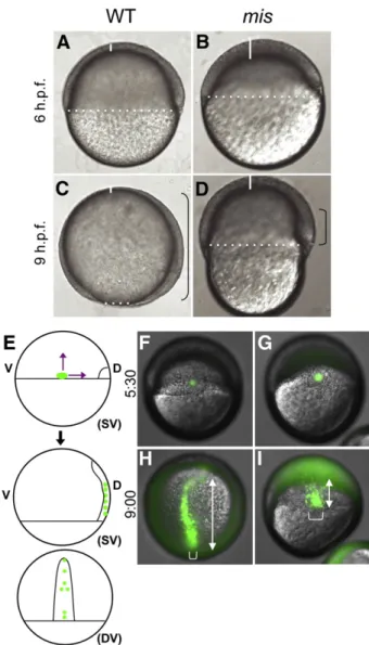
Fig. 1.Gastrulation defects in maternally mutantmisembryos. Wild-type embryos (A,C) andmismutant embryos (B,D).misembryos exhibit reduced epiboly of the blastoderm, as evidenced by the increased thickness of the blastoderm at the animal pole (white solid lines) and reduced coverage of the yolk (white dotted line indicates blastoderm margin). The extent of involution of the hypoblast is also reduced (black brackets). Lateral views (animal top, dorsal right). (E–I) Defects in epiboly and convergence. (E) Labeling strategy, by uncaging a small group of cells at the lateral margin in wild-type (F) and mutant (G) embryos (SV: side view; DV: dorsal view). At a time corresponding to 90% epiboly in the wild-type, the extension of labeled cells along the animal–vegetal axis (double-headed arrows) is reduced in the mutant (I) compared to wild-type (H). In addition, the width of the band of labeled cells (brackets), indicative of cell intercalation during convergence, is reduced in mutant embryos. (F,G) are side views, dorsal to the right. (H,I) are centered on the labeled cells, corresponding to the dorsal view (DV) in (E). Uncaging was carried out at 5:30 h.p.f. and time after fertilization is indicated in h:min. Embryos shown in (F–I) are representative of 5 wild-type and 5 mutant embryos.
controls in A,C; dotted white line indicates blastoderm margin). In addition, the blastoderm fails to thin in mutants, as evidenced by its increased thickness at the animal pole compared to wild-type (solid white lines,Figs. 1B,D, compared to A,C).
In addition to epiboly defects, both involution of the hypoblast cell layer and convergence of cells to the dorsal axis appear to be reduced inmismutants. The hypoblast layer forms from internalization and animalward migration of marginal blastomeres beginning at about 50% epiboly (5.5 h.p.f., reviewed inSolnica-Krezel, 2006). The dorsal hypoblast in the wild-type embryo extends from the vegetal margin toward the animal pole (bracket in Fig. 1C). This animalward extension is clearly reduced in livemismutant embryos (bracket in Fig. 1D). To better visualize this defect, we labeled cells by uncaging a fluorophore at the blastoderm margin at the initiation of gastrulation (50% epiboly, 5 h.p.f.), and observed their spatial distribution at late-gastrulation (9 h.p.f.) (Fig. 1E). Embryos were injected at the 1-cell stage with the cagedfluorophore, which was uncaged in cells at the margin of wild-type andmisembryos (Figs. 1F,G). Marginal cells in wild-type embryos extend along the animal to vegetal axis (Fig. 1H, double-headed arrow) whereas extension inmisembryos is greatly reduced (Fig. 1I). The domain of labeled cells at later stages is also typically broader in mutant embryos (bracket inFig. 1I, compare to H), suggesting defects in the convergence of cells towards the dorsal axis. Patterns of hypoblast layer markerslefty2(lft2;Bisgrove et al., 1999),cyclops(cyc;Feldman et al., 1998; Rebagliati et al., 1998b), and no tail/T/brachyury(ntl) are also consistent with a failure of marginal hypoblast cells to extend animally within the axis and converge towards the dorsal side. By mid-gastrulation in mis mutants, ex-pression of these genes is diminished along the animal–vegetal axis and broadened dorsolaterally (Supp. Fig. 2).
Additional experiments are consistent with a reduction in the ability of mutant cells to move within the blastoderm. Uncaging of a subset of cells at a time coincident with the initiation of epiboly (4.5 h p.f.,Fig. 2A) results in a reduction of cell mixing (Figs. 2C, E, compared to B,D). Observation of wild-type and mutant cells co-transplanted into wild-type hosts prior to the initiation of epiboly (sphere stage, 4 h.p.f.,Fig. 2F) indicate that mutant cells (green;Figs. 2M–R) have a reduced ability to be incorporated into the developing dorsal axis compared to wild-type cells (red;Figs. 2G–L, see also overlay in S–X), again consistent with a reduced ability of cells to undergo cell movements. The microtubule network of the yolk syncytial layer (YSL;Supp. Fig. 3), as well as the f-actin enrichment that occurs at the epibolic margin within the YSL (Supp. Fig. 1), both of which are known to be required for normal epibolic movements (Strähle and Jesuthasan, 1993; Solnica-Krezel and Driever, 1994; Holloway et al., 2009), do not reveal any apparent defects in mutant embryos. Together, our observations indicate thatmisfunction is required in a cell autonomous manner for cell movements during gastrulation. Mission impossible is required for the expression of endoderm-specific zygotic genes
Becausemisaffects processes that occur after initiation of zygotic transcription, we asked whethermismutants exhibit defects in gene expression in pathways involved in embryonic patterning. One of the earliest known patterning events is the induction of the dorsal–
ventral axis in which antagonistic Wnt/β-catenin signaling and BMP signaling establish dorsal and ventral fates, respectively (reviewed in Schier and Talbot, 2005). We assayed for zygotic expression of dorsal specific and ventral specific target genes by whole-mount in situ hybridization.nieuwkoid/bozozok/dharma(nwk), a direct target of the Wnt/ß-catenin pathway (Ryu et al., 2001) is expressed in dorsal domains in both wild-type andmisat 5 h.p.f. (Supp. Fig. 4). We also assayed the expression of downstream targets ofnwk,goosecoid(gsc; Stachel et al., 1993; Schulte-Merker et al., 1994a) andchordin(chd; Fisher et al., 1997; Schulte-Merker et al., 1997), the neuroectodermal
marker foxb1.2 (Odenthal and Nüsslein-Volhard, 1998), and the ventral-specific geneeven skipped(eve;Joly et al., 1993) (Supp. Fig. 4). Overall, expression of these marker genes is properly restricted to dorsal and ventral regions inmismutant embryos indicating correct dorsal–ventral patterning in these embryos.
Next we looked at germ layer-specific genes to determine whether the maternal effect ofmis affects germ layer determination during gastrulation. In situ hybridization of neuroectodermal markerschd and foxb1.2 shows normal levels of transcript expression in mis embryos compared to wild-type up to a time equivalent to 80% epiboly in wild-type, when the embryos begin to undergo lysis due to their abnormal morphogenesis (Figs. 3B,D, compare to A,C), although the characteristic anterior “wings” of expression for these genes (arrows inFigs. 3A,C) appear absent or reduced inmismutants. These data also show that, inmismutant embryos, transcription of at least some genes is active until a time at or close to embryonic lysis.
We also tested for expression of genes in cells fated for mesoderm and endoderm, which are initially intermingled at the blastoderm margin.ntl, snail1a(Hammerschmidt and Nüsslein-Volhard, 1993; Thisse et al., 1993), and lhx1a (Toyama et al., 1995), all genes expressed in this mesendodermal layer, show strong expression around the circumference of the embryo at the margin of both wild-type andmisembryos (Figs. 3F,H,J, compare to E,G,I). On the other hand, expression of endoderm-specific genes, such asfoxA2(Odenthal and Nüsslein-Volhard, 1998) and sox17 (Alexander and Stainier, 1999),is dramatically reduced inmismutant embryos. Endodermal expression for these genes normally displays a dispersed“salt and pepper”pattern in the internal-most cell layer (Figs. 3K,M, brackets), and this expression domain is absent in mutant embryos (Figs. 3L,N). sox17expression normally observed in forerunner cells (arrowhead in Fig. 3M) is also affected in the mutants (Fig. 3N), possibly related to a requirement formisfunction in nodal signaling (see below) and a role for this pathway in forerunner cell formation (Oteíza et al., 2008). Quantitative PCR confirms a nearly 100-fold reduction in sox17 expression relative to ß-actin expression (Supp. Fig. 5). Together, our data indicate a requirement formis function in the expression of endoderm-specific genes.
Defects in maternal mis function do not affect upstream steps within the zygotically-dependent nodal signaling pathway
Because nodal signaling is required for the induction of endoder-mal genes, we assayed the expression of several nodal pathway components by in situ hybridization (Fig. 4). These genes included the Nodal-related genessquint (Feldman et al., 1998; Rebagliati et al., 1998a; Erter et al., 1998) andcyclops(Feldman et al., 1998; Rebagliati et al., 1998b), the mix-type transcription factorbonnie and clyde(bon; Alexander et al., 1999; Kikuchi et al., 2000) and their downstream targetcasanova/sox32(cas;Kikuchi et al., 2001; Dickmeis et al., 2001). At 5 h.p.f.sqttranscripts are induced zygotically at the dorsal margin in both wild-type andmis embryos (Figs. 4A,B) and later expand around the blastoderm margin (data not shown).cyctranscripts are enriched at the blastoderm margin around the entire circumference of the embryo (Figs. 4C,D). Likewise, bon and gata5 transcripts are expressed normally at the blastoderm margin during their initiation until midgastrulation (Figs. 4E,F and data not shown). These results indicate that the mis allele does not affect transcription of these zygotic genes in upstream events within the mesendoderm induction pathway.
In wild-type embryos,casexpression becomes induced indepen-dently of nodal signaling at the YSL margin at 40% epiboly (Fig. 4G), and then later in a nodal-dependent manner in overlying endodermal precursors within the DEL (Fig. 4I, bracket; Kikuchi et al., 2001; Dickmeis et al., 2001). YSL expression ofcasappears to be normally initiated inmisembryos at 40% epiboly (Fig. 4H). However, at 75% epibolymismutant embryos lack the characteristic salt and pepper
endodermalcaspattern (Fig. 4J). Instead, these embryos show an abnormally strong expression of cas at the blastoderm margin (arrowhead). To determine if this marginal expression was located in the DEL or the YSL, we performedfluorescent in situ hybridization ofcasin wild-type andmisembryos and examined these cell layers in
optical sections using confocal microscopy. At 75% epiboly, wild-type embryos show a low level of casexpression in the YSL (Fig. 4K, asterisks), as well as strongcasexpression within a dispersed subset of DEL cells directly overlying the yolk cell (arrows inFig. 4K), the presumed endodermal precursors. In contrast, inmisembryos at this Fig. 2.Defects in cell movements in maternally mutantmisembryos. (A–E) Defect in cell mixing during early epiboly. (A) Labeling strategy, by uncagingfluorescent tracer at the sphere stage. Uncaging of a group of cells at the sphere stage in wild-type (B) andmismutant (C) embryos is followed by extensive cell mixing in the wild-type (D) but little mixing in the mutant (E). (B–E) are side views, tilted toward the animal pole. Time after fertilization as indicated in h:min. Uncaging was carried out at 4:30 h.p.f., and some cell mixing has already occurred in (B,C). Embryos shown in (A–E) are representative of 5 wild-type and 5 mutant embryos. (F–X) Cell autonomy of movement defect. (F) Labeling strategy, by co-transplantation of rhodamine-labeled cells from wild-type (red) andfluorescein-labeled cells from maternally mutantmis(green) donors at the sphere stage (4 h.p.f.) into isochronic unlabeled wild-type host embryos. (G–L) Wild-type cells visualized with the rhodamine channel. (M–R) Mutant cells visualized with thefluorescein channel. (S–X) Merged images. Chimeric embryos were monitored for thefirst 24 h of development, and a subset of time points is shown (h:min p.f., indicated in (S–X)). Wild-type cells populate the embryonic axis of the host, while mutant cells remain in regions overlying the yolk cell. The embryo shown in (G–X) is representative of transplantations into 30 donor embryos. All panels are lateral views with dorsal to the right, except (L,R,X) with dorsal at the top and anterior to the left.
same developmental time point (Fig. 4L), cas continues to be expressed strongly and uniformly in the YSL, andcasDEL expression is drastically reduced. No accumulation of cas expressing cells is observed in marginal cells of the DEL in mutants, ruling out the
possibility that in these embryos a population ofcas-expressing DEL cells is induced at the margin but fails to migrate animalward. Instead, mutants seem to be defective in the induction ofcasin the DEL. These data indicate that mis mutant embryos are defective in dynamic changes incasexpression: although the nodal-independent expres-sion ofcasin the YSL during early gastrulation appears to be initiated normally, by midgastrulation the nodal-dependent induction in the DEL is defective. In addition, the downregulation ofcasexpression in the YSL that occurs during gastrulation is also affected inmismutants (seeDiscussion).
Together, our results indicate that mis is not required for the initiation of the early (zygotic) nodal signaling pathway. Instead,mis appears to be involved in a relatively downstream event or events within this pathway, ultimately leading to reduced activation within the DEL of endoderm genes such ascas, sox17andfoxA2.
mis function is required for nodal-dependent expression of endoderm-specific genes
To determine if endoderm specification by nodal signaling is dependent onmis, we overexpressed the nodal ligand Cyc or Sqt by injection of its mRNA into one-cell stage embryos and then assayed for expression of downstream target genes (similar results were observed in bothcyc-andsqt-injected embryos;Fig. 5and data not shown). Wefirst tested the expression of genes known to respond to ligand overexpression in wild-type embryo,gscand ntl (Chen and Schier, 2001). UponcycmRNA injection at the one-cell stage, bothgsc andntlare ectopically expressed in wild-type embryos at 30% epiboly (5 h.p.f.)(Figs. 5A,E), as expected. Importantly, both of these genes are activated to the same extent in similarly treated mis embryos (Figs. 5B,F). These results suggest thatmismutant embryos are able to normally activate the nodal signaling pathway upon ligand overexpression, resulting in the expression of some nodal-dependent targets. Interestingly, at a later stage (60% epiboly; 7 h.p.f.), ntl expression is largely absent from cyc-injected wild-type embryos (Fig. 5C) while, in stark contrast, similarly injected mis mutant embryos maintain high ntl expression throughout the blastoderm (Fig. 5D). This can be explained by both the demonstrated ability of casto inhibitntlaccumulation (Aoki et al., 2002a; see alsoChen and Schier, 2001) and ourfinding that mis activity is required for cas activity (see below). Additional experiments show thatmisembryos also can respond to the overactivation of other signaling pathways, such as the Wnt/ß-catenin and the FGF signaling pathways (Schier and Talbot, 2005), to induce the ectopic expression of their target genes (Supp. Fig. 6).
Our next experiments directly tested the response of the endoderm-specific genescasandsox17 to ectopic nodal activation in mutant embryos.casshows ectopic induction in response to nodal signaling in both wild-type andmisembryos (Figs. 5G,H), indicating thatmisfunction is not essential forcasactivation. On the other hand, misactivity is essential for the expression ofsox17after ectopic nodal Fig. 3.Induction of endoderm-specific genes is affected in maternally mutantmis embryos. Analysis of germ layer gene markers in wild-type (A,C,E,G,I,K,M) and maternally mutantmisembryos (B,D,F,H,J,L,N). Ectodermal markerschordin(A,B) and foxb1.2(C,D) are induced in wild-type andmismutants at similar levels, although their anterior-most domains of expression (arrows in A,C) are absent inmismutants. Mesodermal marker genesno tail(E,F),snail1a(G,H), andlhx1a(I,J) are also induced in the blastoderm margin at normal levels inmismutants compared to wild-type. On the other hand,mismutant embryos fail to activate endodermal markersfoxA2(K,L) and sox17(M,N) in the “salt and pepper” pattern characteristic of endodermal cells (brackets in K,M, compare to no detectable expression in L,N). The axial mesodermal domain offoxA2(arrowheads in K,L) does become induced in mutant embryos, albeit in an abnormally shaped domain likely caused by the morphogenesis defects associated with the mutation (a similar effect is observed with regards to the axial domain of zlim-1arrowhead in (I); see alsoSupp. Fig. 2). Expression ofsox17in forerunner cells (arrowhead in M) is absent in the mutant (N) (see text). Stages are 5 h.p.f. (E,F), 6 h.p.f. (G–J,M,N), and 8 h.p.f. (A–D,K,L). All images are dorsal views with the animal pole at the top, except (E,F) which are animal views.
activation (Fig. 5J, compare to I; and data not shown). These results further confirm a role for mis function in the nodal-dependent induction of downstream endoderm-specific targets.
The fact that nodal overexpression can effectively inducecasbut notsox17inmisembryos suggests thatmisfunctions at a level at or downstream ofcasin the endoderm specification cascade. To further corroborate this, we injected mRNA encodingcasinto wild-type and misembryos and again assayed for endoderm-specific gene induction (Aoki et al., 2002a). In wild-type injected embryos,sox17is induced throughout the blastoderm (Fig. 5K) butmismutants injected withcas show a reduced ability to inducesox17(Fig. 5L). A requirement formis function is also observed in blastoderm explants with ectopically activated nodal signaling (Fig. 6A;David and Rosa, 2001). Explants from mutant embryos exhibit a reduced ability to induce sox17 (Fig. 6C) compared to the response of wild-type explants (Fig. 6B; David and Rosa, 2001), demonstrating that the requirement formis function is independent of the yolk cell. Together, our results indicate a role for maternally-derivedmisfunction in a relatively downstream event in the zygotic pathway of endoderm induction, specifically at or downstream ofcasfunction but upstream of sox17transcriptional activation.
Mission impossible codes for the RNA helicase Dhx16
To determine the molecular identity ofmis, linkage segregation analysis of the mutation in a genetic background hybrid for two polymorphic strains determined that themis locus is located in a
1.3 cM region between 63.7 cM and 65.0 cM of the MGH zebrafish recombination map (seeMaterials and methods). In addition to the MGH markers, additional polymorphic markers were identified from the established mapping families by sequencing candidate genes or non-coding regions predicted by the genome sequence assembly version 52.7E (http://www.ensembl.org; Supp. Table 1). Segregation analysis in critical recombinant females further narrowed the region containing themislocus to a 0.1 cM interval (3 recombinants out of 2249 total meiotic events;Supp. Fig. 7). According to the available zebrafish genomic databases and excluding genes that include markers genetically separable from the mutation, the resulting region contains 6 known or predicted genes (Supp. Table 2).
In order to identify themislocus within this region, we generated maternal cDNA from 1-cell embryos derived from bothmismutant and wild-typefish and fully sequenced the open reading frames for the six gene candidates within the critical region (Supp. Table 2). Among these genes, only one non-silent mutation was detected: a single nucleotide base change indhx16resulting in an Isoleucine (Ile) to Asparagine (Asn) substitution at amino acid number 430 of the protein (Figs. 7A,B).
Sequence comparison to Dhx16 in other organisms shows that Ile 430, a hydrophobic residue, is conserved in the species Takifugu rubripes, more commonly known as fugu (Fig. 7C). Other eukaryotic Dhx16 sequences, however, possess a Valine at this position, another hydrophobic amino acid. Although Ile is not absolutely conserved in all species, the conservation of its hydrophobicity suggests that this property is important for normal protein function. Comparison of Fig. 4.Defects in endoderm-specific gene expression in maternally mutantmisembryos occur in downstream steps within the pathway. Wild-type (A,C,E,G,I,K) andmismutant (B,D,F,H,J,L) embryos assayed for expressed transcripts encoding endoderm signaling components, including the nodal signalssquint(A,B) andcyclops(C,D), and the transcription factorsbon/mixer(E,F) andcasanova/sox32(cas, G-L). Expression patterns forsquint, cyclops,andbonappear similar in wild-type andmismutant embryos.casbecomes induced in the YSL at 5 h.p.f. in both wild-type andmismutants (G,H). By 8 h.p.f., endodermal cells extending into the hypoblast layer expresscasin a“salt and pepper”distribution in wild-type (I, bracket). However,casexpression inmismutant embryos at this stage is restricted to the blastoderm margin (J, arrowhead). (K,L) Closer analysis ofcasgene expression in the YSL and DEL was carried out by FISH with acasantisense probe (red) and DAPI labeling (blue), followed by confocal microscopy. The YSL layer can be distinguished from the DEL by the less dense arrangement of its nuclei and their larger size, and a white line has been drawn between the two layers. In wild-type embryos at this stage (K), expression ofcasin the YSL is subsiding (asterisks indicate YSL nuclei associated withcasmRNA), while strong expression has begun in interspersed DEL cells tightly adjoining the YSL (arrows), the presumed endodermal precursors. On the other hand,misembryos (L) show abnormally strong and persistentcasexpression in the YSL and very few, if any,cas-expressing cells in the DEL. Stages are 5 h.p.f. (A–D, G,H), 6 h.p.f. (E,F), and 8 h.p.f. (I–L). (A,B,G,H) are lateral views with dorsal to the right, when identifiable. (C–F) are animal pole views (expression in embryos corresponding to these panels is largely symmetric and slight apparent asymmetries in the embryos shown are caused by photographing artifacts). (I,J) are dorsal views. (K,L) are optical sections through the embryonic blastoderm at about 1/3 of the way between the margin and the animal pole.
sequences of the closely related zebrafish proteins Dhx8 and Dhx38 also reveals the presence of Ile at this residue, further corroborating its functional importance (Fig. 7C).
The Zebrafish Dhx16 polypeptide is defined by elements found in the DEAH-box protein family including a DEAH/X helicase domain and putative ATP binding domain (Fig. 7A). Dhx16 is highly similar to other vertebrate and invertebrate molecules includingCaenorhabditis elegansMog-4 (Puoti and Kimble, 2000), and contains some similarity to a maternally expressedArabidopsis thalianaprotein MEE29 (Puoti and Kimble, 2000) and theSaccharomyces cerevisiaeprotein Prp2p (Arenas and Abelson, 1997) (Supp. Table 3). The residue altered by mis is the first of 4 conserved hydrophobic residues adjacent to sequence with strong similarity to the Walker-A motif for ATP binding (Fig. 7A;Walker et al., 1982). This is one of two motifs, the Walker-A and Walker-B motifs,first recognized in the molecules ATP synthase and myosin and subsequently identified in numerous kinases. The Walker-A motif contains the sequence GxxxxGK[S/T] and is thought to be a critical binding site for ATP to DEAH box polypeptides (Hall and Matson, 1999; Tanner and Linder, 2001).
To verify the molecular identity ofmisasdhx16, we synthesized mRNA from a full length wild-type zebrafishdhx16cDNA and injected it into one-cell embryos from wild-type andmisfemales (Fig. 8; Supp. Table 4). Initial experiments indicated that wild-type embryos injected with wild-typedhx16 mRNA appear normal (Fig. 8A and data not shown). mis uninjected embryos showed the expected delayed gastrulation movements and lysed before the completion of epiboly (Fig. 8B).mismutant embryos injected with wild-typedhx16 mRNA, however, showed varied degrees of morphological rescue including a well-defined shield and normal epiboly progression (Fig. 8C), and many were indistinguishable from wild-type uninjected (not shown) or wild-type injected embryos (Fig. 8A). Embryos injected withdhx16mRNA were able to complete gastrulation and survive to 24 h.p.f. (56%, of 129 scored embryos). Of thosemismutant embryos injected with wild-typedhx16mRNA that survived to 24 h p. f., a majority of embryos (68%, of 63 scored embryos) showed a wild-type body morphology (Fig. 8E, compare to D), while others showed Fig. 5.Nodal-dependent activation ofsox17requires maternally-derivedmisfunction.
Wild-type (A,C,E,G,I,K) andmismutant (B,D,F,H,J,L) embryos were injected at the 1-cell stage withcyc (A–D,G–J), sqt (E,F) or cas (K,L) mRNA, and assayed by in situ hybridization for the expression ofno tail(A–D),gsc(E,F),cas(G,H) andsox17(I–L). (A,B) In both wild-type andmismutant embryos injected withcycmRNA, ntlis ectopically induced in the blastoderm at 5 h.p.f. By 7 h.p.f., however,ntlceases to be expressed in injected wild-type embryos (C), but remains strongly expressed inmis mutants (D). (E–H) Activation of nodal signaling results in ectopicgsc(E,F) andcas (G,H) expression at similar levels in both wild-type andmismutant embryos, but leads to the strong induction ofsox17only in wild-type embryos (I) and not inmismutants (J). (K,L) Overexpression ofcasresults insox17expression in wild-type (K) but notmis mutant (L) embryos. All panels are side views, except (E,F), which are animal views.
Fig. 6.Nodal-dependent induction of endoderm-specific genes in blastoderm explants is dependent on maternalmisfunction. (A) Experimental manipulation:cycmRNA was injected at the 1-cell stage, and the blastoderm was manually excised away from marginal cells and the yolk cell at the late blastula stage (sphere stage, 4 h.p.f.), as originally carried out byDavid and Rosa (2001). At 7 h.p.f., explants from wild-type embryos show robustsox17expression (B), while explants frommismutant embryos lacksox17expression (C). Embryos shown are a subset of representative embryos from two experiments with at least 15 wild-type and 15misembryos per experiment. Embryonic explants were only included if they were intact after dissection. Embryo diagrams in (A) are adapted fromKimmel et al. (1995)(images courtesy of ZFIN).
various morphological defects, such a reduction in axis extension, possibly associated with incomplete rescue (Fig. 8F). These genetic rescue data confirm thatmission impossiblecodes fordhx16.
Additional experiments were carried out to compare in parallel the efficiency of rescue of mRNA corresponding to the maternal-effect mist792mutant (dhx16I-NN construct, seeMaterials and methods) and wild-type alleles (Supp. Table 5). As expected, in these experiments mismutant embryos injected with wild-typedhx16mRNA showed marked rescue of the mutant phenotype (66% with normal progress during gastrulation, n = 151). Similarly injected mutant siblings injected with dhx16I-NN mRNA showed no significant rescue, largely exhibiting the delays characteristic of uninjected control mutant embryos (0% with normal progress during gastrulation, n = 71). Expression pattern of mis/dhx16
The expression pattern ofdhx16during embryogenesis has been previously found to be not spatially restricted during early embryo-genesis (Thisse et al., 2004). Presence of the transcripts prior to the mid-blastula transition indicate their maternal inheritance (Supp. Fig. 8), consistent with identification of a maternal-effect mutation in this gene. As development proceeds, transcript levels appear unchanged until gastrulation (Supp. Fig. 8). This suggests the possibility that maternal mRNA may perdure until these later stages, whenmishas a functional role. Levels or distribution of maternally-deriveddhx16mRNA do not appear affected inmismutant embryos, as judged by in situ hybridization analysis (Supp. Fig. 8).mis/dhx16
transcripts can be observed to be ubiquitously expressed throughout gastrulation and somitogenesis, and later become enriched in the head region beginning at 24 h.p.f. (Thisse et al., 2004).
mis/dhx16 is required zygotically for embryonic viability
Previous studies have identified a retrovirally-induced insertional mutation in zebrafishdhx16,dhx16hi4049, which is thought to be a null mutation (Golling et al., 2002). Zygotically homozygousdhx16hi4049 embryos appear morphologically normal during thefirst 24–36 h of development. Moreover, in situ hybridization analysis suggests a normal activation of genes expressed in the mesoderm (ntl) and endoderm (cas, sox17, foxA2)during gastrulation and somitogenesis (assayed until 24 h.p.f., data not shown). The normal establishment of the basic body plan and induction of meso/endodermal layers is consistent with the presence in these embryos of maternal products inherited from the heterozygous mother. However, starting at around 36–48 h.p.f., these embryos show a general necrosis, throughout the body but particularly evident in the brain region, and they invariably lyse between days 3 and 6 of embryonic development (Golling et al., 2002;Fig. 9B, compare to 9A; Supp. Table 6). Defects in these null mutant embryos are likely the result of programmed cell death, as evidenced by increased labeling by the dye acridine orange (Supp. Fig. 9), which labels apoptotic cells (Furutani-Seiki et al., 1996), and the inhibition of this labeling by a general caspase inhibitor (Ikegami et al., 1999; Chan et al., 2001) (Supp. Fig. 9). Labeling ofdhx16hi4049 homozygous mutant embryos to detect key cellular markers, such as Fig. 7.Molecular lesion of the maternally mutantmisallele occurs in a conserved amino acid of thedhx16gene. (A) Amino acid sequence of the zebrafish DEAH Box Polypeptide 16, accession NP_956318 (Amsterdam et al., 2004; Sambrook et al., 2005) shows four domains (denoted by I, II, III, and IV) predicted by Pfam (http://pfam.sanger.ac.uk/;Finn et al., 2008) and the Conserved Domain Database at NCBI (http://www.ncbi.nlm.nih.gov/;Marchler-Bauer et al., 2007). DEAH16 contains the DEXDc domain of the DEAD-like helicase superfamily at residues 429–568 (underlined, I) and a conserved helicase domain for DEAD/H helicases (Pfam family PF00271) at residues 649–743 (underlined, II). The DEAH region contains a stretch of amino acids similar to the Walker A motif for ATP-binding at residues 435–442 (box“a”), and the DEAH box itself (box“b”). The residue Isoleucine 430 affected in themist792
allele is indicated by a red star. A predicted HA2 domain (helicase associated domain; pfam04408) is contained in residues 805–895 (box, III) and may be involved in RNA binding. Another domain of unknown function (DUF1605; pfam07717) found near the C-terminus of DEAD box helicases is identified in residues 929–1028 (box, IV). (B) DNA sequence traces of the wild-type heterozygote and mutant homozygousmist792
genomic DNA. The nucleotide and amino acid substitutions are indicated in red. The region of high conservation in Eukaryota is enclosed by a pair of arrows. (C) Protein sequence comparison showing that the amino acid substitution in themist792
allele occurs in a semi-conserved residue in a highly conserved region. Four conserved hydrophobic residues are underlined in green. Sequence similar to Walker-A motif for ATP binding is underlined in gray (G-X-X-X-X-G-K-T).
microtubules, adhesive membranes and DNA reveals the presence of compact nuclei characteristic of apoptotic cells but no other obvious defects in the cytoskeleton or the cell cycle (data not shown).
We also carried out crosses between individuals carrying the maternal-effect (mist792) and the insertional (dhx16hi4049) alleles. Crosses between individuals carrying these mutations result in a lethal phenotype in the expected Mendelian proportions (Supp. Table 6 and data not shown). A fraction of mist792/dhx16hi4049 transheterozygotes exhibit the necrosis phenotype observed in dhx16hi4049/dhx16hi4049homozygotes (Fig. 9C; Supp. Table 6), further supporting the idea that this phenotype is caused by a reduction in zygoticmis/dhx16function. In addition, functional reduction ofdhx16 with a morpholino-conjugated oligonucleotide directed against the dhx16 translational start results in a similar necrosis phenotype (Fig. 9D and data not shown).
Interestingly, the penetrance and expressivity of the necrosis phenotype is reduced in mist792/dhx16hi4049 transheterozygotes
compared todhx16hi4049/dhx16hi4049homozygotes (Fig. 9C, compare to B; Supp. Table 6). Nevertheless, most if not all transheterozygotes appear to not be viable at day 5 of development, as reflected by a lack of swimbladder inflation (Supp. Table 6 and data not shown), a phenotypic endpoint associated with a wide range of late embryonic lethal conditions (see, for example,Haffter et al., 1996; Carney et al., 2006). The reduced phenotypic strength in mist792/dhx16hi4049 transheterozygotes is consistent with the mist792 allele retaining partial function.
In agreement with insertional site assignment byGolling et al. (2002), injection of wild-type dhx16 mRNA at the one-cell stage rescues the zygotic necrosis phenotype normally observed in clutches derived fromdhx16hi4049heterozygous parents (Supp. Fig. 10). We used this assay to test whether the maternal-effectmist792mutation (dhx16I-NN) may constitute a dominant-negative allele, as the zygotic null phenotype for other genes can be significantly enhanced by the expression of dominant negative products (presumably through their interference with maternally-inherited wild-type product, seeYabe et al., 2009). Injections of mRNA coding for the dhx16I-NN product at the one-cell stage do not significantly enhance the strength of the zygotic phenotype compared to uninjected controls (Supp. Fig. 10), arguing against a dominant-negative nature for this allele. This is further supported by the fully recessive nature of this allele in a large number of mapping crosses (Supp. Fig. 7and data not shown), as well as the lack of significant effects caused by expression of dhx16I-NN product into wild-type embryos (data not shown). Instead, our genetic complementation analysis suggests that the mist792 allele codes for a hypomorphic (partial loss-of-function) allele. In the context of increased functional requirements often observed during the rapid divisions characteristic of cleavage stages (e.g.Yabe et al., 2007, 2009), this hypomorphic nature may explain the isolation of an apparently maternal-specific allele in a gene that has both zygotic and maternal requirements.
Discussion
Here, we present the functional analysis and molecular identifi -cation of the genemission impossible,originally identified through a maternal-effect mutation that results in defects in embryonic inviability during gastrulation.misfunction appears to be required within zygotic pathways that act relatively late in embryogenesis, well after the activation of the zygotic genome at the midblastula transition, and our results provide a striking example of the interweaving of maternally- and zygotically-derived functions to control the vertebrate egg-to-embryo transition. We further show that mission impossible encodes the RNA helicase Dhx16. RNA helicases have been known to be important for the determination of the primordial germ cells (Hay et al., 1988; Roussell and Bennett, 1993; Gruidl et al., 1996; MacArthur et al., 2000; Navarro et al., 2001; Palacios et al., 2004; Kotaja et al., 2006; Kloc and Chan, 2007; Salinas et al., 2007) and various other processes in somatic tissues (Tijster-man et al., 2002; Audhya et al., 2005; Yang et al., 2006, 2007; Meignin and Davis, 2008; Hubert and Anderson, 2009). This study adds to this growing list by demonstrating a role for an RNA helicase in the expression of specific target genes in the early zebrafish embryo. Mission impossible encodes Dhx16
Positional cloning and genetic analysis of a mutation inmission impossible,initially identified as a recessive maternal-effect mutation, indicate that this gene encodes the RNA helicase Dhx16. First,mis maps to a critical 0.1 cM region that includes Dhx16. Second, sequencing Dhx16 in the chromosome containing themis mutant allele reveals an amino acid substitution in an evolutionarily conserved residue in the Dhx16 protein, while other genes in the critical region do not contain any non-synonymous changes. Third, Fig. 8.Rescue of the maternally mutantmisphenotype with wild-typedhx16mRNA.
(A–C) Live embryos observed at 8 h.p.f., corresponding to 75% epiboly stage in unaffected wild-type embryos. Dotted line shows the blastoderm margin, indicative of the front of epiboly; a vertical solid white line indicates the thickness of the animal-most cell layer, which normally thins out during gastrulation; and brackets demarcate the approximate extent of the involuted cell layer. (A) Wild-type embryos injected with dhx16mRNA appear normal, shown here during gastrulation. (B) Control uninjectedmismutant embryos show morphogenetic defects characteristic of the mutation, including a reduction in epiboly, a thickened animal cap and reduced involution. (C) Threemismutant embryos injected with dhx16mRNA at the 1-cell stage, showing largely normal morphogenesis during gastrulation. (D–F) Live embryos observed at 24 h.p.f. (D) Wild-type embryo at 24 h.p.f. (E–F)mismutant embryos with injecteddhx16mRNA survive gastrulation. A majority of these embryos exhibit a wild-type morphology (E), indicative of full genetic rescue. Some embryos, likely those with partial genetic rescue, survive gastrulation but have a shortened axis (F). Focal plane in (F) allows observing an abnormal undulation of the notochord (arrowhead), which may reflect remaining defects in dorsal convergence. As expected, uninjectedmismutant sibling embryos lyse during epiboly (not shown). Side views, animal pole to the top and dorsal side to the right in (A–C), and anterior to the left and dorsal to the top in (D–F).
injection of mRNA corresponding to the wild-typedhx16allele into one-cell stagemismutant embryos rescues the embryonic phenotype characteristic of this mutation. Finally, the maternal effect mis mutation fails to complement an insertional mutation in thedhx16 gene. We conclude thatmission impossiblecorresponds todhx16.
The mutation in the maternal-effect mis allele is a missense mutation in a highly evolutionarily conserved amino acid in the RNA helicase Dxh16. This missense mutation results in the substitution of a nonpolar amino acid found in all Dhx16 homologues from plants to humans to a negatively charged residue, in a conserved hydrophobic core just 5 residues upstream of the Walker A motif involved in ATP binding (Walker et al., 1982). This amino acid change may alter the efficiency of the ATP-dependent helicase activity of this protein and lead to partial loss of function of the mutant protein.
Zebrafish mis/dhx16 gene is essential for embryonic development Our analysis of the mission impossible/dhx16gene indicates an essential role for this gene in zebrafish embryogenesis. Some of the essential functions are dependent on maternally derived products. One such maternally-dependent function appears to be important for
proper cellular movements during gastrulation. Cells in maternally mutantmisembryos undergo slower vegetalward movements during epiboly, a defect that can be observed during early epiboly (5.3 h.p.f.). Movements that begin somewhat later during epiboly, such as involution (5.7 h.p.f.) and dorsal convergence (6–7 h.p.f.) also appear reduced in maternally mutant mis embryos. Transplantation of mutant cells into wild type embryos indicates that these processes are cell autonomous. These multiple defects may be most easily explained if maternally mutantmiscells have a general defect in cell migration. We cannot rule out, however, the possibility that such a generalized defect in cell movement stems from an overall reduction in cell viability due to a general cellular requirement formis/dhx16. Nevertheless, a possible general requirement formis/dhx16function is not supported by the robust expression of various genes at later stages of development (see below) as well as the absence of signs of cell necrosis or apoptosis during gastrulation.
A second, perhaps more specific, function for maternally-derived Mis product appears to be in the activation of specific downstream genes within the endoderm induction pathway. This defect appears to be relatively specific to downstream endodermal genes, as both ectodermal genes and mesodermal genes appear to be induced to Fig. 9.Complementation analysis and functional reduction ofmis/dhx16. Live embryos at day 3 p.f. (A,A’) heterozygous for thedhx16hi4049mutation have a wild-type morphology.
(B,B’) Homozygousdhx16hi4049
/dhx16hi4049
embryos exhibit necrosis and overall reduction in the head region, as well as a shortened and bent body axis. (C,C’) Necrosis, head reduction and body shape defects are present inmist792
/dhxhi4049
transheterozygotes, albeit not to the extent found indhx16hi4049
homozygotes. (D,D’) Embryos injected with 0.25–1 ng (shown here with 0.25 ng) of morpholino-conjugated oligonucleotide directed against thedhx16translational start site develop a phenotype similar to that of dhx16hi4049
/dhx16hi4049
homozygotes. Embryos injected with similar amounts of a control morpholino appear normal (not shown). In (B–D), the necrosis phenotype could be observed as early as day 2 p.f. as a darkening in the head (not shown), developed at day 3 p.f. into the smaller head (sometimes still associated with darkening) shown in these panels, and the embryos invariably lyse between days 3 and 6 of development. Side views, anterior left, dorsal up. Right column is a higher-magnification view of the left column focusing on the head region.
normal levels in mutant embryos. Moreover, activation of endodermal (but not mesodermal) gene expression after overexpression of nodal signals is affected inmismutant embryos.
Several lines of evidence suggest that the defects in morphogenetic movements and endoderm induction observed in mis mutant embryos are separable. On one hand, defects in cellular movements can be observed as early as the initiation of epiboly (5 h.p.f.), which begins hours prior to the induction of the affected endodermal genes that normally occurs at 75% epiboly (8 h.p.f.). On the other hand, defects in the induction of endoderm-specific genes in maternally mutant mis embryos occur even under conditions of nodal over-expression in blastoderm explants which do not seem to undergo any of the cellular movements characteristic of gastrulation. However, we cannot entirely rule out that the observed set of defects, in morphogenesis and gene expression, is not causally related by processes that we do not currently understand.
The analysis of the nullmis/dhx16 mutant phenotype suggests additional roles for this gene during development. Embryos homozy-gous for an insertional allele indhx16,dxh16hi4049exhibit a general necrosis observable starting at 24–36 h of development. This indicates thatmis/dhx16 also has a function essential for cell viability later during development, in this case relying on zygotically-derivedmis/ dhx16. It is presently unclear whether the apparent increased severity of this defect in the brain region represents a greater requirement for mis/dxh16 function in this region, or instead reflects a general requirement formis/dhx16function in cell viability that is revealed by the faster rates of cell division and exhaustion of maternal product supply in this tissue (see, for example,Wehman et al., 2007; Yabe et al., 2009).
Dependence of the zygotically-driven nodal signaling pathway on maternally-derived mis/dhx16 function
A striking aspect of the phenotype of maternally mutant mis embryos is a defect in the activation of a subset of endodermal genes. It is remarkable that, althoughmisfunction is required maternally for endoderm induction, our analysis indicates that its function occurs in downstream steps within the zygotically driven nodal signaling pathway. In maternally mutantmisembryos, the nodal-related genes squintandcyclops,and their immediate downstream target genebon are initiated normally. However, in these mutants expression of more downstream genes in the endoderm induction pathway, such ascas, sox17andfoxA2is defective.
Expression analysis in embryos overexpressing nodal ligands suggests that the requirement formis/dhx16function occurs down-stream ofcasactivation and is essential for the expression ofsox17. This is further supported by thefinding thatsox17expression does not occur inmismutant embryos expressing Cas. Thus, our observations suggest a role formis/dhx16 function in a step downstream ofcas function but upstream ofsox17gene activation (Fig. 10). Moreover, misfunction is essential for the induction of endodermal genes in blastoderm explants, thus ruling out the possibility thatmisfunction is essential for target gene activation by mediating a signal from the yolk cell.
While a requirement formisfunction incasactivation is apparent in unperturbed embryos, mutant embryos with exogenously activated nodal signaling appear to expresscasnormally. This may be explained by the role ofmis/dhx16function downstream of Cas activity and the previously postulated function of Cas in the maintenance of its own expression (Dickmeis et al., 2001). However, our data do not fully rule out a separate effect of themis/dhx16mutation oncasinduction that is only apparent in the unperturbed embryo.
In contrast to defects in the induction of endoderm-specific genes in the DEL, mis mutant embryos do not exhibit defects in the activation ofcas in the YSL layer. This is in agreement with our findings thatmismediates downstream events in the nodal pathway,
sincecasYSL expression has been previously shown to be nodal-independent (Dickmeis et al., 2001; Kikuchi et al., 2001). However, mis mutants do show persistent levels of cas in the YSL by midgastrulation, a time when this expression has largely subsided in wild-type embryos. This suggests a possible scenario for the dynamic regulation ofcasexpression, where nodal/mis-independent YSL expression precedes nodal/mis-dependent DEL expression, and negative feedback from nodal/mis-dependent genes in the DEL is important for subsequent downregulation ofcasexpression in the YSL.
Molecular mechanisms of potential mis/dhx16 homologues suggest a requirement for this gene in posttranscriptional regulation of gene targets Studies on the function of Dxh16 homologues in other organisms offer clues as to the potential function of zebrafishmis/dhx16. In yeast, thedhx16homologuesprp8inS. pombeandprp2inS. cerevisiae,are involved in spliceosome function (Yean and Lin, 1991; Kim et al., 1992; Lundgren et al., 1996). Expression of the human homologue DBP2 partially rescues lethality inS. pombe prp8mutants (Imamura et al., 1998), indicating functional conservation between yeast and human proteins. In the nematodeC. elegans, thedhx16orthologmog-4 is among a group of genes shown to have an essential role in the translational repression offem-3, required for the sperm-to-oocyte transition in this organism by FBF (Gallegos et al., 1998; Graham and Kimble, 1993; Graham et al., 1993; Puoti and Kimble, 1999, 2000; Belfiore et al., 2002), which directly binds regulatory elements in the fem-3 3′-UTR (Ahringer and Kimble, 1991; Zhang et al., 1997; Crittenden et al., 2002). However, the molecular basis for the requirement formoggenes in this process has not been reported. Interestingly, mutant combinations that allow the production of oocytes fromC. elegans mogmutant females reveal maternal-effect morphogenesis defects caused by mog mutations (Graham and Fig. 10.Model for the site of action ofmis/dhx16function within the endoderm induction pathway. Lack of induction ofsox17expression inmismutants expressing nodal ligand or Cas suggests a role for maternally-providedmisfunction, downstream ofcasexpression but upstream ofsox17expression. Induction of mesodermal genes, in unperturbed embryos and embryos expressing nodal ligand, is unaffected inmis mutants. Effects ofmismutation on the endogenous expression ofcasmay reflect a requirement for Cas activity for its own expression (Dickmeis et al., 2001; not shown in diagram).
Kimble, 1993; Graham et al., 1993), reminiscent of those observed in zebrafishmismutants. These observations and our own data suggest that zebrafishmis/dhx16may be required for the regulation of a subset of genes involved in early development, possibly at the level of posttranscriptional regulation. Indeed, the large number of maternal transcripts deposited in the zebrafish egg, which await their activation at the right time and place during early embryonic development, poses an impending need for precise posttranscrip-tional regulation. However, the precise molecular mechanism of Mis/ Dhx16 action remains a question for future studies.
Further studies will also be necessary to identify the targets or sets of targets regulated by mis/dhx16. Lack of maternal spg/pouf51 function results in defects in morphogenesis and gene expression defects similar to those observed in maternally mutant mis/dhx16 embryos (Lunde et al., 2004; Reim et al., 2004). Thus, it is possible that spg/pouf51is a target of Mis/Dhx16. Another potential Mis/Dhx16 target is the mRNA for the gene eomesodermin, which has been implicated in the transcriptional regulation ofcasand whose maternal mRNA becomes localized to the blastodisc margin during early embryonic cell divisions (Bjornson et al., 2005). In addition, Eomes and its target gene mtx2 are also required for proper epibolic movements (Bruce et al., 2005; Wilkins et al., 2008). However, in situ hybridization analysis indicates that maternally mutant mis embryos do not exhibit readily apparent changes in maternaleomes mRNA localization (our unpublished data). We are currently investigating whether mis is involved in the regulation of these candidate genes at other posttranscriptional levels, such as splicing or translational control. In addition, genome-wide studies will be essential to determine, in an unbiased manner, potential targets for mis/dhx16function.
In summary, we show that the zebrafish maternal-effect gene mission impossiblecodes for an RNA helicase, Dhx16, that is essential for embryonic development, including the expression of downstream endoderm-specific genes. The molecular identity of this gene suggests a function of this helicase in the regulation of mRNAs during the transition between the maternal and zygotic control in vertebrate embryogenesis.
Supplementary materials related to this article can be found online atdoi:10.1016/j.ydbio.2011.03.001.
Acknowledgments
We are grateful to Drs. Rebagliati (Stowers Institute), Schier (Harvard U.), Stainier (UCSF), and other members of the zebrafish community for plasmids, and Drs. Hopkins and Amsterdam (MIT) for the dhx16hi4049 mutant line. We also thank Drs. Jamie Lyman-Gingerich and Sreelaja Nair for sharing unpublished data relevant to this work, as well as for their comments and those of other members of the Pelegri laboratory and anonymous reviewers on the manu-script. We are also grateful to Drs. Xiaoyan Ge and Sreelaja Nair for assistance with experiments during the revision of this manuscript, and to Dr. Scott Kennedy (U.W. Madison) for access to the Real-Time PCR system. Research in the Pelegri laboratory is funded by grant R01 GM065303 from NIH.
References
Ahringer, J., Kimble, J., 1991. Control of the sperm-oocyte switch inCaenorhabditis eleganshermaphrodites by thefem-33′untranslated region. Nature 349, 346–348. Alexander, J., Stainier, D.Y.R., 1999. A molecular pathway leading to endoderm
formation in zebrafish. Curr. Biol. 9, 1147–1157.
Alexander, J., Rothenberg, M., Henry, G.L., Stainier, D.Y.R., 1999.casanovaplays an early and essential role in endoderm formation in zebrafish. Dev. Biol. 215, 343–357. Amsterdam, A., Nissen, R.M., Sun, Z., Swindell, E., Farrington, S., Hopkins, N., 2004.
Identification of 315 genes essential for early zebrafish development. Proc. Natl. Acad. Sci. U. S. A. 101, 12792–12797.
Aoki, T.O., David, N.B., Minchiotti, G., Saint-Etienne, L., Dickmeis, T., Persico, G.M., Strähle, U., Mourrain, P., Rosa, F.M., 2002a. Molecular integration ofcasanovain the nodal signalling pathway controlling endoderm formation. Development 129, 275–286. Aoki, T.O., Mathieu, J., Saint-Etienne, L., Rebagliati, M.R., Peyriéras, N., Rosa, F.M., 2002b.
Regulation of Nodal signalling and mesendoderm formation by TARAM-A, a TGFß-related type I receptor. Dev. Biol. 241, 273–288.
Arenas, J.E., Abelson, J.N., 1997. Prp43: an RNA helicase-like factor involved in spliceosome disassembly. Proc. Natl. Acad. Sci. U. S. A. 94, 11798–11802. Audhya, A., Hyndman, F., McLeod, I.X., Maddox, A.S., Yates III, J.R., Desai, A., 2005. A
complex containing the Sm protein CAR-1 and the RNA helicase CGH-1 is required for embryonic cytokinesis inCaenorhabditis elegans. J. Cell Biol. 171, 267–279. Belfiore, M., Mathies, L.D., Pugnale, P., Moulder, G., Barstead, R., Kimble, J., Puoti, A., 2002. The
MEP-1 zincfinger protein acts with MOG DEAH box proteins to control gene expression via thefem-33′untranslated region inCaenorhabditis elegans. RNA 8, 725–739. Bisgrove, B.W., Essner, J.J., Yost, H.J., 1999. Regulation of midline development by
antagonism of lefty and nodal signaling. Development 126, 3253–3262. Bjornson, C.R., Griffin, K.J., Farr III, G.H., Terashima, A., Himeda, C., Kikuchi, Y., Kimelman,
D., 2005. Eomesodermin is a localized maternal determinant required for endoderm induction in zebrafish. Dev. Cell 9, 523–533.
Brand, M., Granato, M., Nüsslein-Volhard, C., 2002. Keeping and Raising Zebrafish. In: Nüsslein-Volhard, C., Dahm, R. (Eds.), Zebrafish - A Practical Approach, Vol. 261. Oxford University Press, Oxford, pp. 7–37.
Bruce, A.E.E., Howley, C., Dixon Fox, M., Ho, R.K., 2005. T-box geneeomesoderminand the homeobox-containing Mix/Bix genemtx2regulate epiboly movements in the zebrafish. Dev. Dyn. 233, 105–114.
Carney, S.A., Heideman, W., Peterson, R.E., Prasch, A., 2006. Understanding dioxin developmental toxicity using the zebrafish model. Birth Defects Res. Clin. Mol. Teratol. 76, 7–18.
Chan, Y.M., Wu, W., Yip, H.K., So, K.F., Oppenheim, R.W., 2001. Caspase inhibitors promote the survival of avulsed spinal motoneurons in neonatal rats. Neuroreport 12, 541–545. Chen, Y., Schier, A.F., 2001. The zebrafish Nodal signal Squint functions as a morphogen.
Nature 411, 607–610.
Cinalli, R.M., Rangan, P., Lehmann, R., 2008. Germ cells are forever. Cell 132, 559–562. Crittenden, S.L., Bernstein, D.S., Bachorik, J.L., Thompson, B.W., Gallegos, M., Petcherski, A.G., Moulder, G., Barstead, R., Wickens, M., Kimble, J., 2002. A conserved RNA binding protein controls germline stem cells inCaenorhabditis elegans. Nature 417, 660–663. David, N.B., Rosa, F.M., 2001. Cell autonomous commitment to an endodermal fate and
behaviour by activation of Nodal signalling. Development 128, 3937–3947. Dickmeis, T., Mourrain, P., Saint-Etinne, L., Fischer, N., Aanstad, P., Clark, M., Strähle, U.,
Rosa, F., 2001. A crucial component of the endoderm formation pathway, CASANOVA, is encoded by a novelsox-related gene. Genes Dev. 15, 1487–1492. Erter, C.E., Solnica-Krezel, L., Wright, C.V.E., 1998. Zebrafish nodal-related 2 encodes an
early mesendodermal inducer signaling from the extraembryonic yolk syncytial layer. Dev. Biol. 204, 361–372.
Feldman, B., Gates, M.A., Egan, E.S., Dougan, S.T., Rennebeck, G., Sirotkin, H.I., Schier, A.F., Talbot, W.S., 1998. Zebrafish organizer development and germ-layer formation require nodal-related signals. Nature 395, 181–185.
Ferg, M., Sanges, R., Gehrig, J., Kiss, J., Bauer, M., Lovas, A., Szabo, M., Yang, L., Strähle, U., Pankratz, M.J., Olasz, F., Stupka, E., Müller, F., 2007. The TATA-binding protein regulates maternal mRNA degradation and differential zygotic transcription in zebrafish. EMBO J. 26, 3945–3956.
Finn, R.D., Tate, J., Mistry, J., Coggill, P.C., Hotz, H.R., Ceric, G., Forslund, K., Eddy, S.R., Sonnhammer, E.L., Bateman, A., 2008. The Pfam protein families database. Nucleic Acids Res. 36, D281–D288.
Fisher, S., Amacher, S.L., Halpern, M.E., 1997. Loss ofcerebumfunction ventralizes the zebrafish embryo. Development 124, 1301–1311.
Furutani-Seiki, M., Jiang, Y.-J., Brand, M., Heisenberg, C.-P., Houart, C., Beuchle, D., van Eeden, F.J.M., Granato, M., Haffter, P., Hammerschmidt, M., Kane, D.A., Kelsh, R.N., Mullins, M.C., Odenthal, J., Nüsslein-Volhard, C., 1996. Neural degeneration mutants in the zebrafish,Danio rerio. Development 123, 229–239.
Gallegos, M., Ahringer, J., Crittenden, S., Kimble, J., 1998. Repression by the 3′UTR of fem-3, a sex-determining gene, relies on a ubiquitousmog-dependent control in Caenorhabditis elegans. EMBO J. 17, 6337–6347.
Giraldez, A.J., Mishima, Y., Rihel, J., Grocock, R.J., Van Dongen, S., Inoue, K., Enright, A.J., Schier, A.F., 2006. Zebrafish MiR-430 promotes deadenylation and clearance of maternal mRNAs. Science 312, 75–79.
Golling, G., Amsterdam, A., Sun, Z., Antonelli, M., Maldonado, E., Chen, W., Burgess, S., Haldi, M., Artzt, K., Farrington, S., Lin, S.-Y., Nissen, R.M., Hopkins, N., 2002. Insertional mutagenesis in zebrafish rapidly identifies genes essential for early vertebrate development. Nat. Genet. 31, 135–140.
Graham, P.L., Kimble, J., 1993. The mog-1 gene is required for the switch from spermatogenesis to oogenesis inCaenorhabditis elegans. Genetics 133, 919–931. Graham, P.L., Schedl, T., Kimble, J., 1993. Moremoggenes that influence the switch from
spermatogenesis to oogenesis in the hermaphrodite germ line ofCaenorhabditis elegans. Dev. Genet. 14, 471–484.
Griffin, K.J.P., Amacher, S.L., Kimmel, C.B., Kimelman, D., 1998. Molecular identification of spadetail: regulation of zebrafish trunk and tail mesoderm formation by T-box genes. Development 125, 3379–3388.
Gritsman, K., Zhang, J., Cheng, S., Heckscher, E., Talbot, W.S., Schier, A.F., 1999. The EGF-CFC protein one-eyed pinhead is essential for nodal signaling. Cell 97, 121–132. Gruidl, M.E., Smith, P.A., Kuznicki, K.A., McCrone, J.S., Kirchner, J., Roussell, D.L., Strome,
S., Bennett, K.L., 1996. Multiple potential germ-line helicases are components of the germ-line-specific P granules ofCaenorhabditis elegans. Proc. Natl. Acad. Sci. U. S. A. 93, 13837–13842.
Haffter, P., Granato, M., Brand, M., Mullins, M.C., Hammerschmidt, M., Kande, D.A., Odenthal, J., van Eeden, F.J.M., Jiang, Y.-J., Heisenberg, C.-P., Kelsh, R.N.,