Prenat Diagn 2008;28: 525–530. Published online in Wiley InterScience
(www.interscience.wiley.com)DOI:10.1002/pd.2018
The best approach for early prediction of fetal gender by
using free fetal DNA from maternal plasma
Elena Picchiassi, Giuliana Coata, Alessia Fanetti, Michela Centra, Luana Pennacchi and Gian Carlo Di Renzo*
Department of Obstetrics and Gynecology and Centre of Perinatal and Reproductive Medicine, University of Perugia, Perugia, Italy
Objectives Detection of free fetal DNA (ffDNA) in maternal blood during pregnancy has given rise to the possibility of developing new noninvasive approaches for early prenatal diagnosis.
On a large-scale study, two protocols of real-time polymerase chain reaction (PCR) were compared in order to establish which Y-specific locus, either multicopy DYS14 or single copy SRY sequence, was the most suitable for developing a test with high diagnostic efficiency for early fetal gender assessment. The second aim was to assess whether the combination of the two detection systems could increase the performance of the prenatal test.
Methods We analyzed 145 plasma samples from healthy pregnant women between 11 and 12 weeks of singleton gestation. For each sample, fetal gender was determined by using both protocols (DYS14 andSRY) during the same real-time PCR run.
Results The data obtained by theDYS14 andSRY assays showed an efficiency in fetal gender prediction of 97.9 and 80%, respectively. It is not advisable to combine the two protocols because this association does not help in further improvements in fetal gender prediction.
Conclusions DYS14 assay is the best approach for early fetal gender assessment because it is more sensitive, accurate, and efficient than theSRY assay. Copyright2008 John Wiley & Sons, Ltd.
KEY WORDS:SRY;DYS14; real-time PCR; fetal gender; noninvasive prenatal diagnosis
INTRODUCTION
Since the presence of free fetal DNA (ffDNA) in mater-nal plasma and serum during pregnancy has been dis-covered, prenatal diagnosis of genetic diseases by means of noninvasive approaches has been getting increasingly closer to becoming a reality in clinical practice.
Loet al. (1998) demonstrated that ffDNA circulating in maternal blood has a relatively high concentration in early pregnancy, which increases during gestation and degrades in a few hours after delivery (Loet al., 1999; Angert et al., 2003; Benachi et al., 2003); therefore, it cannot interfere with prenatal diagnosis of subsequent pregnancies.
As the use of ffDNA in noninvasive prenatal diagnosis is possible only for fetal DNA sequences absent in the mother, the potential clinical applications are concerned with the prediction of RhD genotype (Singleton et al., 2000; van der Schootet al., 2003; Clausenet al., 2005), Mendelian diseases (Amicucciet al., 2000; Chiu et al., 2002; Gonzalezet al., 2002), and fetal gender (Rijnders et al., 2003; Cremonesiet al., 2004; Birchet al., 2005). Several authors have demonstrated the possibility of detecting Y-specific sequences in maternal blood dur-ing gestation usdur-ing real-time quantitative polymerase chain reaction (PCR) and different types of primers and
*Correspondence to: Prof. Gian Carlo Di Renzo, Department of OB/GYN, Policlinico Monteluce, University of Perugia, Via Brunamonti, 51, 06122 Perugia, Italy.
E-mail: [email protected]; [email protected]
probes specific for chromosome Y, such as the multicopy DYS14 sequence located within theTSPY gene (Zhong et al., 2001, 2002; Zimmermannet al., 2005; Gerovassili et al., 2007) and the single copy SRY gene (Lo et al., 1998; Hromadnikovaet al., 2003).
Nevertheless, the encouraging results obtained have been generated without following a standardized and reproducible protocol and conclusions have been mostly based on a small number of samples analyzed. In addition, confounding factors that could impact routine clinical application may be undetected in small-scale studies (Chiuet al., 2001).
Therefore, before introducing this approach of nonin-vasive prenatal diagnosis of fetal gender, it is necessary to detect its accuracy and reproducibility and to under-stand the variables that could affect its performance.
In this study, we have analyzed a large number of samples by using two different protocols of real-time PCR, theDYS14 andSRY assays, to establish which is the most suitable in terms of sensitivity, specificity, and predictability for the diagnosis of fetal gender. More-over, we aimed to assess whether the combination of the two assays could increase the performance of the test.
MATERIALS AND METHODS
Patients
Among pregnant women who attended the Department of Obstetrics and Gynecology of the University of
Perugia for the routine check-up of pregnancy, 145 patients were enrolled following specific criteria of selection. Gestational age was between 11 and 12 weeks of gestation, calculated from last menstruation and confirmed by ultrasound. All the women were healthy and had a singleton and physiological pregnancy.
All patients provided informed consent after they were made aware of the purpose and experimental nature of the study.
Drawing of blood samples
Peripheral blood (5 mL) was drawn and collected into tubes containing ethylenediaminetetraacetic acid (EDTA) as an anticoagulant. The samples were treated within 4 h from the sampling and until their treatment they were stored at+4◦C.
The blood samples were centrifuged at 1600g for 10 min, then the supernatant was collected in a 1.5 mL tube and it was centrifuged again at 16 000 gfor 10 min to pellet any remaining cellular debris. The plasma samples obtained were divided in aliquots of 500 µL and stored at−20◦C until their use.
The samples were blinded to all personnel involved with the preparation and analysis.
DNA extraction
Genomic DNA from 500µL of maternal plasma was extracted by using QIAmp DSP Virus kit (Qiagen, Germany). The manufacturer’s instructions were amended in that we modified the incubation time as fol-lows: 56◦C for 20 min with the Lysis buffer, room tem-perature for 10 min with ice ethanol 96–100%, 56◦C for 5 min after dry centrifugation, and room tempera-ture for 5 min before DNA nucleic acid elution. We used centrifugation, instead of vacuum system, as suggested by manufacturer’s instructions, to minimize the risk of contamination.
The DNA was eluted into 60µL of sterile and DNase-free water of which 5µL was used as a template for the PCR. Extracted DNA was stored at+4◦C until real-time PCR analysis.
Real-time PCR
The real-time PCR analysis was performed using Real-time PCR 7300 detection system (Applied Biosystems, USA).
Extracted DNA was analyzed for DYS14, SRY, and telomerase loci. The multicopyDYS14sequence located onTSPY gene and the SRY gene were used to measure the quantity of fetal male DNA, and the telomerase sequence was used to confirm the presence and quality of total (fetal and maternal) DNA in each sample.
Primers and TaqMan probes forDYS14 (Zhonget al., 2001, 2002; Zimmermannet al., 2005; Gerovassiliet al., 2007) and SRY (Lo et al., 1998; Hromadnikova et al., 2003) sequences were selected among the most utilized and promising in literature.
Primers and TaqMan probes specific for the DYS14 andSRY sequences and telomerase house-keeping gene were as follows:
DYS14 forward primer 5-GGG CCA ATG TTG TAT CCT TCT C-3;
DYS14 reverse primer 5-GCC CAT CGG TCA CTT ACA CTT C-3;
DYS14 probe 5-(FAM) TCT AGT GGA GAG GTG CTC (TAMRA)-3;
SRY 109 forward primer 5-TGG CGA TTA AGT CAA ATT CGC-3;
SRY 245 reverse primer 5-CCC CCT AGT ACC CTG ACA ATG TAT T-3;
SRY probe 5-(FAM) AGC AGT AGA GCA GTC AGG GAG GCA GA (TAMRA)-3.
Telomerase forward primer 5-GGT GAA CCT CGT AAG TTT ATG CAA-3;
Telomerase reverse primer 5-GGC ACA CGT GGC TTT TCG-3;
Telomerase probe 5-(FAM) TCA GGA CGT CGA GTG GAC ACG GTG (TAMRA)-3.
The singleplex reactions were set up in a total volume of 25µL, using 12.5µL of TaqMan Universal 2X PCR Master Mix (Applied Biosystems), 5µL of extracted DNA and optimized primers, and TaqMan probes (Applied Biosystems). In particular, for the detection system DYS14, primers and probes at final concentration of 900 and 200 nM, respectively were used; and for the detection systemsSRY and telomerase, primers and probes at final concentration of 300 and 100 nM, respectively were employed.
Cycling conditions for all reactions consisted of 2 min at 50◦C and 10 min at 95◦C, followed by 50 cycles of 95◦C for 15 s and 60◦C for 1 min.
To determine the copy number of male DNA present in the plasma sample, a standard curve using serial dilutions of the reference human genomic male DNA (Promega, USA) was employed.
Levels of ffDNA were calculated according to Lo et al. (1998) and expressed in genome equivalents per milliliter (GE/mL) of maternal plasma by using 6.6 pg as conversion factor.
Each sample was analyzed in triplicate, multiple-negative reaction blanks, and positive controls of male DNA pool were included in every analysis. A calibration curve and each DNA sample was tested for the DYS14 and SRY markers on the same reaction plate for each PCR run.
After real-time PCR runs, samples were designated as female fetus if no positive replicates were obtained, and as male fetus if they had at least two replicates with positive amplifications. When only one positive replicate occurred, the result was considered inconclu-sive and the analysis repeated again. After this PCR run repetition, when one positive replicate occurred, a fetus was designated as female by using the DYS14 assay, and as male by using the SRY assay. This different interpretation of the results for the two assays is the consequence of the higher sensitivity of DYS14 than the SRY assay, observed previously (data not shown)
by analyzing standard curve of both systems: the low-est amount of standard male DNA was amplified by the DYS14 assay in more than 1 well whileSRY amplified it in none or 1 well.
Fetal gender of each sample was determined by anal-ysis of karyotype from invasive procedures or confirmed phenotypically at birth.
Anticontamination measures
The blood sampling, plasma preparation, DNA extrac-tion and real-time PCR analysis were carried out by female staff and in separate areas to minimize the risk of contamination.
All equipment and work areas were swabbed with fresh 10 mL/L hypochlorite. In addition, all tubes and filtertips used were sterile. UV cross-linking of pipettes was performed before starting DNA extrac-tion and real-time PCR mixture preparaextrac-tion. The Taq-Man Universal 2X PCR Master Mix used contained UracilN-glycosilase in combination with deoxyuridine-5-triphosphate (dUTP) to prevent contamination by the carry-over of PCR products.
Statistical analysis
The results were expressed as median values with range and interquartile range (IQR) for descriptive statistics performed by Excel software (Microsoft).
Moreover, we determined diagnostic sensitivity, speci-ficity, positive predictive value (PPV) and negative pre-dictive value (NPV), and efficiency of the two tests on the basis of true and false positive/negative results obtained.
RESULTS
DNA extraction protocol had an optimal performance as demonstrated by the presence of telomerase amplicons, detected in all samples examined.
In order to evaluate the analytical sensitivity and reproducibility of the DYS14 and SRY assays, we ana-lyzed the data obtained from the standard curve of each experiment. Our results demonstrated a higher sensitiv-ity and reproducibilsensitiv-ity of theDYS14 assay compared to theSRY’s.
While the first assay allowed reproducible amplifica-tion of the target amount as low as 0.886 GE input, the determination of the SRY sequence was limited up to only 8.86 GE input. The Ct values of the DYS14 assay
were at least four cycles less than the SRY assay. The coefficient of variation (CV), calculated on ffDNA con-centration of three replicates of the same sample, showed higher reproducibility in DYS14 in respect to the SRY amplification system (Table 1).
Cytogenetic analyses or phenotype at birth revealed that among 145 women enrolled in this study, 82 were bearing male fetuses and the remaining 63 patients were bearing female fetuses. By using the DYS14 and SRY assays, we observed a concordance of 97.9% (142/145) and 80% (116/145), respectively, with regard to fetal gender determination.
TheDYS14 assay detected male DNA in 81 out of 82 samples from pregnant women carrying male fetuses, whereas the SRY assay detected male DNA in only 54 samples out of 82 showing a higher sensitivity for DYS14 (98.8%) in respect to theSRY assay (65.9%).
By using theDYS14 assay, we correctly detected 61 out of 63 samples from pregnant women carrying female fetuses showing a specificity of fetal gender detection of 96.8%; but while using theSRY assay the specificity was 98.4%. Thus, the specificity in fetal gender detection of the two protocols was comparable.
Among 145 samples analyzed by using the DYS14 assay, only 18 were repeated twice after having obtained inconclusive results. PCR assay repetition showed that fetal gender was correctly determined in all samples. By using theSRY assay, it was necessary to repeat the test on 24 samples of which 6 samples still showed incorrect fetal gender determination.
Fetal gender detection performed by theDYS14 proto-col gave two false positive results and one false negative result, whereas theSRY analysis gave one false positive result and 28 false negative results. Therefore, PPV and NPV ofDYS14 analysis protocol for fetal gender deter-mination were 97.6 and 98.4%, respectively, while for the SRY analysis protocol for fetal gender determina-tion were 98.2 and 68.9%, respectively. Therefore, while PPVs of both assays were similar, NPV ofDYS14 was nearly 1.5 times higher than that of theSRY assay. The results of sensitivity, specificity, PPV, NPV and effi-ciency of the DYS14 and SRY assays are summarized in Table 1.
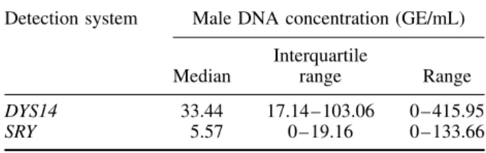
Among the 82 samples from pregnant women bearing male fetuses, the median ffDNA concentration, detected by the DYS14 assay was 33.44 GE/mL of maternal plasma (IQR: 17.14–103.06 GE/mL; range 0–415.95 GE/mL); instead, the median ffDNA concentration, detected by the SRY assay, was 5.57 GE/mL of mater-nal plasma (IQR: 0–19.16 GE/mL; range 0–133.66 GE/mL). These data (Table 2) evidenced howSRY was less sensitive in male ffDNA detection compared to the DYS14 assay and suggested that, as early as at 11 and
Table 1 — Comparison of performance in fetal gender prediction between theDYS14 andSRY assays
Detection system Gestational age (weeks) Total No. of patients Sensitivity (%) Specificity (%) PPV (%) NPV (%) Efficiency (%) CV of DNA quantity (%) DYS14 11 – 12 145 98.78 96.82 97.59 98.39 97.93 23.55 SRY 11 – 12 145 65.85 98.41 98.18 68.89 80.00 60.54
Table 2 — Fetal DNA concentration expressed as genome equivalents of male DNA/mL of maternal blood (GE/mL) calculated on samples from male fetuses by using theDYS14
andSRY detection systems
Detection system Male DNA concentration (GE/mL) Median
Interquartile
range Range
DYS14 33.44 17.14 – 103.06 0 – 415.95
SRY 5.57 0 – 19.16 0 – 133.66
12 weeks of gestation, ffDNA was present in all the sam-ples tested except one, indicating that DNA extraction protocol worked with a high performance.
Re-examining the two false positive results obtained by theDYS14 protocol, we observed that Ct values were at least 3 cycles higher, and consequently, ffDNA con-centration was about 10 times lower for these patients than for others in which a male fetus was predicted.
The unique sample from male fetal pregnancy detected as false negative by the DYS14 assay, did not produce any positive amplification by using theSRY assay. How-ever, extracting the DNA again from another plasma aliquot of this sample, which resulted in a false negative result, the follow-up real-time PCR analysis correctly revealed a male fetus.
DISCUSSION
This study was carried out by using real-time PCR in order to assess ffDNA in maternal plasma and to ver-ify whether two Y-amplification assays (the multicopy DYS14 sequence and the single copy SRY gene) could be technically employed in fetal gender determination; and to verify whether the association of the two detec-tion assays could improve the performance of the test. To our knowledge, this is the first study performed on a large number of patients at an early gestational age in order to compare these two assays in predicting fetal gender from ffDNA present in maternal plasma.
Our results showed the great technical performance of both protocols employing real-time PCR. In particular, theDYS14 assay demonstrated a higher sensitivity and reproducibility in male DNA detection compared to the SRY assay. This evidence is in keeping with the findings observed by Zimmermannet al. (2005) and Zhonget al. (2006).
With regard to fetal gender prediction, our data obtained by theDYS14 assay showed a high diagnostic concordance (97.9%) with the results verified by analy-sis of karyotype from invasive procedures, or confirmed phenotypically at birth. We have not reached 100% effi-ciency because we revealed two false positive results and one false negative result. By using theSRY detec-tion system, we observed a concordance of 80% having incorrectly diagnosed 29 samples, of which 1 false pos-itive result and 28 false negative results were detected.
The lower SRY diagnostic efficiency, in comparison with the DYS14 assay, is due to the different number
of false negative samples detected by using the two amplification systems, as also observed by Zimmermann et al. (2005). The smaller number of false negative results obtained from the DYS14 assay in comparison withSRY could be justified by the fact that a multicopy sequence (DYS14) in respect to a single copy sequence (SRY) could be able to detect male DNA also in those samples at very low ffDNA concentration. Therefore, the data suggested that ffDNA, coming from pregnant women at 11–12 weeks of gestation, is too scarce to be detected by theSRY assay, but is an adequate amount to be identified by the use of multicopy sequence. On the other hand, Zhong et al. (2006) demonstrated that the ffDNA concentration detectable during second trimester is sufficient to be revealed by both amplification systems. This discrepancy could be due to the different gestational age in which Zhong’s and our studies were carried out in the second and the first trimester of pregnancy, respectively.
The results of our study are of greater importance than those of Zimmermannet al. (2005) who obtained similar findings but in a much smaller population.
The data regarding the lowSRY diagnostic efficiency could seem to be in disagreement with those of others (Lo et al., 1998; Guibert et al., 2003; Hromadnikova et al., 2003; Rijnders et al., 2003) who showed 100% accuracy and specificity; however, it should be pointed out that all the studies performed involved less than 50 patients. Authors, such as Cremonesi et al., 2004; Rijnders et al., 2004 and Birch et al., 2005, achieved high diagnostic accuracy and specificity of the SRY amplification system by analyzing a bigger population, however, they had considered a much larger gestational age interval ranging from as low as 5 up to 41 weeks. We presume that their preference forSRY-based PCR results could be justified by the higher concentration of ffDNA in maternal blood at advanced gestational weeks, which could enable easier detectability of ffDNA allowing the use of a lower sensitive system such as a single copy, in respect to a multicopy system.
Moreover, our results obtained by using the DYS14 system, are of great interest in this field for those who want to use this technology in a clinical setting because they are widely intra- and interlaboratory reproducible as demonstrated by the multicenter studies (Legleret al., 2007) in which the researchers got similar data as ours. For the widespread introduction of noninvasive pre-natal diagnosis in clinical service, false negative results should be avoided because they are more clinically worrisome than false positives. Since the unique false negative result from the DYS14 assay was correctly revealed as a male fetus after a second sample extraction and PCR analysis, we think that it would be advis-able in carrying out two individual DNA extractions or increasing two-fold input volume of maternal plasma for extraction.
Regarding the false positives, we observed that the DYS14 assay could reach 100% specificity by select-ing a cut-off at 1.5 GE/mL of maternal plasma, while for the SRY assay this cut-off is not feasible because the male ffDNA concentration does not differentiate between false positive and true positive cases. Another
possible measure to reduce the incidence of false posi-tive results is the combination of our genetic test with early ultrasound scan in order to determine the presence of a singleton or twin pregnancy, and therefore, avoid possible bias in case of a vanishing twin.
This approach for fetal gender determination is useful for all pregnant women at risk of X-linked disorders or metabolic conditions associated with ambiguous devel-opment of external genitalia. Since a specific diagnostic test is available only for a limited number of the X-linked diseases, for all the others fetal gender is the only useful diagnostic information for first-step screening. In case of pregnancies at risk of metabolic disorders associ-ated with ambiguous development of external genitalia, the knowledge of fetal gender is important to establish as to which of the cases needs to be pharmacologically treated.
From all these observations, we believe that 11–12 weeks of gestation is the best sampling period to achieve the optimal compromise between accuracy and precocity in test performing. Before this gestational age, the test could give a consistent number of false neg-ative results due to the insufficient quantity or absence of ffDNA in the sample analyzed, whereas after this period, it might not be possible to assure the adequate management of pregnancy, either for the use of invasive procedures or for an optimal pharmacological treatment. For example, in case of pregnancies at risk of hemophilia or Duchenne Syndrome, if the fetus is male, the detec-tion at 12 weeks allows the performance of convendetec-tional prenatal diagnosis by invasive procedures, whereas in case of a female fetus, the pregnant woman should not undergo any invasive procedures with the related risk of fetal loss. On the other hand, in pregnancies at risk of metabolic disorders, such as congenital adrenal hyper-plasia (CAH), the early prenatal fetal gender prediction would be extremely important for carrying out steroid prophylaxis only on women with female fetuses in order to prevent their fetal virilization.
From our results, it is clear that the DYS14 assay has been demonstrated as the most adequate and most promising to be employed in the diagnostic routine of fetal gender prediction, and to be considered the gold standard of prenatal noninvasive fetal sex diagnosis because it is sensitive, accurate, and efficient, as con-firmed by Sekizawaet al., 2001 and Hondaet al., 2002. It is not advisable to combine the two protocols because this association does not reach further improvements in fetal gender prediction as demonstrated by the lower sensitivity and specificity of this combination in respect to those obtained from theDYS14 assay.
Consequently, this test could be recommended to all pregnant women with previous sons except for twin pregnancies with at least one male fetus. This exception is due to the impossibility of establishing which fetus is male, in other words, to which twin fetus the male ffDNA belongs.
We are convinced that fetal gender determination, by using real-time PCR and ffDNA from maternal plasma, represents the very near future of noninvasive prenatal diagnosis because it is feasible, rapid to perform, has a low cost, and mainly detects fetal gender at an early
gestational age in a robust manner. Nevertheless, further studies on a large number of maternal plasma samples are required to define firmly cut-off values and to standardize the whole protocol.
ACKNOWLEDGEMENTS
The European Commission for the Special Non-invasive Advances in Fetal and Neonatal Evaluation (SAFE) Network of Excellence (LSHB-CT-2004-503243), from which this study was partially funded, is gratefully acknowledged.
We are deeply indebted to Prof. Antonio Tabilio, MD, Dr Mauro Di Ianni, MD, and Lorenzo Moretti, BSc, from the Department of Experimental Medicine and Oncology of University of Perugia, for providing sug-gestions and support in the development and application of the real-time PCR technology.
Moreover, we are also deeply indebted to Zena Baccaille and Nadia Belia, midwives, for their help in collecting blood samples and data from patients enrolled in our study.
REFERENCES
Amicucci P, Gennarelli M, Novelli G, Dallapiccola B. 2000. Prenatal diagnosis of myotonic dystrophy using fetal DNA obtained from maternal plasma.Clin Chem46: 301–302.
Angert RM, LeShane ES, Lo YM, Chan LY, Delli-Bovi LC, Bianchi DW. 2003. Fetal cell-free plasma DNA concentrations in maternal blood are stable 24 hours after collection: analysis of first and third-trimester samples.Clin Chem49(1): 195–198.
Benachi A, Steffann J, Gautier E,et al. 2003. Fetal DNA in maternal serum: does it persist after pregnancy?Hum Genet113(1): 76–79. Birch L, English CA, O’Donoghue K, Barigye O, Fisk NM, Keer JT. 2005. Accurate and robust quantification of circulating fetal and total DNA in maternal plasma from 5 to 41 weeks of gestation.
Clin Chem51(2): 312–320.
Chiu RW, Poon LL, Lau TK, Leung TN, Wong EM, Lo YM. 2001. Effects of blood-processing protocols on fetal and total DNA quantification in maternal plasma.Clin Chem47(9): 1607–1613. Chiu RW, Lau TK, Cheung PT, Gong ZQ, Leung TN, Lo YM. 2002.
Non invasive prenatal exclusion of congenital adrenal hyperplasia by maternal plasma analysis: a feasibility study.Clin Chem48(5): 778–780.
Clausen FB, Krog GR, Rieneck K, et al. 2005. Reliable test for prenatal prediction of fetal RhD type using maternal plasma from RhD negative women.Prenat Diagn25: 1040–1044.
Cremonesi L, Galbiati S, Foglieni B,et al. 2004. Feasibility study for a microchip-based approach for non-invasive prenatal diagnosis of genetic diseases.Ann N Y Acad Sci1022: 105–112.
Gerovassili A, Garner C, Nicolaides KH, Lay Thein S, Rees DC. 2007. Free fetal DNA in maternal circulation: a potential prognostic marker for chromosomal abnormalities? Prenat Diagn 27(2): 104–110.
Gonzalez MC, Garcia-Hoyos M, Trujillo MJ, et al. 2002. Prenatal detection of a cystic fibrosis mutation in fetal DNA from maternal plasma.Prenat Diagn22: 946–948.
Guibert J, Benachi A, Grebille AG, Ernault P, Zorn JR, Costa JM. 2003. Kinetics of SRY gene appearance in maternal serum: detection by real time PCR in early pregnancy after assisted reproductive technique.Hum Reprod18(8): 1733–1736.
Honda H, Miharu N, Ohashi Y,et al. 2002. Fetal gender determina-tion in early pregnancy trough qualitative and quantitative analysis of fetal DNA in maternal serum.Hum Genet110: 75–79. Hromadnikova I, Houbova B, Hridelova D, et al. 2003. Replicate
sensitivity of non-invasive fetal sex determination. Prenat Diagn
23: 235–238.
Legler TJ, Liu Z, Mavrou A, et al. 2007. Workshop report on the extraction of foetal DNA from maternal plasma. Prenat Diagn
27(9): 824–829.
Lo YMD, Zhang J, Leung TN, Lau TK, Chang AMZ, Hjelm NM. 1999. Rapid clearance of fetal DNA from maternal plasma. Am J Hum Genet64: 218–224.
Lo YMD, Tein MSC, Lau TK, et al. 1998. Quantitative analysis of fetal DNA in maternal plasma and serum: Implications for non-invasive prenatal diagnosis.Am J Hum Genet62: 768–775. Rijnders RJ, Christiaens GC, Soussan AA, van der Schoot CE. 2004.
Cell-free fetal DNA is not present in plasma of nonpregnant mothers.Clin Chem50(3): 679–681.
Rijnders RJ, Van Der Luijt RB, Peters ED, et al. 2003. Earliest gestational age for fetal sexing in cell-free maternal plasma.Prenat Diagn23(13): 1042–1044.
Sekizawa A, Kondo T, Iwasaki M, et al. 2001. Accuracy of fetal gender determination by analysis of DNA in maternal plasma.Clin Chem47(10): 1856–1858.
Singleton BK, Green CA, Avent N, et al. 2000. The presence of an RhD pseudogene containing a 37 base pair duplication and a
nonsense mutation in africans with the RhD-negative blood group phenotype.Blood95: 12–18.
van der Schoot CE, Tax MG, Rijnders JP, de Haas M, Christiaens GC. 2003. Prenatal typing of Rh and Kell blood group system antigens: the edge of a watershed.Transfus Med Rev17(1): 31–44. Zhong XY, Holzgreve W, Hahn S. 2006. Direct quantification of fetal cells in maternal blood by real-time PCR.Prenat Diagn 26: 850–854.
Zhong XY, Holzgreve W, Hahn S. 2002. Cell-free fetal DNA in the maternal circulation does not stem from the transplacental passage of fetal erythroblasts.Mol Hum Reprod8(9): 864–870.
Zhong XY, Laivouri H, Livingston JC,et al. 2001. Elevation of both maternal and fetal extracellular circulating deoxiribonucleic acid concentration in the plasma of pregnant women carrying fetus with preeclampsia.Am J Obstet Gynecol184: 414–419.
Zimmermann B, El-Sheikhah A, Nicolaides K, Holzgreve W, Hahn S. 2005. Optimized real-time quantitative PCR measure-ment of male fetal DNA in maternal plasma. Clin Chem 51(9): 1598–1604.