Development and Function of the Human Fetal
Adrenal Cortex: A Key Component in the
Feto-Placental Unit
Hitoshi Ishimoto and Robert B. JaffeDepartment of Obstetrics and Gynecology (H.I.), Specialized Clinical Science, Tokai University School of Medicine, Kanagawa 259-1193, Japan; and Center for Reproductive Sciences (R.B.J.), Department of Obstetrics, Gynecology and Reproductive Sciences, University of California, San Francisco, San Francisco, California 94143-0556
Continuous efforts have been devoted to unraveling the biophysiology and development of the human fetal adrenal cortex, which is structurally and functionally unique from other species. It plays a pivotal role, mainly through steroidogenesis, in the regulation of intrauterine homeostasis and in fetal development and maturation. The ste-roidogenic activity is characterized by early transient cortisol biosynthesis, followed by its suppressed synthesis until late gestation, and extensive production of dehydroepiandrosterone and its sulfate, precursors of placental estro-gen, during most of gestation. The gland rapidly grows through processes including cell proliferation and angio-genesis at the gland periphery, cellular migration, hypertrophy, and apoptosis. Recent studies employing modern technologies such as gene expression profiling and laser capture microdissection have revealed that development and/or function of the fetal adrenal cortex may be regulated by a panoply of molecules, including transcription factors, extracellular matrix components, locally produced growth factors, and placenta-derived CRH, in addition to the primary regulator, fetal pituitary ACTH. The role of the fetal adrenal cortex in human pregnancy and parturition appears highly complex, probably due to redundant and compensatory mechanisms regulating these events. Mount-ing evidence indicates that actions of hormones operatMount-ing in the human feto-placental unit are likely mediated by mechanisms including target tissue responsiveness, local metabolism, and bioavailability, rather than changes only in circulating levels. Comprehensive study of such molecular mechanisms and the newly identified factors implicated in adrenal development should help crystallize our understanding of the development and physiology of the human fetal adrenal cortex.(Endocrine Reviews32: 317–355, 2011)
I. Introduction
II. Structural and Functional Adrenal Development A. Prenatal adrenal development
B. Postnatal involution of the fetal zone of the adrenal cortex
C. Prenatal adrenal growth and zonation
D. Functional development: ontogeny of steroidogen-esis and functional zonation
E. Angiogenesis and its regulation
III. Candidate Molecules Implicated in Human Fetal Adre-nal Development and Function
A. Gene expression profiles of human fetal and adult adrenals
B. Markers unique to zonal cellular subtypes C. Extracellular matrix (ECM) environment D. Transcriptional regulators
E. Role of growth factors
F. Placental factors: role of estrogens
IV. Regulation by ACTH, Pituitary Proopiomelanocortin-Derived Peptides, and Angiotensin-II
A. ACTH and pituitary proopiomelanocortin (POMC)-derived peptides
B. Angiotensin-II (AT-II)
V. The Fetal Adrenal Cortex in the Human Feto-Placental Unit
ISSN Print 0021-972X ISSN Online 1945-7197 Printed in U.S.A.
Copyright © 2011 by The Endocrine Society
doi: 10.1210/er.2010-0001 Received January 26, 2010. Accepted October 4, 2010. First Published Online November 4, 2010
Abbreviations: ABC, ATP-binding cassette; ACTHR, ACTH receptor; Ang, angiopoietin; AT1, II type 1 receptor; AT2, II type 2 receptor; AT-II, angiotensin-II; CAH, congenital adrenal hyperplasia; CAP, contraction-associated protein; Cbp, cAMP response element binding protein; CITED2, Cbp/p300-interacting transactivator, with Glu/ Asp-rich carboxy-terminal domain, 2; COUP-TF, chicken ovalbumin upstream promoter transcription factor; CYP11A, cytochrome P450 cholesterol side-chain cleavage; CYP11B1, 11-hydroxylase; CYP11B2, aldosterone synthase; CYP17, 17␣-hydroxylase/17,20-lyase; CYP19, aromatase; CYP21, 21-hydroxylase; DAX1, dosage-sensitive sex reversal-adrenal hypoplasia congenita critical region on the X chromosome; DHEA, dehydroepiandros-terone; DHEAS, DHEA sulfate; dpc, days post-conception; DZ, definitive zone; ECM, ex-tracellular matrix; EGF, epidermal growth factor; ER, estrogen receptor; FGD, familial glu-cocorticoid deficiency; FGF, fibroblast growth factor; FZ, fetal zone; HFA, human fetal adrenal; HPA, hypothalamic-pituitary-adrenal; HSD11B, 11-hydroxysteroid dehydroge-nase; HSD17B, 17-hydroxysteroid dehydrogenase; HSD3B2, type 2 3-hydroxysteroid dehydrogenase/⌬4 –5isomerase; LDL, low-density lipoprotein; MC2R, melanocortin 2 recep-tor; MPS-1, metallopanstimulin-1; MRAP, MC2R accessory protein; NCAM, neural cell adhe-sion molecule; NGFI-B, nerve growth factor-induced clone B; NOV, nephroblastoma overex-pressed; PBX1, pre-B cell leukemia transcription factor 1; POMC, proopiomelanocortin; PR, progesterone receptor; SALL1, human homolog ofspalt(sal), a region-specific homeotic gene inDrosophila melanogaster; SF1, steroidogenic factor-1; SP, side population; SPARC, secreted protein acidic and rich in cysteine; StAR, steroid acute regulatory protein; SULT2A1, DHEA sulfotransferase; TZ, transitional zone; VEGF, vascular endothelial growth factor.
Endocrine Reviews, June 2011, 32(3):317–355 edrv.endojournals.org 317
A. Glucocorticoids: roles in fetal maturation and parturition
B. Progesterone and estrogens: roles in parturition C. CRH and the human fetal adrenal cortex VI. Summary and Future Perspectives
I. Introduction
T
he human fetal adrenal (HFA) cortex shares its unique structural and functional organization with higher primates such as the rhesus monkey and baboon. The March 1997 issue ofEndocrine Reviewsreviewed the de-velopment and function of the primate fetal adrenal cortex (1). The HFA cortex is an active endocrine organ in which most steroidogenic activity is exerted in a specialized cor-tical compartment known as the fetal zone (FZ), a unique feature of fetal adrenals in humans and some higher pri-mates but not in other species such as rodents and sheep. Elegant studies during the 1950s and 1960s brought to light the essential role of the HFA in the high estrogenic milieu of pregnancy and led Diczfalusy (2) and coinvesti-gators to propose the concept of the “feto-placental unit,” in which the FZ of the HFA produces large amounts of adrenal androgens that are used by the placenta for estro-gen biosynthesis. However, the roles of the estroestro-genic mi-lieu in human pregnancy still remain unclear. During the 1960s, Ligginset al. (3, 4) demonstrated that in the sheep, maturation of the fetal hypothalamic-pituitary-adrenal (HPA) axis occurs late in gestation, and cortisol secreted by the fetal adrenal cortex stimulates maturation of fetal organs and initiates the cascade of events leading to par-turition. Similarly, the HFA cortex begins to produce cor-tisol late in gestation. However, it was soon realized that, although cortisol promotes fetal maturation in humans as it does in sheep, the role of cortisol in the regulation of human parturition has not been completely elucidated be-cause fundamental differences exist between sheep and humans in terms of regulation of parturition. Thus, the precise roles of these steroid hormones in the maintenance of pregnancy, fetal maturation, and development, and in the initiation of parturition have been controversial. Neverthe-less, recent research developments have allowed signifi-cant progress in clarifying many aspects of the biophysi-ology of the human feto-placental unit.Consistent with its endocrine capabilities, the human fetus has very large adrenals. Accumulating data indicate that growth of the HFA appears to involve cellular pro-liferation, hypertrophy, apoptosis, and migration. In ad-dition, angiogenesis, another fundamental process for or-gan growth, has been recently investigated in the HFA. Genetic studies in mice and humans, as well as studies using materials from human and subhuman primates, and the application of modern technologies, such as
gene arrays and laser capture microdissection, have contributed greatly to our understanding of the genes and gene products that play key roles in HFA develop-ment and function.
It is well accepted that ACTH is the primary regulator for the development and function of the HFA. However, this tenet does not argue against physiological roles for ACTH-independent mechanisms in the regulation and fine-tuning of the gland. Indeed, such ACTH-independent mechanisms have been identified through recent observations.
Therefore, it seemed appropriate to review the progress in the field since the previous review appeared in 1997. We introduced the topic of the previous review by indicating that “steroid hormones produced by the fetal adrenal cor-tex regulate intrauterine homeostasis, the maturation of fetal organ systems necessary for extrauterine life, and in some species, the timing of parturition. Appropriate de-velopment and function of the fetal adrenal cortex there-fore are critical for fetal maturation and perinatal survival. Moreover, the fetal adrenal cortex must itself undergo maturation in preparation for its essential role postnatally, i.e., production of glucocorticoids, androgens, and min-eralocorticoids for fetal intrauterine homeostasis and to insure adrenal cortical autonomy once the placenta has separated.” Those comments remain valid. However, as mentioned above, there have been many advances in fur-ther understanding the physiological and pathological bi-ology of the initiation of the onset of labor, the interactions of the fetus and placenta, the development of the HFA, the relationship between fetal adrenal architecture and its function, and the role of steroid hormones in intrauterine homeostasis and preparation for extrauterine life. This remains a fascinating area of investigation, which not only will advance our understanding of the biology of the HFA, but should also pave the way for new improvements in treatment of such disorders as premature labor, abnor-malities of fetal and newborn adrenal function, and a more comprehensive view of this unique organ.
In this article, we will provide a brief synopsis of earlier studies and present an overview of recent key observations from our laboratories as well as those of other investiga-tors that serve to gain a better understanding of the de-velopmental processes and functional aspects of the organ.
II. Structural and Functional Adrenal Development
The interplay of cell proliferation, differentiation, ap-optosis, and cellular senescence affects the development of the HFA. In this section, we summarize the current knowledge of the structural and functional develop-ment of the HFA.
A. Prenatal adrenal development
Cells of the human adrenal cortex arise from the inter-mediate mesoderm. The earliest recognizable manifesta-tion of the adrenal gland is called “the adrenal blastema” (5) or “the adrenal primordium,” which appears distinct from surrounding structures at 33 days post-conception (dpc) (6, 7), lying posteromedial to the urogenital ridge. Hanleyet al.(6 – 8) have demonstrated, usingin situ hy-bridization, that the transcription factors, steroidogenic factor-1 (SF1; also known as Ad4BP or NR5A1) and DAX1 (dosage-sensitive sex reversal-adrenal hypoplasia congenita critical region on the X chromosome; NR0B1), are expressed in this adrenal anlage and that hybridization signals from SF1 transcripts are stronger than those from DAX1 at all embryonic stages studied (i.e., 33 through 52 dpc) (7). By the eighth week of gestation, cells in the ad-renal blastema organize into anastomosing cords and show ultrastructural characteristics consistent with ste-roidogenic capability. By 50 –52 dpc, the developing HFA acquires two rudimentary, but distinct, zones: the inner FZ that consists of large eosinophilic cells, and the outer definitive zone (DZ), which is comprised of small, densely packed basophilic cells (7, 9). The origin or lineage rela-tionship of cells of both zones remains unknown (5, 10). At about the ninth week of gestation, the developing HFA is completely encapsulated.
During the fetal period, the morphology of the adrenal cortex remains relatively constant. The DZ is composed of a narrow band of small (10 –20 mm) cells that exhibit structural characteristics typical of cells in a proliferative state. Inner layers of the DZ form arched cords with finger-like columns of cells reaching the outer rim of the FZ. Cells in the DZ are lipid-poor during midgestation. As gestation advances, the cells accumulate cytoplasmic lipid and begin to resemble steroidogenically active cells. The FZ consists of large (20 –50 mm) cells with ultrastructural character-istics typical of steroidogenic cells. In the outer regions of the FZ, the cells are arranged in tightly packed cords. In the central portion, the FZ forms a reticular pattern, with cells spaced more widely and separated by numerous vascular sinusoids. Ultrastructural studies also have revealed a third zone between the DZ and FZ, which we have named the transitional zone (TZ) (11). Cells in this zone show intermediate characteristics (12). TZ cells have the capac-ity to synthesize cortisol, being analogous to cells of the zona fasciculata of the adult adrenal cortex. By the 30th week of gestation, the HFA cortex manifests a rudimen-tary form of the adult adrenal cortex; the DZ and TZ begin to resemble the zona glomerulosa and the zona fasciculata, respectively (13).
As early as 6 wk of gestational age, pheochromoblasts derived from the neural crest migrate through the fetal
adrenal cortex to form the medulla at a later stage of de-velopment (10, 14 –16). The medulla is not recognized as a distinct structure in the HFA throughout most of gesta-tion, except for small clusters or nests of chromaffin cells scattered throughout the body of the cortex (17–19). A more structurally discrete medulla does not form until af-ter birth (10, 17).
Chromaffin cells appear to establish part of their phe-notype as early as 6 wk gestation, as evidenced by the expression of chromogranin A and tyrosine hydroxylase (16, 20). The widely accepted concept of a key role for adrenal glucocorticoids in chromaffin cell differentiation (21, 22) has been challenged by a recent study in mice lacking the glucocorticoid receptor (23); chromaffin cells in such mice develop quite normally (for review, see Refs. 24 and 25). Studies on interactions between the adrenal cortical cells and chromaffin cells have been reviewed else-where (22). However, such interactions of two cell types in the development of the human embryonic and fetal ad-renal gland remain to be characterized.
B. Postnatal involution of the fetal zone of the adrenal cortex
Soon after birth, the HFA undergoes rapid involution due to the rapid disappearance of the FZ, with a decrease in androgen secretion (13, 14, 26 –29). As a consequence, the total weight of the glands decreases by approximately 50% (12, 30). The atrophy of the FZ appears to occur by apoptosis (31). The number of apoptotic nuclei increases markedly during the postnatal period, whereas it is very low in midgestation HFAs.
There is controversy about whether the timing of the fetal adrenal involution is determined by gestation or by birth. A morphometric study in autopsy cases and an ul-trasonographic study during antenatal and neonatal pe-riods indicate that the adrenal gland may shrink more rapidly in infants born at full term (32, 33). Longitudinal observations of infants born preterm showed that urinary excretion of FZ steroids (3-OH-5ene steroids) persists until term and then decreases as it does in full-term infants, suggesting that the HFA involution is related to gesta-tional age rather than birth (34, 35). In contrast, a more recent study has demonstrated a similar pattern of rapid adrenal involution and/or remodeling in all neonates ex-amined, regardless of their gestational age at birth (26 –35 wk) (36). The size of the adrenal gland, as measured by ultrasonography, decreased to its normal infantile size within the first 2 wk after birth (36).
A gender difference does not appear to exist in FZ re-gression/involution in humans, at least in terms of hor-mone secretion, given the testicular secretion of androgens in male infants. In a study of Ben-Davidet al.(36), serum levels of cortisol, dehydroepiandrosterone (DHEA)
fate (DHEAS), and androstenedione sharply decreased during the first week of life. However, only in males, an-drostenedione levels increased as of d 21. Garagorriet al. (37) reported that plasma levels of adrenal steroids, with the exception of cortisol, decreased progressively from birth to 6 months of age. Plasma levels of 17-hydroxy-progesterone, 11-desoxycortisol, and cortisol did not re-veal gender differences, whereas testosterone and andro-stenedione levels were higher in males and DHEAS levels were higher in females.
Dramatic remodeling of the postnatal adrenal gland involves a complex combination of FZ regression and de-velopment of the zonae glomerulosa and fasciculata (13, 32). Because morphological studies have identified rudi-mentary zonae glomerulosa and fasciculata during late gestation, the development of these zones may occur from their primordial structures, although there has been a gen-eral belief (14) that the adult cortical zones develop from the persistent DZ.
C. Prenatal adrenal growth and zonation
The weight of the developing human adrenal gland in-creases almost 10-fold from 8 to 10 wk post-conception (9). Although the relative increase in adrenal weight is the greatest during the first trimester (38), the HFA grows rapidly thereafter until term. By 20 wk gestation, the gland becomes as large as the fetal kidney and by 30 wk achieves a relative size 10- to 20-fold that of the adult adrenal gland. A further doubling in fetal adrenal weight occurs thereafter, and by full term the HFA weighs approxi-mately 3–5 g (5, 14).
Numerous studies on development of the adrenal cor-tex have generated at least three general hypothetical mod-els on zonation of the adrenal cortex: 1) the cell migration model; 2) the transformation field model; and 3) the zonal model (for review, see Ref. 39). An increasing number of studies support the cell migration model of adrenal cor-tical cytogenesis. In this model, each zone is derived from a common pool of progenitor cells, which then migrate and differentiate to populate the cortical zones. It is pre-sumed that the progenitor cells are located in the periphery of the postnatal adrenal gland (i.e., zona glomerulosa/ zona fasciculata boundary and/or subcapsular layer) (40 – 43). In the human adult adrenal cortex, immunoreactivity of Ki-67, an antigen associated with cell proliferation, was observed predominantly in the outer zona fasciculata but also in the zonae glomerulosa and reticularis (44). The adult rat has a layer of cells between the zonae glomerulosa and the fasciculata/reticularis that is nonsteroidogenic and is postulated to play a progenitor role (40, 41, 45). A similar layer of putative progenitor cells also has been visualized in sheep (46) and marmoset (47) adrenals. Stud-ies on adrenals of chimeric and transgenic rodents show
variegated expression of chimeric or reporter genes in ra-dial cords of cells from the subcapsular outer rim extend-ing into the cortex, indicatextend-ing a clonal origin of cells within each radial cord and supporting the migration theory (48 – 51). Cells in the outer DZ of the HFA exhibit structural characteristics typical of cells in a proliferative state (i.e., small cytoplasmic volume containing free ribosomes; small, dense mitochondria with lamelliform cristae and scant lipid) (52). Most of them are positive for the prolif-eration markers proliferating cell nuclear antigen and Ki-67 in the midgestation HFA (31, 53). In contrast, cells in the inner FZ are less positive for these markers. Cen-tripetal migration of lipid-containing cells from the DZ to the FZ in the HFA was reported in earlier studies (10, 14). Jirasek (5) described the presence of daughter cells that result from mitoses in the DZ and form cords invading the outer layers of the FZ. Thus, in human fetuses, it is likely that cells proliferate in the periphery of the cortex and subsequently migrate centripetally (by active migration or passive mitotic pressure) and populate the rest of the gland (Fig. 1).
The origin of progenitor cells during embryonic and fetal adrenal development is unclear. Kim and Hammer (43, 54) and coinvestigators have hypothesized that the precursors in the adrenal primordia (adrenal blastema or the fetal cortex) of the early developing mouse adrenal give rise to Sf1-negative stem cells that reside in the adrenal capsule. In response to mitogenic/morphogenic signals, such Sf1-negative capsular stem cells would exit from the
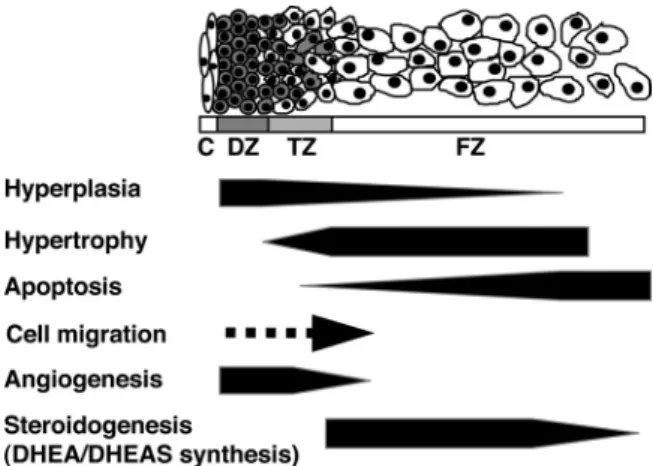
FIG. 1. Hypothetical model of development of the midgestation HFA gland. The schema shows the HFA cortex structure, which consists of the DZ, TZ, and FZ. The medulla is not recognized as a distinct structure in the HFA throughout most of gestation. Previous studies from our laboratory and others have demonstrated that the HFA cortex is a dynamic organ: hyperplasia occurs primarily in the DZ; hypertrophy occurs primarily in the FZ; and apoptosis occurs primarily in the central portion of the FZ. Thus, cells appear to proliferate in the periphery, migrate centripetally, differentiate to form the specialized cortical compartments, and then undergo senescence when they reach the center of the cortex. Recent studies indicate that angiogenesis (formation of new capillaries from preexisting blood vessels) occurs at the periphery of the gland. C, Capsule.
capsular niche into the subcapsular environment, where they commence Sf1 expression and proliferate.
The rapid HFA growth is almost entirely due to en-largement of the FZ; by midgestation (16 –20 wk), the FZ clearly dominates in the gland. In contrast to the DZ, mi-totic figures in the FZ are scant. The cell number of the FZ is not necessarily higher, but the size of it is much larger than that of the DZ. In the fetal rhesus monkey, Coulteret al.(55) demonstrated that growth of the FZ occurs primarily by hypertrophy, in response to increased endogenous ACTH secretion provoked by metyrapone treatment. Collectively, the FZ appears to grow by hypertrophy under limited cell proliferation.
Apoptosis also appears to occur in the developing HFA cortex. Jirasek (5) provided evidence of cellular apoptosis, determined by morphological criteria, in the HFA primar-ily in the central portions of the FZ. Spenceret al.(31) detected apoptotic cells byin situanalysis of DNA frag-mentation and found that the labeling index of apopto-tic nuclei is greater in the central areas of the FZ than in the DZ.
The disparate level of cell proliferation between the DZ and FZ and evidence of centripetal migration favor the migration theory of adrenal cortical development and sug-gest that the DZ is a pool of progenitor cells from which the inner cortical zones are derived. Thus, we have pro-posed that the HFA cortex is a dynamic organ in which cells proliferate in the periphery, migrate centripetally, and differentiate to form the specific cortical zones during their inward migration (and possibly continue to prolif-erate within the zones), only to undergo senescence when they reach the center of the gland (1, 31) (Fig. 1). The size of the fetal adrenal cortex and its constituent, specialized cortical compartments, represents the net effect of forces that modulate these dynamic parameters of growth.
D. Functional development: ontogeny of steroidogenesis and functional zonation
The precise onset of function (i.e., steroidogenesis) and the functional zonation of the HFA cortex have been of great academic and potentially clinical interest. We pre-viously reviewed the literature regarding this issue pub-lished before 1997 (1). Evidence is accumulating that the HFA cortex has the capability to produce steroids early in gestation. To avoid repetition, we will focus on recent findings and issues of importance in this section.
1. Cortisol production in early pregnancy
One of the major unsolved questions concerning func-tion of the HFA is when it begins to produce cortisol. Early onset of cortisol production has been suggested by obser-vations of infants affected with deficiency of 21-hydrox-ylase (CYP21), a steroidogenic enzyme in the cortisol
syn-thesis pathway (Fig. 2) (for review, see Ref. 56). In such patients, the fetal adrenal cortex cannot synthesize ade-quate amounts of cortisol. The suppressed cortisol inhibits negative feedback at the fetal anterior pituitary, which leads to a compensatory increase in ACTH secretion. The elevated ACTH causes fetal adrenal hyperplasia and in-creases production of DHEAS because its biosynthesis is not affected by CYP21 deficiency. In the first trimester when sexual differentiation occurs, there is a relative lack of aromatase (CYP19) activity in contrast to high placen-tal aromatase activity seen later in gestation. Thus, the primary clinical manifestations of CYP21 deficiency are those of androgen excess, which are first expressed in utero, resulting in virilization of the external genitalia of female fetuses. In this context, a recent case report by Men-donca et al. (57) provides important insights. They re-ported the first female case with homozygous disruption of the glucocorticoid receptor as well as haploinsufficiency of the CYP21 gene. Although patients with haploinsuffi-ciency of CYP21 do not usually present with an abnormal genital phenotype (56), the Mendoncaet al.(57) case was born with female pseudohermaphroditism. Thus, this case highlights the importance of a defective negative feedback at the corticotroph and of elevated ACTH levels in the pathogenesis of female virilization in CYP21 deficiency. Moreover, maternal administration of the synthetic glu-cocorticoid dexamethasone, which crosses the placenta, initiated before the seventh to eighth week of gestation, appears to prevent virilization of a female fetus with CYP21 deficiency (58, 59). Because sexual differentiation of external genitalia begins at wk 7 of gestation and is complete by wk 10 (60), the androgen excess observed in CYP21 deficiency most likely occurs during this time window.
Recently, Gotoet al.(9) performed a series of localiza-tion and funclocaliza-tional studies that strongly support the con-cept of early fetal adrenal cortisol synthesis. The authors demonstrated by immunohistochemistry that steroid acute regulatory protein (StAR), cytochrome P450 cho-lesterol side-chain cleavage (CYP11A), 17␣-hydroxylase/ 17,20-lyase (CYP17), CYP21, and 11-hydroxylase (CYP11B1)/aldosterone synthase (CYP11B2) are not present at 41 dpc. However, at 50 –52 dpc they appear within the nascent inner FZ. In the outer DZ, expressions of StAR, CYP11A, CYP21, and CYP11B1/CYP11B2 are lower than in the inner FZ. CYP17 appears largely absent in the DZ. Up to 14 wk post-conception, their expression profiles persist in the FZ, whereas CYP17 is also weakly expressed in the DZ. The ontogenic expression profile of CYP17 is consistent with their previous study, in which scattered expression of CYP17 was noted in the DZ at 18 wk gestation (7). Our previous study (61) using
tation HFAs (15–24 wk gestation) also demonstrated some isolated DZ cells with CYP17 staining. Adrenalde novocortisol synthesis requires expression and activity of type 2 3-hydroxysteroid dehydrogenase/⌬4 –5isomerase (HSD3B2) (Fig. 2). Goto et al. (9) demonstrated that HSD3B2 protein is not positive at 41 dpc. However, at 50 –52 dpc, HSD3B2-positive cells appear mostly at the interface between the DZ and FZ. The HSD3B2 expres-sion becomes more widespread throughout the gland and peaks at 8 –9 wk post-conception. Thereafter, HSD3B2 immunoreactivity declines, and no protein can be detected at 14 wk post-conception. In parallel with this decreasing pattern of HSD3B2 expression, cortisol content per tissue weight in the first-trimester fetal adrenal, which is 9- to 18-fold higher than in the fetal kidney, decreases by ap-proximately 50% between 8 and 10 wk post-conception (9). Gotoet al.(9) further performedex vivotissue culture of human first-trimester fetal adrenals under conditions in which culture does not artificially cause HSD3B2 up-reg-ulation, which usually matters inin vitroexperiments (1), and showed that ACTH and forskolin increase adrenal cortisol secretion. Additionally, they demonstrated that human fetal corticotrophs at 8 wk post-conception secrete significant amounts of ACTH, which can be suppressed by dexamethasone. Thus, these studies provide not only proof that the HFA can produce cortisol, likelyde novo from cholesterol, early in gestation, but also furnish a ra-tionale for prenatal treatment with dexamethasone for
female fetuses with CYP21 deficiency. Physiological significance of adrenal cortisol synthesis in the first-trimester fetus remains unclear. Goto et al. (9) hypothesize that the transient, early ad-renal cortisol synthesis exerts a nega-tive-feedback effect on ACTH secretion by the anterior pituitary corticotroph, thereby minimizing ACTH-induced an-drogen secretion to safeguard normal fe-male sexual development.
2. Spatiotemporal expression of ste-roidogenic pathway components
Attempts have been made to exam-ine spatiotemporal expression of com-ponents of steroidogenic pathways in the HFA. Previous (1) and recent data regarding the HFA expression of HSD3B2 are largely consistent with each other but somewhat conflicting in details. A synopsis is as follows: 1) there is a transient HSD3B2 expression in the first trimester (9, 62); 2) during most of the second trimester, the fetal adrenal expression of HSD3B2 is suppressed (11, 62– 64); 3) how-ever, after 23–24 wk gestation, HSD3B2 expression is de-tectable in the DZ and TZ (62, 64, 65); and 4) except in the first trimester, HSD3B2 is not expressed in the FZ (9, 62, 64, 66). A recent cDNA microarray study demonstrated that HSD3B2 mRNA expression in HFAs (15–20 wk) is 21-fold lower than that of adult adrenals (67). Although human adrenal HSD3B2 expression is spatially and tem-porally regulated during fetal life, the mechanisms regu-lating HSD3B2 expression remain poorly defined. An in-triguing characteristic of FZ cells is that they do not express HSD3B2in vivobut will readily express itin vitro when stimulated by ACTH (for detailed discussion, see Ref. 1). Thus, there may be a specific repressor that in-hibits up-regulation of HSD3B2in vivoin the HFA. Al-ternatively, a specific inducer for HSD3B2 expression may exist. The transcription factor nerve growth factor-in-duced clone B (NGFI-B) is likely one such inducer (68) (see Section III.D.2). Other potential candidates as HSD3B2 regulators include GATA-4 and GATA-6, SF1, and the related molecule liver receptor homolog 1 and signal transducer and activator of transcription family member 5 (for review of HSD3B2 regulation, see Ref. 69).
CYP11A protein is expressed between 14 and 22 wk gestation only in the FZ and TZ, but not in the DZ, and becomes detectable in the DZ after 23–24 wk (11, 64). CYP21 protein localizes in the FZ and TZ between 14 wk
FIG. 2. Principal pathways of human steroid biosynthesis. Pathways for biosynthesis of progesterone, mineralocorticoids, glucocorticoids, androgens, and estrogens. Names of the enzymes that catalyze each step are indicated inboxes.17-OH Preg,
17-Hydroxypregnenolone; 17-OH Prog, 17-hydroxyprogesterone. *, 11-Deoxycorticosterone is converted to corticosterone by 11-hydroxylase activity of CYP11B2 (in zona glomerulosa) or by CYP11B1 (in zona fasciculata). #, The conversion of 17-hydroxyprogesterone to androstenedione by CYP17 is limited in humans.
and term, and in the DZ starting at 24 wk and continuing to term (64, 70). The distribution of CYP17 is almost limited to the TZ and FZ throughout gestation; CYP17 is largely absent in the DZ (7, 11, 64). Using an antibody that recognizes both CYP11B1 and CYP11B2, Coulter and Jaffe (70) found CYP11B1/B2 protein localization in the TZ and FZ of HFAs (13–24 wk), with higher expression in the FZ than the TZ. However, the DZ lacks CYP11B1/B2 protein. In this study, an antibody that rec-ognizes both CYP11B1 and CYP11B2 was used. The au-thors further showed that, in the fetal rhesus monkey be-tween 109 d and term, CYP11B1/B2 protein is detectable in all cells of the TZ and FZ but is absent in the DZ until near term. In addition, metyrapone-induced ACTH stim-ulation in the monkey induces CYP11B1/B2 expression in the DZ and up-regulates its expression in the TZ and FZ (70). Freijeet al. (71) detected CYP11B1 mRNA in both the DZ and FZ of the midgestation HFA using RNase protection assays and RT-PCR. By RNase protection as-says, CYP11B2 mRNA was weakly positive after pro-longed exposure in the DZ but was not detectable in the FZ, whereas the more sensitive RT-PCR detected CYP11B2 mRNA in both the DZ and FZ. Narasakaet al. (64) also investigated protein localization of other steroid-ogenic components in HFAs (14 – 40 wk). DHEA sulfo-transferase (SULT2A1), which converts DHEA to DHEAS, localizes to the TZ and FZ, but not to the DZ. P450 oxidoreductase and cytochrome b, which regulate the activity of CYP17 (72, 73), are distributed to FZ and TZ cells. In the DZ, both proteins are absent until 22 wk but emerge after 23 wk. SF1 protein is expressed in almost all adrenocortical cells in the DZ, TZ, and FZ (64). Sig-nificance of SF1 in the development of the HFA will be discussed inSection III.D.1. StAR protein, which regu-lates the rate-limiting step in steroid biosynthesis (for re-view, see Refs. 74 and 75), i.e., the intramitochondrial transport of cholesterol is found in FZ and TZ cells, but not in DZ cells until 22 wk gestation (64).
The current data regarding the ontogeny of expression of steroidogenic components would account for zonal dif-ferential steroidogenic activity and its onset. The HFA cortex after midgestation is composed of three function-ally distinct zones, each of which expresses different com-binations of steroidogenic enzymes and cofactors (Fig. 3): 1) the DZ, which is the likely site of aldosterone synthesis late in gestation because of the persistent lack of CYP17, and the eventual expression of HSD3B2, CYP11A, CYP21, and probably of CYP11B2; 2) the TZ, which ap-pears to be the site of cortisol production late in gestation based on the persistent expression of CYP17, CYP11A, CYP21, and the eventual expression of HSD3B2, and probably of CYP11B1; and 3) the FZ, which expresses
CYP11A and CYP17 but not HSD3B2, is the site of⌬ 5-steroid production, particularly DHEA and DHEAS, throughout most of gestation. The localization and on-togeny of the steroidogenic enzymes and cofactors fit well with the concept that the DZ develops to form the zona glomerulosa, the TZ is the equivalent of the zona fascicu-lata, and the FZ is analogous to the zona reticularis. 3. Ontogeny of steroidogenic activity
Data on the ontogeny of HFA steroidogenic activity are available from previous studies that include hormone de-terminations from cord blood and amniotic fluid,in vitro incubation and superfusion studies of HFA tissues, and perfusion of previable human fetuses with radiolabeled hormones. For detailed description of those studies, the reader is referred to our previous review (1). Based on such data, along with the recent comprehensive analysis of on-togenic expression of the components of the steroidogenic machinery, the ontogeny of steroidogenic activity in the HFA is summarized as follows: 1) DHEAS production appears to begin at around 8 –10 wk gestation, continues thereafter, and increases considerably during the second and third trimesters, such that by term the HFA produces around 200 mg of DHEAS per day; 2)de novocortisol
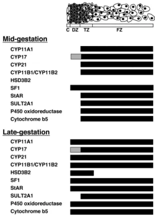
FIG. 3. Zonal expression of steroidogenic components in the HFA cortex during mid- and late gestation. Schema shows the specific zonal expression of components of the steroidogenic machinery.Black crossbardenotes expression, whereas absence of a bar represents lack of expression.Graydepicts low levels of expression. Figure is derived from previous studies (7, 11, 64, 70).
production likely occurs transiently early in gestation (around 7–10 wk gestation); 3) due to the lack of HSD3B2 expression,de novocortisol biosynthesis ap-pears to be suppressed until late gestation when cortisol production escalates (76 –78); and 4) aldosterone syn-thesis in the HFA may be suppressed during midgesta-tion due to the probable lack of CYP11B2 expression, but likely becomes active by term because 80% of al-dosterone in human fetal blood at term appears to orig-inate from the fetal adrenal (79).
In addition, Gotoet al.(9) described the capacity of the HFA at 8 wk post-conception to secrete androgens. An-drostenedione and testosterone were detected in the media of HFA tissue cultured overnight. In addition, the fetal adrenal secretions of such androgens are responsive to ACTH or forskolin. Because such adrenal androgen pro-duction is perfectly timed to influence the development of external genitalia, the investigators suggested that this ACTH-responsive androgen synthesis by the HFA may be partly responsible for the pathogenesis of virilization of the external genitalia seen in female infants with CYP21 deficiency. They also examined expression of the 17 -hydroxysteroid dehydrogenase (HSD17B) isoforms, which can convert androstenedione to testosterone by RT-PCR and found that the type 5 isoform (HSD17B5; officially named AKR1C3) was more readily detected in the early HFA than HSD17B3 that is responsible for testosterone synthesis in the testis.
E. Angiogenesis and its regulation
The HFA is one of the most highly vascularized organs in the human fetus (12, 80). The development of an ex-tensive vasculature in the organ is essential for organ growth and delivery of tropic agents (e.g., ACTH) and steroid hormone precursors to the gland and secretion of hormone products into the peripheral circulation. Here we review studies on HFA vasculature, angiogenesis (the pro-cess of formation of new capillaries from preexisting blood vessels), and its regulation.
1. Vasculature of the HFA gland
The extraorganic and intraorganic vascular systems of the human adult adrenal gland have been described (38, 81, 82). In general, the adrenal gland is supplied by arteries that arise from the renal and inferior phrenic arteries and the aorta. Earlier studies of the adult adrenal vasculature demonstrated significant variability in both origin and number of the adrenal arteries, which was consequently confirmed by a selective angiographic study of the adrenal glands in living adults. The adrenal arteries supply blood to a network of arterioles in the adrenal capsule, the sub-capsular arteriolar plexus, which gives rise to sub-capsular
capillaries that are then in continuity with the thin-walled sinusoids of the zonae glomerulosa, fasciculata, and re-ticularis. The subcapsular arteriolar plexus also branches into medullary arteries, which traverse the cortex, and conveys blood directly to the adrenal medulla. Studies of the arterial blood supply to the HFA are limited. How-ever, the sources of vessels for the HFA are similar to those for the adult adrenal (83, 84). The arterial blood supply to the HFA also is extremely variable in both the origin and number of the adrenal arteries (84).
The vasculature of the HFA gland is established by the eighth week of gestation when the adrenal is supplied by arteries from the descending aorta, and the capillary si-nusoids within the gland form a continuum with the gen-eral circulation (5). Gotoet al.(9) indicated that vascular channels positive for CD34, a vascular endothelial cell marker, penetrate the human embryonic adrenal gland between the aorta and mesonephros as early as 41 dpc, and vessel density increases at the periphery of the gland at 50 dpc. This timing of adrenal vascular establishment may be critical because the same investigators also provided evi-dence that the HFA can secrete cortisol after 8 wk post-conception (9) (seeSection II.D.1.).
The general vascular pattern within the HFA appears to correspond to that previously described for the adult gland (82, 85– 87). Pityn´skiet al.(87), in a study using micro-corrosion casts and scanning electron microscopy, de-scribed a dense vascular tree that shows a clear centripetal blood flow pattern. Thus, blood from superficial arteries and their branches would run through irregular capillaries of the subcapsular arteriolar plexus and the DZ, and then through the sinusoidal network of the FZ to the central vein. The authors also described the rare presence of med-ullary arterioles and absence of any portal systems. In ad-dition, they noted the presence of circular impressions on casts of superficial arteries, suggesting existence of mus-cular sphincters, a cirmus-cular arrangement of intimal con-nective tissue fibers, or smooth muscles (87). Thus, the HFA gland has a dense vascular system very similar to that of adults, which enables proximity between adrenocytes and endothelial cells, facilitating delivery of tropic agents, steroid hormone precursors, and hormone products, and is consistent with the fact that the organ is an active en-docrine organ during fetal life.
2. Angiogenesis and its regulation
Angiogenesis, the process of formation of new capil-laries from preexisting blood vessels, is considered an in-tegral process for organ growth. Because the HFA under-goes a phase of rapid growth in midgestation, angiogenesis likely is essential for the rapid growth of the HFA.
Angiogenesis is partly mediated by proangiogenic fac-tors such as fibroblast growth factor (FGF)-2 and vascular
endothelial growth factor (VEGF)-A, both of which are potent mitogens for endothelial cells (88). Angiopoietin (Ang)-1 and Ang2 belong to a more recently identified family of endothelial cell-specific growth factors that also play a crucial role in angiogenesis (88, 89). Ang1 is ex-pressed in a variety of tissues, whereas Ang2 expression is found primarily at sites of vascular remodeling, including the reproductive tract and placenta (90, 91). Both Ang1 and Ang2 bind their Tie2 receptor with high affinity. The currently accepted hypothesis is that Ang1 signals via Tie2 and promotes vessel stabilization and maturation, whereas Ang2 antagonizes Ang1/Tie2 signaling and destabilizes vessels, leading to either angiogenesis or vessel regression, depending on the presence of angiogenic stimuli such as FGF-2 and VEGF-A (88, 89). Studies from our laboratory demonstrated that VEGF-A, FGF-2, Ang1, Ang2, and Tie2 are expressed in the midgestation HFA gland (53, 92–95). Tie2 localizes exclusively to endothelial cells of the organ (95). A recent study by Ishimotoet al.(53) re-vealed that mRNA expression and protein localization of Ang2 and FGF-2 in the HFA gland show outer-zone pre-dominance. VEGF-A protein localizes throughout the gland, whereas mRNA of VEGF-A follows the outer-zone predominance. In parallel to the outer-zone dominant proangiogenic factor expression, endothelial cell prolifer-ation was restricted to the outer region of the HFA gland. Collectively, these results indicate that the periphery of the HFA is the primary site of angiogenesis (Fig. 1) and that locally produced angiogenic factors such as Ang2, VEGF-A, and FGF-2 likely play a key role in angiogenesis of the HFA. We hypothesize that the outer DZ may benefit from a plastic vascular state to accommodate its prolifer-ative phenotype, and that interactions between adrenal cortical and endothelial cells ensure a coordinate expan-sion of the vascular network as DZ cells proliferate and the organ grows.
Regulation of proangiogenic factors has been investi-gated in isolated adrenocortical cells from HFAs (53, 92, 94, 95). ACTH up-regulates VEGF-A in HFA cortical cells (94, 95). ACTH also increases mRNAs encoding Ang1 and Ang2 in HFA cortical cells, with an altered Ang bal-ance in which Ang2 predominates and stimulates Ang2 protein expression (95). Because the growth-stimulatory actions of ACTH in the HFA are likely mediated, at least in part, by locally produced growth factors, acting in an autocrine and/or paracrine fashion (92, 96), and ACTH is not an angiogenic factorper se, these angiogenic factors may mediate the tropic action of ACTH, exerting parallel control over the fetal adrenal vasculature. In addition, Ang2 mRNA can be induced by FGF-2 in HFA cortical cells (53), partly explaining the parallelism observed in
the outer-zone predominant expression patterns of Ang2 and FGF-2.
Taken together, these studies highlight the importance of coordinated organ and vasculature growth by interac-tions between adrenal cortical cells and endothelial cells in which FGF-2 and the vascular endothelial specific growth factors, Ang2 and VEGF-A, likely are involved.
III. Candidate Molecules Implicated in Human Fetal Adrenal Development and Function
Intensive efforts have been made to identify genes and gene products implicated in human adrenal development and function. The recent emergence of new technologies, such as cDNA microarray, laser-capture microdissection, and suppression subtractive hybridization, has provided re-searchers with new tools to tackle this issue.
A. Gene expression profiles of human fetal and adult adrenals
Marked structural and functional differences are ob-served between the human fetal and adult adrenal glands. Raineyet al.(67) have contrasted human fetal (15–20 wk) and adult adrenal gene expression profiles using cDNA microarrays. Among 69 transcripts that have a greater than 2.5-fold difference in expression between fetal and adult adrenals, the largest differences are observed for transcripts encoding IGF-II (25-fold higher in the fetal ad-renal) and HSD3B2 (21-fold higher in the adult adad-renal). Additionally, the transcript expression of the cholesterol biosynthetic enzyme, 24-dehydrocholesterol reductase, exhibits the strongest signal intensity in fetal adrenals. For adult adrenal RNA, the transcript of CYP11B1 has the highest signal intensity ranking (97).
Rehmanet al.(98), using real-time RT-PCR, compared fetal and adult gene expression levels of components of the adrenal steroidogenic pathway. The study revealed higher fetal adrenal levels of transcripts encoding the components that favor enhanced synthesis of DHEA and DHEAS, the distinct phenotype of the HFA. Thus, transcripts were higher in the HFA for cytochrome b5 and P450 oxi-doreductase that may increase P450c17 activity and SULT2A1 that catalyzes DHEA sulfurylation. In contrast, HSD3B2 mRNA was 127-fold higher in the adult adrenal, consistent with the previous result from the cDNA mi-croarray experiments (67).
More recently, Xinget al.(99) compared mRNA levels of a variety of G protein-coupled receptors between hu-man fetal and adult adrenal glands. They found that tran-script levels of six G protein-coupled receptors, including GnRH receptor, angiotensin-II (AT-II) type 2 receptor (AT2), and melanocortin 2 receptor [MC2R; or the ACTH
receptor (ACTHR)], were significantly higher in fetal than adult adrenals.
B. Markers unique to zonal cellular subtypes
As noted, the HFA is comprised initially of two zones, the DZ and the FZ. A third zone, the TZ, develops between the two zones after midgestation. Identification of cellular markers unique to each zone of the HFA should be useful for purifying cells of the zone of interest and/or providing insight into the mechanisms underlying zonation of the HFA, which facilitates detailed molecular, cellular, and structural analysis toward a better understanding of HFA development and function (Fig. 4).
1. DZ cell markers
Ratcliffeet al.(100) used laser-capture microdissection to obtain cells from the DZ and FZ. They performed sub-tractive hybridization and found two previously described growth regulatory proteins, nephroblastoma overex-pressed (NOV) and metallopanstimulin-1 (MPS-1) that are expressed almost exclusively in the DZ (100).
NOV (CCN3) is a recently described member of the cysteine-rich 61, connective tissue growth factor, NOV
(CCN) family of growth regulatory proteins. The family members have described roles in regulating mitosis, cell adhesion, migration, growth arrest, apoptosis, differenti-ation, and tumorigenesis (101–103). In human adults, NOV is more strongly expressed in the adrenal cortex than in other endocrine tissues (103). Consistent with the no-tion that NOV is a DZ cell marker, immunohistochemical studies have demonstrated DZ-specific or DZ-predomi-nant localization of NOV in HFAs from first and second trimesters (103, 104). The role of NOV in HFA develop-ment remains to be clarified. Several lines of evidence sup-port its role in inhibiting growth while promoting differ-entiation (105). The amount of NOV increases in benign adrenocortical tumors, whereas it decreases in malignant adrenocortical tumors, suggesting a role for NOV as a growth regulatory protein (103). Alternatively, NOV may be a differentiating factor because, in NCI-H295R adre-nocortical cells, expression of NOV is down-regulated by TGF-1 (106), which negatively regulates adrenal steroid-ogenesis. NOV also is negatively regulated by WT-1, the Wilm’s tumor suppressor gene product (107). Of interest, mice lacking WT-1 fail to develop adrenals (107, 108).
Less is known about MPS-1, a zinc-finger phosphopro-tein with DNA-binding properties belonging to the S27E family of ribosomal proteins (109). MPS-1 is highly ex-pressed in a wide variety of actively proliferating cells, cancer cell lines, and malignant tumor cells, whereas it is generally underexpressed in normal adult cells (109 –111). Therefore, MPS-1 could play a role in modulating cell proliferation in the DZ. Alternatively, MPS-1 possibly me-diates the suppressive effects of TGF-1 on steroidogen-esis in the DZ, because MPS-1 can be up-regulated by TGF-1 in mammary carcinoma cells (109).
Ratcliffe et al. (100) also performed immunohisto-chemical screens and identified P-glycoprotein, the prod-uct of the multidrug resistance 1 (MDR1; or ABCB1) gene, as a DZ marker. P-glycoprotein is the prototype member of the family of ATP-binding cassette (ABC) transporters, which are ATP-dependent membrane proteins predomi-nantly expressed in excretory organs, such as the liver, intestine, blood-brain barrier, blood-testis barrier, pla-centa, and kidney (112, 113). The ABC transporters are implicated in the absorption, distribution, and excretion of drugs, xenobiotics, and endogenous compounds. P-glycoprotein is expressed abundantly in the adult and fetal adrenal gland in many species, including human (114 – 116). Its role in the adrenal gland is unknown. Several studies indicate that P-glycoprotein is involved in active efflux of steroids, particularly aldosterone and cortisol, from steroidogenic cells (117–119). The HFA expression of P-glycoprotein occurs after 22 wk (100). Of particular interest, consistent with this timing of P-glycoprotein
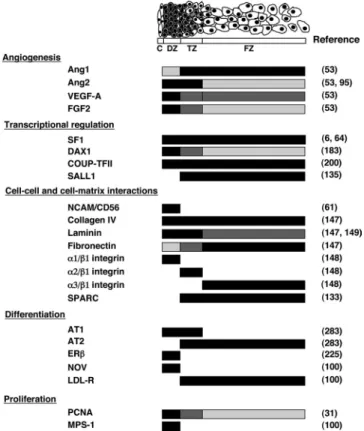
ex-FIG. 4. Zonal expression of putative factors implicated in HFA development and/or function. Schema shows specific zonal expression of putative factors implicated in HFA development and/or function.
Black crossbardenotes high levels of expression, whereas absence of a bar represents lack of expression.Dark graydepicts moderate levels of expression, andlight graydepicts low levels. LDL-R, LDL receptor; PCNA, proliferating cell nuclear antigen. Figure was derived from studies shown in right column.
pression, the steroidogenic enzymes necessary for aldoste-rone synthesis begin to be expressed in DZ cells (11). In addition to its pump actions, P-glycoprotein also has been implicated in the protection of cells against apoptosis pro-voked by a variety of stimuli such as cytotoxic drugs and TNF-␣(120, 121). A role for P-glycoprotein during cel-lular differentiation has also been proposed. P-glycopro-tein and another ABC transporter, breast cancer resistance protein (ABCG2), are highly expressed in an enriched pop-ulation of primitive stem cells: the side poppop-ulation (SP) (113). SP cells were originally discovered in bone marrow by their capacity to exclude rhodamine 123 and Hoechst dye 33342; however, mounting evidence also revealed their presence in other nonhematopoietic tissues. The ex-pression of breast cancer resistance protein and P-glyco-protein are under strict control and may determine the differentiation of SP cells toward other more specialized cell types. Thus, P-glycoprotein expression is down-regu-lated during differentiation of pluripotent stem cells along the myeloid lineage (122), and overexpression of P-glyco-protein in mouse stem cells induces a myeloproliferative syndrome (123). In this context, P-glycoprotein expres-sion in the DZ is consistent with our hypothesis that cells in the DZ may comprise a progenitor population, some of which migrate centripetally to populate the rest of the gland (1, 61, 100). P-glycoprotein, therefore, may be a useful cell surface marker to distinguish between those cells that have left the progenitor pool and differentiated into steroid-producing cells and the truly undifferentiated progenitor cells.
Recently, Muenchet al.(61) found that the neural cell adhesion molecule (NCAM; CD56) can serve as a marker of DZ cells through an extensive series of phenotyping experiments using fluorescence-activated cell sorting. In isolated HFA cells, NCAM was expressed on a cell pop-ulation of small, dense, low side-scatter cells that lack markers of hematopoietic origin (CD31, CD34, and CD235a), and this cell population was enriched for cells expressing NOV and MPS-1, the DZ cell markers (61). NCAM is widely expressed in the nervous system and plays important roles in neurodevelopment (124, 125). NCAM also has been localized in nonneural tissues, in-cluding endocrine organs (126 –128). Functions of NCAM in the HFA remain to be clarified. NCAM can modulate cell motility and migration of glioma cells (129). It can also act as a receptor for a diffusible growth factor (130). In the context of the latter possibility, a recent study (131) indicating that NCAM may be a receptor for the heparin-binding growth factor midkine is of particular interest because midkine stimulates selective proliferation of DZ cells (132) where NCAM is localized at the mem-brane. Muench et al. (61) also described islands of
NCAM-positive cells that likely represent chromaffin cells. Chromaffin cells are a major component of the ad-renal medulla. However, during primate fetal life, a well-formed medulla is not present before birth (17, 18). It is evident that functions of NCAM in HFA development should be explored in future studies.
2. FZ cell markers
Immunohistochemical studies have revealed several proteins that localize exclusively in the FZ. The low-den-sity lipoprotein (LDL) receptor is one such protein. De-spite its FZ-specific immunoreactivity (100, 133), analysis of zonal mRNA expression of the LDL receptor, using laser-capture microdissection and real-time RT-PCR, demonstrates only a 2-fold difference in LDL receptor mRNA levels between the FZ and DZ (133), suggesting that the expression of the LDL receptor may be regulated mainly at the translational level. Given the important role of the LDL receptor inde novosteroidogenesis in the ad-renal gland and the DZ’s lack of steroidogenic activity until close to term (11), DZ cells would not be expected to express the LDL receptor at midgestation. SALL1 [human homolog ofspalt(sal), a region-specific homeotic gene in Drosophila melanogaster] protein is a zinc finger tran-scriptional repressor and may be part of the NuRD histone deacetylase complex. Defects in the gene SALL1 are a cause of Townes-Brocks syndrome, a disorder with mul-tiorgan dysgenesis including renal and genital malforma-tions (134). Expression of SALL1 appears confined to the pituitary-adrenal-gonadal axis and the placenta (135). Distribution of SALL1 is restricted to the FZ of the mid-gestation HFA, whereas it is observed in all zones of the adult adrenal cortex, suggestive of its association with steroidogenesis. Interestingly, in a recent study using hu-man adrenocortical H295R cells (136), SALL1 expression was induced by AT-II. Furthermore, SALL1 was shown to decrease expression of CYP11B1 and CYP11B2 in cotransfection reporter assays. SPARC (secreted protein acidic and rich in cysteine), a matricellular protein, also localizes strictly to the FZ of the midgestation HFA (133) (seeSection III.C).
3. G protein␣-subunits
Heterotrimeric (␣␥) G proteins are central compo-nents of the primary mechanism to receive, interpret, and respond to a variety of extracellular stimuli (137, 138). Breaultet al.(139) described differential expression of G protein␣-subunits in the HFA. Interestingly, the␣ -sub-units had a specific pattern of distribution, other than the ␣ssubunit that was detected in all adrenal cell types except
for endothelial cells. The␣il-2subunit was restricted to the DZ, whereas␣i3staining was mainly seen in the FZ. The
␣qsubunit was localized in vascular endothelial cells at the
periphery and in FZ cells at the center of the gland. Chro-maffin cells expressed␣s,␣q, and␣o1, but not␣o2nor␣i. G protein-coupled receptors, including the ACTH recep-tor and AT-II receprecep-tors, play a crucial role in HFA devel-opment and function (1, 99). Therefore, the differential expression of G protein␣-subunits may partly explain the zonal or cell-type specific responses to stimuli and the uniqueness of the HFA phenotype.
C. Extracellular matrix (ECM) environment
Mounting evidence indicates that the extracellular mi-croenvironment can orchestrate functions such as cell pro-liferation, migration, differentiation, and apoptosis, es-sential components of organogenesis (140, 141).
Earlier studies showed growth-promoting effects of ECM components on bovine and human fetal adrenocor-tical cells in culture (142–146). Crickard et al. (142) showed that isolated fetal adrenocortical cells maintained on an ECM prepared from bovine corneal endothelial cells had a higher growth rate than cells maintained on plastic alone. Recently, Chamouxet al.(147) revisited this issue and demonstrated that isolated HFA cells grown on col-lagen IV and, to a lesser extent, on laminin favor cell pro-liferation, whereas cells grown on fibronectin become more apoptotic. In an earlier study, the same group of investigators examined the spatial distribution of these structural components of the ECM in second-trimester HFAs (148). Collagen IV was localized evenly throughout the midgestation HFA. The distribution of fibronectin and laminin exhibited a mirror image of each other; fibronec-tin was more abundant in the central portion, and laminin was mainly found at the periphery of the gland (148). In addition, several integrin subunits, which can serve as re-ceptors for ECM components, have been localized in the HFA. The␣1- and␣2-subunits were found mainly in the DZ and in the TZ, respectively. The␣3-subunit that binds both fibronectin and laminin was detected only in the FZ. The 2-subunit was observed exclusively in chromaffin cells (148). Similar results for integrin localization were obtained in the other study where distribution of laminin chains, laminin-binding integrins, and non-integrin recep-tors was examined in the HFA (149). Laminin␣2 and␣5 chains were weakly localized in the DZ, and laminin␣4, 1, and␥1 chains were observed around vessels. A punc-tate distribution of dystroglycan was seen mainly in the DZ and weakly in the FZ. Chamouxet al.(147) also found that cell morphology can be affected by environmental cues; for example, HFA cells grown on laminin appear rounded, clustered, and smaller, resembling the morphol-ogy of DZ cells. They further demonstrated that the ECM components modulate the profile of steroidogenesis by HFA cells. Collagen IV favors ACTH- or AT-II-stimulated
cortisol secretion and elevated DHEA secretion stimulated by an agonist for AT2. In contrast, laminin and fibronectin diminished responsiveness to ACTH in relation to cortisol production while increasing ACTH-induced DHEA and DHEAS secretion. Collectively, the differential distribu-tion of the ECM structural proteins may explain some aspects of the zone-specific cellular behavior seen in the HFA.
Matricellular proteins, a term coined by Bornstein (150), are another class of ECM components. They are secreted macromolecules that interact with cell-surface receptors, ECM, growth factors, and/or proteases but do not themselves subserve strictly structural roles, un-like other ECM proteins such as laminin and fibronec-tin. They disrupt cell-matrix interactions and are in-volved in tissue remodeling, morphogenesis, and vascular growth. The group includes thrombospondins, tenascin C, and SPARC. Among them, SPARC shows abundant expression in the midgestation HFA and a unique FZ-specific localization (133). SPARC modulates cell prolif-eration and migration. Its expression appears limited largely to tissues undergoing remodeling, morphogenesis, or tissue repair (151). Thus, SPARC could exert an anti-proliferative and antimigratory effect, reflected by the se-lective expression of SPARC in the FZ, where cell prolif-eration is less and cell migration should terminate rather than be initiated.
D. Transcriptional regulators
Studies of experimental animal models and humans are beginning to unravel the mechanisms of transcriptional regulation responsible for organogenesis of the human ad-renal cortex (for review, see Refs. 152 and 153). An in-creasing number of transcription factors has been impli-cated in adrenal development. Among them, SF1 and DAX1, which have been most extensively investigated and play essential roles in normal adrenocortical differentia-tion and development (154 –159). In addidifferentia-tion, SF1 is a key transcriptional activator of numerous genes involved in steroidogenesis (154, 158), whereas DAX1 acts as a neg-ative regulator of SF1-induced transactivation (157, 159). 1. SF1 and DAX1
An extensive body of evidence delineates essential roles for SF1 in regulating adrenocortical differentiation and function. SF1 is expressed in the HFA from its earliest stages of development—initially in the adrenogonadal precursors, and subsequently in both the FZ and DZ of the gland (7). SF1 is expressed in all zones of the human adult adrenal cortex. SF1 regulates the transcription of multiple genes involved in steroidogenesis, reproduction, and male sexual differentiation (154, 158). For example, an SF1
response element has been identified within the proximal promoter region of the genes encoding the ACTH recep-tor, StAR, CYP11A1, CYP17, HSD3B2, CYP21, CYP11B, and SULT2A1 (158, 160). Intriguingly, a recent study of Yazawa et al. (161) demonstrated that stable transfection with an SF1 expression vector, with the aid of cAMP, can induce the differentiation of bone marrow-derived mesenchymal stem cells into steroidogenic cells such as adrenocortical cells and Leydig cells.
SF1 knockout mice lack adrenals and gonads and die shortly after delivery due to severe adrenal insufficiency (162, 163). These mice also exhibit male-to-female sex reversal of their genitalia, impaired gonadotropin expres-sion, and agenesis of the ventromedial hypothalamic nu-cleus (162, 164). Heterozygous mice with one disrupted allele of the SF1 gene show a decrease in adrenocortical volume, coupled with decreased stress-induced corticoste-rone response (165). Thus, in mice, SF1 regulates funda-mental events in adrenal and gonadal differentiation. Re-cent evidence suggests a similar role for SF1 in the regulation of adrenal development in humans. SF1 muta-tions have been identified in an increasing number of pa-tients with impaired adrenal development and/or sexual differentiation disorder (166 –175). The first three cases presented with early onset of adrenal insufficiency with or without gonadal dysgenesis (166 –168). Biason-Lauber and Schoenle (168) described a prepubertal girl with heterozygous mutation of the SF1 gene that presented with adrenal insufficiency but apparently had normal ovaries, suggesting that ovarian development requires only one functional SF1 allele. More recent individuals presented with various degrees of gonadal dysgenesis with normal adrenal function (171). The underlying mechanisms by which SF1 mutations give rise to gonadal dysgenesis while not affecting adrenal development and function remain to be elucidated.
DAX1 is another important regulator of adrenal de-velopment. Mutations in the DAX1 gene give rise to X-linked adrenal hypoplasia congenita, an inherited disorder characterized by manifestations in infancy of adrenal in-sufficiency and hypoplasia of the HFA with an absence or near absence of the DZ and a structural disorganization of the FZ (176, 177). The tissue distribution of DAX1 (ad-renal cortex, gonads, hypothalamus, and pituitary) over-laps that of SF1. Thus, SF1 and DAX1 may be coregulators of steroidogenic tissue development and function. In hu-man embryos, both SF1 and DAX1 are expressed through-out the developing adrenal gland from its inception at 33 dpc (7).In situhybridization signal intensities of SF1 tran-scripts were greater than those for DAX1 at all embryonic stages. Lower levels of SF1 and DAX1 expression per-sisted throughout the HFA cortex at 18 wk gestation (7).
These concordant expression patterns very likely reflect the interplay between SF1 and DAX1. DAX1 is one of the target genes for SF1 (178), and DAX1, in turn, inhibits SF1-mediated transcription, likely by recruiting corepres-sors to the transcriptional complex (179, 180). The adre-nal insufficiency in SF1 haploinsufficient mice was par-tially rescued by knockout of DAX1 (180), suggestive of the importance of a functional antagonism between SF1 and DAX1 in adrenal development. On the other hand, DAX1 expression is unimpaired in SF1 null mice (181), and no change was noted in SF1 expression in the adrenals of DAX1 knockout male mice (180). The mechanism that underlies the differential interplay between SF1 and DAX1 remains unclear.
Recent studies reveal the complexities of DAX1 regu-lation and function. These include identification of an al-ternatively spliced variant, DAX1A, and of DAX1 ho-modimers and heterodimers (177). Niakan et al. (182) have proposed a novel role for DAX1 in the maintenance of a relatively undifferentiated state. They demonstrated that knockdown of murine DAX1 expression by RNA interference, as well as conditional knockout of the DAX1 gene, induced differentiation in murine embryonic stem cells. Battistaet al.(183) examined DAX1 localization in the midgestation HFA. DAX1 protein is localized mainly in the nucleus of DZ cells and in the cytoplasm of FZ cells, whereas the number of DAX1-positive cells decreases from the periphery to the center of the gland. Lehmannet al.(184) reported that the localization of DAX1 adrenal hypoplasia congenita mutant proteins is significantly shifted toward the cytoplasm and that cytoplasmic local-ization of DAX1 adrenal hypoplasia congenita missense mutants directly correlates with the magnitude of the im-pairment in their transcriptional repression activity. DAX1 functions as a potent negative regulator of steroid-ogenesis (157). Thus, nuclear DAX1 in the DZ may rep-resent the undifferentiated, nonsteroidogenic phenotype of DZ cells. Because the X-linked form of adrenal hyp-oplasia congenita is associated with the absence of the DZ, these results are consistent with the hypothesis that the DZ is a pool of undifferentiated, progenitor/stem cells. 2. NGFI-B family members
Members of the NGFI-B family of transcription factors, particularly NURR1 and NGFI-B (also termed Nurr77 or NR4A1), regulate transcription of the steroidogenic en-zymes CYP21, HSD3B2, and CYP11B2 (68, 185–188). Immunoreactivity of NURR1 and NGFI-B is detected at high levels in the DZ of the HFA and zona glomerulosa in the postnatal adrenal cortex (188). A microarray study shows that NGFI-B mRNA expression is 7.5-fold higher in adult than fetal adrenals (67). By using transient transfec-tions into NCI-H295R adrenocortical cells, Bassettet al.
(188) found that NGFI-B stimulates HSD3B2 reporter ac-tivity without an effect on a CYP17 promoter construct. In addition, NGFI-B expression parallels that of HSD3B2 in the human fetal and adult adrenal glands. Likewise, Goto et al.(9) found that HSD3B2 and NGFI-B share a remark-ably similar transient pattern of expression in the early HFA. Of particular interest, infection of primary human adrenal FZ cells, which express little or no HSD3B2, with adenovirus containing NGFI-B increased cortisol secre-tion by 8-fold and induced HSD3B2 mRNA expression by 26-fold over that observed in mock infected cells. These results support a role for NGFI-B as a regulator of the temporal and zone-specific expression of HSD3B2 in the HFA, whereas there still remains the question of the fac-tors controlling the characteristic expression profile of NGFI-B.
3. Other transcription regulators
Expression of several transcription factors is found in the HFA. As described inSection III.B.2, immunoreactive SALL1 is restricted to the FZ of the midgestation HFA, whereas it is observed in all zones of the adult adrenal cortex (135). The GATA family of transcription factors are emerging as novel regulators involved in the develop-ment and differentiation of steroidogenic tissues (189, 190). Among the family members, GATA-4 and GATA-6 are expressed in the HFA cortex (191). Adrenal GATA4 expression is down-regulated postnatally, whereas GATA-6 expression persists. GATA-4 and GATA-6 reg-ulate transcription of genes encoding steroidogenic en-zymes and cofactors, including CYP17, HSD3B2, SULT2A1, and cytochromeb5 (192–195). Cbp/p300-in-teracting transactivator, with Glu/Asp-rich carboxy-ter-minal domain, 2 (CITED2) is a transcriptional coregulator of cAMP response element binding protein (Cbp) and p300 (196). Immunoreactive CITED2 in an 8-wk human adrenal was restricted to the DZ (197). In NCI-H295R cells, FGF-2, but not ACTH, stimulates CITED2 promoter activity, mRNA, and protein expression (197). Recentin situ hybridization studies showed that transcripts of CITED2 and pre-B cell leukemia transcription factor 1 (PBX1), a homeobox protein (198), were expressed throughout the adrenal gland in a 10-wk human fetus (199). The study also demonstrated, using adrenocortical NCI-H295R cells, that transcription of the CITED2 and PBX1 genes is activated by SF1, and PBX1 is synergisti-cally activated by SF1 and DAX1. Immunoreactivity of chicken ovalbumin upstream promoter transcription fac-tor (COUP-TF)-II is found in the DZ and FZ of the HFA, as well as in all three zones of the adult adrenal cortex, but not in the medulla (200, 201). COUP-TFII immunoreac-tivity is intense in the zona glomerulosa and weak in the zonae fasciculata and reticularis from 7 months to 8 yr of
age, but it subsequently decreases in all of the zones (200). In vitrostudies show that COUP-TFs suppress the tran-scriptional activity of SF1 (202), CYP17 gene transcrip-tion in Y-1 murine adrenocortical cells (203), and StAR protein expression in bovine adrenal glomerulosa cells (204). Therefore, COUP-TFs may negatively regulate steroidogenesis.
Studies in mutant mice and human subjects have iden-tified more molecules involved in early stages of adrenal development (i.e., differentiation of the adrenogonadal primordium from the urogenital ridge and formation and differentiation of the adrenal primordium). These mole-cules, most of which are transcription factors, include GLI3, SALL1, FoxD2, WT1, WNT4, PBX1, Acd, and CITED2 (152, 153). Mice lacking or having defects in each of those genes exhibit an adrenal phenotype. In hu-mans, most of those genes demonstrate no adrenal phe-notype when mutated, or human mutations have not been reported. An exception is the Pallister-Hall syndrome, an autosomal dominant disorder caused by GLI3 frameshift mutations that generate a truncated protein. Some pa-tients with Pallister-Hall syndrome manifest absence or hypoplasia of the adrenal glands, as well as kidney mal-formation (205, 206). The reasons for the phenotypical differences between mice and humans remain unknown. For detailed descriptions of the mutant mice and genes, the reader is referred to recent excellent reviews (152, 153).
E. Role of growth factors
ACTH, the primary regulator of fetal adrenocortical development, stimulates proliferation of adrenocortical cellsin vivo, whereas it is not a mitogen for adrenocor-tical cells in vitro. Therefore, it seems likely that the trophic actions of ACTH may be mediated by locally pro-duced growth factors that act in an autocrine and/or para-crine fashion. Numerous studies have examined in vivo andin vitroexpression of such growth factors, their func-tions, and regulation. These include FGF-2 (also called basic FGF), epidermal growth factor (EGF), IGFs, ac-tivins/inhibins, and TGF-. The literature before 1997 on these issues was reviewed previously (1, 207). In summary, in vitrostudies indicate that FGF-2, EGF, and IGF-I/II act as mitogens for human fetal adrenocortical cells, whereas activin-A and TGF-inhibit proliferation. ACTH up-reg-ulates expression of FGF-2, IGF-II, and the activin-inhibin subunits by isolated human fetal adrenocortical cells. Ad-ditionally, IGF-II, activin-A, and TGF-modulate ACTH-induced steroidogenesis. This section focuses on recent findings.
A comparative tissue microarray analysis revealed genes that are expressed much higher in the HFA than in adult adrenals (67). Among them, the gene for IGF-II is most highly expressed in the HFA compared with the adult