Prognostic Factors in Patients with Multiple Myeloma
B. V. Afanasiev 1, E. I. Podoltzeva 2, E. V. ~orozova 1, V. A. Almazov 2,L. S. Zubarovskaya 1, and V. M. Kravzova
Introduction
Multiple myeloma (MM) is a malignant clonal B-Iymphoproliferative disease. Survival ranges from a few months to many years [7]. The determination of prognosis for the disease's course is im-portant in therapeutic choice. At least two factors determine the course of MM: 1) the biology of myeloma cells and 2) the interrelation of malignant cells and
host organism.
That is why the morphology of myeloma cells is one of the most important prog-nostic factors of the disease [4]. There are also other prognostic factors as-sociated with both malignant cell charac-teristics and the status of the host's im-munocompetent system [7]. Taking into account the data on the decrease in colony-forming units - granulocyte/mac-rophage (CFU-OM) in MM [2, 11], an~
the antitumor effect of tumor necrOSIS factor alpha (TN Fa) [8, 9] and interleukin-2 (1L-2) [10], we investigated myeloma cell morphology, the level of serum P2-microglobulin (SP2-M) and CFU-OM, TNF production by peri-pheral blood monocytes, and IL-2 pro-duction by peripheral blood mononu-clear cells in different types ofMM course in order to determine their prognostic significance.
1 Petrov Research Institute of Oncology,
Leningrad, USSR.
2 The 1st Leningrad Medical Institute,
Leningrad, USSR.
50 Haematology and Blood Transfusion Vol. 35 Modern Trends in Human Leukemia IX
Materials and Methods
We investigated 101 patients with MM. Diagnosis was made according to previ-ously described criteria [5].
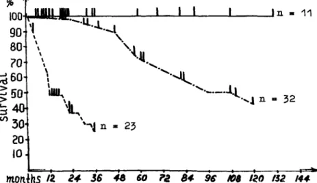
Clinical Classification. It is impossible to predict individual prognosis in MM ac-cording to the stage of the disease [6] at the first presentation. That is why we classified our patients according to their life duration, which determined the type of the disease. This classification is a result of prospective investigation. We divided the patients into three groups: first - indolent myeloma, second - active myeloma, third - aggressive myeloma. The diagnosis was made on the following criteria:
1) Indolent myeloma. Patients who pre-sented with Kyle's criteria [7], patients who had been in remission for more than 5 years without chemotherapy, and patients with monoclonal gam-mopathy ofundetermine~ signi~ca~ce who had been under InvestIgatIOn from 2 to 5 years (Fig. 1).
2) Active myeloma. Patients in who~ fir~t
line chemotherapy was effectIve In achieving remission or the plateau stage. Median survival was 96 months.
3) Aggressive myeloma. Patients who had primary or secondary resistance to chemotherapy. Median Survival was 12 months.
Morphological Classification. The degree of myeloma cell maturation :was ~ete~
IOOL..rlIolMI:1W~n-:---L-~--L---L-_---JL---' n - 11 Il" '-.Ll..J. 90
'I,
·-l'.
80 \ '.~ 70 \.!t.,
~60\
',
. ..Jt.
,
"
..1.t
> .-' fiO > '-: j lUU\t ._._. . ... .1 n .. 32 " \ V) 30 20 10 """J n • 23monihs
12. 24 .36 48 60 1'2 8t1 9&f(),
I~O 132 /4t1Fig. 1. Survival of patients with various courses of multiple myeloma. The solid line
shows the indolent course; the dash-dot line,
of the morphological classification sys-tem for MM [4]:
1) mature - comprising more than 50 % mature plasma cells;
2) immature - comprising more than 50 % proplasmacytes and mixed cell myeloma where none of the morpho-logically distinct plasma cell types ex-ceed 50 % (the proportion of lympho-plasmacytoid cells was always less than 50 % of the bone marrow plasma cells);
3) plasmablastic - comprising more than 50 % plasmablasts.
Serum P2-Microglobulin. SP2-M was measured by 1251 radioimmunoassay. Colony-Forming Unit-Granulocyte/Mac-rophage. The cloning of hemopoietic cells was performed in a agar drop-liquid me-dium system [1].
Biological Activity ofIL-2. The biological activity of IL-2 in conditioned media of peripheral blood mononuclear cells was assayed according to the method re-ported by Bockman and Ropo [3]. TNF a Activity. The spontaneous and
pyrogen-stimulated (50 Ili/ml) produc-tion of TNF a by peripheral blood
mono-the active course, mono-the line of dashes, the ag-gressive course
cytes was assayed by exploring the im-munoenZyme kits (performed by the In-stitute of High Purified Preparations (HPP) Leningrad, USSR), based on monoclonal and polyclonal antibodies to TNF a' In order to form the calibration
curve in each case TNF a was used (made
by NPO "Ferment", Tallin, USSR).
Results
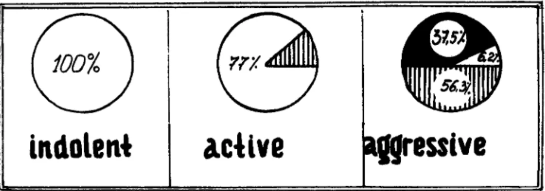
It was revealed that mature plasma cells were the morphological substrate in indo-lent myeloma (Fig. 2) at the same time in active myeloma, mature cells predom-inated in 77% of the patients, whereby in the remaining 23 % of the patients, the cells were immature. In aggressive myeloma, immature cells were the main morphological type in 56.3 % of the pa-tients, plasmablasts prevailed in 37.5 % of the patients, and mature cells were in 6.2 % of the patients.
We discovered that the level of SP2-M correlated with the course ofMM in most of the patients (Fig. 3). There was a significant difference between the level of
SP2-M in patients with indolent myeloma
(3.3 + 0.4 mg/l) and in healthy controls
(1. 7 + 0.4 mg/l; p < 0.05). We revealed a significant difference between the level of
iRc1olen~
clc4lve
Fig. 2. Morphology and course of MM. The
white area shows the percentage of mature
mg/l
2520
15
Ifreulve
cells; the hatched area, the immature cells; and the black area, the plasmoblastic cells
•
•
•
•
10•
.Ie
normal level;
~
:
_307:;j?1UDIZZJ.JIIIJ7T!!Juli1Z7211/?_
o
aC'Lve
a.,ressive
Types of
courseFig. 3. Serum P2-microglobulin and types of courses in patients with multiple myeloma
SP2-M in patients with indolent and
ac-tive myeloma (4.9
+
0.4 mg/l; p < 0.05). There was also significant difference between the level of SP2-M in patients with active and aggressive myeloma(8.6
+
1.2 mg/l; p < 0.05) and a signifi-cant difference between patients with aggressive and indolent courses (p < 0.01; Fig. 4).Studies on the c1onogenic ability of CFU-GM in patients with an indolent course gave unusual results. In most patients with an indolent course, the number of CFU-GM was higher than in healthy controls (Fig. 5). The number of colony-forming cells in agar culture ranged from 30 to 300 per 105 bone
marrow cells, whereas in normal bone marrow cells it ranged from 40 to 60 per 105 bone marrow cells. High levels of
CFU-GM were also found in some pa-tients with active myeloma as compared with healthy controls. The number of colony-forming cells in agar culture in active myeloma ranged from 42 to 420 per 105 bone marrow cells. In patients
with aggressive myeloma the number of colony-forming cells in agar culture ranged from 0 to 25 per 105 bone marrow
cells, which was much less than in pa-tients with indolent and active myeloma and less than in normal controls.
There was an insignificant difference in IL-2 production between healthy
con-the course
Sp~M[mQ/l]
indolent
5,3:!:.q4
p~q051
active
4,9±O,4
P<
0,01
oQQresslve
8,6:!: 1,2
p<'Q05
Fig.4. Serum P2-microglobulin and the course of multiple myeloma
troIs (13.9
+
1.2 units of activity; p < 0.05) and patients with indolent(12.9
+
1.1 units of activity) (Figs. 6, 7) and active courses (11.9+
0.7 units of activity) and between the indolent and active courses. Then significant differ-ences were found in the IL-2 production between patients with active and aggres-sive myeloma (8.6+0.6 units of activity;420t
•
'001
•
"0•
130•
t20•
tlO 100•
•
(/l 90-
~ (1J 80 u In 70 0--
I
•
"-(/) 60 .~ 50 c 0 .. 0--
0 0 30•
20 10p < 0.01) and also between aggressive and indolent myeloma and healthy con-trols (p < 0.01).
An insignificant difference between the pyrogen-stimulated production of TNF a
in patients with an indolent course
(1.5
+
0.3 ng/ml) and healthy controls(2.2
+
0.38 ng/ml) was detected (p> 0.05;Fig. 8).
A significant difference was found between the pyrogen-stimulated produc-tion of TNF a in patients with indolent
and active courses (6.1
+
1.1 ng/ml; p < 0.01). There was also a significant different in TNFa production between patients with active myeloma and healthy controls (p < 0.05), and also in TNFa production in patients with active and aggressive myeloma (2.0+
1.2 ng/ml; p < 0.05). There was an insignificant dif-ference between TNFa production in pa-tients with aggressive and indolent courses and healthy controls (p > 0.05;Fig. 9).
••
I
lndolent
actLve
a~resslve('l'ypes of course)
Fig. 5. Colony-forming unit granulocyte and macrophage of bone marrow and the multiple myeloma courseFig. 6. Production of interleukin-2 by peri-pheral blood mononuclear cells in patients
the course
n-atn·oJoct.
indolent
Q3
tO,4
P~q,05
active
4,9±Q4
p< 0,01OQVressive
8,6:tQ6
p<q,Q5.
Fig. 7. Interleukin-2 and the course of multi-ple myeloma
Conclusion
Our data suggest that:
1) Morphology of myeloma cells is the most important prognostic factor which, without a doubt, can be easily reproduced, and is widely used in clin-ical practice.
2) The level of SP2-M may be used to determine the MM course type. 3) CFU-GM probably may also be used
as a prognostic factor. An increased or normal level of CFU -G M confirms the
with different types of courses in multiple myeloma
indolent or active course ofMM, while a decreased level of CFU-GM reflects the aggression of MM.
4) Decreased production of IL-2 by peri-pheral blood monocytes is character-istic of an aggressive MM course. 5) TN Fa has a prognostic significance ifit
is used together with myeloma cell morphology as
a) mature myeloma cells and normal production of TNFa by peripheral blood monocytes are typical of an indolent course;
b) mature or immature morphology of myeloma cells and increased pro-duction of TN Fa are typical of an active course; and
c) immature or plasmablastic mor-phology and normal production of TN Fa are typical of an aggressive course.
The studies mentioned above were ne-cessary in order to find out the role of these factors in MM pathogenesis.
6 4
E
... 2 «1 E '-~Obldolell4
I
•
•
I
•
•
aclive
agfressive
Types
of course
Fig. 8. Pyrogenal-stimulated tumor necrosis factor-alpha production by peripheral blood monocytes in patients with multip~e myeloma
the
course
TNfcL[RWml1
lndolent
1.5tOJ~P<b01
I
f'
actlve
ql
:tl,1
P>
0,05aQ9resslve
2,O±1,2
P<.O,OSI
Fig. 9. Tumor necrosis factor-alpha and the course of multiple myeloma
References
1. Afanasiev BV, Tiranova SA, Kulibaba TG, Zubarovskaya LS, Bolshakova GD, Zabelina TS (1983) Cloning of human hemopoietic cells in the "agar drop-liquid medium" system. Ter Arkh 8:114
2. Almazov V A, Afanasiev BV, Zaritsky A, Chichkov A (1981) Leukopenia, Lenin-grad, "Medicine"
3. Bockman RS, Ropo MA (1981) Lympho-kinemediated bone resorption requires endogenous prostaglandin synthesis. J Exp Med 154:529-534
4. Carter A, Hocherman J, Linn S, Cohen J, Tatarsky J (1987) Prognostic significance of plasma cell morphology in multiple myeloma. Cancer 60: 1060 -1 065
. 5. Chronic Leukaemia-Myeloma Task Force of the National Cancer Institute (1968) Guidelines for protocol studies. II. Plasma cells myeloma. Cancer Chemother Rep 1: 17
6. Durie BGM, Salmon SE (1975) A clinical staging system for multiple myeloma: cor-relation of measured myeloma cell mass with presenting clinical features, response to treatment, and survival. Cancer 36:842 7. Kyle RA (1988) Prognostic factor in
multi-ple myeloma. Hematol Oncol 6: 125 -130 8. Palladino MA Jr, Shalaby MF, Kramer SM, et al. (1987) Characterization of the antitumor activities of human tumor necrosis factor-a and the comparison with other cytokines: induction of tumor spec-ific immunity. J Immunol138:4023 9. Price G, Brenner MK, Prentice HG,
New-lands AJ (1987) Cytotoxic effects of tumour necrosis factor and gamma inter-feron on myeloid leukaemia blast cells. Br J Cancer 55: 287
10. Rosenberg SA, Lotze MT, Muul LM, et al. (1987) A progress report on the treatment of 157 patients with advanced cancer using lymphokine-activated killer cells and interleukin-2 or high-dose interleukin-2 alone. N Engl fMed 316:889-897 11. Takahashi T, Lim B, Jamal N, Tritchler D,
Lochwood G, McKinney S, Bergsagel DE, Messner HA (1985) Colony growth and self renewal of plasma cell precursora in multiple myeloma. J Clin Oncol 3: