Skeletal Scintigraphy of Young Patients with
Low-Back Pain and a Lumbosacral Transitional
Vertebra
Leonard P. Connolly, MD1; Pierre A. d’Hemecourt, MD2; Susan A. Connolly, MD1; Laura A. Drubach, MD1; Lyle J. Micheli, MD2; and S. Ted Treves, MD1
1Division of Nuclear Medicine, Department of Radiology, Children’s Hospital, Boston, Massachusetts; and2Department of
Orthopedics, Children’s Hospital, Boston, Massachusetts
Lumbosacral transitional vertebrae can alter the biomechanics of weight transfer at the affected spinal segment. Low-back pain may result. This study assessed what skeletal scintigraphy reveals about stress associated with a lumbosacral transitional vertebra in young patients with low-back pain.Methods:The study population included 48 patients (30 male, 18 female; age range, 6 –19 y; mean age, 15.7 y) with low-back pain and a lumbosacral transitional vertebra. Skeletal scintigraphy was correlated with plain radiographs in all, CT in 12, and MRI in 11.
Results:High uptake was shown at the articulation between the transverse process of a lumbosacral transitional vertebra and the sacrum in 39 (81%) of the patients. In 23 (59%) of the 39 patients with high uptake, this finding was shown only by SPECT. In 13 (81%) of the 16 for which the high uptake was shown by planar imaging, the anterior projection was more valuable than the posterior projection. In 9 (23%) of the 39 patients with high uptake at the transverse–sacral articulation, the lumbosacral transitional vertebra had not been noted in a radiographic report before skeletal scintigraphy but was identi-fied through reevaluation or repetition of radiographs after skel-etal scintigraphy. Radiographs showed sclerosis along the transverse–sacral articulation in only 8 (21%) of the 39 patients with high uptake. Findings indicating stress or motion at the joint were shown by CT in 6 (55%) of 11 and by MRI in 5 (63%) of 8 patients with high uptake at the transverse–sacral articula-tion who underwent these examinaarticula-tions.Conclusion:Skeletal scintigraphy often indicates stress at the transverse–sacral ar-ticulation of young patients with low-back pain and a lumbosa-cral transitional vertebra. Showing evidence of stress is best accomplished using SPECT. Changes are usually not radio-graphically evident, but there is a trend for MRI and CT to show findings that imply stress or motion at the articulation. The unique ability of skeletal scintigraphy to provide this physiologic information supports its use in these patients.
Key Words:skeletal scintigraphy; low-back pain; spine
J Nucl Med 2003; 44:909 –914
L
ow back pain is a common problem in young athletes (1). The differential diagnosis is extensive. Stress injuries account for the vast majority of cases. Diagnoses vary considerably in frequency with patient age at our pediatric and adult sports medicine clinics (2). Most patients younger than 21 y have stress injuries of the posterior elements, mainly the partes interarticulares. These injuries are rare in older patients. Conversely, low-back pain in young athletes is infrequently due to disk abnormalities, degenerative changes, and muscular strain, which are the leading diag-noses in adults.The presence of a structural anomaly adds to the diag-nostic challenge of evaluating low-back pain. A relatively common anomaly is a lumbosacral transitional vertebra, which is present in 3%–21% of humans (3–9). It is charac-terized by a large transverse process that follows the contour of the sacral ala and fuses with the sacrum, most often at a diarthrodial joint and sometimes by an ossified union. The large transverse process is usually unilateral. A disk space, which is typically vestigial, is present below the vertebral body (7,10). The anomalous vertebra is commonly referred to as a sacralized L5 or as a lumbarized S1 on the basis of the number of non–rib-bearing vertebrae shown by radio-graphs that include the thoracolumbar junction (11).
Early in the last century, radiographic and clinical obser-vations led Bertolotti to conclude that lumbosacral transi-tional vertebrae contribute to low-back pain (12,13). Although this proposal has been variably accepted (7–10,14 –17), low-back pain in the presence of a lumbosa-cral transitional vertebra is often referred to as Bertolotti’s syndrome (10).
Because it articulates or fuses with the sacrum, a lumbo-sacral transitional vertebra is an important part of the weight-bearing platform (18). Repetitive flexion and exten-sion stress the transitional lumbosacral osseous complex (1), which is delineated superiorly by the disk space above the vertebra and inferiorly by the sacroiliac joint (18). Stress is greatest at the superior disk space and the articulation between the transitional transverse process and the sacrum.
Received Aug. 26, 2002; revision accepted Feb. 13, 2003.
For correspondence or reprints contact: Leonard P. Connolly, MD, Division of Nuclear Medicine, Department of Radiology, Children’s Hospital, 300 Longwood Ave., Boston, MA 02115.
Treatment for stress-related pain involving the transitional lumbosacral osseous complex may include use of a lumbo-sacral orthosis, physical therapy to stretch and stabilize the spine, and sport-specific training to avoid or limit exposure to the mechanism of injury. These steps are usually suc-cessful (1). When they are not, corticosteroid injection of the disk above the transitional vertebra or the transverse– sacral articulation and, for very rare cases, surgical fusion or a resection of the articulating transitional transverse process can be performed (19).
After radiography, CT and MRI (10,20) are the most widely used imaging tests in patients with low-back pain and a lumbosacral transitional vertebra. This is largely based on what has been reported in adults. Disk bulge or herniation, spinal stenosis, and nerve root canal stenosis at the space above the lumbosacral transitional vertebra have been shown in over 50% of cases. These abnormalities have been attributed to hypermobility at that disk space, similar to what occurs in spinal segments adjacent to a fusion mass or block vertebra. At the transverse–sacral articulation, peri-articular sclerosis and osteophyte formation indicative of degenerative change can be shown by CT and subchondral edema indicative of stress can be shown by MRI. These findings have been identified less than one fifth as often as abnormalities at the superior disk space (10).
Skeletal scintigraphy, which is highly sensitive for de-tecting stress-induced changes in bone, is an important part of the evaluation of young athletes with low-back pain largely because the technique often indicates radiographi-cally occult pars interarticularis stress (21–25), the identi-fication of which facilitates early intervention that can pro-mote healing and lessen morbidity (26). Is skeletal scintigraphy also effective in indicating stress involving the transitional lumbosacral osseous complex without changes being shown radiographically? We report what skeletal scintigraphy reveals in symptomatic young athletes with this anomaly.
MATERIALS AND METHODS
The study population included 48 patients (30 male, 18 female; age range, 6 –19 y; mean age, 15.7 y) with low-back pain who were consecutively referred for skeletal scintigraphy over a 4-y interval and who had a lumbosacral transitional vertebra.
We performed skeletal scintigraphy 3– 4 h after intravenous administration of 99mTc-methylene diphosphonate. The
adminis-tered dose was 7.4 MBq/kg (maximum, 740 MBq). Planar anterior and posterior images of the spine and pelvis (500,000 counts) and SPECT images of the lumbosacral spine and pelvis were acquired using a dual-head gamma camera (Ecam; Siemens Gammasonics) equipped with a high-resolution collimator. For SPECT, images were acquired on a 128⫻128 matrix. Pixel size was 4.8 mm. A noncircular 360° orbit with 240 stops (120 stops of 15 s per detector) was used, and a Butterworth filter was applied (cutoff frequency of 0.4 – 0.5 cycle per centimeter).
We recorded sites of abnormally high spinal and pelvic uptake that 1 of 3 pediatric nuclear medicine specialists had described in
the diagnostic reports. Skeletal scintigraphy reports were corre-lated with radiographic reports for all patients and with the reports of CT, MRI, or both for 19 patients, 8 of whom underwent CT only, 7 of whom underwent MRI only, and 4 of whom underwent both CT and MRI within 4 wk of skeletal scintigraphy. A pediatric nuclear medicine specialist and a pediatric radiologist retrospec-tively verified the reported results.
RESULTS
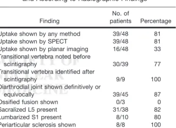
High uptake was present at the articulation between the transverse process of the lumbosacral transitional vertebra and the sacrum (Figs. 1–3) in 39 (81%) of the 48 patients. In 30 (63%) of the 48 patients, this was the only uptake abnormality. In 23 (59%) of the 39 patients with high uptake at the transverse–sacral articulation, the abnormality was shown only by SPECT (Fig. 1). For 13 (81%) of the 16 patients with high uptake by planar imaging, the finding was shown better or only by the anterior projection (Fig. 2). The uptake abnormalities were unilateral in all but one patient, who was the only one with bilateral transverse–sacral artic-ulations shown by radiographs. In 9 (23%) of the 39 patients with high uptake at the transverse–sacral articulation, a lumbosacral transitional vertebra had not been noted in a radiographic report before skeletal scintigraphy and was identified through reevaluation of radiographs after skeletal scintigraphy.
FIGURE 1. (A) Transitional vertebra is shown on radiograph of 17-y-old girl with 7-mo history of progressive low-back pain that was exacerbated with hyperextension. Sclerosis is not evident along articulation of right transverse process with sacrum, which is denoted by arrows. (B) No focal uptake abnormality is demonstrated by posterior (left) and anterior (middle) planar images from skeletal scintigraphy, but SPECT (axial [top right] and coronal [bottom right] images) shows high uptake at trans-verse–sacral articulation (arrows).
For all patients with high uptake at the transverse–sacral articulation, radiographs either showed a diarthrodial joint (n⫽ 28) at the fusion or were equivocal (n⫽11) for this determination because bowel contents obscured the region. None of 3 patients whose radiographs indicated that fusion between the lumbosacral transitional vertebra and the sa-crum was ossified had high uptake in the region of the fusion. Radiographs showed sclerotic bone at the
trans-FIGURE 3. (A) Radiograph of 15-y-old girl with extension-based low-back pain shows sclerosis along left-sided transverse–sacral articulation (arrows). (B) Posterior (left) and anterior (middle) planar images and SPECT (axial [top right] and coronal [bottom right] images) show high uptake (arrows) along articulation. (C) Coronal T1-weighted MRI after gadolinium shows enhancement (arrows) indicating marrow edema on each side of articulation.
Š
FIGURE 2. (A) Skeletal scintigram shows high uptake at left-sided transverse–sacral articulation (arrows) of 17-y-old female athlete who had been experiencing low-back pain for 2 y. Abnor-mality is shown better on anterior (middle) than posterior (left) planar image and is seen better still on SPECT images (axial [top right] and coronal [bottom right]). (B) Radiography findings had been interpreted as normal for this patient, perhaps because bowel gas and contents obscured transverse–sacral articulation, which was clearly shown (arrows) on repeated radiographs (C) after skel-etal scintigraphy. Bone along medial margin of transverse–sacral articulation appears sclerotic. Axial (D) and coronal (E) reformatted CT images show cystic changes and sclerosis on either side of transverse–sacral articulation, which is indicated by arrow.
verse–sacral articulation (Figs. 2 and 3) in 8 (21%) of the 39 patients with and none of the 9 patients without high uptake at the articulation. The frequency with which high uptake was shown at the articulation did not differ between patients with a sacralized L5 and patients with a lumbarized S1. Table 1 summarizes the proportions of patients with high uptake at the transverse–sacral articulation for the entire population and according to radiographic findings.
The only finding that skeletal scintigraphy showed apart from the transverse–sacral articulation was evidence of pars interarticularis stress. This was identified in 10 (21%) of the 48 patients: 4 by SPECT only and 6 by both planar imaging and SPECT. A single vertebral level (8 unilateral and 2 bilateral) was affected in each of these patients. The affected pars interarticularis involved an articulation with the lum-bosacral transitional vertebra and corresponded to the fourth or fifth lumbar level in 9 (90%) of the 10 patients. In the other patient, the uptake abnormality was 2 levels above the lumbosacral transitional vertebra at a level considered L3. Of the 12 partes interarticulares with high uptake, 9 were ipsilateral to a transverse–sacral articulation. These in-cluded 1 patient with a unilateral uptake abnormality in the pars interarticularis and bilateral fusion. Of the 10 patients with evidence of pars interarticularis stress, 9 (90%) also had high uptake at the transverse–sacral articulation. No other vertebral or pelvic uptake abnormalities were shown. Among patients with high uptake at the transverse–sacral articulation, CT showed sclerosis, joint surface irregularity, or subchondral cysts (Fig. 2) in 6 (55%) of 11 patients and MRI showed periarticular high T2 signal or postgadolinium enhancement indicative of marrow edema (Fig. 3) in 5 (63%) of 8 patients. These findings were not identified in the few patients without uptake abnormalities at the transverse– sacral articulation who underwent CT (n⫽1) or MRI (n⫽ 4). The only finding shown by CT or MRI, apart from sites
where skeletal scintigraphy showed abnormalities, was disk bulging, which was defined as diffuse extension of the outer contour of the annulus beyond the disk space. This finding was shown in 7 patients: 6 by MRI and 1 by CT. In all, the bulge was less than 3 mm. In 5 patients, disk bulge was shown at both the contiguous space above and the space 2 levels above the lumbosacral transitional vertebra. One or the other of these spaces was affected in 1 patient each. Six of 7 patients with disk bulge had high uptake at the trans-verse–sacral articulation. Focal displacement of disk mate-rial, which would constitute radiographic evidence of her-niation (27), was not appreciated in any patient.
DISCUSSION
Just over 80% of young patients with low-back pain and a lumbosacral transitional vertebra in this series had high uptake at the transverse–sacral articulation (Figs. 1–3). This finding suggests that localized stress at the articulation contributes to low-back pain. This possibility is supported by the fact that high uptake at the transverse–sacral articu-lation was the only uptake abnormality in 63% of the patients and by the fact that the only structural abnormality shown by CT or MRI, apart from the articulation, was relatively mild disk bulging, which is present in about 20% of asymptomatic young adults (28,29). The potential signif-icance of the high uptake at the transverse–sacral articula-tion is also suggested by the trend for MRI and CT to show findings that imply stress or motion (10) in patients with high uptake at the articulation (Figs. 2 and 3).
Nearly 60% of the uptake abnormalities at transverse– sacral articulations were shown only by SPECT (Fig. 1). This finding is similar to what has been reported when skeletal scintigraphy is used for diagnosis of stress injury to the pars interarticularis and supports the recommendation that SPECT be performed when young patients with low-back pain are referred for skeletal scintigraphy (21–24). When shown by planar imaging, the abnormality was usu-ally seen better or only on the anterior projection (Fig. 2). Stress at the transverse–sacral articulation is usually not evident by radiography, which showed sclerosis in only 21% of the patients with high uptake at the articulation. Additionally, a lumbosacral transitional vertebra may be undocumented before skeletal scintigraphy (Fig. 2). In 23% of our patients, high uptake was shown at a transverse– sacral articulation without the lumbosacral transitional ver-tebra having been noted in prior radiographic reports. In some cases, this may have been attributable to obscuration of the lumbosacral junction by bowel contents. In others, it may have reflected a radiologist’s choice not to note an anatomic variant. Inclusion of patients whose lumbosacral transitional vertebra was identified radiographically only after skeletal scintigraphy showed that an abnormality added a selection bias to this study. The presence of high uptake at the articulation in 77% of the patients whose lumbosacral transitional vertebra was noted before skeletal
TABLE 1
Proportions of Patients with High Uptake at Transverse–Sacral Articulation for Entire Population
and According to Radiographic Findings
Finding
No. of
patients Percentage
Uptake shown by any method 39/48 81
Uptake shown by SPECT 39/48 81
Uptake shown by planar imaging 16/48 33
Transitional vertebra noted before
scintigraphy 30/39 77
Transitional vertebra identified after
scintigraphy 9/9 100
Diarthrodial joint shown definitively or
equivocally 39/45 87
Ossified fusion shown 0/3 0
Sacralized L5 present 31/38 82
Lumbarized S1 present 8/10 80
Periarticular sclerosis shown 8/8 100
scintigraphy indicates that the results were not significantly affected by this bias. Lumbosacral transitional vertebrae may also not be recognized through MRI (15), but we could not assess this possibility in our patients because the anom-aly had always been identified before MRI.
Evidence of stress injury to a pars interarticularis was shown less frequently than was evidence of stress at the transverse–sacral articulation. Our results indicate that stress injury to a pars interarticularis is neither more nor less likely to occur when a lumbosacral transitional vertebra is present (22,30). Although the observed preferential involve-ment of partes interarticulares at or adjacent to the lumbo-sacral transitional vertebra might suggest a specific suscep-tibility, this preference is also consistent with the known preponderance of spondylolysis at the L4 and L5 levels (31,32).
Skeletal scintigraphy should be most useful in young patients with a lumbosacral transitional vertebra when stress at a transverse–sacral articulation or stress injury to a pars interarticularis is suspected. The 2 conditions may coexist, and patients with either can be expected to present with extension-based pain. In some patients, localized tenderness and evidence of sacroiliac irritation may point to stress at the transverse–sacral articulation. When clinical findings suggest disk herniation, spinal stenosis, or foraminal steno-sis as the cause for low-back pain, MRI should be more advantageous than skeletal scintigraphy. These structural pathologies have a different distribution but not a higher incidence in patients with lumbosacral transitional verte-brae. The intervertebral disk level immediately above the anomalous vertebra is predominantly affected (10,33). Be-cause the association of pars interarticularis stress with extension-based pain is well known, patients with such pain were likely preferentially referred for skeletal scintigraphy. This likelihood prevents us from drawing conclusions about the relative frequencies of different causes for low-back pain in patients with a lumbosacral transitional vertebra.
Our clinical data do not substantiate our belief that stress at the transverse–sacral articulation contributes to low-back pain, and we have not established the frequency with which high uptake is shown at the transverse–sacral articulation in asymptomatic individuals. We did not attempt to assess clinical parameters because the patients’ evaluation, treat-ment, and follow-up were not standard among the referring clinicians and we did not believe that the available clinical data allowed us to reliably assess a possible correlation between the characteristics of the patients’ pain and the findings shown by skeletal scintigraphy. We expect that some asymptomatic individuals would have evidence of stress at the transverse–sacral articulation, just as many young athletes have asymptomatic lower-extremity stress changes (34).
Although an uptake abnormality alone does not establish a cause for low-back pain, the data indicate that stress at the transverse–sacral articulation should be considered before the pain is attributed to nonspecific or muscular etiologies.
This indication differs from what has been suggested on the basis of experience with the anatomic imaging modalities CT and MRI in adults (10). Identification of high uptake at the transverse–sacral articulation may assist with initiation of therapeutic measures, but how to optimally use skeletal scintigraphy in management needs further investigation.
CONCLUSION
Skeletal scintigraphy often indicates stress at the trans-verse–sacral articulation of young patients with low-back pain and a lumbosacral transitional vertebra. Showing evi-dence of stress is best accomplished using SPECT. Changes are usually not radiographically evident, but there are trends for MRI and CT to show findings that imply stress or motion at the articulation. The unique ability of skeletal scintigraphy to provide this physiologic information sup-ports its use in these patients.
REFERENCES
1. d’ Hemecourt PA, Gerbino PG II, Micheli LJ. Back injuries in the young athlete.
Clin Sports Med. 2000;19:663– 679.
2. Micheli LJ, Wood R. Back pain in young athletes: significant differences from adults in causes and patterns. Pediatr Adolesc Med. 1995;149:15–18. 3. Sutherland CG. A roentgenographic study of development anomalies of the spine.
J Radiol. 1922;3:357–364.
4. Moore S. On the incidence of the sacralized transverse process and its signifi-cance. Radiology. 1924;2:287–301.
5. Giles RG. Vertebral anomalies. Radiology. 1931;17:1262–1266.
6. Southworth JD, Bersack SR. Anomalies of the lumbosacral vertebrae in five hundred fifty individuals without symptoms referable to low back pain. AJR. 1950;64:624 – 634.
7. Tini PG, Wieser C, Zinn WM. The transitional vertebra of the lumbosacral spine: its radiological classification, incidence, prevalence, and clinical significance.
Rheumatol Rehabil. 1977;16:180 –185.
8. Dai L. Lumbosacral transitional vertebrae and low back pain. Bull Hosp Jt Dis. 1999;58:191–193.
9. Hahn PY, Strobel JJ, Hahn FJ. Verification of lumbosacral segments on MR images: identification of transitional vertebrae. Radiology. 1992;182:580 –581. 10. Elster AD. Bertolotti’s syndrome revisited: transitional vertebrae of the lumbar
spine. Spine. 1989;14:1373–1377.
11. Wigh RE. The thoracolumbar and lumbosacral transitional junctions. Spine. 1980;5:215–222.
12. Bertolotti M. Contribution to the knowledge of the defects of regional differen-tiation of the vertebral column with special attention to the fusion of the fifth lumbar vertebra to the sacrum [in Italian]. Radiologique Medica. 1917;4:113– 144.
13. Bertolotti M. Lumbo-ischial syndromes of vertebral origin: their morphologic, radiologic, and clinical aspects [in French]. Rev Neurol (Paris). 1922;38:1112– 1125.
14. Wigh RE, Anthony HF Jr. Transitional lumbosacral discs: probability of herni-ation. Spine. 1981;6:168 –171.
15. O’Driscoll CM, Irwin A, Saifuddin A. Variations in morphology of the lumbo-sacral junction on sagittal MRI: correlation with plain radiography. Skeletal
Radiol. 1996;25:225–230.
16. Magora A, Schwartz A. Relation between the low back pain syndrome and x-ray findings. 2. Transitional vertebra (mainly sacralization). Scand J Rehab Med. 1978;10:135–145.
17. Santiago FR, Milena GL, Herrera RO, Romero PA, Plazas PG. Morphometry of the lower lumbar vertebrae in patients with and without low back pain. Eur Spine
J. 2001;10:228 –233.
18. Wigh RE. The transitional lumbosacral osseous complex. Skeletal Radiol. 1982; 8:127–131.
19. Jonsson B, Stromqvist B, Egund N. Anomalous lumbosacral articulations and low-back pain: evaluation and treatment. Spine. 1989;14:831– 834.
20. Hashimoto M, Watanabe O, Hirano H. Extraforaminal stenosis in the lumbosa-cral spine: efficacy of MR imaging in the coronal plane. Acta Radiol. 1996;37: 610 – 613.
21. Bodner RJ, Heyman S, Drummond DS, Gregg JR. The use of single photon emission computed tomography (SPECT) in the diagnosis of low-back pain in young patients. Spine. 1988;13:1155–1160.
22. Bellah RD, Summerville DA, Treves ST, Micheli LJ. Low back pain in adoles-cent athletes: detection of stress injuries to the pars interarticularis with SPECT.
Radiology. 1991;180:509 –512.
23. Papanicolaou N, Wilkinson RH, Emans JB, Treves ST, Micheli LJ. Bone scintigra-phy and radiology in young athletes with low back pain. AJR. 1985;145:1039 –1044. 24. Collier BD, Johnson RP, Carrera GF, et al. Painful spondylolysis or spondylolis-thesis studied by radiography and single-photon emission computed tomography.
Radiology. 1985;154:207–211.
25. Sty JR, Wells RG, Conway JJ. Spine pain in children. Semin Nucl Med. 1993; 23:296 –320.
26. Micheli LJ. Low back pain in the adolescent: differential diagnosis. Am J Sports
Med. 1979;7:362–364.
27. Brant-Zawadzki MN, Jensen MC, Obuchowski N, Ross JS, Modic MT. Interob-server and intraobInterob-server variability in interpretation of lumbar disc abnormalities: a comparison of two nomenclatures. Spine. 1995;20:1257–1263.
28. Milette PC. Classification, diagnostic imaging, and imaging characterization of a lumbar herniated disk. Radiol Clin North Am. 2000;38:1267–1292.
29. Boden SD, Davis DO, Dina TS, Patronas NJ, Wiesel SW. Abnormal magnetic-resonance scans of the lumbar spine in asymptomatic subjects: a prospective investigation. Patient Care Manag. 1990;72:403– 408.
30. Feldman DS, Hedden DM, Wright JG. The use of bone scan to investigate back pain in children and adolescents. J Pediatr Orthop. 2000;20:790 –795. 31. Lonstein JE. Spondylolysis and spondylolisthesis. In: Morrisy RT, Weinstein SL,
eds. Lovell and Winter’s Pediatric Orthopedics. 5th ed. Philadelphia, PA: Lip-pincott-Raven; 2001:777–798.
32. Ozonoff MB. Pediatric Orthopedic Radiology. 2nd ed. Philadelphia, PA: W.B. Saunders; 1992:1–116.
33. Castellvi AE, Goldstein LA, Chan DP. Lumbosacral transitional vertebrae and their relationship with lumbar extradural defects. Spine. 1984;9:493– 495.
34. Drubach LA, Connolly LP, D’Hemecourt P, Treves ST. Assessment of the clinical significance of asymptomatic lower extremity uptake abnormalities in athletes. J Nucl Med. 2001;42:209 –212.