CONCISE GUIDANCE TO GOOD PRACTICE
A series of evidence-based guidelines for clinical management
NUMBER 7
The prevention, diagnosis, referral and
management of melanoma of the skin
CONCISE GUIDELINES
Guideline Development Group
These guidelines were prepared by a
multidisciplinary Guideline Development Group convened by the British Association of
Dermatologists in conjunction with the Clinical Standards Department of the Royal College of Physicians.
Julia Newton BishopMD FRCP (Chair), Professor of Dermatology St James’s University Hospital, Leeds
Veronique BatailleMD PhD FRCP, Consultant Dermatologist West Herts NHS Trust, Hertfordshire
Alice GavinMB ChB MRCGP DPD, General Practitioner Bradford
Marko LensMD PhD FRSC, Consultant Plastic Surgeon King’s College London, Genetic Epidemiology Unit, London
Jerry MarsdenFRCP, Consultant Dermatologist University Hospital, Birmingham
Tania Mathews, Lay representative
Anthony Ormerod, MB MD FRCP (Edin) FRCP (Lond), Affiliation Reader in Dermatology, University of Aberdeen
Catherine WheelhouseRGN BSc (Hons), Skin Cancer Nurse Specialist Bradford Teaching Hospitals Foundation Trust
Clinical Standards Department
The purpose of the Clinical Standards Department of the Royal College of Physicians is to improve patient care and healthcare provision by setting clinical standards and monitoring their use. We have expertise in the development of evidence-based guidelines and the organisation and reporting of multicentre comparative performance data. The department has three core strategic objectives: to definestandards around the clinical work of physicians; to measureand evaluate the implemenation of standards and its impact on patient care; and to effectively implementthese standards.
Our programme involves the relevant specialist societies, patient groups and national bodies including the National Institute for Health and Clinical Excellence (NICE), the Healthcare Commission and the Health Foundation.
Concise Guidance to Good Practice series
The concise guidelines in this series are intended to inform those aspects of physicians’ clinical practice which may be outside their own specialist area. In many instances the guidance will also be useful for other clinicians including GPs and other healthcare professionals.
The guidelines are designed to allow clinicians to make rapid, informed decisions based wherever possible on synthesis of the best available evidence and expert consensus gathered from practising clinicans and service users. A key feature of the series is to provide both recommendations for best practice, and where possible practical tools with which to implement it.
Series Editor: Lynne Turner-StokesFRCP
Royal College of Physicians
11 St Andrews Place, London NW1 4LE
www.rcplondon.ac.uk
Registered Charity No 210508 ISBN 978-1-86016-301-2
Review date: 2010
Designed and typeset by the Publications Unit of the Royal College of Physicians
Printed in Great Britain by Sarum ColourView Group, Salisbury, Wiltshire
Citation: Royal College of Physicians and British Association of Dermatologists. The prevention, diagnosis, referral and management of melanoma of the skin: concise guidelines.Concise guidance to good practice series, No 7. London: RCP, 2007.
Copyright: All rights reserved. No part of this publication may be reproduced in any form (including photocopying or storing it in any medium by electronic means and whether or not transiently or incidentally to some other use of this publication) without the written permission of the copyright owner. Applications for the copyright owner’s permission to reproduce any part of this publication should be addressed to the publisher.
Contents
Guideline Development Group ii Methodology 1
Summary of the guidelines 2 Introduction 3
What is melanoma and what is its epidemiology? 3
Types of melanoma 3
Who is at risk of melanoma? 4 Where do they occur on the body? 4 What is the relationship between moles and melanoma? 4
What are the symptoms and signs of a melanoma? 5
What are the diagnostic signs of a melanoma? 6
Management 7 References 7 Appendices 8
1 Guideline development process 8
2 Grading system for recommendations adapted from the SIGN methodology 9
Figures 10
Methodology
These guidelines were developed by a
multiprofessional Guideline Development Group including representatives from specialist nursing, general practice, plastic surgery, in addition to dermatology.
A literature search was carried out using the following databases: Medline, Embase, Cochrane Library. The literature was appraised and articles not published in English were excluded. Much of the advice is based on expert opinion and practice because of a lack of other evidence.
These guidelines were developed by a multidisciplinary group to promote the early diagnosis of melanoma of the skin. This increasingly common tumour often has a slow early growth rate during which curable lesions may be detected and removed. Physicians therefore have the potential to reduce mortality and these guidelines are intended to aid physicians, GPs and other health professionals in their ability to recognise melanoma. The majority of melanomas occur in white-skinned people. The most common risk factors are pale sun-sensitive skin and the presence of increased numbers of melanocytic naevi (moles). Melanoma is more common in women than men and the mean age of onset is 50 years but a fifth of cases occur in young adults. In the UK population the most common site is the lower leg in women, and on the back in men. The predictors of melanoma are progressive change in the shape, size and colour of moles. These guidelines provide a series of
photographs of moles, melanomas and other skin lesions, which may resemble melanomas. The purpose of these guidelines is to provide physicians, GPs and other relevant healthcare professionals with a practical guide to the recognition of melanoma on the skin.
The guidelines were prepared in accordance with the principles laid down by the AGREE Collaboration (Appraisal of Guidelines for Research and Evaluation). A summary of the guideline development process is given in Appendix 1.
The system used to grade the evidence and guidance recommendations has been adapted from that used by SIGN (Appendix 2). These are indicated in bold type throughout the text, such as [IIB].
Recommendation Grade
1 Identifying people at risk B
People should be considered to have higher risk (approximately 10-fold) of malignant melanoma if they have:
•
>100 normal moles•
atypical moles•
two or more cases of melanoma in first degree relatives. Lower (approximately 2- to 3-fold) levels of risk are associated with:•
freckles•
red hair or skin which burns in the sun•
any family history of malignant melanoma.2 Primary prevention B
•
People at risk of skin cancer should protect their skin from the sun by avoidance and clothing primarily.•
They should also use a sun protection factor (SPF) of 20 to 30, and five star ultraviolet A (UVA)protection as an adjunct.
3 Secondary prevention C
•
People who are in any of these higher risk (10-fold) categories above should be referred for risk estimation and education directed towards self examination with a dermatologist specialising in moles and pigmented lesions (routine appointment).•
Base-line photography is a useful aid to monitoring moles.4 Urgent referral to dermatologist B
The following should be regarded as suspicious lesions requiring urgent referral to a dermatologist within two weeks:
•
a new mole which is growing quickly over the age of puberty•
a long-standing mole which is changing progressively in shape or colour regardless of age•
any mole which has three or more colours or has lost its symmetry•
any new nodule which is growing and is pigmented or vascular in appearance•
a new pigmented line in a nail•
something growing under a nail•
a mole which has changed in appearance and which is also itching or bleeding.Introduction
Melanoma of the skin is an increasingly common tumour, which often has a slow early growth rate during which curable lesions may be detected and removed. In the UK, melanoma is diagnosed at a mean age of around 50 years but a fifth of cases occur in young adults. So while it is one of the less common forms of cancer, it has a large impact in terms of years of life lost. These guidelines have been developed to help healthcare professionals recognise early predictors of melanoma, using the series of photographs provided, and thus to help reduce mortality.
What is melanoma and what is its
epidemiology?
Melanoma of the skin is a malignant tumour, which arises from cutaneous melanocytes. Current UK lifetime risk is about 1:150 for men and 1:120 for women. It is therefore an uncommon cancer in the UK, so most physicians have a limited experience of it and the majority have not had the opportunity to
develop diagnostic skills. The incidence of melanoma continues to increase.
Melanoma is a tumour predominantly of white-skinned people1and the incidence correlates with latitude of residence,1providing strong evidence that sun exposure is causal. Although it is uncertain what decrement in UV dose reduction might produce a decrement in melanoma risk, conventional advice to white-skinned populations at risk is to minimise sun exposure.
The recent interest in vitamin D as a putative protective agent for other forms of cancer2makes it important that the advice to avoid sun exposure should be targeted at those most at risk [IIB]. It may also be sensible to advise those who avoid the sun to reduce their risk of skin cancer that they should ensure compensatory dietary intake of vitamin D.
Types of melanoma
Table 1 lists characteristics of the four clinical subtypes of melanoma. Figs 1–13 at the end of the booklet illustrate each type (pp10–12.)
Table 1. Types of melanoma (see Figs 1–13).
Type Description
Superficial spreading Slow growing initially: develop horizontally at first for months before they acquire melanomas (Figs 5 and 6) the capacity for invasion. The physician therefore has the potential to diagnose at a
curable phase.
Commonest type of melanoma.
Nodular melanomas Grow more rapidly – vertical growth phase (having acquired the capacity for invasion)
(Fig 10) from the beginning.
More common in older individuals.
Lentigo maligna melanomas Slow-growing at first then rapid when become invasive.
(Fig 11) Develop in a slow-growing precursor pigmented macule called a lentigo maligna or Hutchinson’s freckle, which may remain in situ and incapable of metastasis for very many years. However, once a melanoma develops these variants are as aggressive as others of similar thickness. Much more common in people over 60 years old.
Acral lentiginous melanomas Rarest type. Found on the soles or palms or under the nail (subungual).
Who is at risk of melanoma?
The most important phenotypic markers are an above-average mole count and sun-sensitive skin (Table 2). Individuals with either are at increased risk. The phenotypic markers of sun sensitivity are hair colour (red or blonde hair), blue eyes and particularly freckles [IIA].3
The mean age of diagnosis in the UK is around 50 years, but the age distribution curve is relatively flat and 20% of cases occur in young adults aged 15 to 39 years old.7Although melanoma remains one of the less common forms of cancer, its occurrence in young people means that it has a large impact in terms of years of life lost.
Where do they occur on the body?
Melanomas may occur anywhere on the skin but they occur particularly as described here:
•
Women most commonly develop melanomas on the lower limb (50% of women, 18% of men).7•
Men who develop superficial spreading or nodular melanomas most commonly develop them on the trunk (35% of men, 14% of women), especially the back.•
Patients with chronic sun exposure through life commonly develop their melanomas on the head and neck.What is the relationship between
moles and melanoma?
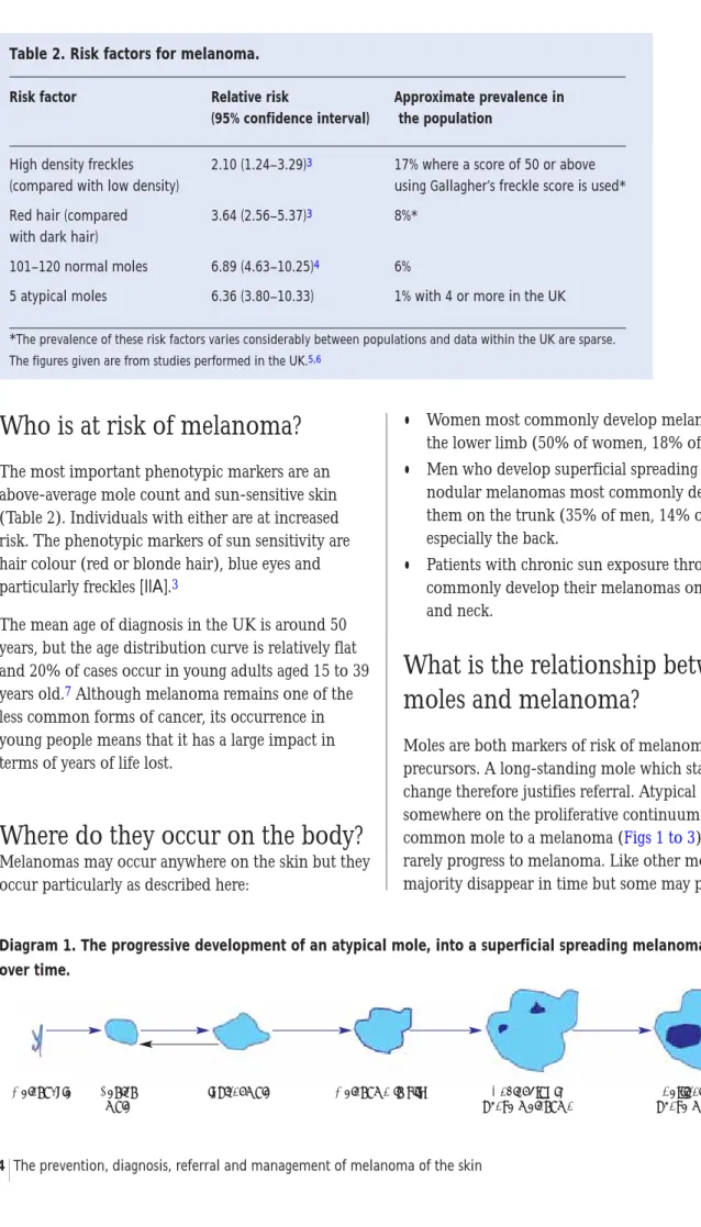
Moles are both markers of risk of melanoma4and precursors. A long-standing mole which starts to change therefore justifies referral. Atypical moles are somewhere on the proliferative continuum from a common mole to a melanoma (Figs 1 to 3) but they rarely progress to melanoma. Like other moles the majority disappear in time but some may progress.
Melanocyte Benign Atypical mole Melanoma in situ Radial growth Vertical growth
mole phase melanoma phase melanoma
Diagram 1. The progressive development of an atypical mole, into a superficial spreading melanoma over time.
Table 2. Risk factors for melanoma.
Risk factor Relative risk Approximate prevalence in (95% confidence interval) the population
High density freckles 2.10 (1.24–3.29)3 17% where a score of 50 or above (compared with low density) using Gallagher’s freckle score is used* Red hair (compared 3.64 (2.56–5.37)3 8%*
with dark hair)
101–120 normal moles 6.89 (4.63–10.25)4 6%
5 atypical moles 6.36 (3.80–10.33) 1% with 4 or more in the UK *The prevalence of these risk factors varies considerably between populations and data within the UK are sparse. The figures given are from studies performed in the UK.5,6
An atypical mole which is changing in size, shape or colour should be reviewed by a specialist (see Fig 3). The progression in Diagram 1 is important to recognise, although it must be emphasised that the majority of such moles never progress and indeed disappear as the patient ages. Thus, atypical moles are those that must be monitored but the absolute risk of melanoma is so low that prophylactic excision is not justified.8The indication for removal is to exclude melanoma [IIB].
Patients with many moles, especially clinically atypical moles, are at sufficiently increased risk of melanoma (relative risk around 10-fold compared with those with few moles, lifetime risk in the order of 1 in 15) to merit referral for assessment. Patients with two or more atypical moles should be referred to a dermatologist for risk assessment and education about self-monitoring, based on the risk of
melanoma reviewed in a recent meta-analysis [IIA].4 In the UK around 2% of the population have this phenotype [IIB].6
What are the symptoms and signs
of a melanoma?
Most melanomas are asymptomatic, although some patients report that their melanoma itches or tingles. Vertical growth phase melanomas may also bleed if ulcerated. However, normal moles may itch,
particularly if they are inflamed, and they may bleed if caught. Both these symptoms are therefore poor discriminators and are unlikely to be of note unless the mole has changed in appearance over time or has the clinical features of irregularity of shape and colour. Only about 50% of melanomas develop in pre-existing moles – a large proportion appear de novo. A patient who reports a history of progressive change in the shape, size or colour of a mole should be referred to a dermatologist promptly even if the lesion looks relatively banal. It is very important to realise that melanoma may look unremarkable early in its evolution – simply an asymmetrical and bigger version of the benign mole from which it may have grown. The clue to the diagnosis may lie only in the history.
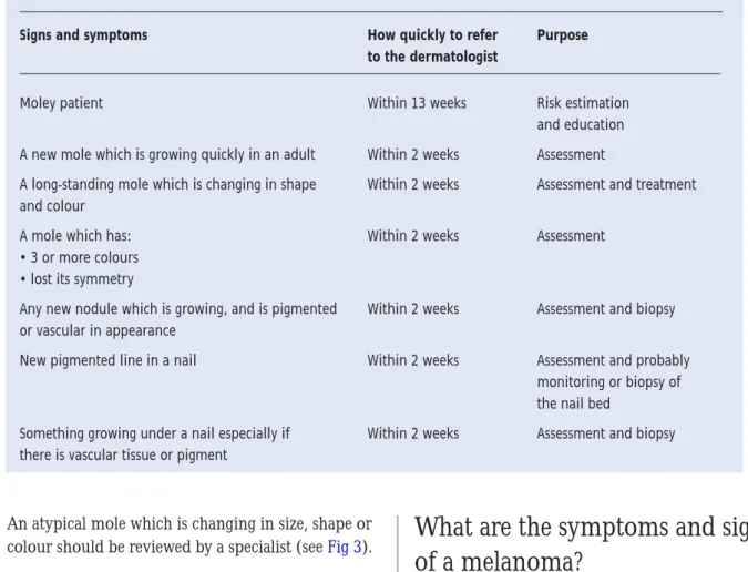
Table 3. Who and when to refer.
Signs and symptoms How quickly to refer Purpose to the dermatologist
Moley patient Within 13 weeks Risk estimation
and education A new mole which is growing quickly in an adult Within 2 weeks Assessment
A long-standing mole which is changing in shape Within 2 weeks Assessment and treatment and colour
A mole which has: Within 2 weeks Assessment
• 3 or more colours • lost its symmetry
Any new nodule which is growing, and is pigmented Within 2 weeks Assessment and biopsy or vascular in appearance
New pigmented line in a nail Within 2 weeks Assessment and probably monitoring or biopsy of the nail bed
Something growing under a nail especially if Within 2 weeks Assessment and biopsy there is vascular tissue or pigment
What are the diagnostic signs of a
melanoma?
Superficial spreading melanomas (Figs 5 and 6) are usually:
•
larger in surface area than normal moles and increase in size progressively over time. Most (but not all) are therefore 7 mm or greater in diameter when diagnosed•
irregular in colour: commonly three or more different colours (browns, reds, blacks or blue-blacks)•
irregular in shape: lacking in symmetry, with geographical borders.Nodular melanomas often also have irregular colour (Fig 10) but may be more uniform in colour than superficial spreading melanomas. Such tumours are usually black or red and may be ulcerated. Lentigo maligna melanomas (Fig 11) may have the appearance of a superficial spreading or nodular melanoma but they arise in a longstanding flat pigmented precursor lesion known as a lentigo maligna. This precursor lesion has the appearance of a freckle but one which is larger, more sharply defined and darker than a normal freckle.
Acral lentiginous melanomas (Fig 12) appear on the palm and sole initially as flat pigmented lesions resembling moles, which start to increase in size and which ultimately develop irregular areas of
pigmentation and which sometimes ulcerate. Such melanomas often present late partly because the patient may be unaware of the appearance of the sole of the foot and partly because they may be
misdiagnosed as an ulcer, particularly in a diabetic. They may also be hidden by a layer of reactive keratin, appearing as a callus (corn). The diagnostic clue here may be bleeding.
Melanomas which grow under the nail (subungual) (Fig 13) are most common under the thumb nail or the nail of the great toe. They appear always to arise from the nail matrix and therefore first appear at the proximal nail fold. The classical subungual melanoma arises as a narrow pigmented band in the nail, which slowly widens and ultimately produces a subungual mass (often amelanotic and friable) and which lifts up the nail plate and tends to bleed. This form of
melanoma is very easy to misdiagnose, commonly as a fungal infection or a pyogenic granuloma.
Management
Accurate diagnosis is most likely to result from an accurate history recording and lesion examination, comparison with a previous photograph, and prompt complete excision of suspicious lesions. This clinico-pathological correlation is important because melanoma may clinically and histologically mimic benign lesions and vice versa. It is strongly
recommended that incisional or incomplete excisions be avoided, particularly because sampling error may result in inaccurate diagnoses. The British
Association of Dermatologists and the Melanoma Study Group guidelines describe the management of melanoma.9
Tables 3 and 4 provide details on referral and
management for different types of moles and lesions.
Table 4. Who not to refer.
Lesions which should not be referred Suggested management
A typical seborrhoeic wart (basal cell papilloma) None needed unless inflamed when referral would be appropriate
(Fig 9) or curettage with histology
A maturing mole (Fig 2) which is slowly becoming Reassurance provided information is given (ideally written) to dome shaped and losing its colour seek advice if significant change subsequently occurs
References
1 Parkin DM, Whelan SL, Ferlay J et al. Cancer incidence in five
continents. IARC Scientific Publications No 143, Lyon:
International Agency for Research on Cancer, 1997. II. 2 Garland CF, Garland FC, Gorham ED et al. The role of
vitamin D in cancer prevention. Am J Public Health, 2005:96(2);209–10.
3 Gandini S, Sera F, Cattaruzza MS et al. Meta-analysis of risk factors for cutaneous melanoma: III. Family history, actinic damage and phenotypic factors. Eur J Cancer
2005:41(14);2040–59.
4 Gandini S, Sera F, Cattaruzza MS et al. Meta-analysis of risk factors for cutaneous melanoma: I. Common and atypical naevi. Eur J Cancer 2005:41(1);28–44.
5 Bertram CG, Gaut RM, Barrett JH et al. An assessment of the CDKN2A variant Ala148Thr as a nevus/melanoma susceptibility allele. J Invest Dermatol 2002:119(4);961–5. 6 Bataille V, Bishop JA, Sasieni P et al. Risk of cutaneous
melanoma in relation to the numbers, types and sites of naevi: a case-control study. Br J Cancer 1996:73(12);1605–11. 7 Quinn MJ, Babb P, Brock A et al. Cancer trends in England
and Wales 1950–1999. Office for National Statistics Single
Market Programme in Services No 66. London: TSO, 2004. 8 Tsao H, Bevona C, Goggins W et al. The transformation rate
of moles (melanocytic nevi) into cutaneous melanoma: a population-based estimate. Arch Dermatol
2003:139(3);282–8.
9 Roberts DLL, Anstey AV, Barlow RJ et al. UK guidelines for the management of cutaneous melanoma. Br J Dermatol 2002:146(1);7–17.
Appendix 1. Guideline development process
The guidelines have been developed in accordance with the principles laid down by the AGREE collaboration (Appraisal of Guidelines for Research and Evaluation).
Scope and purpose
Overall objective of the guidelines To improve the prognosis for melanoma patients by increasing the awareness of melanoma in physicians and therefore by promoting secondary prevention. The patient group covered: Individuals at risk of melanoma.
Target audience All clinicians, including general physicians, GPs and other health professionals who are involved in examining patients and therefore their skin.
Clinical areas covered How to recognise melanoma; what to do if the diagnosis is suspected.
Stakeholder involvement
The Guideline Development Group A multidisciplinary group comprising:
professionals: consultants in dermatology, plastic surgery, a melanoma specialist nurse and a general practitioner
user representation: a patient, Tania Mathews.
Funding This guideline was commissioned and edited by the British Association of Dermatologists and the Clinical Standards Department of the Royal College of Physicians.
Conflicts of interest No external funding has been sought or obtained. All authors and group members have declared, and provided details, of any actual or potential conflicts of interest.
Rigour of development
Evidence gathering Evidence for these guidelines was provided by review of Cochrane Library, Medline, Embase, conference proceedings and other guidelines up to October 2005. Articles not published in English were excluded. Much of the advice is based on expert opinion and practice because of a lack of other evidence.
Links between evidence and The system used to grade the evidence and guidance recommendations is adapted from recommendations that published by the Scottish Intercollegiate Guidelines Network. These are indicated in
bold type throughout the text, such as [IIB].
Piloting and peer review. Not yet piloted.
Implementation
Plans for update Review is planned for 3 years time, and forms part of the work undertaken by the British Association of Dermatologists.
Appendix 2. Grading system for recommendations adapted from the Scottish
Intercollegiate Guidelines Network (SIGN) methodology
Level Type of evidence Grade of recommendation
IA Meta-analysis of randomised clinical trials (RCT) or inception cohort studies A
IB At least one RCT or well-designed cohort studies with good follow-up A
IIA At least one well-designed controlled study without randomisation or a meta-analysis of B case control studies
IIB At least one study with quasi-experimental design or case-control study B
III At least one non-experimental study (such as descriptive study) C
Fig 1. Junctional moles: these are normal moles which develop from early childhood. They are normally less than 5 mm in diameter.
The risk of melanoma from any such mole is extremely low but patients should be aware that they should seek advice if there is any change in shape, size or colour.
(a) (b) CM (c)
Fig 2. Mature ‘dermal’ moles. Over time, particularly on the trunk and face, moles often mature into smooth dome-shaped lesions. This
change in shape may cause concern, but the symmetry and slow rate of change is reassuring. These moles have an extremely low risk of change and should be left in situ.
(a) (b) (c)
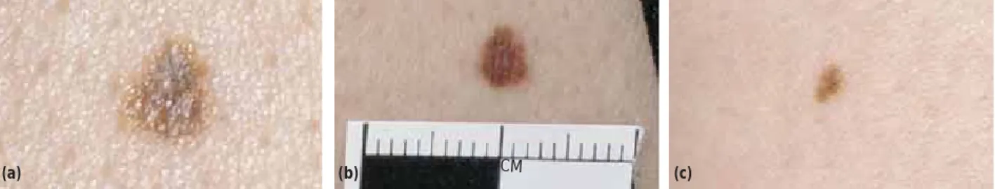
Fig 3. Moderately atypical moles. These are moles which are behaving atypically in that they are becoming irregular in shape, larger
(usually over 5 mm in diameter) and may have variable colours. These moles are more likely to evolve into melanoma and should therefore be reviewed at 3 months for change, although the absolute risk of malignant change is very low.
(a) (b) (c)
Fig 4. Severely atypical moles/melanoma in situ. These moles are evolving into
melanoma and show more marked variation in shape and colour. Early melanomas often look inflamed, as can be seen in both these examples. Lesions like these should be referred
urgently (within two weeks) to a dermatologist. (a) (b)
Fig 5. Superficial spreading melanomas progressively become more irregular in shape and colour over time. Two-week rule referral is
essential.
(a) (b) (c)
CM
CM
Fig 6. Superficial spreading melanoma: showing irregularity of shape and colour. Melanomas tend to have several different colours:
browns, blacks, greys and reds. The continuum of progressive change in appearance is evident.
(a) (b) (c)
Fig 7. Superficial spreading melanomas with nodular elements. Superficial spreading
melanoma evolves slowly over time: often over many months, if not years. Ultimately, however, nodular elements develop corresponding to a vertical growth phase,
associated with much higher risk of metastasis. (a) CM (b)
CM
Fig 8. Differential diagnosis dermatofibromas are firm dermal nodules which appear slowly, often on the limbs. Although they may have a
pigmented halo as seen in the lesion on the left, they are symmetrical. The symmetry, firm nature and very slow change are reassuring. Such lesions do not require referral.
(a) (b)
Fig 9. Differential diagnoses: seborrhoeic warts (basal cell papillomas) may be variably pigmented but have a ‘stuck on’ appearance. Refer if the pigment is variable: rarely nodular melanomas may be difficult to distinguish from warts.
(a) (b) (c)
Fig 10. Nodular melanomas: the melanoma on the left is amelanotic. There is no visible pigment. Nodular melanomas
exhibit vertical growth phase from the beginning and therefore tend to be thicker and to have a poorer prognosis than superficial
spreading melanomas. (a) (b)
Fig 11. Lentigo maligna melanomas arise in precursor in situ lesions called lentigo maligna or Hutchinson’s freckle. The
growth rate of the precursor lesion is very slow but once invasive disease develops these melanomas behave prognostically like
superficial spreading melanomas. (a) (b)
Fig 13. Early subungual melanomas are difficult to diagnose. Image (a) shows a
melanoma which has disrupted the normal growth of the nail but in which there is no clear pigment. More typically there is a narrow pigmented longitudinal band which gets wider over time. The other two images
(b and c) show advanced tumours. (a) (b)
Fig 12. Acral lentiginous melanomas: an early lesion is seen in (c). There is subtle pigmentation which is extensive and irregular. The
other two melanomas are much more advanced and there is reactive hyperkeratosis. It is easy to miss these tumours. Beware of scaly lesions or chronic ulcers on the sole of the foot and refer to the dermatologist for advice if in doubt.
(a) (b) (c)
(c)