PDF hosted at the Radboud Repository of the Radboud University
Nijmegen
The following full text is a publisher's version.
For additional information about this publication click this link.
http://hdl.handle.net/2066/118452
Please be advised that this information was generated on 2017-12-05 and may be subject to
change.
Function and prognostic value of LAMP3 in cancer Anika Nagelkerke, 2013
ISBN: 9789461085054
Printed by: Gildeprint Drukkerijen, Enschede, the Netherlands
Cover: Xenograft of a head and neck squamous cell carcinoma stained against LAMP3 (red), hypoxia (green) and vessels (blue).
FUNCTION AND PROGNOSTIC VALUE OF LAMP3 IN CANCER
proefschrift
Ter verkrijging van de graad van doctor aan de Radboud Universiteit Nijmegen
op gezag van de rector magnificus prof. mr. S.C.J.J. Kortmann, volgens besluit van het college van decanen
in het openbaar te verdedigen op dinsdag 12 november 2013 om 10.30 uur precies
door
Adriana Pieternella (Anika) Nagelkerke Geboren op 16 april 1986
Promotor Prof. dr. C.G.J. Sweep Copromotoren Dr. P.N. Span Dr. J. Bussink Manuscriptcommissie
Prof. dr. N. Hoogerbrugge-van der Linde (voorzitter) Prof. dr. J.H.J.M. van Krieken
TABLE OF CONTENTS
TABLE OF CONTENTS
CHAPTERONE 11
General introduction
CHAPTERTWO 25
Hypoxic regulation and prognostic value of LAMP3 expression in breast cancer
CHAPTERTHREE 43
Generation of multicellular tumour spheroids of breast cancer cells: How to go three-dimensional
CHAPTERFOUR 53
Hypoxia stimulates migration of breast cancer cells via the PERK/ATF4/LAMP3-arm of the unfolded protein response
CHAPTERFIVE 71
Hypoxic activation of the PERK/eIF2α-arm of the unfolded protein response promotes metastasis through induction of LAMP3
CHAPTERSIX 95
Hypoxic regulation of the PERK/ATF4/LAMP3-arm of the unfolded protein response in head and neck squamous cell carcinoma
CHAPTERSEVEN 109
LAMP3 is involved in tamoxifen resistance in breast cancer through modulation of autophagy
CHAPTEREIGHT 127
The PERK/ATF4/LAMP3-arm of the unfolded protein response affects radioresistance by interfering with the DNA damage response
CHAPTERNINE 141
Constitutive expression of γH2AX has prognostic relevance in triple negative breast cancer
CHAPTERTEN 153
Constitutive γH2AX expressing triple negative breast cancers exhibit telomere dysfunction and have no increased sensitivity to anti-cancer drugs
CHAPTERELEVEN 167
TABLE OF CONTENTS
CHAPTERTWELVE 181
Summary/Samenvatting
LIST OF ABBREVIATIONS AND SYMBOLS 193
LIST OF PUBLICATIONS 195
CURRICULUM VITAE 197
CHAPTER ONE
CHAPTER ONE
GENERALINTRODUCTION
13
Cancer is a multifactorial disease. Environmental, genetic and behavioural factors all contribute to its development. Cancer cells are characterised by their aberrant functioning. In 2000, HANAHAN and WEINBERG described a number of features cancer cells are likely to present with and that contribute to their malignancy: the hallmarks of cancer (see Figure 1)1. A major
characteristic of cancer cells is their uncontrollable and infinite cell division. In order to establish this, cancer cells have become self-sufficient in growth signals and are insensitive to growth-inhibitory signals. In addition, cancer cells continuously attempt to evade death (apoptosis) and in order to sustain growth, cancer cells need continuous angiogenesis for the supply of oxygen and nutrients. To be able to spread through their host, tumour cells can invade surrounding tissue and metastasise.
As cancer research proceeds, new hallmarks are being recognised2. Evidence is emerging that
cancer cells reprogram their energy metabolism and attempt to evade destruction by the immune system. In addition, tumour-promoting inflammation is also of interest for tumourigenesis as it can contribute in acquiring other cancer hallmarks. Another important feature is that cancer cells often have an unstable genome (genomic instability), characterised by increased mutation rates, chromosomal rearrangements and aberrant chromosome numbers. As the human body consists of billions of cells that are susceptible to become malignant, the chance of developing cancer seems enormous. However, most of these insults are averted by specialised cellular mechanisms: the DNA damage repair pathways.
FIGURE 1 Hallmarks of cancer. The concept of cancer hallmarks describes a set of capabilities or enabling characteristics that are involved in the pathogenesis of cancer. Growth and progression of most - if not all - tumours is stimulated by these characteristics. So far, ten hallmarks have been formulated. Adapted from 2, © 2011 Elsevier, reprinted
with permission.
DNA damage repair
The DNA damage repair pathways play a vital role in maintaining genome integrity and represent essential biological mechanisms to identify and repair DNA damage or DNA aberrations3. DNA integrity is continuously threatened by both exogenous and endogenous
factors, such as UV irradiation, chemicals, ionising radiation, but also by reactive oxygen species that are part of normal cell metabolism4. There are different types of DNA damage
that can occur and therefore, different kinds of repair mechanisms exist. All these pathways have in common that they are composed of four core components: sensors, mediators,
CHAPTER ONE
14
transducers and effectors5,6. After damage is inflicted, the sensors detect and mark the
damaged region. Mediators and transducers are recruited to pass the signal on to effectors. These factors will in turn execute repair. Of the repair pathways described to date, base-excision repair (BER), nucleotide-base-excision repair (NER) and mismatch repair (MMR) are responsible for the repair of damage associated with single bases and nucleotides7. Whereas in
these instances only one strand of the DNA double helix is affected, so-called double-strand breaks (DSBs) represent a more lethal form of DNA damage. If left unrepaired, DSBs can cause cell death, but can also lead to chromosomal rearrangements and genomic instability, and eventually cancer8,9. Cells have two mechanisms to repair DSBs: non-homologous end-joining
(NHEJ) and homologous recombination (HR). In HR, the sister chromatid is used as a template for repair7. Consequently, HR can only occur during S phase or G2. NHEJ can be
used throughout the cell cycle, but is more error-prone. Another vital part of the DNA damage response is the activation of cell cycle checkpoints. This leads to cell cycle arrest, giving cells time to repair damage before it becomes lethal or can be passed on to daughter cells. If the damage is too severe, cell death pathways will be activated.
The DNA damage response is an important tool to prevent genomic instability and therefore it can act as a barrier against tumourigenesis10,11. Deficiencies or mutations in the DNA damage
repair pathways can predispose to cancer and other malignancies. Cells and organisms with defective DNA repair are also more radiosensitive12,13. This is exemplified by the presence of
syndromes in individuals defective for genome maintenance components. Mutations in
BRCA1 or BRCA2 (breast cancer susceptibility protein type 1/2) predispose humans to breast and ovarian cancer. Defects of the ATM (ataxia telangiectasia mutated) protein kinase lead to the cancer-prone, X-ray-sensitive syndrome ataxia telangiectasia. NBS1 deficiency gives rise to Nijmegen breakage syndrome, which leads to higher levels of cancer3. In contrast, evidence is
emerging that cancers can also aberrantly express certain components of the DNA damage repair signalling10,14-17. This ‘pathological’ activation of the DNA damage response could
already be observed in pre-malignant lesions10,11 and has been the incentive to regard the DNA
damage repair pathways as anti-tumour barriers18.
Besides having potential detrimental effects, DNA damage can also be used as a cancer therapy. Radiotherapy and a variety of chemotherapeutic drugs can target DNA integrity to cause cytotoxicity. Both treatment strategies cause either direct or indirect DNA damage. The aim is to generate such a large amount of damage that it is irreparable for the tumour cells with cell death as the end result. Both radio- and chemotherapy can induce DSBs, the type of damage most complex to repair and therefore most lethal to the cell. The efficacy of these treatments has been acknowledged for years. Nevertheless, the effectiveness of cancer therapies is highly influenced by the exact (sub)cellular organisation of the tumour: the tumour microenvironment.
The tumour microenvironment and hypoxia
Tumours are more than a collection of proliferating cancer cells. Instead, tumours are complex structures, in which multiple cell types are present. Together, these cells constitute what is
GENERALINTRODUCTION
15
called the tumour microenvironment, consisting of blood vessels, stromal cells (fibroblasts) and immune cells (lymphocytes, natural killer cells and antigen presenting cells)19.
Hypoxia is an important feature of the microenvironment of most solid tumours. In a rapidly proliferating tumour, vascularisation is often compromised. This may result in regions with an insufficient supply of blood and therefore an inadequate supply of oxygen and nutrients. Two manners in which hypoxia arises have been defined. Diffusion-limited or chronic hypoxia occurs when cells are located at a distance from the blood vessels beyond which oxygen cannot diffuse20. Perfusion-limited or acute hypoxia is caused by structural abnormalities in the
vasculature, leading to a temporarily reduced, fluctuating and dynamic oxygen availability21.
The occurrence of tumour hypoxia has multiple consequences. Oxygen plays a crucial part in the efficient production of energy. Therefore, hypoxia can lead to alterations in cellular and metabolic processes. As a result, hypoxia can induce the expression of genes involved in adaptation of tumour cells to hypoxic conditions22. Low oxygen levels can also promote
genomic instability. The frequency of mutations is found to be higher in cells exposed to hypoxia than in normoxic cells23,24. This could be caused by reduced DNA repair during
hypoxic conditions25,26 or elevated DNA damage by reactive oxygen species once
reoxygenation takes place27. Hypoxia can also select for cells with low sensitivity to
apoptosis28,29 and low oxygen can promote invasive phenotypes30-32. Furthermore, hypoxia is
known to reduce the efficacy of cancer therapies, like radio- and chemotherapy33 and can
affect the response to anti-cancer drugs by inducing the expression of multidrug transporters34,35. Alterations in gene expression during hypoxia are coordinated by several
independent pathways36: the hypoxia-inducible factor (HIF) pathway, the unfolded protein
response (UPR) and by the mammalian target of rapamycin (mTOR) (see Figure 2). The HIF pathway mediates a transcriptional response, whereas the UPR and mTOR pathways focus more on altering translation of mRNA.
HIF pathway
The HIF pathway is considered the master regulator of the hypoxic response. Several HIFs have been described, of which HIF1 is most frequently associated with tumour hypoxia. HIF1 is a heterodimer, consisting of a constitutively expressed β-subunit and an O2 regulated
α-subunit. The α-subunit is rapidly degraded under normoxic conditions. However, during hypoxia ubiquitination of HIF1α is decreased, stabilising the α-subunit37,38. HIF1 can regulate
transcription of hundreds of genes during hypoxia by binding to hypoxia-responsive element sequences39,40.
UPR
The UPR is induced by a broader range of stressors besides hypoxia. Various physiological and pathological conditions can cause accumulation of misfolded proteins in the endoplasmic reticulum (ER), resulting in ER-stress. The main function of the UPR is to alleviate this stress and to restore homeostasis36. Recently, ionising radiation has been shown to induce ER-stress
as well41. The UPR consists of multiple pathways operating in parallel. To date, three arms
CHAPTER ONE
16
requiring enzyme 1) and ATF6 (activating transcription factor 6) arms36,42,43. Activation of all
three arms of the UPR depends on a factor named glucose-related protein 78 (GRP78, also known as BiP)43. GRP78 binds to the luminal domain of both PERK, IRE1 and ATF6,
thereby blocking their activation. GRP78 can sense unfolded proteins in the ER. During stress conditions, GRP78 will bind to these proteins, thereby leaving PERK, IRE1 and ATF6 free for activation.
FIGURE 2 Hypoxia responsive pathways.Schematic representation of the three main hypoxia regulated pathways. On the right, the HIF pathway is depicted, which transcriptionally activates a large number of genes. In the middle, the UPR is shown. The three arms of the UPR coordinate a response that leads to general inhibition of protein translation, as well as transcription and translation of specific genes. On the left, the mTOR pathway is presented, which is also involved in inhibition of general translation. Together, activation of these pathways leads to adaptation to hypoxia and cell survival.
Upon activation of PERK, serine 51 of eIF2α (eukaryotic initiation factor 2α) is phosphorylated44. This will lead to an overall halt of protein synthesis, aimed at relieving the
protein load of the ER45. In contrast, specific mRNAs can be upregulated, including ATF4
(activating transcription factor 4)46,47. In turn, ATF4 can activate several genes aimed at
restoring normal ER function48. ATF4 also activates growth arrest and DNA damage-inducible
protein 34 (GADD34), which dephosphorylates eIF2α, thereby serving as a negative feedback loop49.
GENERALINTRODUCTION
17
IRE1 and ATF6 are responsible for the transcriptional response of the UPR. IRE1 can activate XBP1 (x-box-binding protein 1)50,51, which in turn activates genes involved in ER protein
maturation and ER-associated degradation51,52. Other results have indicated that IRE1 can also
signal through JNK (c-jun N-terminal kinase)53. Upon activation, ATF6 is translocated to the
nucleus and functions as a transcription factor activating numerous genes of which those encoding ER chaperone proteins are an example54.
mTOR
mTOR is a central factor in the integration of metabolic signals36. An important function is the
regulation of protein synthesis. mTOR receives input for several signalling pathways and can regulate cell survival and proliferation. mTOR is also hypoxia responsive55. mTOR can be
hypophosphorylated by hypoxia, rendering it less active and consequently not able to sustain universal initiation of translation.
Autophagy
Protein degradation in cells can be accomplished by two complementary, reciprocally regulated mechanisms: the ubiquitin-proteasome-system and autophagy56,57. Typically, the proteasome is
responsible for the selective degradation of short-lived proteins, whereas autophagy is responsible for the less-selective degradation of long-lived proteins, protein aggregates and even entire cellular organelles. Autophagy, also known as macroautophagy, therefore represents a metabolic pathway in which cells can recycle their own components and remove defective structures58. It is evolutionary conserved as yeast, plants and animals have
homologous systems58,59. Autophagy guards homeostasis and perturbations in normal function
of the cell (stress) can activate autophagy60. Examples of such stresses are nutrient starvation,
hypoxia, oxidative stress61, radiation62-64, anti-cancer drugs or pathogen infection. Autophagy
can be induced by hypoxia through multiple mechanisms. HIF1 can induce autophagy via the mitochondrial associated BNIPs (Bcl-2/adenovirus E1B 19-kDa interacting proteins) BNIP3 and BNIP3L65. In addition, a HIF-independent autophagy response can be launched through
mTOR66-68 or the UPR60,69-71, where autophagy can help to alleviate ER-stress.
The autophagic cascade, represented in Figure 3, is initiated by the formation of so-called phagophores, pre-autophagosomal structures, which elongate and mature into autophagosomes72,73. Cytosolic proteins and organelles are sequestered in these
double-membrane vesicles. Multiple autophagy-related proteins (ATG proteins) are involved in the formation of the autophagosome. One of the key ATG proteins is microtubule-associated protein 1 light chain 3B (MAP1LC3B or LC3B, a mammalian homologue of yeast ATG8). LC3B is transformed from its free form (LC3BI) to its membrane-bound, lipidated form (LC3BII) and coats the autophagosome74. To degrade their content, autophagosomes fuse with
lysosomes, which contain hydrolases, disintegrating components back to their constituents. Autophagy is tightly regulated by the action of various kinases, phosphatases and GTPases (guanosine triphosphatases)59 and deregulation can have dramatic consequences. Elevated
autophagy activity has been associated with Parkinson’s and other neurodegenerative diseases, whereas decreased autophagy is linked with cardiomyopathy59,75. Autophagy is also of
CHAPTER ONE
18
increasing interest in cancer76-80, but its role in this disease is seemingly controversial. Although
proposed as a cell survival mechanism, several oncogenes were found to inhibit autophagy, whereas tumour suppressors could induce it81. Also beclin 1 (BECN1), the mammalian
homologue of ATG6, was found to be mono-allelically deleted in numerous cancers82. Mice
with a heterozygous disruption of BECN1 show a high incidence of spontaneous tumourigenesis83-85. This suggests that autophagy can suppress initial cancer development, but
increases survival of tumour cells once they are present. Therefore, the exact role of autophagy in cancer depends on tumour type, context and stage.
FIGURE 3 The autophagic process.Schematic representation of the cellular degradation pathway autophagy. Proteins and cellular organelles can be recycled to generate energy if required during times of stress. Cargo is selected by proteins such as p62 and delivered to autophagosomes. These double-membrane vesicles are coated with LC3BII. Autophagosomes fuse with lysosomes, upon which the cargo, including p62 is degraded by lysosomal hydrolases. Metastasis
The impact tumour cells have on their host can be detrimental. The World Health Organisation (WHO) reported that in 2008 7.6 million people died of cancer worldwide. This number is expected to rise to an annual 13.1 million cancer-related deaths by 2030. The spread of tumour cells through their host represents the most life-threatening aspects of cancer: it is predicted that 90% of cancer-related deaths can be attributed to metastasis86,87. In order to
acquire a metastatic phenotype, cells undergo massive transformations in their gene expression88-91. The actual process of metastasis is complex92,93. Cancer cells first need to
disrupt the contact with neighbouring cells. They have to cross the basement membrane and migrate through the extracellular matrix. Next, cells need to infiltrate the lymphatic and/or haematogenous system, endure the host defence system and implant themselves in unfamiliar tissue in order to proliferate and initiate neo-vascularisation. Knowledge of these processes might allow for the pharmacological targeting of crucial steps in metastasis in the treatment of cancer. Interestingly, the aggressive metastatic tumour phenotype has been linked to hypoxia 94-96. A number of genes implicated in metastasis are hypoxia responsive88,91,97,98. Recently, the
metastasis-associated factor LAMP3 was found to be under the control of hypoxia via the UPR99.
GENERALINTRODUCTION
19
LAMP3
In this thesis, the role of lysosome-associated membrane protein 3 (LAMP3) in cancer biology is examined. LAMP3 is a highly glycosylated, type I integral membrane protein, first described in 1998 by two independent research groups. LAMP3 was named DC-LAMP by DE SAINT-VIS
et al.100, who showed that it was a marker of mature dendritic cells with a possible role in
migration of dendritic cells from the periphery into lymph vessels. In addition, LAMP3 was called TSC403 by OZAKIet al.101. This group found that LAMP3 was expressed specifically in
lung tissue but that in several carcinomas it was overexpressed. A subsequent study showed that LAMP3 was prognostic in a small cohort of cervical cancer patients and that it was associated with metastasis102. LAMP3 is also induced by hypoxia as part of the
PERK/ATF4-arm of the UPR99. However, the precise role of LAMP3 in cancer biology remains unclear.
This thesis aims to shed further light on the function of LAMP3 in cancer. Outline of this thesis
In CHAPTER TWO the effect of hypoxia on LAMP3 is examined in breast cancer and the prognostic value of LAMP3 in breast tumours is evaluated. CHAPTER THREE describes the difficulties of culturing multicellular tumour spheroids from breast cancer cells. In CHAPTER FOUR breast cancer spheroids are used to explore migration of cancer cells. The role of PERK, ATF4 and LAMP3 in hypoxia-induced migration of breast cancer cells is studied. CHAPTER FIVE extends these data by examining LAMP3 mediated hypoxia-induced metastasis in an orthotopic xenograft model of cervical cancer. In CHAPTER SIX the prognostic value of LAMP3 is examined in head and neck squamous cell carcinoma. In addition this chapter deals with the hypoxic regulation of the UPR in these cancers. The involvement of LAMP3 and the UPR in resistance to therapy is studied in the following two chapters. CHAPTER SEVEN deals with the effect of LAMP3 on resistance to the anti-oestrogen tamoxifen in breast cancer patients. In CHAPTER EIGHT the association between the UPR and resistance to radiotherapy in breast cancer cells is explored. In CHAPTER NINE the interesting phenomenon of constitutively active DNA repair is studied in a cohort of breast cancer patients. The value of the findings of this is further elaborated upon in CHAPTER TEN. Here, the origin of constitutively active DNA repair and the consequences for drug-sensitivity are examined. Finally, CHAPTER ELEVEN discusses the data presented in this thesis in light of previous knowledge in the field of cancer research and will give recommendations for future studies. CHAPTER TWELVE gives a summary of the work described in this thesis.
REFERENCES
1. HANAHAN,D., WEINBERG,R.A. The hallmarks of cancer. Cell 2000; 100: 57-70.
2. HANAHAN,D., WEINBERG,R.A. Hallmarks of cancer: the next generation. Cell 2011; 144: 646-674. 3. HOEIJMAKERS,J.H. Genome maintenance mechanisms for preventing cancer. Nature 2001; 411: 366-374. 4. JACKSON,A.L., LOEB,L. A. The contribution of endogenous sources of DNA damage to the multiple
mutations in cancer. Mutat Res 2001; 477: 7-21.
5. PETRINI,J.H., STRACKER,T.H. The cellular response to DNA double-strand breaks: defining the sensors and mediators. Trends Cell Biol 2003; 13: 458-462.
6. SANCAR,A., LINDSEY-BOLTZ,L.A., UNSAL-KACMAZ,K. et al. Molecular mechanisms of mammalian DNA repair and the DNA damage checkpoints. Annu Rev Biochem 2004; 73: 39-85.
CHAPTER ONE
20
8. VAN GENT,D.C., HOEIJMAKERS,J.H., KANAAR,R. Chromosomal stability and the DNA double-stranded break connection. Nat Rev Genet 2001; 2: 196-206.
9. MORGAN, W. F., CORCORAN, J., HARTMANN, A. et al. DNA double-strand breaks, chromosomal rearrangements, and genomic instability. Mutat Res 1998; 404: 125-128.
10. BARTKOVA,J., HOREJSI,Z., KOED,K. et al. DNA damage response as a candidate anti-cancer barrier in early human tumorigenesis. Nature 2005; 434: 864-870.
11. GORGOULIS,V.G., VASSILIOU,L.V., KARAKAIDOS,P. et al. Activation of the DNA damage checkpoint and genomic instability in human precancerous lesions. Nature 2005; 434: 907-913.
12. BASSING,C.H., CHUA,K.F., SEKIGUCHI,J. et al. Increased ionizing radiation sensitivity and genomic instability in the absence of histone H2AX. Proc Natl Acad Sci U S A 2002; 99: 8173-8178.
13. CELESTE,A., PETERSEN,S., ROMANIENKO,P.J. et al. Genomic instability in mice lacking histone H2AX. Science 2002; 296: 922-927.
14. BARTKOVA,J., HOREJSI, Z., SEHESTED,M. et al. DNA damage response mediators MDC1 and 53BP1: constitutive activation and aberrant loss in breast and lung cancer, but not in testicular germ cell tumours. Oncogene 2007; 26: 7414-7422.
15. BARTKOVA,J., TOMMISKA,J., OPLUSTILOVA,L. et al. Aberrations of the MRE11-RAD50-NBS1 DNA damage sensor complex in human breast cancer: MRE11 as a candidate familial cancer-predisposing gene. Mol Oncol 2008; 2: 296-316.
16. DITULLIO,R.A.,JR., MOCHAN,T.A., VENERE,M. et al. 53BP1 functions in an ATM-dependent checkpoint pathway that is constitutively activated in human cancer. Nat Cell Biol 2002; 4: 998-1002.
17. TOMMISKA,J., BARTKOVA,J., HEINONEN,M. et al. The DNA damage signalling kinase ATM is aberrantly reduced or lost in BRCA1/BRCA2-deficient and ER/PR/ERBB2-triple-negative breast cancer. Oncogene 2008; 27: 2501-2506.
18. VENKITARAMAN,A.R. Medicine: aborting the birth of cancer. Nature 2005; 434: 829-830.
19. PAVLIDES,S., VERA,I., GANDARA,R. et al. Warburg meets autophagy: cancer-associated fibroblasts accelerate tumor growth and metastasis via oxidative stress, mitophagy, and aerobic glycolysis. Antioxid Redox Signal 2012; 16: 1264-1284.
20. THOMLINSON,R.H., GRAY,L.H. The histological structure of some human lung cancers and the possible implications for radiotherapy. Br J Cancer 1955; 9: 539-549.
21. BROWN,J.M. Evidence for acutely hypoxic cells in mouse tumours, and a possible mechanism of reoxygenation. Br J Radiol 1979; 52: 650-656.
22. DACHS,G.U., TOZER,G.M. Hypoxia modulated gene expression: angiogenesis, metastasis and therapeutic exploitation. Eur J Cancer 2000; 36: 1649-1660.
23. REYNOLDS,T.Y., ROCKWELL,S., GLAZER,P.M. Genetic instability induced by the tumor microenvironment. Cancer Res 1996; 56: 5754-5757.
24. PAPP-SZABO,E., JOSEPHY,P.D., COOMBER,B.L. Microenvironmental influences on mutagenesis in mammary epithelial cells. Int J Cancer 2005; 116: 679-685.
25. CHAN, N., KORITZINSKY, M., ZHAO, H. et al. Chronic hypoxia decreases synthesis of homologous recombination proteins to offset chemoresistance and radioresistance. Cancer Res 2008; 68: 605-614.
26. BINDRA,R.S., CROSBY,M.E., GLAZER,P.M. Regulation of DNA repair in hypoxic cancer cells. Cancer Metastasis Rev 2007; 26: 249-260.
27. HAMMOND,E.M., DENKO,N.C., DORIE,M.J. et al. Hypoxia links ATR and p53 through replication arrest. Mol Cell Biol 2002; 22: 1834-1843.
28. GRAEBER,T.G., OSMANIAN,C., JACKS,T. et al. Hypoxia-mediated selection of cells with diminished apoptotic potential in solid tumours. Nature 1996; 379: 88-91.
29. HOCKEL,M., SCHLENGER,K., HOCKEL,S. et al. Hypoxic cervical cancers with low apoptotic index are highly aggressive. Cancer Res 1999; 59: 4525-4528.
30. PENNACCHIETTI,S., MICHIELI,P., GALLUZZO,M. et al. Hypoxia promotes invasive growth by transcriptional activation of the met protooncogene. Cancer Cell 2003; 3: 347-361.
31. SULLIVAN,R., GRAHAM,C.H. Hypoxia-driven selection of the metastatic phenotype. Cancer Metastasis Rev 2007; 26: 319-331.
32. VAUPEL,P. Hypoxia and aggressive tumor phenotype: implications for therapy and prognosis. Oncologist 2008; 13 Suppl 3: 21-26.
33. VAUPEL,P., THEWS,O., HOECKEL,M. Treatment resistance of solid tumors: role of hypoxia and anemia. Med Oncol 2001; 18: 243-259.
34. COMERFORD,K.M., WALLACE,T.J., KARHAUSEN,J. et al. Hypoxia-inducible factor-1-dependent regulation of the multidrug resistance (MDR1) gene. Cancer Res 2002; 62: 3387-3394.
35. WARTENBERG,M., LING,F.C., MUSCHEN,M. et al. Regulation of the multidrug resistance transporter P-glycoprotein in multicellular tumor spheroids by hypoxia-inducible factor (HIF-1) and reactive oxygen species. FASEB J 2003; 17: 503-505.
GENERALINTRODUCTION
21
36. WOUTERS,B.G., KORITZINSKY,M. Hypoxia signalling through mTOR and the unfolded protein response in cancer. Nat Rev Cancer 2008; 8: 851-864.
37. MAXWELL,P.H., WIESENER,M.S., CHANG,G.W. et al. The tumour suppressor protein VHL targets hypoxia-inducible factors for oxygen-dependent proteolysis. Nature 1999; 399: 271-275.
38. HUANG,L.E., GU,J., SCHAU,M. et al. Regulation of hypoxia-inducible factor 1alpha is mediated by an O2-dependent degradation domain via the ubiquitin-proteasome pathway. Proc Natl Acad Sci U S A 1998; 95: 7987-7992.
39. GOONEWARDENE,T.I., SOWTER,H.M., HARRIS,A.L. Hypoxia-induced pathways in breast cancer. Microsc Res Tech 2002; 59: 41-48.
40. WENGER,R.H. Cellular adaptation to hypoxia: O2-sensing protein hydroxylases, hypoxia-inducible transcription factors, and O2-regulated gene expression. FASEB J 2002; 16: 1151-1162.
41. ZHANG,B., WANG,Y., PANG,X. et al. ER stress induced by ionising radiation in IEC-6 cells. Int J Radiat Biol 2010; 86: 429-435.
42. RON,D., WALTER,P. Signal integration in the endoplasmic reticulum unfolded protein response. Nat Rev Mol Cell Biol 2007; 8: 519-529.
43. VERFAILLIE,T., SALAZAR,M., VELASCO,G. et al. Linking ER Stress to Autophagy: Potential Implications for Cancer Therapy. Int J Cell Biol 2010; 2010: 930509.
44. KOUMENIS,C., NACZKI,C., KORITZINSKY,M. et al. Regulation of protein synthesis by hypoxia via activation of the endoplasmic reticulum kinase PERK and phosphorylation of the translation initiation factor eIF2alpha. Mol Cell Biol 2002; 22: 7405-7416.
45. HARDING,H.P., ZHANG,Y., RON,D. Protein translation and folding are coupled by an endoplasmic-reticulum-resident kinase. Nature 1999; 397: 271-274.
46. AMERI,K., LEWIS,C.E., RAIDA,M. et al. Anoxic induction of ATF-4 through HIF-1-independent pathways of protein stabilization in human cancer cells. Blood 2004; 103: 1876-1882.
47. BLAIS,J.D., FILIPENKO,V., BI,M. et al. Activating transcription factor 4 is translationally regulated by hypoxic stress. Mol Cell Biol 2004; 24: 7469-7482.
48. HARDING,H.P., ZHANG,Y., ZENG,H. et al. An integrated stress response regulates amino acid metabolism and resistance to oxidative stress. Mol Cell 2003; 11: 619-633.
49. NOVOA,I., ZENG,H., HARDING,H. P. et al. Feedback inhibition of the unfolded protein response by GADD34-mediated dephosphorylation of eIF2alpha. J Cell Biol 2001; 153: 1011-1022.
50. CALFON,M., ZENG,H., URANO,F. et al. IRE1 couples endoplasmic reticulum load to secretory capacity by processing the XBP-1 mRNA. Nature 2002; 415: 92-96.
51. YOSHIDA,H., MATSUI,T., YAMAMOTO,A. et al. XBP1 mRNA is induced by ATF6 and spliced by IRE1 in response to ER stress to produce a highly active transcription factor. Cell 2001; 107: 881-891.
52. LEE,A.H., IWAKOSHI,N.N., GLIMCHER,L.H. XBP-1 regulates a subset of endoplasmic reticulum resident chaperone genes in the unfolded protein response. Mol Cell Biol 2003; 23: 7448-7459.
53. URANO,F., WANG,X., BERTOLOTTI,A. et al. Coupling of stress in the ER to activation of JNK protein kinases by transmembrane protein kinase IRE1. Science 2000; 287: 664-666.
54. HAZE, K., YOSHIDA, H., YANAGI, H. et al. Mammalian transcription factor ATF6 is synthesized as a transmembrane protein and activated by proteolysis in response to endoplasmic reticulum stress. Mol Biol Cell 1999; 10: 3787-3799.
55. ARSHAM,A.M., HOWELL,J.J., SIMON,M.C. A novel hypoxia-inducible factor-independent hypoxic response regulating mammalian target of rapamycin and its targets. J Biol Chem 2003; 278: 29655-29660.
56. HERSHKO,A., CIECHANOVER,A. The ubiquitin system. Annu Rev Biochem 1998; 67: 425-479.
57. LEVINE,B., KLIONSKY,D.J. Development by self-digestion: molecular mechanisms and biological functions of autophagy. Dev Cell 2004; 6: 463-477.
58. KLIONSKY,D.J., EMR,S.D. Autophagy as a regulated pathway of cellular degradation. Science 2000; 290: 1717-1721.
59. CUERVO,A.M. Autophagy: in sickness and in health. Trends Cell Biol 2004; 14: 70-77.
60. KOUROKU,Y., FUJITA,E., TANIDA,I. et al. ER stress (PERK/eIF2alpha phosphorylation) mediates the polyglutamine-induced LC3 conversion, an essential step for autophagy formation. Cell Death Differ 2007; 14: 230-239.
61. ZHANG,H., BOSCH-MARCE,M., SHIMODA,L.A. et al. Mitochondrial autophagy is an HIF-1-dependent adaptive metabolic response to hypoxia. J Biol Chem 2008; 283: 10892-10903.
62. PAGLIN,S., HOLLISTER,T., DELOHERY,T. et al. A novel response of cancer cells to radiation involves autophagy and formation of acidic vesicles. Cancer Res 2001; 61: 439-444.
63. CHAACHOUAY,H., OHNESEIT,P., TOULANY,M. et al. Autophagy contributes to resistance of tumor cells to ionizing radiation. Radiother Oncol 2011; 99: 287-292.
64. APEL,A., HERR,I., SCHWARZ,H. et al. Blocked autophagy sensitizes resistant carcinoma cells to radiation therapy. Cancer Res 2008; 68: 1485-1494.
CHAPTER ONE
22
65. BELLOT,G., GARCIA-MEDINA,R., GOUNON,P. et al. Hypoxia-induced autophagy is mediated through hypoxia-inducible factor induction of BNIP3 and BNIP3L via their BH3 domains. Mol Cell Biol 2009; 29: 2570-2581. 66. CODOGNO,P., MEIJER,A.J. Autophagy and signaling: their role in cell survival and cell death. Cell Death Differ
2005; 12 Suppl 2: 1509-1518.
67. BLOMMAART,E.F., LUIKEN,J.J., BLOMMAART,P.J. et al. Phosphorylation of ribosomal protein S6 is inhibitory for autophagy in isolated rat hepatocytes. J Biol Chem 1995; 270: 2320-2326.
68. JUNG,C.H., RO,S.H., CAO,J. et al. mTOR regulation of autophagy. FEBS Lett 2010; 584: 1287-1295. 69. ROUSCHOP,K.M., VAN DEN BEUCKEN,T., DUBOIS,L. et al. The unfolded protein response protects human
tumor cells during hypoxia through regulation of the autophagy genes MAP1LC3B and ATG5. J Clin Invest 2010; 120: 127-141.
70. RZYMSKI,T., MILANI,M., SINGLETON,D.C. et al. Role of ATF4 in regulation of autophagy and resistance to drugs and hypoxia. Cell Cycle 2009; 8: 3838-3847.
71. OGATA,M., HINO,S., SAITO,A. et al. Autophagy is activated for cell survival after endoplasmic reticulum stress. Mol Cell Biol 2006; 26: 9220-9231.
72. MEHRPOUR,M., ESCLATINE,A., BEAU,I. et al. Overview of macroautophagy regulation in mammalian cells. Cell Res 2010; 20: 748-762.
73. WANG,C.W., KLIONSKY,D.J. The molecular mechanism of autophagy. Mol Med 2003; 9: 65-76.
74. KABEYA,Y., MIZUSHIMA,N., UENO,T. et al. LC3, a mammalian homologue of yeast Apg8p, is localized in autophagosome membranes after processing. EMBO J 2000; 19: 5720-5728.
75. HUANG,J., KLIONSKY,D.J. Autophagy and human disease. Cell Cycle 2007; 6: 1837-1849.
76. BRECH,A., AHLQUIST,T., LOTHE,R.A. et al. Autophagy in tumour suppression and promotion. Mol Oncol 2009; 3: 366-375.
77. HIPPERT,M.M., O'TOOLE,P.S., THORBURN,A. Autophagy in cancer: good, bad, or both? Cancer Res 2006; 66: 9349-9351.
78. MATHEW,R., WHITE,E. Autophagy in tumorigenesis and energy metabolism: friend by day, foe by night. Curr Opin Genet Dev 2011.
79. ROY,S., DEBNATH,J. Autophagy and tumorigenesis. Semin Immunopathol 2010; 32: 383-396. 80. LIANG,C., JUNG,J.U. Autophagy genes as tumor suppressors. Curr Opin Cell Biol 2010; 22: 226-233. 81. MAIURI,M.C., TASDEMIR,E., CRIOLLO,A. et al. Control of autophagy by oncogenes and tumor suppressor
genes. Cell Death Differ 2009; 16: 87-93.
82. AITA,V.M., LIANG,X.H., MURTY,V.V. et al. Cloning and genomic organization of beclin 1, a candidate tumor suppressor gene on chromosome 17q21. Genomics 1999; 59: 59-65.
83. LIANG,X.H., JACKSON,S., SEAMAN,M. et al. Induction of autophagy and inhibition of tumorigenesis by beclin 1. Nature 1999; 402: 672-676.
84. QU,X., YU,J., BHAGAT,G. et al. Promotion of tumorigenesis by heterozygous disruption of the beclin 1 autophagy gene. J Clin Invest 2003; 112: 1809-1820.
85. YUE,Z., JIN,S., YANG,C. et al. Beclin 1, an autophagy gene essential for early embryonic development, is a haploinsufficient tumor suppressor. Proc Natl Acad Sci U S A 2003; 100: 15077-15082.
86. GUPTA,G.P., MASSAGUE,J. Cancer metastasis: building a framework. Cell 2006; 127: 679-695. 87. STEEG,P.S. Tumor metastasis: mechanistic insights and clinical challenges. Nat Med 2006; 12: 895-904. 88. CHAN,D.A., GIACCIA,A.J. Hypoxia, gene expression, and metastasis. Cancer Metastasis Rev 2007; 26: 333-339. 89. LE,Q.T., DENKO,N.C., GIACCIA,A.J. Hypoxic gene expression and metastasis. Cancer Metastasis Rev 2004;
23: 293-310.
90. XIE,K., HUANG,S. Regulation of cancer metastasis by stress pathways. Clin Exp Metastasis 2003; 20: 31-43. 91. LUNT,S. J., CHAUDARY,N., HILL,R. P. The tumor microenvironment and metastatic disease. Clin Exp
Metastasis 2009; 26: 19-34.
92. PANTEL,K., BRAKENHOFF,R.H. Dissecting the metastatic cascade. Nat Rev Cancer 2004; 4: 448-456. 93. MACK,G.S., MARSHALL,A. Lost in migration. Nat Biotechnol 2010; 28: 214-229.
94. FYLES,A.W., MILOSEVIC,M., WONG,R. et al. Oxygenation predicts radiation response and survival in patients with cervix cancer. Radiother Oncol 1998; 48: 149-156.
95. HOCKEL,M., SCHLENGER,K., ARAL,B. et al. Association between tumor hypoxia and malignant progression in advanced cancer of the uterine cervix. Cancer Res 1996; 56: 4509-4515.
96. SUNDFOR,K., LYNG,H., ROFSTAD,E.K. Tumour hypoxia and vascular density as predictors of metastasis in squamous cell carcinoma of the uterine cervix. Br J Cancer 1998; 78: 822-827.
97. CHAUDARY,N., HILL,R.P. Hypoxia and metastasis in breast cancer. Breast Dis 2006; 26: 55-64.
98. GORT,E.H., GROOT,A.J., VAN DER WALL,E. et al. Hypoxic regulation of metastasis via hypoxia-inducible factors. Curr Mol Med 2008; 8: 60-67.
99. MUJCIC,H., RZYMSKI,T., ROUSCHOP,K.M. et al. Hypoxic activation of the unfolded protein response (UPR) induces expression of the metastasis-associated gene LAMP3. Radiother Oncol 2009; 92: 450-459.
GENERALINTRODUCTION
23
100. DE SAINT-VIS,B., VINCENT,J., VANDENABEELE,S. et al. A novel lysosome-associated membrane glycoprotein, DC-LAMP, induced upon DC maturation, is transiently expressed in MHC class II compartment. Immunity 1998; 9: 325-336.
101. OZAKI,K., NAGATA,M., SUZUKI,M. et al. Isolation and characterization of a novel human lung-specific gene homologous to lysosomal membrane glycoproteins 1 and 2: significantly increased expression in cancers of various tissues. Cancer Res 1998; 58: 3499-3503.
102. KANAO,H., ENOMOTO,T., KIMURA,T. et al. Overexpression of LAMP3/TSC403/DC-LAMP promotes metastasis in uterine cervical cancer. Cancer Res 2005; 65: 8640-8645.
CHAPTER TWO
HYPOXIC REGULATION AND PROGNOSTIC VALUE OF LAMP3 EXPRESSION IN BREAST CANCER
ANIKA NAGELKERKE
HILDA MUJCIC
JOHAN BUSSINK
BRADLY G. WOUTERS
HANNEKE W.M. VAN LAARHOVEN
FRED C.G.J. SWEEP
PAUL N. SPAN
Cancer 2011; 117: 3670-3681.
CHAPTERTWO
26
ABSTRACT Background
LAMP3 is a newly described hypoxia-regulated gene of potential interest in hypoxia-induced therapy resistance and metastasis. The prognostic value of LAMP3 in breast cancer was investigated.
Methods
Expression levels of LAMP3 in breast cancer cell lines and patient tissues were determined by real time quantitative polymerase chain reaction and in a tissue microarray by immunohistochemistry. Immunofluorescent staining was used to evaluate the distribution of LAMP3 in tumour xenografts relative to pimonidazole. Kaplan-Meier analysis as well as multivariate Cox regression survival analyses were performed.
Results
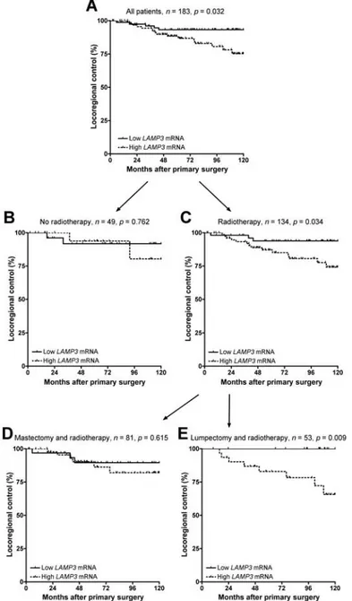
LAMP3 was variably expressed in breast cancer cell lines and induced in an oxygen concentration-dependent manner. LAMP3 protein expression colocalised with hypoxic areas in breast cancer xenografts. LAMP3 mRNA was higher in breast tumours from patients with node-positive (p = 0.019) and/or steroid hormone receptor-negative tumours (p < 0.001). Breast cancer patients with high LAMP3 mRNA levels had more locoregional recurrences (p = 0.032 log-rank). This was limited to patients treated with lumpectomy and radiotherapy as primary treatment (n = 53, p = 0.009). No association with metastasis-free survival was found. In multivariate Cox regression analysis, LAMP3 remained as a statistically independent prognostic factor for locoregional recurrence (hazard ratio, 2.76; 95% confidence interval, 1.01 - 7.5; p = 0.048) after correction for menopausal status, histologic grade, tumour size, nodal status, therapy and steroid hormone receptor status. LAMP3 protein in breast cancer tissue proved also to be of prognostic relevance.
Conclusions
Evidence is provided for an association of LAMP3 with tumour cell hypoxia in breast cancer xenografts. In the current breast cancer cohorts, LAMP3 has independent prognostic value.
KEYWORDS
LAMP3ANDBREASTCANCER
27
INTRODUCTION
Lysosome-associated membrane protein 3 (LAMP3) was first identified in 1998 independently by two research groups. DE SAINT-VIS et al. named it DC-LAMP, a marker of mature dendritic cells1 with a possible role in migration of dendritic cells from the periphery into lymph vessels2, whereas OZAKI et al. described it as TSC403 with specific expression in the lung and overexpression in carcinomas of the breast, amongst others3. Significant similarities of TSC403 with LAMP1 and LAMP2 were found3. LAMPs are highly glycosylated type I integral membrane proteins, which reside in lysosomal membranes1-4. Their function is still largely unknown. In non-cancerous cells, LAMP1 and LAMP2 are rarely expressed on the cell surface5. However, in cancer cells their presence on the plasma membrane is frequently observed, with an increased incidence in more metastatic cell lines6,7. Experiments with overexpression of LAMP3 indicated similar characteristics for this protein: a strong association between LAMP3 and the promotion of metastatic potential was described both in vitro and in vivo2. This suggests that LAMP3 expression might be related to metastasis. Indeed, high LAMP3 mRNA levels correlated significantly with poor prognosis in a small cohort of cervical cancers2.
For several decades, hypoxia (oxygen deprivation) has been recognised as a hallmark of solid tumours. A low blood and oxygen supply to cancers complicates their (local) treatment. Hypoxia can decrease the effectiveness of both radiotherapy and chemotherapy and induce treatment resistance8. Especially for radiotherapy, hypoxia poses problems, as oxygen is required for radiation to cause DNA damage9. In addition, several studies have reported that less well-oxygenated tumours show a worse prognosis and a higher incidence of metastases compared with better oxygenated tumours10-12. Hypoxia has therefore been linked to a more aggressive and metastatic tumour phenotype. Several hypoxia-regulated genes have been described that influence metastasis of tumours, including factors that enhance dissociation and migration of tumour cells13. Recently, LAMP3 was shown to be upregulated by hypoxia in a panel of tumour cells, via the unfolded protein response (UPR)14. The UPR is a mechanisms of adaptation to endoplasmic reticulum stress and has been demonstrated to contribute to hypoxic adaptation in tumours through multiple mechanisms15. This adaptation may facilitate the survival of treatment-resistant hypoxic cells, leading to a poor prognosis of patients16. Whether LAMP3 is associated with treatment resistance or metastasis in solid tumours is unknown. In this study, we investigated the correlation between hypoxia and LAMP3 expression in breast cancer xenografts and we assessed the prognostic value of LAMP3 mRNA and protein expression in breast cancer patients.
MATERIALS AND METHODS Cell culture
MDA-MB-231 and SKBR-3 breast tumour cells were obtained from LGC Promochem (London, UK) and cultured at 37 ˚C with 5% CO2 in Dulbecco’s modified Eagles medium (Lonza, Biowhittaker, Walkersville, MD, USA) supplemented with 10% (v/v) heat-inactivated foetal bovine serum, 10 U/ml penicillin, 10 µg/ml streptomycin, 2 mM L-glutamine, 20 mM Hepes (all from Invitrogen, Carlsbad, CA, USA) and 1× non-essential amino acids (Promocell,
CHAPTERTWO
28
Heidelberg, Germany). Cells were harvested and reseeded at 100% confluence. cDNA from a panel of breast cancer cell lines (n = 16) was a gift from DR. M. SCHUTTE (Department of Medical Oncology, Josephine Nefkens Institute, Erasmus University Medical Centre, Rotterdam, the Netherlands)17,18.
Cell culture in hypoxic conditions
For culturing under hypoxic conditions, MDA-MB-231 cells were seeded at 300,000 cells per 10-cm dish and were allowed to recover overnight. Hypoxia was induced using a H35 Hypoxystation (Don Whitley Scientific Ltd., Shipley, UK), set at 0.5%, 0.2% or 0.1% oxygen. After incubation of up to 72 hours, cells were harvested.
RNA isolation, cDNA synthesis and quantitative polymerase chain reactions (RT-qPCR)
RNA from hypoxic cultured cells was isolated using Norgen’s Total RNA purification kit (Norgen Biotek Corp., Thorold, Canada), according to the manufacturer’s instructions with addition of an on-column DNase treatment. Isolated RNA was stored at -80 ˚C until further processing. cDNA synthesis was performed by reverse transcribing 1 µg of RNA using the Reverse Transcription System (Promega, Madison, WI, USA) according to the manufacturer’s instructions. The primers used for LAMP3 RT-qPCR were FW
5’-TGAAAACAACCGATGTCCAA-3’ and RV 5’-TCAGACGAGCACTCATCCAC-3’. mRNA expression
was analysed on a CFX96 realtime PCR detection system (Bio-Rad Laboratories Inc., Richmond, CA, USA) using SYBR green (Applied Biosystems, Foster City, CA, USA). Hypoxanthine phosphoribosyl transferase 1 (HPRT1) was used as the reference gene, analysed with a pre-developed assay (Applied Biosystems).
Western blot analysis
Cells were harvested in RIPA buffer (1% NP-40, 0.5% sodium desoxycholate and 0.1% sodium dodecylsulfate) to which phosphatase and protease inhibitors (Roche, Indianapolis, IN, USA) were added. Protein concentrations were determined using the Pierce BCA assay (Thermo Fisher Scientific, Rockfold, IL, USA), after which 30 µg of protein was fractionated on 12% Criterion XT Bis-Tris gels (Bio-Rad). After running, samples were transferred to PVDF membranes (Millipore Immobilon, Millipore, Bedford, MA, USA) for 2 hours at 4 ˚C. After blocking with 4% non-fat dry milk (Bio-Rad), membranes were incubated overnight at 4 ˚C with rabbit anti-human-LAMP3 IgG (AP1827a, 0.25 mg/ml, Abgent, San Diego, CA, USA) in a dilution of 1:100 or rabbit anti-β-actin IgG (#4970, Cell Signaling Technology, Beverly,
MA, USA) in a dilution of 1:1,000, both diluted in 5% (w/v) bovine serum albumin (Sigma Aldrich, St. Louis, MO, USA) in tris-buffered saline (TBS) with 0.1% Tween-20 (w/v) (Sigma). Next, membranes were incubated for 1 hour at room temperature with horseradish peroxidase (HRP)-labelled goat anti-rabbit immunoglobulin G (IgG; 1:2,000, #7074, Cell Signaling). Proteins were detected using chemiluminescent peroxidase substrate (Sigma) and imaged on a ChemiDoc XRS+ imaging system (Bio-Rad).
LAMP3ANDBREASTCANCER
29
Growth of breast cancer cell xenografts
Xenografts of breast cancer cells were grown on nu/nu BALB/c athymic mice, kept in a specific pathogen-free unit at the central animal facility of the Radboud University Nijmegen. All animal procedures were approved by the local ethics committee. In short, a cell suspension of MDA-MB-231 or SKBR-3 cells was diluted in Matrigel (BD Bioscience, Bedford, MA, USA) 2:1 and injected subcutaneously into the flank of the mice. At an average diameter of 8 mm to 9 mm, mice were injected intraperitoneally with the hypoxic cell marker pimonidazole (1-[(2-hydroxy-3-piperidinyl)propyl]-2-nitroimidazole hydrochloride, Natural Pharmacia International Inc., Burlington, MA, USA). One hour post-injection, mice were sacrificed and tumours were harvested and immediately stored in liquid nitrogen until further processing.
Immunohistochemistry
Frozen 5 µm tumour sections were fixed in acetone for 10 minutes at 4 ˚C, after which they were rehydrated for 30 minutes in phosphate-buffered saline (PBS) pH 7.4 (Klinipath, Duiven, the Netherlands) and subjected to staining. All antibodies were dissolved in primary antibody diluent (PAD, AbD Serotec, Oxford, UK). Between all incubations, sections were rinsed three times in PBS. Sections were incubated overnight at 4 ˚C with goat anti-human-LAMP3 IgG (AF4087, 0.2 mg/ml, R&D Systems Inc., Minneapolis, MN, USA) used in a dilution of 1:100. Subsequently, sections were treated with Cy3-conjugated donkey anti-goat IgG (705-166-147, 1.5 mg/ml, Jackson ImmunoResearch Laboratories Inc., West Grove, PA, USA) in a 1:600 dilution for 30 minutes at 37 ˚C as the secondary antibody. Vessels were visualised by incubating sections with undiluted 9F1 supernatant, a rat monoclonal antibody to mouse endothelium (Department of Pathology, Radboud University Nijmegen Medical Centre) for 45 minutes at 37 ˚C. Then sections were incubated with rabbit anti-pimonidazole (J.A. RALEIGH)
diluted 1:1,000 for 30 minutes at 37 ˚C. Finally, sections were incubated with 647-conjugated chicken anti-rat IgG (A21472, 2 mg/ml, Invitrogen) diluted 1:100 and Alexa-Fluor-488-conjugated donkey anti-rabbit IgG (A21206, 2 mg/ml, Invitrogen) diluted 1:600 for 60 minutes at 37 ˚C. Nuclei were visualised using Hoechst 33342 (1 mg/ml) 1:3,000 for 5 minutes. Slides were mounted in Fluorostab (ICN Pharmaceuticals Inc., Zoetermeer, the Netherlands).
Tissue microarray (TMA) of breast cancer patients
For the TMA construction, formalin fixed paraffin embedded (FFPE) tumour sections were marked on haematoxylin and eosin-stained slides of the primary tumours by a pathologist. TMAs were constructed with a tissue arrayer using a two mm diameter punch (Beecher Instruments, Silver Spring, MD, USA). Staining for LAMP3 was performed as described in the next section and scored blinded for outcome semiquantitatively as being either absent/low or present/high.
Immunohistochemical assessment of LAMP3 in FFPE sections
FFPE sections were dewaxed in Histosafe (Adamas Instruments BV., Leersum, the Netherlands) and rehydrated in graded alcohols. Antigen retrieval was performed by boiling
CHAPTERTWO
30
sections in 10 mM citrate buffer pH 6.0 (Dako, Glostrup, Denmark) for 30 minutes after which endogenous peroxidase was blocked for 10 minutes using 3% H2O2 in methanol. Sections were incubated for 30 minutes with 5% normal donkey serum (Jackson) in PAD and subsequently incubated overnight at 4 ˚C with goat anti-human-LAMP3 (R&D systems), diluted 1:100 in PAD. Next, sections were incubated for 60 minutes with biotin-conjugated donkey anti-goat IgG (705-066-147, 1.5 mg/ml, Jackson), diluted 1:200 in PBS, after which they were incubated for 30 minutes with avidin-biotin complex reagent (Vector Laboratories Inc., Burlingame, CA, USA). After rinsing with deionised water, sections were incubated with diaminobenzidine (Invitrogen) for 10 minutes. Next, sections were rinsed with tap water and counterstained with haematoxylin (Klinipath) for one minute, after which they were dehydrated and mounted in mounting medium (Klinipath).
Microscopy
All images were acquired using IP-lab imaging software (Scanalytics Inc., Fairfax, VA, USA) in combination with a Leica DM 6,000 (fluorescence) microscope.
Patients
Coded tumour tissues were used in accordance with the Code of Conduct of the Federation of Medical Scientific Societies in the Netherlands (Code for Proper Secondary Use of Human Tissue in the Netherlands, http://www.fmwv.nl). The study adhered to all relevant institutional and national guidelines and was reported according to REMARK guidelines19. A series of 183 patients with unilateral, resectable breast cancers who had undergone resection of their primary tumour between November 1987 and December 1997 were selected based on the availability of RNA in the tumour bank in the Department of Laboratory Medicine of the Radboud University Nijmegen Medical Centre20. This bank contains material from breast cancer patients of seven different hospitals of the Comprehensive Cancer Centre East in the Netherlands. The inclusion and exclusion criteria have been described earlier20. Post-operative radiotherapy was given to the breast after an incomplete resection or after breast-conserving treatment, or parasternal when the tumour was medially localised. Axillary irradiation was given in the case of positive lymph nodes. A TMA was constructed from FFPE tissues as described above from another cohort of 61 breast cancer patients who had undergone lumpectomy and radiotherapy as primary treatment of their tumours between 1991 and 1996 at the Rijnstate Hospital, Arnhem, the Netherlands. These patients had no involved axillary lymph nodes and received no systemic adjuvant therapy, as was the practice at the time. Other clinicopathologic characteristics (steroid hormone receptor status, tumour size and histologic grade) were essentially similar to those of the cohort from which RNA was extracted.
Statistical analysis
Statistical analyses were carried out using SPSS 16.0 software (SPSS Benelux BV., Gorinchem, the Netherlands). Normality of data distribution was tested by Kolmogorov-Smirnov testing. An analysis of variance (ANOVA) with post-hoc Tukey’s honestly significant difference (HSD) testing was used to assess the effect of hypoxia on LAMP3 mRNA levels.
Non-LAMP3ANDBREASTCANCER
31
parametric Mann-Whitney U tests or Kruskal-Wallis tests were used to assess differences in LAMP3 mRNA levels between categories of patients. Locoregional control (defined as the time from surgery until diagnosis of locoregional recurrent disease), disease-free survival (DFS) time (defined as the time from surgery until diagnosis of recurrent disease), metastasis-free survival (defined as the time between date of surgery and diagnosis of a distant metastasis) and overall survival (OS) time (defined as the time between date of surgery and death by any cause) were used as follow-up end points. Survival curves were generated using the Kaplan-Meier method. Equality of survival distributions was tested using log-rank testing and Cox univariate and multivariate regression analyses. Variables were selected for the multivariate survival analyses by backward stepwise selection with removal testing based on the probability of the likelihood-ratio statistic, at a p > 0.10. Two-sided p-values < 0.05 were considered to be statistically significant.
RESULTS
Expression of LAMP3 in breast cancer cell lines
We first measured the expression of LAMP3 mRNA in a panel of 16 different breast cancer cell lines under standard (normoxic) culturing conditions (see Figure 1). Three cell lines (SUM44PE, MPE600 and ZR75-30) displayed no measurable LAMP3 mRNA expression. In the other breast cell lines, levels varied >100-fold between SKBR-3 and MDA-MB-361, with OCUB-F cells exhibiting even higher LAMP3 levels. No association between LAMP3 expression and cancer subtype or oestrogen receptor (ESR), progesterone receptor (PGR) or human epidermal growth factor receptor 2 (ERBB2) expression was found (see Figure 1).
FIGURE 1 LAMP3 mRNA expression in a panel of breast cancer cell lines. Gene expression levels are shown relative to phosphoribosyl transferase 1 (HPRT1). L indicates luminal; E, ERBB2; N, normal-like; B, basal-like; O, other subtype; −, not detectable; +, expressed; ++, overexpressed.18
CHAPTERTWO
32
Expression of LAMP3 in breast cancer cells under hypoxic conditions
In MDA-MB-231 cells, which show moderate basal LAMP3 mRNA expression levels, LAMP3 was strongly induced under hypoxic conditions in an oxygen-dependent manner (see Figure 2A). The highest induction (i.e., four-fold) was observed after 48 hours of incubation under severe hypoxic conditions (0.1% O2, p = 0.003). An incubation of 72 hours at 0.1% O2 led to a reduction in LAMP3 expression compared with 48 hours. At 0.2% O2 there was a two-fold increase overall as a maximum, whereas at 0.5% O2 no statistically significant change was seen. Thus, the level of LAMP3 mRNA induction is dependent on the level of hypoxia, with severe hypoxia leading to a stronger induction of LAMP3 mRNA than moderate hypoxia. This dependency is consistent with the known oxygen dependency of UPR activation21. Protein expression was found to be induced in a similar manner (see Figure 2B).
FIGURE 2 Induction of LAMP3 in MDA-MB-231 cells under hypoxic conditions. mRNA expression (A) was found to be dependent on the level of hypoxia (0.1 [dark gray], 0.2 [gray], or 0.5 [white] % O2). Gene expression levels are shown relative to phosphoribosyl transferase 1 (HPRT1). Data (n = 6) are presented as box-and-whisker plots with median values, 25 and 75 percentiles and minimal and maximal values. A similar effect was observed for protein expression (B). The multiple bands are distinct forms of LAMP3 carrying different glycosylations.
Colocalisation of LAMP3 with hypoxic areas in breast cancer xenografts
Next, xenografts of SKBR-3 (low endogenous expression) and MDA-MB-231 breast cancer cells were established in immunodeficient mice. After subcutaneous injection of the hypoxic cell marker pimonidazole, mice were sacrificed and xenografted tumours were stained for LAMP3 and hypoxia. In agreement with our in vitro expression data, we found only limited staining of the SKBR-3 tumours for LAMP3 in normoxic areas (see Figures 3A-C).
LAMP3ANDBREASTCANCER
33
Simultaneous staining for LAMP3 and pimonidazole revealed a regional colocalisation of intense LAMP3 staining with areas of severe hypoxia, distant from perfused blood vessels (see Figure 3D). A haematoxylin and eosin staining showed the epithelial cell origin of this breast cancer xenograft and LAMP3 expressing cells (see Figure 3E). The MDA-MB-231 xenografted tumours were well oxygenated and did not show hypoxic areas or LAMP3 expression (results not shown).
FIGURE 3 Single-field pseudo-coloured immunofluorescent images of a SKBR-3 xenograft at 100× magnification. A.
LAMP3 (red) B. pimonidazole binding (green) and C. vessels (white) and nuclei (blue). D. superimposed image of LAMP3, pimo and vessels and E. haematoxylin and eosin staining of an adjacent tumour section. Yellow arrows indicate vessels. Bar equals 200 µm.
LAMP3 mRNA expression in breast cancer
LAMP3/HPRT mRNA values were assessed in 183 human invasive breast cancer tissues and were found to be log-normally distributed with a >100-fold variation in values, similar to the range in the breast cancer cell lines. There was no association between LAMP3 and menopausal status, type of surgery, histologic grade, or tumour size (pT, see Table 1). However, LAMP3 was significantly higher in node-positive patients (p = 0.019, Kruskal-Wallis, see Figure 4A), steroid hormone receptor-negative tumours (p < 0.001, Mann-Whitney U, see Figure 4B), patients receiving radiotherapy as part of their primary treatment (p = 0.022) and patients receiving adjuvant systemic chemotherapy (p = 0.050). Thus, LAMP3 is
CHAPTERTWO
34
associated with poor prognosis characteristics (node positive, steroid hormone receptor negative) and therefore with more aggressive treatment (radiotherapy, chemotherapy).
TABLE 1 LAMP3 mRNA levels in different clinicopathological subgroups of breast cancer patients
n LAMP3 mRNA
Total 183 Median Interquartile range p
Menopausal status 0.196a pre 48 10,825 21,005 post 135 9,883 15,524 Histological grade 0.082a I/II 70 9,403 14,898 III 65 13,998 23,164 Tumour size 0.533b pT1 45 9,409 15,340 pT2 115 9,986 16,148 pT3 16 16,973 25,150 pT4 6 7,772 10,803 Nodal status 0.019b 0 78 9,017 14,348 1-3 57 13,998 18,262 ≥ 4 26 10,129 15,667 Surgery 0.904a lumpectomy 55 9,884 16,389 mastectomy 128 9,889 16,440 Radiotherapy 0.022a no 49 7,292 11,895 yes 134 10,688 17,814 Adjuvant therapy 0.195b none 82 9,017 15,967 endocrine 67 10,670 15,070 chemo 24 14,104 25,642 both 10 15,317 15,200 ESR <0.001a neg 55 21,499 42,924 pos 126 8,336 10,630 PGR <0.001a neg 71 15,751 28,429 pos 111 8,932 12,471 aMann-Whitney U test bKruskall-Wallis test
Prognostic value of LAMP3 mRNA expression
Patients were dichotomised based on LAMP3 levels in their primary tumour and evaluated for response parameters. Patients with higher levels of LAMP3 (n = 99) had more locoregional recurrences than those with low LAMP3 mRNA levels (n = 84), p = 0.032 log-rank (see Figure 5A). In Cox regression analysis, this amounted to a hazard ratio (HR) of 2.85 (95% CI, 1.05 - 7.8). There was no association with distant metastasis-free or overall survival. In exploratory subgroup analyses, we found that the association of LAMP3 with locoregional control was limited to patients who had received radiotherapy as part of their primary treatment. In
LAMP3ANDBREASTCANCER
35
patients who had not received radiotherapy, LAMP3 showed no association with locoregional control (n = 49, p = 0.762, see Figure 5B), whereas in patients who had received radiotherapy, a significant association was found (n = 134, p = 0.034, see Figure 5C). Within the patient cohort that had received radiotherapy, no association was found if this was part of a modified radical mastectomy procedure (n = 81, p = 0.615, see Figure 5D). LAMP3 was significantly associated with locoregional control but only in those patients who received radiotherapy as part of breast-conserving lumpectomy (n = 53, p = 0.009, see Figure 5E). In multivariate Cox regression analysis, only LAMP3 remained as an independent prognostic factor for locoregional recurrences (HR = 2.76, 95% CI, 1.01 - 7.5, p = 0.048) after correction for menopausal status, histologic grade, tumour size, nodal status, therapy and steroid hormone receptor status (see Table 2).
FIGURE 4 Differences in LAMP3/phosphoribosyl transferase 1 (HPRT1) values in (A) patients with 0, 1 to 3 or ≥ 4 positive lymph nodes (p = 0.019) and (B) ESR-negative and ESR-positive (p < 0.001) tumours. Horizontal bars represent median values. Open symbols represent outliers above the range of the chosen y-axis.
Immunohistochemical validation of prognostic value of LAMP3 mRNA
We set out to validate the data on LAMP3 mRNA as being prognostic in breast cancer patients who were treated with lumpectomy and radiotherapy on the protein level. For this, LAMP3 protein was semiquantitatively scored in TMA of a series of FFPE sections of breast cancer patients that were only treated with lumpectomy and radiotherapy. These patients received no adjuvant systemic therapy, making it possible to distinguish a prognostic from a predictive value of LAMP3 in these patients. Of the 61 patients, 21 (34%) had low or absent levels of LAMP3 protein (see Figure 6A) and 40 (66%) had high levels (see Figure 6B) of LAMP3 protein in their tumour. Despite the clear difference in the number of events between these subgroups (1 vs. 8 in the LAMP3 low and high cohort, respectively), the power was too low to obtain a significant difference in locoregional control (p = 0.121). These two categories of patients did, however, differ significantly in both disease-free survival (DFS) (p = 0.024, see
CHAPTERTWO
36
Figure 6C) and overall survival (OS) time (p = 0.019, see Figure 6D), confirming the prognostic value of LAMP3 mRNA levels in this patient group on the protein level.
FIGURE 5 Prognostic value of LAMP3 mRNA levels. A. Patients with high LAMP3 mRNA levels (dashed line) in their primary tumour experienced more locoregional recurrences (p = 0.032, log-rank). B. There is no association with locoregional control in patients who had no radiotherapy (n = 49, p = 0.762), but (C) only in patients who had received radiotherapy (n = 134, p = 0.034). D. LAMP3 was not prognostic if radiotherapy was part of a modified radical mastectomy procedure (n = 81, p = 0.615), but (E) only as part of a breast-conserving lumpectomy (n = 53, p = 0.009).
LAMP3ANDBREASTCANCER
37
TABLE 2 Uni- and Multivariate Cox Regression survival analyses for locoregional control Univariate analysis Multivariate analysis
HR 95% CI p HR 95% CI p Menopausal status 0.827 0.942 post vs. pre 0.827 0.320 - 2.134 0.964 0.360 - 2.579 Histological grade 0.459 0.598 III vs. I/II 1.433 0.553 - 3.718 1.622 0.605 - 4.352 Tumour size 0.571 0.368 pT2 vs. pT1 1.905 0.630 - 5.768 1.989 0.640 - 6.185 pT3 vs. pT1 0.974 0.108 - 8.746 1.125 0.118 - 10.74 pT4 vs. pT1 2.783 0.309 - 25.07 6.663 0.646 - 68.72 Nodal status 0.596 0.669 1-3 vs. 0 1.531 0.621 - 3.771 1.414 0.104 - 19.283 ≥ 4 vs. 0 0.906 0.194 - 4.227 0.523 0.027 - 10.197 Surgery 0.766 0.545 mastectomy vs. lumpectomy 0.873 0.360 - 2.117 0.761 0.314 - 1.845 Radiotherapy 0.561 0.718 yes vs. no 1.381 0.465 - 4.108 0.826 0.215 - 3.172 Adjuvant therapy 0.462 0.952 endocrine vs. none 1.173 0.465 - 2.956 1.384 0.546 - 3.506 chemo vs. none 1.311 0.353 - 4.868 1.196 0.319 - 4.477 both vs. none -a - ESR 0.497 0.641 pos vs. neg 0.726 0.293 - 1.798 1.371 0.364 - 5.155 PGR 0.488 0.699 pos vs. neg 0.736 0.310 - 1.749 0.833 0.329 - 2.107 LAMP3 0.028 0.048 high vs. low 2.853 1.044 - 7.792 2.758 1.010 - 7.534 a too few events
DISCUSSION
In this study, the occurrence and prognostic relevance of LAMP3 in breast cancer was evaluated. We present evidence indicating that LAMP3 mRNA levels are elevated in aggressive ESR-negative breast tumours and in tumours that have already spread to the axillary lymph nodes. Patients with high LAMP3 mRNA levels also have more locoregional recurrences than those with low levels, a phenomenon that remains after correction for menopausal status, tumour size, histologic grade, nodal status, steroid hormone receptor status, adjuvant therapy and radiotherapy. The prognostic value of LAMP3 for locoregional control is limited to patients treated with breast-conserving lumpectomy and radiotherapy. The prognostic value of LAMP3 mRNA is also seen for LAMP3 protein as assessed by immunohistochemistry. In 2004, TREILLEUX et al. examined the potential prognostic role of LAMP3 in breast cancer
tissue using a semiquantitative immunohistochemical analysis22. They found that the presence of mature LAMP3 positive dendritic cells within clusters of lymphocytes at the margin of the tumour correlated with lymph node involvement and tumour grade, although no association with prognosis was found. Our immunohistochemical data indicate that tumour hypoxia is also an important regulator of LAMP3 in the epithelial cancer cells. LAMP3 expression in breast cancer xenografts colocalises with tumour cell hypoxia as revealed by pimonidazole labelling. Together with our immunohistochemical data on actual breast tumours, this indicates that