lication in the following source:
Zeraid, Ferial M., Jawkhab, Asma A., Al-Tuwairqi, Waleed S., &Osuagwu, Uchechukwu L.
(2014)
Visual rehabilitation in low-moderate keratoconus : intracorneal ring seg-ment implantation followed by same-day topography-guided photorefrac-tive keratectomy and collagen cross linking.
International Journal of Ophthalmology,7(5), pp. 800-806.
This file was downloaded from: http://eprints.qut.edu.au/79422/
c
Copyright 2014 International Journal of Ophthalmology Press
Notice: Changes introduced as a result of publishing processes such as copy-editing and formatting may not be reflected in this document. For a definitive version of this work, please refer to the published source:
IJO-2013-0462
Visual rehabilitation in low-moderate keratoconus: intracorneal ring segment implantation followed by same-day topography-guided photorefractive keratectomy and collagen cross linking
Running Title: Visual Improvements after same day two step keratoconus surgery.
Ferial M. Zeraid, [1] Asmaa A Jawkhab, [1] Waleed S Al-Tuwairqi, [2] Uchechukwu L. Osuagwu.[3] 1
Department of Optometry&Vision Sciences, College of Applied Medical Sciences, King Saud University. P.O. Box 10219 Riyadh 11433, Kingdom of Saudi Arabia
2
Senior Ophthalmology Consultant, Elite Medical & Surgical Center, Ulaya. P.O Box 65197, Riyadh 11556, Kingdom of Saudi Arabia.
3
School of Optometry & Vision Science and Institute of Health & Biomedical Innovation, Queensland University of Technology, 60 Musk Avenue, Kelvin Grove Q 4059, Australia
Correspondence to: Uchechukwu L. Osuagwu. School of Optometry & Vision Science and Institute of Health & Biomedical Innovation, Queensland University of Technology, 60 Musk Avenue, Kelvin Grove Q 4059, Australia. Email: osuagoul@aston.ac.uk. Phone: +61 7 3138 6152; Fax: +61 7 3138 6030
Abbreviation of authors’ names: Zeraid FM, Jawkhab AA, AlTuwairgi WS, Osuagwu UL. Abstract
AIM: To present the results of same-day topography-guided photorefractive keratectomy (TG-PRK) and corneal collagen crosslinking (CXL) after previous intrastromal corneal ring segment (ISCR) implantation for keratoconus.
METHODS: An experimental clinical study on twenty-one eyes of 19 patients aged, 27.1±6.6 years (range: 19 – 43 years), with low to moderate keratoconus who were selected to undergo customized TG-PRK immediately followed by same-day CXL, 9 months after ISCR implantation in a university ophthalmology clinic. Refraction, uncorrected (UDVA) and corrected distance visual acuities (CDVA), keratometry (K) values, central corneal thickness (CCT) and coma were assessed 3 months after TG/PRK and CXL.
RESULTS: After TG-PRK/CXL: the mean UDVA (logMAR) improved significantly from 0.66±0.41 to 0.20±0.25 (P<0.05); K flat value decreased from: 48.44±3.66 D to 43.71±1.95 D; K steep value decreased from 45.61±2.40 D to 41.56±2.05D; K average also decreased from 42.42±2.07 D to 47.00±2.66 D (P<0.05 for all). The mean sphere and cylinder decreased significantly postsurgery from, 3.10±2.99 D to 0.11±0.93 D and from, -3.68±1.53 to -1.11±0.75D respectively, while the CDVA, CCT and coma showed no significant changes. Compared to post-ISCR, significant reductions (P ˂ 0.05 or all) in all K-values, sphere and cylinder were observed after TG-PRK/CXL.
CONCLUSION: same-day combined topography-guided PRK and corneal crosslinking following placement of ICRS is a safe and potentially effective option in treating low-moderate keratoconus. It significantly improved all visual acuity, reduced keratometry, sphere and astigmatism, but caused no change in central corneal thickness and coma.
KEYWORDS: keratoconus; astigmatism; photorefractive keratectomy; corneal collagen crosslinking; intrastromal corneal rings
INTRODUCTION
Keratoconus is a chronic non inflammatory, asymmetric and usually bilateral cornea degeneration[1], characterized by localized corneal thinning, visual distortion, corneal steepening, and central corneal scarring.[2] Its onset is usually in young adult hood [3] but often becomes apparent during the second decade of life[4], progressing until the fourth decade of life, when it usually stabilizes.[5,6] Although, predicting the severity and time of progression of the disease is hard, progress is usually more rapid in the first affected eye [5] and happens in a period of 3 to 8 years.[7]
The ocular symptoms and signs of the disease vary widely with the disease severity.[3] At the earliest stage, unless corneal topography is assessed, keratoconus can go unnoticed by the practitioner, due to the absence of symptoms.[8] There is a significant loss of visual acuity (VA) which cannot be corrected with spectacles, and the appearance of a ‘scissor-like’ reflex motion on retinoscopy, suggesting the development of irregular astigmatism. Keratometer readings are commonly within the normal range but may have irregular mires. Corneal topography may reveal corneal thinning with the thinnest part of the cornea located outside the visual axis.[1] In the later (Moderate to advanced) stage of the disease, Fleischer’s ring, Vogt’s striae, Rizzuti’s sign, Munson’s sign, and the increased visibility of cornea nerves become apparent.[9] Corneal scarring may eventually develop in the majority of contact lens wearers.
Treatment options comprise first: the use of spectacle or rigid gas permeable contact lenses at the early stages; second, restoring the integrity of the cornea by surgical techniques such as intracorneal ring segments[9] and collagen crosslinking (CXL).[10] However, lamellar or penetrating keratoplasty[11] are considerable options in the advanced stages of keratoconus, where the aim is often to improve patient’s quality of life.
Collagen crosslinking treatment uses riboflavin and ultraviolet-A (UVA) irradiation to strengthen the cornea by augmenting the crosslinks between collagen fibrils,12 thereby stabilizing the condition. Studies on CXL[13-15] have reported stability in progression of the disorder, but, with minimal improvement in vision quality of patients. For this reason, combined procedures have been proposed to maximize the results from CXL: conductive keratoplasty followed by CXL[16]; Intrastromal corneal ring (ISCR) implantation and subsequent PRK/CXL[17-19]; and simultaneous wavefront-guided PRK combined with CXL.[20] Recent studies[17-19, 21-24] have reported promising results in the improvement of visual outcomes, and stability of the disease condition using a combined treatment
of topography-guided photorefractive keratectomy (TG-PRK) and CXL. Kanellopoulos[23] showed that same-day
simultaneous topography-guided PRK and CXL was superior to sequential CXL with later PRK in the visual rehabilitation of progressing keratoconus. In this study, we present the results of same-day topography-guided photokeratectomy combined with CXL, performed three months after ISCR implantation in the treatment of progressive keratoconus.
SUBJECTS AND METHODS
Study Population Twenty-one eyes of 19 keratoconus patients [fifteen males (79%) and four females (21%)] of age 19 - 43 years were recruited in this study. All patients underwent ICSR implantation followed by same-day TG-PRK and CXL. These patients were recruited randomly from patients already scheduled to undergo the surgery technique in our hospital after the research protocol was explained, and informed consent obtained, from each subject. The Hospital research review board approved the study protocol and study procedures conformed to the tenets of the declaration of Helsinki (1975), as revised in Edinburgh 2000.
Patients were included if: they were intolerant to contact lens wear, showed low-grade to moderate keratoconus, and progression in keratoconus observed over the previous 6 months; and excluded in the presence of any of the following conditions: central or paracentral corneal scarring; central pachymetry less than 400 µm, as measured by ultrasound pachymeter (DGH
Technology Inc, Exon, PA); pregnancy and lactation; severe dry-eye disease; systemic autoimmune disease; and/or a history of herpetic keratitis. Keratoconus was graded based on Amsler-Krumeich classification in accordance with the distribution area of the ectasia as has been described by Gómez-Miralles et al.[25] However, progression of KC was defined as one or more of the following changes over a period of 6 months: an increase of ≥1.00D in K-max, an increase of ≥1.00D manifest cylinder, or an increase of ≥0.50D in manifest refraction spherical equivalent. All patients were seeking for relief from their refractive error.
Data Collection Clinical evaluation of general and ocular health was performed pre-operatively. For all patients, the same Optometrists also assessed the following visual parameters, at baseline, after ICSR, at last follow-up (average of 9 months) after ICSR but prior to TG-PRK/CXL, and three months after TG-PRK/CXL: uncorrected distance visual acuity (UDVA [logMAR]), corrected distance visual acuity (CDVA[logMAR]) were obtained by the Snellen projected eye chart; mean manifest cylinder and mean sphere by objective refraction were thrice obtained and averages recorded; topographical keratometry values (D) and Coma (μm) were once obtained by the Schwind Corneal wavefront Analyzer (Eye-tech-Solutions, GmbH & Co. Kleinostheim, Germany). All data were analyzed for normality. All data were entered into a Microsoft Excel 2007 spreadsheet (Microsoft, Inc, Redmond, Washington, USA). The mean, standard deviation, minimum and maximum values were calculated and presented descriptively in a table and figure where applicable. To study the differences in all tested parameters pre- and post-treatments, a student t-test analysis was conducted to compare the baseline with first-step; baseline with second step; first step with second step, post-surgical outcome values. All statistical analyses were conducted using the Graphpad Instat software (version 3.00–Graph pad Software Inc., San Diego, CA). A P value <0.05 (α) was considered statistically significant, and with 20 eyes the study had a power of 80% as calculated using the G power software 3.1.3 version.
Surgical Procedures All procedures-the first step (ICSR) and the second step (PRK, CXL) - in all patients were performed in the University Ophthalmology clinic by the same Ophthalmologist (WA) under sterile conditions. As a first step, all eyes underwent femtosecond laser–enabled (Intralase FS, Intralase Corp./Abbott Medical Optics, Inc. Abbott Park, Illinois) placement of ICRs using the Keraring (Mediphacos, Brazil). Segment sizes were determined according to the nomogram provided by the manufacturer. Depth of the ring channels was set at 75-80% of the thinnest pachymetry reading. At the end of the surgery, antibiotic and corticosteroid eye drops were administered, a bandage contact lens was fitted, and slit lamp examination was conducted. About three to five days later, the bandage contact lens was removed after the epithelial defect at the site of incision had closed. The patients were followed after ICRS implantation for at least 3 months and at most 24 months until stabilization of refraction was achieved. Subsequently, the patients underwent a second step topography-guided PRK followed by same-day riboflavin-UVA CXL during same session. De-epithelialization was performed in all cases by phototherapeutic keratectomy (PTK) using the Schwind Amaris laser platform (SCHWIND Eye-tech-Solutions, GmbH & Co. Kleinostheim, Germany), so as to smooth the anterior irregular cornea. The PTK ablation was performed to remove 50µ of the central 6.5mm of corneal epithelium prior to performing TG-PRK treatments with a 6mm optical zone. The aim was to normalize the cornea by reducing irregular astigmatism while treating
part of the refractive error. This depth was chosen to minimize tissue ablation and reduce the risk of iatrogenic ectasia as was reported in a previous study.10 Mitomycin C 0.02mg/ml was then applied for 30 sec for all TG-PRK procedures.
Shortly after PRK, the CXL treatment was initiated by instilling 0.1% Riboflavin solution in the center of the cornea every 2 to 3min for 30min. Then UVA irradiation was performed at the central 8.0mm diameter of the cornea using a UVX system (Peshke, Inc.) of wavelength 365nm for 30min at irradiance of 3.0mJ/cm2; during this time, riboflavin drops were instilled every 2min. After CXL, the corneal surface was irrigated with a cold balanced salt solution kept at 4˚C, antibiotic and corticosteroid eye drops were administered, a bandage contact lens was fitted until full re-epithelialization, and the eyes were examined at the slitlamp. The bandage contact lens was removed when total epithelialization was observed (between 3 days to 5 days) and post-operative medication included: diclofenac sodium 0.1% (Voltaren) drops for 2 days and tobramycin/dexamethasone drops 4 times daily until bandage contact lens was removed. In all cases, a silicon-hydrogel lens (Air Optix [Ciba Vision] of material lotrafilcon B, diameter 14.0 mm, base curve 8.6mm, and Dk of 140 barrers, was used as bandage contact lens. Safety Index was calculated by the ratio: Post-keraring placement CDVA/baseline CDVA (in logMar). Efficacy index was calculated by the ratio: Post-keraring placement UDVA/baseline CDVA (in logMar).
RESULTS
The descriptive statistics of the mean±standard deviation values of the visual and refractive outcomes at baseline (pre-ICRS), after the first-step (post-ICRS), pre-2nd step (pre-TG-PRK/CXL) and at after the second step (post-TG-PRK/CXL), and the results of the paired t-test analysis has been shown in Table 1.
Comparison of Baseline and Post-ICRS (1st step) and Post-TG-PRK/CXL (2nd step) Outcomes Mean follow-up was 9.0±5.7 months (range: 3 to 24 months) post-ICRS and 3 months post TG-PRK/CXL. From Table 1, it could be deduced that: compared to baseline (pre-ICRS), the mean UDVA, the mean K-values, the mean sphere, and the mean cylinder, all showed statistically significant improvements after the first step and after the second step (Paired t-test: P<0.05, for both steps); the K- values (K max, K min, K average) showed significant decreases, so did the spherical and cylindrical refractive components, 3 months after PRK/CXL. The improvement in mean CDVA from baseline to post-ICRS was not statistically significant (Paired t-test: P>0.05); however, the improvement post-TG-PRK/CXL was statistically significant (Paired t-test: P=0.03). There was no significant change from baseline (pre-ICRS) to after treatment (post-ICRS and post-TG-PRK/CXL) in the mean CCT and in the mean coma, both of which decreased.
However, between the first step (post-ICRS) and the second step (post-TG-PRK/CXL), the improvements in mean UDVA, mean CDVA; the reductions in mean coma and mean CCT, were not statistically significant (Paired t-test: P>0.05, for the four comparisons), while the decrease in K-values, the decrease in mean spherical and mean cylindrical refractive components were all statistically significant (Paired t-test: P<0.05, for the five comparisons) three months after TG-PRK/CXL (Table 1). Patient topographic improvements can be seen in the Schwind wavefront analyzer after ISCR (Figure 1) and TG-PRK/CXL (Figure 2). There were no intraoperative or postoperative complications.
Figure 1 An example of a patient’s left eye topographic maps (obtained by Schwind eye-tech-solutions, wavefront analyzer) after intra-stromal corneal ring implantation, (before 1st step-ISCR) treatment for keratoconus, showing significant improvement.
Figure 2 An example of the same patient’s left eye topographic maps (obtained by Schwind eye-tech-solutions, wavefront analyzer) after same-day combined photorefractive keratectomy and corneal collagen-crosslinking (after 2nd step) treatment for keratoconus, showing significant improvement.
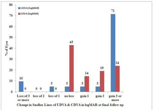
Figure 3 Bar graph showing change in uncorrected distance visual acuity (UCVA), and corrected distance visual acuity (CDVA), before and after the second step procedure (topography-guided photorefractive keratectomy followed by corneal collagen cross-linking) for low to moderate keratoconus. It shows that approximately 71% of eyes and 24% of eyes gained at least three lines of UCVA and CDVA respectively, at last follow-up examination.
Table 1 Descriptive statistics and results of paired t-test conducted on mean±standard deviation (range) values obtained from 21 eyes with low-grade-moderate keratoconus pre-operatively (baseline), post-intracorneal ring segment implantation (1st step), and subsequent same-day topography-guided photorefractive keratectomy&corneal collagen cross linking (2nd step)
3 P 2 P Post-2nd step Pre-2nd step 1 P Post-1st step Baseline Outcomes 0.01 0.17 0.20±0.25 (0 to 1) 0.28±0.37 (0.00 to 1.00) 0.01 0.31±0.18 (0.00 to 0.70) 0.66±0.41 (0.05 to 1.30) UDVA (logMar) 0.03 0.10 0.03±0.04 (0 to 0.15) 0.28±0.73 (0 to 1.00) >0.05 0.28±0.73 (0 to 1.00) 0.21±0.22 (-0.10 to 0.70) CDVA (logMar) <0.05 0.0007 43.71±1.95 (39.59 to 46.68) 44.18±2.90 (40.66 to 50.58) <0.0001 45.98±2.53 (43.42 to 51.30) 48.44±3.66 (42.40 to 54.42) K flat (D) <0.05 0.0005 41.56±2.05 (37.9 to 45.90) 45.98±2.50 (43.54 to 51.30) 0.0003 44.18±2.90 (40.66 to 50.58) 45.61±2.40 (41.74 to 50.27) K steep (D) <0.05 0.0001 42.42±2.07 (39.18 to 46.29) 47.00±2.66 (41.57 to 52.35) 0.0001 45.01±2.34 (42.21 to 50.80) 47.00±2.66 (41.57 to 52.35) K average (D) <0.05 0.02 -0.11±0.93 (-2.75 to 1.50) -1.67±1.97 (-5.75 to +1.50) 0.004 -1.33±1.51 (-3.25 to +0.5) -3.10±2.99 (-9 to +3) Sphere (D) <0.05 0.02 -1.11±0.75 (-3.00 to 0.00) -2.10±1.45 (-6.0 to -0.50) <0.0001 -1.81±1.49 (-0.5 to -6) -3.68±1.53 (-6.0 to -1.25) Cylinder (D) >0.05 >0.14 1.58±0.75 (0.68 to 2.97) 1.57±0.68 (0.70 to 2.90) >0.05 1.65±0.97 (0.50 to 3.86) 2.13±1.25 (1.4 to 4.54) Coma (µm) 0.27 0.29 469.82±33.60 (425 to 526) 492.90±15.02 (477 to 521) 0.38 492.90±15.02 (477 to 521) 496.94±31.39 (443 to 565) CCT (µm) 1,2,3
is for P values of post-hoc analysis 1st step versus baseline, 1st step versus 2nd step, baseline versus 2nd step respectively. UDVA: uncorrected distance visual acuity in logMAR; CDVA: corrected distance visual acuity in logMAR; CCT=central corneal thickness; D: diopter; 1st step=intrastromal corneal ring implantation; 2nd step=Topography-Guided Photorefractive Keratectomy and Collagen Cross Linking.
Regarding the method safety; 71% (15/21) of eyes gained three or more lines, 5% (1/21) each gained one and two lines respectively, while 10% (2/21) and 5% (1/21) of eyes each lost more than three lines and one line of UDVA respectively (which was corrected with spectacles), three months after PRK/CXL. Generally, UDVA was available in 17 out of 21 eyes at final follow-up (Figure 3) and an improvement in CDVA of at least one Snellen acuity line (57% [12/21] of eyes). After the second step, the safety index was estimated to be 1.33 and efficacy index to be 0.46. As shown in Figure 1, at the last follow-up examination, 5% (1/21) eyes lost 1 line of BCVA. DISCUSSION
Collagen crosslinking with riboflavin and UVA have been shown to be effective in stalling the progression of keratoconus and pellucid marginal degeneration, but with minimal improvement in visual quality, as a result of apical keratometry reduction and consequent corneal regularization.[14, 26, 27] Better results have been shown with a
combination of CXL with techniques that improve visual acuity. Topography-guided PRK in combination with CXL was found to yield improved topographic indices and improved UDVA and CDVA, in patients with mild to moderate keratoconus.[18-20, 28, 29] Lovieno et al [28] noted significant improvements in UDVA, CDVA, refraction, keratometry, and total aberrations, after six months of Intacs plus PRK/CXL in five eyes. In their study,[28] neither a loss of lines of CDVA, nor a development of haze in any eye, was observed. Another study[20] also reported significant improvements in mean UDVA and CDVA, no loss of lines of CDVA, a significant reduction in mean cylinder, mean apex K-value, and mild haze was observed in 11.1% of treated eyes, after 12 months of PRK-CXL. They[20] also observed that these visual outcomes did not differ significantly between 6 months and 12-months after the procedure. In a more recent study[19] conducted on thirteen low-moderate keratoconic eyes, significant improvements were observed in UDVA, CDVA, mean sphere, mean astigmatism, average K, and coma, after six months of TG-PRK/CXL. Also, while no eyes lost lines of CDVA, 63% of eyes gained >2 lines of CDVA, and there was no change in CDVA in ∼27% of the treated eyes.[19] Kanellopoulos[23] compared the visual functions of 325 eyes that underwent same-day simultaneous topography-guided PRK/CXL, to those who had
sequential CXL with later PRK. They[23] showed that, same-day simultaneous topography-guided PRK/CXL was
superior to sequential CXL with later PRK, in the visual rehabilitation of progressing keratoconus. In that study,[23] the simultaneous group did better (P<0.05) in all fields evaluated: greater significant improvements in UCVA and BSCVA, greater mean reduction in spherical equivalent refraction and keratometry, and lesser corneal haze, after about 36 months of the performing the procedures. While all these studies[9, 18-20, 23, 28, 29] have shown similar significant improvements in most of the visual functions examined in relation to the current study, they have either been conducted on a different population sample, and/or have not completely agreed in all findings, partly because of the variation in the surgical protocol, data collection/analysis method or follow-up duration. In the current study, ICRS implantation with the aid of femtosecond laser was performed in 21 eyes of nineteen patients with low to moderate keratoconus, as a first-step procedure. Then, after a follow up period of nine months (when refraction and cornea regularity have stabilized), a second-step procedure was performed which involved a topography guided-PRK and CXL, performed on the same day, and in the same session. The satisfactory visual, keratometry and topography results obtained after three months of this triple procedure is an indication that, the 3 procedures complement one another.
The results showed a significant reduction in the pre-operative mean cylinder, mean sphere, steep K, flat K, and average K-values three months after the second step procedure. Significant improvement in the UDVA was also observed in 71% (15/21) of eyes, and in the CDVA, 57% (12/21) of eyes showed improvement of at least one Snellen acuity line, 3 months after combined therapy. No change in CDVA was observed in 43% of eyes (Figure 1). Similar results have been recently reported.[19] However, the study[19] enrolled lesser number of eyes (thirteen eyes), and their follow-up period was longer (six months), than that of the current study. From their observation[19] we can therefore deduce that, the improvements in visual functions observed in our subjects, will remain stable or even improve even after 6 months and beyond. In addition, in relation to the first step (post-ICSR) procedure, the current study observed that the second step combined therapy significantly reduced all keratometry values and refractive components, but no significant change in UDVA and CDVA, despite the slight improvement shown. Similarly, CCT and coma were further reduced by about 21µm (P>0.05) and 2µ (P>0.05) respectively (Table 1). Because the results of postoperative data varied significantly between ICRS and PG-PRK/CXL, we conclude that the same-day combined technique yielded better visual outcomes in patients with mild to moderate keratoconus (Figure 1 vs Figure 2). This also agrees with previous reports.[17, 19, 23, 24, 28, 29]
The first combination of a TG-PRK treatment with CXL on patients with keratoconus was described by Kanellopoulos and Binder[22], and one study[23] observed that, the technique was more effective when applied on the same day. Subsequently, studies[16, 22] have shown significant improvements in visual outcomes with
simultaneous topography guided PRK followed by CXL in patient series. Kymionis and cohorts[17] then reported
even more improved visual outcomes on performing on the same day, a combination of PRK/CXL treatment after previous ISCR implantation on a patient with pellucid marginal degeneration. Although, the current study observed some loss of lines of UDVA, this was corrected with spectacles, three months after PRK/CXL. Nevertheless, these eyes showed no post-surgical complications, and with a recent study[30] reporting significant improvement of 2.7 lines in UDVA even after one year of paired ICRS combined with CXL, there is a greater tendency that these eyes will also improve in UDVA, few months after our data collection.
Even though the result of this study agrees well with previous studies[17, 19, 23, 24, 28, 29, 30], it is limited by the relatively short period of follow up after the second step procedure, despite the observed significant visual improvements, which seem to be even more promising in the long term. Studies with longer follow-up period are needed to further confirm these findings. Again, recruiting a control group may be necessary to better understand the cause of the changes observed in the current study.
In conclusion, the placement of intracorneal ring segments followed by same-day combined TG-PRK/CXL is a safe procedure that offers patients a functional vision. However, while this procedure appears to stabilize the
cornea ecstasia in selected patients with keratoconus, there is also the need to monitor these eyes over a period of at least one year, in order to confirm this.
Acknowledgement: The authors extend their appreciation to the Research Centre, College of Applied Medical Sciences and the Deanship of Scientific Research at King Saud University for funding this research.
REFERENCES
1. Romero-Jiménez M, Santodomingo-Rubido J, Wolffsohn JS. Keratoconus: A Review. Contact Lens &
Ant Eye 2010;33(4):157-166
2. Wagner H, Barr JT, Zadnik K. Collaborative Longitudinal Evaluation of keratoconus (CLEK) Study:
Methods and Findings to date. Cont Lens Anterior Eye 2007;30(4):223-232
3. Robinowitz YS. Keratoconus. Surv Ophthalmol 1998;42(4):297-319
4. Kennedy RH, Bourne WM, Dyer JA. A 48-year clinical and epidemiologic study of keratoconus. Am J
Ophthalmol 1986; 101(3): 267-273
5. Woodward EG. Keratoconus-epidemiology. J Br Contact Lens Assoc 1984;7:64-76
6. Duke-Elder S, Leigh AG. Keratoconus. In: Duke-Elder S, editor. System of ophthalmology. St Louis: Mosby: 1965;8:964-976
7. Bilgin LK, Yilmaz S, Araz B, Yüksel SB, Sezen T. 30 years of contact lens prescribing for keratoconic
patients in Turkey. Contact Lens & Ant Eye 2009; 32(1): 16-21
8. Arntz A, Duran JA, Pijoan JI. Subclinical keratoconus diagnosis by elevation topography. Arch Soc Esp
Oftalmol 2003;78(12):659-664
9. Kymionis GD, Siganos CS, Tsiklis NS, Anastasakis A, Yoo SH, Pallikaris AI, Astyrakakis N, Pallikaris
IG. Long-term follow-up of Intacs in keratoconus. Am J Ophthalmol 2007;143:(2)236-244
10. Tan DT, Por YM. Current treatment options for corneal ectasia. Curr Opin Ophthalmol
2007;18(4):284-289. Review
11. Yildiz EH, Cohen EJ, Virdi AS, Hammersmith KM, Laibson PR, Rapuano CJ. Quality of life in
keratoconus patients after penetrating keratoplasty. Am J Ophthalmol 2010;149(3):416-422
12. Wollensak G. Crosslinking treatment of progressive keratoconus: new hope. Curr Opin Ophthalmol
2006;17(4):356-360. Review
13. Wittig-Silva C, Whiting M, Lamoureux E, Lindsay RG, Sullivan LJ, Snibson GR. A randomized
controlled trial of corneal collagen cross-linking in progressive keratoconus: preliminary results. J Refract Surg 2008;24(7):720-725
14. Raiskup-Wolf F, Hoyer A, Spoerl E, Pillunat LE. Collagen crosslinking with riboflavin and ultraviolet-A
light in keratoconus: long-term results. J Cataract Refract Surg 2008;34(5):796-801
15. Coskunseven E, Jankov MR 2nd, Hafezi F. Contralateral eye study of corneal collagen cross-linking with
riboflavin and UVA irradiation in patients with keratoconus. J Refract Surg 2009;25(4):371-376
16. Kymionis GD, Karavitaki AE, Kounis GA, et al. Management of pellucid marginal corneal degeneration
with simultaneous customized photorefractive keratectomy and collagen crosslinking. J Cataract Refract Surg 2009; 35:1298-1301
17. Kymionis GD, Karavitaki AE, Kounis GA, Portaliou DM, Yoo SH, Pallikaris IG. Photorefractive
keratectomy followed by same-day corneal collagen crosslinking after intrastromal corneal ring segment implantation for pellucid marginal degeneration. J Cataract Refract Surg 2010; 35(7):1783-1785
18. Tuwairqi WS, Sinjab MM. Safety and efficacy of simultaneous corneal collagen cross-linking with topography-guided PRK in managing low-grade keratoconus: 1-year follow-up. J Refract Surg 2012; 28: 341-345
19. Al-Tuwairqi W, Sinjab MM. Intracorneal Ring Segments Implantation Followed by Same-day
Topography-guided PRK and Corneal Collagen CXL in Low to Moderate Keratoconus. J Refract Surg
2013;28(5):59-64
20. Kremer I, Aizenman I, Lichter H, Shayer S, Levinger S. Simultaneous wavefront-guided photorefractive
keratectomy and corneal collagen crosslinking after intrastromal corneal ring segment implantation for keratoconus. J Cataract Refract Surg 2012;38(10):1802-1807
21. Krueger RR, Kanellopoulos AJ. Stability of Simultaneous Topography-guided Photorefractive
Keratectomy and Riboflavin/UVA Cross-linking for Progressive Keratoconus: Case Reports. J Refract Surg 2010;
26(10): 827-832
22. Kanellopoulos AJ, Binder PS. Collagen cross-linking (CCL) with sequential topography-guided PRK: a
temporizing alternative for keratoconus to penetrating keratoplasty. Cornea 2007;26(7):891-895
23. Kanellopoulos AJ. Comparison of sequential vs same-day simultaneous collagen cross-linking and
topography-guided PRK for treatment of keratoconus. J Refract Surg 2009;25(9):812-818
24. Kanellopoulos AJ, Binder PS. Management of corneal ectasia after LASIK with combined, same-day, topography-guided partial transepithelial PRK and collagen cross-linking: the Athens protocol. J Refract Surg 2011;27(5):323-331
25. Gómez-Miralles M, Peris-Martínez C, Pastor-Pascual F. Biomechanical corneal response measurement after manual insertion of intrastromal rings in patients with keratoconus. J Emmetropia 2010;1:206-212
26. Spadea L. Corneal collagen cross-linking with ribofl avin and UVA irradiation in pellucid marginal degeneration. J Refract Surg 2010;26(5):375-377
27. Vinciguerra P, Albè E, Trazza S, Seiler T, Epstein D. Intraoperative and postoperative effects of corneal
collagen cross-linking on progressive keratoconus. Arch Ophthalmol 2009; 127(10):1258-1265.
28. Iovieno A, Légaré ME, Rootman DB, Yeung SN, Kim P, Rootman DS. Intracorneal Ring Segments
Implantation Followed By Same-day Photorefractive Keratectomy and Corneal Collagen Cross linking in Keratoconus. J Refract Surg 2011;27(12):915-918.
29. Kanellopoulos AJ. The management of cornea blindness from severe corneal scarring, with the Athens Protocol (transepithelial topography-guided PRK therapeutic remodeling, combined with same-day, collagen cross-linking). Clin Ophthalmol 2012;6:87-90.
30. Yeung SN, Ku JY, Lichtinger A, Low SA, Kim P, Rootman DS. Efficacy of single or paired intrastromal
corneal ring segment implantation combined with collagen crosslinking in keratoconus. J Cataract Refract Surg