Supporting Information
Copyright Wiley-VCH Verlag GmbH & Co. KGaA, 69451 Weinheim, 2014
A General Strategy for Site-Directed Enzyme Immobilization by
Using NiO Nanoparticle Decorated Mesoporous Silica
Daishun Ling,[a, b] Liqian Gao,[c, e] Jianpeng Wang,[c] Mohammadreza Shokouhimehr,[a, b] Jiahui Liu,[c] Yongsheng Yu,[c] Michael J. Hackett,[a, b]Pui-Kin So,[d] Bo Zheng,[d]
Zhongping Yao,[d] Jiang Xia,*[c] and Taeghwan Hyeon*[a, b]
1
Abbreviations:
Fmoc: 9-fluorenylmenthoxycarbonyl
HBTU: 2-(1H-benzotriazol-1-yl)-1, 1, 3, 3-tetramethyluronium hexafluorophosphate HOBt: N-hydroxybenzotriazole
DMF: N, N-dimethylformamide DIEA: N, N-diisopropylethylamine DCM: dichloromethane
TFA: trifluoroacetic acid EDT: 1,2-Ethanedithiol TIS: triisopropylsilane DMSO: dimethyl sulfoxide RT: room temperature
SDS-PAGE: Sodium dodecyl sulfate – polyacrylamide gel electrophoresis Tris: tri-hydroxymethylaminomethane
PBS: phosphate buffered saline DTT: dithiothreitol
Tricine: N-(2-Hydroxy-1,1-bis(hydroxymethyl)ethyl)glycine Tris: tri-hydroxymethylaminomethane
2
Materials and Instruments
Unless otherwise noted, all reagents were purchased from commercial sources and used without further purification. Ala-Wang resin, Ile-Wang resin, Rink Amide-ChemMatrix resin, Fmoc-protected amino acids, and D-biotin for solid-phase peptide synthesis were obtained from GL Biochem Ltd (Shanghai, China). The fluorophore 5(6)-FAM was purchased from Life Technologies (USA). Other reagents were purchased from commercial suppliers, Labscan Limited (Thailand), Meryer Technologies Co., Ltd (Shenzhen, China), Chem-Impex International Inc. (USA), Sigma-Aldrich Co. (USA). Mass spectra of peptides were obtained on 4800 Plus MALDI TOF/TOF™ Analyzer (AB SCIEX, USA) and QSTAR® Elite Hybrid LC/MS/MS System (AB SCIEX, USA). Water was deionized by a Barnsted Nano Pure System. Synthetic DNA oligonucleotides for cloning were obtained from Invitrogen (HK). Restriction endonucleases were purchased from Takara Biotech Co., Ltd (Dalian, China). PCR products and products of restriction digests were purified by gel electrophoresis and extraction using the gel extraction kit from Takara. Plasmid DNA was purified from overnight cultures by using the Takara Plasmid Miniprep Kit. Sequencing reactions were analyzed at BGI (HK). Transmission electron microscope (TEM) and high resolution TEM (HRTEM) images were obtained using a JEOL JEM-2030 microscope at an acceleration voltage of 200 kV. Powder X-ray diffraction was obtained with a Rigaku D/Max-3C diffractometer, equipped with a rotating anode and a Cu Kα radiation source (λ= 0.15418 nm). The surface area and pore size was measured using a micromeritics surface area measurement analyzer (mi-micrometrics 3 Flex-surface characterization analyzer).
Preparation of SBA-15
The synthesis of SBA-15 followed a reported procedure.1 Briefly, 2.0 g of Pluronic P123 was dissolved in
30 g of water and 120 g of 2 M HCl with stirring at 35 °C. Then 4.4 g of Tetraethyl orthosilicate (TEOS) was added with stirring at 35 °C for 24 h. The mixture was aged at 100 °C overnight without stirring. The solid product was recovered, washed with de-ionized water and ethanol and air-dried at RT. Calcination was carried out by slowly increasing the temperature from RT to 500 °C over 8 h and sustaining heating at 500 °C for 6 h. The as-calcined white SBA-15 powders were then used as the supports for these studies.
Synthesis of SBA-NiO
SBA-NiO were prepared by wet impregnation. In a typical procedure, 0.1 g of an aqueous solution of NiCl2 (1.1 wt %) were impregnated with 1 g of dry SBA carrier. The mixture was stirred at RT overnight,
then the product was collected via centrifugation. The resulting materials were dried and heated to 500 oC in air for 5 h so Ni2+ salts were oxidized to ultrafine NiO NPs yielding SBA-NiO.
3
Plasmid construction
DNA fragment coding for PSP was amplified from GST-PSP DNA plasmid (a generous gift from Prof. Shannon Au, department of biochemistry, CUHK) using the following primers: forward: 5’-GGGAAGCTTGCGGACCAAACACAGAATTTGCAC-3’ and reverse: 5’-GGGCTCGAGTT-ATTGTTTCTCTACAAAATATTG-3’. The fragment was inserted into pMD-18 T vector and sub-cloned into pET28m-EGFP at Hind III and Xho I restriction enzyme sites to yield pET28m-EGFP-PSP.
Protein expression and purification
The plasmid pET28-EGFP-PSP was transformed into E. coli. BL21 (DE3) competent cells. Colonies were picked and grown in LB medium at 37oC overnight and the start culture was inoculated into 600 mL LB medium at a dilution of 1:100. The cell culture was allowed to grow at 37oC until OD600 reached 0.6
and then followed by the induction by Isopropyl β-D-1-thiogalactopyranoside (IPTG) at a final concentration of 1 mM. After growing at 16oC for 20 hours, cells were harvested by centrifugation at 6000 g, and the cell pellet re-suspended in 25 mL lysis buffer (50 mM Tris, 300 mM NaCl, 4 mM β-mercaptoethonol and 10 mM imidazole, pH 7.5). The cell suspension was solubilized by sonication on ice followed by centrifugation at 20,000 g for 1 hour. The supernatant was collected, filtered and incubated with Ni-NTA Sepharose (GE healthcare, USA) for 40 min. After washing with the lysis buffer, his-tagged-EGFP-PSP was eluted by increasing concentration of imidazole buffer (50 mM Tris, 300 mM NaCl, 4 mM β-mercaptoethanol, 50 to 500 mM gradient imidazole, with pH adjusted to 7.5). Protein eluents were collected, ultra-centrifuged and buffer exchanged to the storage buffer (50 mM Tris, 150 mM NaCl, 1 mM EDTA, 1mM DTT, pH 7.0) and stored in aliquots at -20oC. Other his-tagged proteins
such as EGFP and mCherry were expressed and purified using the same protocol. The emission spectra of the fluorescent protein was measured by a Hitachi F-7000 fluorescence spectrophotometer.
Peptide synthesis and purification
Peptides were manually synthesized based on standard FMOC solid phase peptide synthesis protocol on Rink Amide-ChemMatrix® resins (PCAS BioMatrix, Canada). Briefly, coupling steps were done using a solution containing Fmoc-protected amino acid, HBTU, HOBt, and DIEA in DMF (1: 1: 1: 2: 5, w/w/w/v/v). Deprotection of Fmoc was done in 20% piperidine in DMF (v/v). The final cleavage of the peptide from the solid support together with the side-chain deprotection was carried out in 2 mL cocktail solution containing TFA, TIS and H2O (v/v/v=95:2.5:2.5) at RT for 2 h followed by precipitation in cold
4
diethyl ether. The peptides were dissolved in 500 μL 50% acetonitrile in H2O (v/v) and analyzed by
RP-HPLC (Shimadzu, DGU 20A5, Japan) using an Apollo C18 column (Grace Alltech, PN36511, USA). The eluents were 0.1% TFA in H2O (A) and 0.1% TFA in MeCN (B) with a flow rate of 1 mL/min and a
gradient of 0-95% B over 25 min. For peptide purification, a semi-prep RP-HPLC column (Grace Alltech, 218TP510, USA) was used at a total flow rate of 3 mL/min using a linear gradient of 0-45% B over 24 min. The peptide peaks were collected, lyophilized and re-dissolved in DMSO to give a stock concentration of 20 mM before being validated by ESI-MS. LEVLFQGP, [M+H]+ found: 901.9, calculated: 902.1.
Protein absorption and elution measurement
Different amounts of his-tagged EGFP or mCherry were incubated with fixed amounts of SBA-NiO material in the storage buffer (50 mM Tris, 150 mM NaCl, 1 mM EDTA, 1mM DTT, pH 7.0) at RT. The solutions were collected after centrifugation and resolved on an SDS-PAGE gel and stained by coomassie blue. The relative amount of protein leftover in solution was quantified based on the pixels counted by Image J software using 0.4 nmol protein as a standard (Figure 2C lane 6).
For the protein elution experiment, 40 μl his-tagged mCherry protein was incubated with 1 mg SBA-NiO materials at RT for 2 h. The solid material was quickly precipitated and the supernatant decanted. Next, 40 μl of 250 mM imidazole was added to incubate with SBA-NiO. After being sonicated for 10 min, 20 μl of the solution was removed, thermally denatured and subjected to separation on SDS-PAGE.
Enzymatic activity assay and reaction kinetics measurement
His-tag-EGFP-PSP protein (50 μg) was incubated with a selected amount of SBA-NiO materials for 30 min in storage buffer. Subsequently, the peptide substrate was added to give a final concentration of 1 mM. The catalytic reaction was allowed to proceed at RT. At different reaction time points, an aliquot was removed and thermally denatured to quench the reaction. The solution was precipitated by centrifugation at 10,000 g for 20 min to remove SBA-NiO. The supernatant containing peptides was filtered and analyzed using analytical RP-HPLC as described above. The lines show curve fitting to a kinetic equation, [Product] = 1 – exp(− kobs × time).2
5
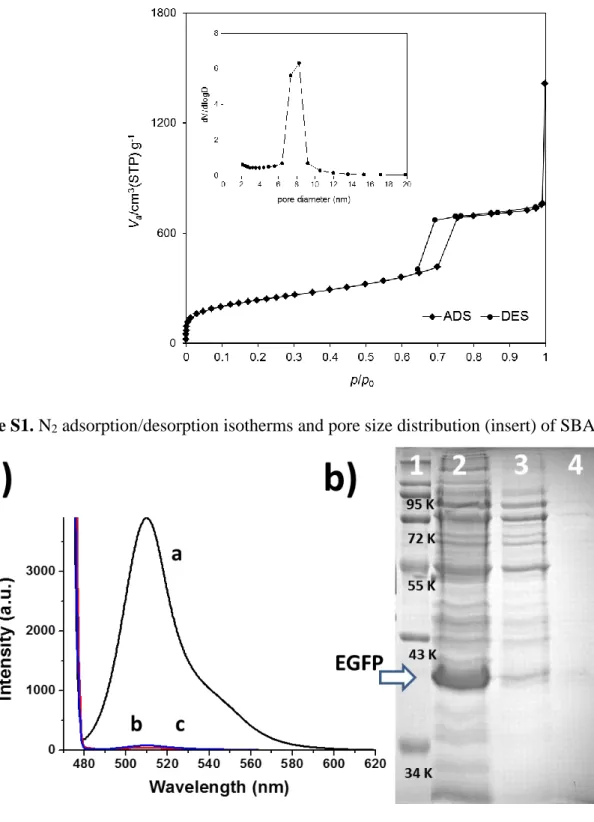
Figure S1. N2 adsorption/desorption isotherms and pore size distribution (insert) of SBA-15.
Figure S2. Immobilization of His-tagged EGFP. a) Immobilization and elution of purified his-tagged EGFP monitored by the fluorescent emission of the solution. a, fluorescent signal corresponding to EGFP solution at 41 μg/mL; b, red curve, fluorescent signal of the solution upon incubation of the EGFP solution with 1 mg SBA-NiO; c, fluorescent spectrum of the solution eluted from SBA-NiO by 1 M Im. b) Immobilization and elution of EGFP in crude cell lysate monitored by SDS-PAGE. Lane 1, molecular weight marker; lane 2, crude cell lysate (The arrow indicates overexpressed EGFP); lane 3, 2× dilution
6
of the supernatant of the mixture of crude lysate and SBA-NiO; lane 4, 1 M Im eluent of EGFP-SBA-NiO.
Figure S3. Confocal images of a) SBA-NiO without his-tag-EGFP and b) SBA-NiO with his-tag-EGFP immobilized.
7
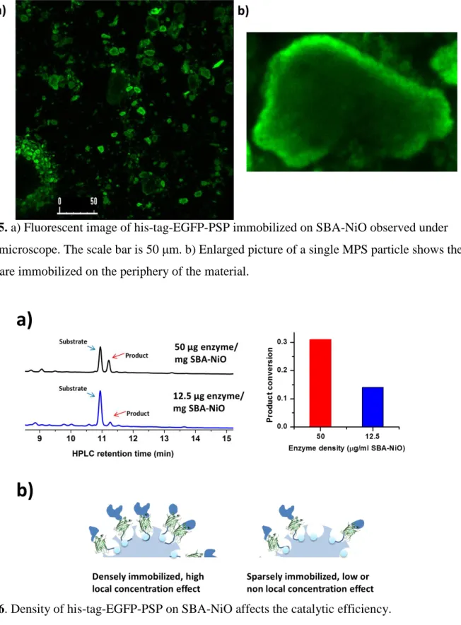
Figure S5. a) Fluorescent image of his-tag-EGFP-PSP immobilized on SBA-NiO observed under confocal microscope. The scale bar is 50 μm. b) Enlarged picture of a single MPS particle shows the enzymes are immobilized on the periphery of the material.
Figure S6. Density of his-tag-EGFP-PSP on SBA-NiO affects the catalytic efficiency.
Reference
(1) D. Zhao, Q. Huo, J. Feng, B. F. Chmelka and G. D. Stucky. J. Am. Chem. Soc.1998,120, 6024-6036. (2) L. Gao, H. Sun, S. Q. Yao. Biopolymers2010, 94, 810-819.