http://dx.doi.org/10.4236/wja.2014.44043
How to cite this paper: Adungo, F.O., Gicheru, M.M., Adungo, N.I., Matilu, M.M., Lihana, R.W. and Khamadi, S.A. (2014) Diversity of Human Immunodeficiency Virus Type-1 Subtypes in Western Kenya. World Journal of AIDS, 4, 365-372. http://dx.doi.org/10.4236/wja.2014.44043
Diversity of Human Immunodeficiency
Virus Type-1 Subtypes in
Western Kenya
F. O. Adungo
1, M. M. Gicheru
2, N. I. Adungo
1, M. M. Matilu
1, R. W. Lihana
3,
S. A. Khamadi
3*1Centre for Infectious and Parasitic Diseases Control Research, Kenya Medical Research Institute, Busia, Kenya 2Department of Zoological Sciences, Kenyatta University, Nairobi, Kenya
3Centre for Virus Research, Kenya Medical Research Institute, Nairobi, Kenya
Email: *skhamadi@gmail.com
Received 10 September 2014; revised 5 October 2014; accepted 1 November 2014
Copyright © 2014 by authors and Scientific Research Publishing Inc.
This work is licensed under the Creative Commons Attribution International License (CC BY).
http://creativecommons.org/licenses/by/4.0/
Abstract
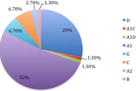
Background: HIV/AIDS is the principal pandemic in the world today. Two viral types (HIV-1 and HIV-2), with numerous groups (M, N and O for HIV-1 and A through H for HIV-2) have emerged. These have further proliferated into numerous subtypes, sub-subtypes and circulating recombi-nant forms (CRF) over the last 30 years. HIV-1 variants circulate together within a geographical region providing an opportunity for recombination of viral strains within infected individuals. In Kenya, at least nine different genetic HIV-1 subtypes and several recombinant forms have been defined within group M, which accounts for the majority of cases in the AIDS pandemic. Objective: To determine the genetic diversity of HIV-1 in the western region of Kenya bordering Uganda. Methodology: A cross sectional study was carried out at Busia District Hospital between 2007 and 2009. A total of 75 patients were sampled randomly from a cohort of 1000 clients on antiretroviral therapy. Blood samples were analysed at the HIV Laboratory, Kenya Medical Research Institute, Nairobi, Kenya. PCR was carried out on the Pol region of HIV, sequenced and analysed by BLAST for subtypes. Results: BLAST analysis revealed the following circulating subtypes: 40/75 (53.30%) were HIV-1 group M subtype A1; 21/75 (28.0%) were subtype D; 5/75 (6.7%) were subtype G; 4/75 (5.30%) were subtype C; and 2/75 (2.70%) were subtype A2. Only one isolate was identified for the other subtypes viz: 1/75 (1.30%) resembled subtype B; 1/75 (1.30%) was A1/C, and 1/75 (1.30%) was A1/D. Conclusion: The study showed increasing HIV-1 diversity along the Kenya- Uganda border with the emergence of A1/C and A1/D recombinants. Such HIV-1 diversity vis a vis the recent expanded access to antiretroviral therapy in resource limited settings calls for
nuous evaluation of anti-HIV regimens. There is need therefore, for regular surveillance and moni- toring for mutations that are likely to lead to drug resistance if we have to achieve successful treatment outcomes.
Keywords
HIV-1, Subtypes, Genetic Diversity
1. Introduction
Human Immunodeficiency Virus (HIV) infection is today one of the most devastating infections globally. Ever since the discovery of the virus some two decades ago, there has been a dramatic increase in infections and deaths due to this virus.
HIV is a member of the retroviridae family, which has a single stranded RNA genome. Two major types of HIV are currently recognised: HIV type 1 (HIV-1), and HIV type 2 (HIV-2). HIV-1 is further divided into three genetic groups: group M (major or main), group O (outlier) and group N (new or non-M, non-N) [1]. While groups O and N are restricted to countries of Central Africa, notably Cameroon; HIV-1 group M is widely dis-tributed worldwide. It is responsible for the AIDS pandemic, which accounts for over 90% of HIV infections [2]. HIV-2 is restricted to countries in West Africa, where it also represents a minority of viral infections (3% of to-tal HIV infections). Fortunately, it is decreasing in prevalence over time [3].
1.1. Molecular Epidemiology of HIV
HIV-1 subtype diversity is the highest in the world. Over nine pure subtypes of HIV-1 group M are currently known (A-D, F-H, J and K). Some of these subtypes are further subdivided into sub-subtypes e.g. subtypes F (F1 and F2); A (A1, A2 and A3). Intrasubtype divergence of up to 20% also occurs. Furthermore, intersubtype divergence of about 25% to 35% occurs, for the env amino acid sequences [1] [4] [5]. This intermixture of HIV-1 variants that circulate together within a geographical region provides an opportunity for recombination of virus strains within dually or multiply infected individuals [6]. Some of these recombinant forms may further achieve epidemic relevance, giving rise to unknown circulating recombinant forms (CRFs) thus complicating therapy. To date, over 40 CRFs are recognized in diverse parts of the world [7]. It is currently believed that HIV-1 M subtypes and CRFs are the result of founder effects in different geographic locales, followed by local-ised evolution. As a consequence, such HIV-1 forms are heterogeneously spread out worldwide [2].
1.2. Distribution of HIV-1 Subtypes
Africa has all HIV-1 subtypes, although A and C predominate [8]. In East Africa, subtypes A and D have domi-nated Uganda from mid 1980s [9]. These subtypes A and D have also been isolated from various parts of Tan-zania [10]. However, in the Southern Tanzania town of Mbeya, the predominant subtype is C [11]. This is be-lieved to have been introduced from Southern African countries where subtype C is predominant [12].
HIV-1 epidemic in Kenya like the rest of the world shows regional heterogeneity. On the basis of the env
C2-V3, Neilson et al., 1999; found that subtype A predominates (71% - 87%), with significant components of subtype D (7% - 29%) and subtype C (7% - 17%). Carr et al., 2005 [14] has identified a full-length subtype G in Kenya. On the other hand, it has been established that HIV-1 subtypes circulating in the Northern Kenya town of Moyale mainly comprise subtype A (50%), subtype C (39%) and subtype D (11%); i.e. subtypes A and C are predominant. This is probably influenced by neighbouring Ethiopia which is dominated by HIV-1 subtype C. It has been suggested that this scenario is indicative of cross-border movements influencing circulation of subtypes in northern Kenya [13]. Evidence of increasing HIV-1 diversity is emerging. For example, Lihana et al., 2006
[15] identified four circulating recombinant forms: between A1, A2, and D; A2 and D; A1 and D; A1 and G; A1 and C; A1, C and D have been documented among STI patients in Nairobi.
out-comes. Thus, this study was conducted in western Kenya bordering Uganda.
2. Materials and Methods
2.1. Study Site
The study was conducted in western Kenya at sites within the border area of Kenya and Uganda. The five sites: Butula, Busia, Budalangi, Funyula and Mumias were selected to reflect representative number of patients in the cohort. This region has an adult HIV prevalence of 6.7% (NASCOP 2007).
2.2. Study Population and Sampling
The study covered a cohort of 1000 patients on antiretroviral therapy supported by the MedecinsSansFrontieres
(MSF) programs in Busia district hospital between 2007 and 2009. From this cohort, 75 persons were randomly selected. The samples analysed come from persons resident in the rural villages (Table 1) who, since they tend to travel less often, were very likely to have acquired their HIV infection in their villages. This facility serves patients from both Kenya and Uganda. Inclusion criteria comprised having been enrolled on antiretroviral ther-apy for at least 6 months.
2.3. Blood Samples
After obtaining informed consent, venous blood collection was performed according to the protocol previously approved by the Kenya Medical Research Institute Scientific Steering Committee and Ethical Review Board (Ref. KEMRI SSC No. 1127). At least 5ml of whole blood were collected into vacutainer tubes from each study subject. The blood was packed and transported to the HIV lab in Nairobi for further analysis.
2.4. DNA Extraction
Peripheral blood mononuclear cells (PBMCs) were extracted from whole blood by density gradient centrifuga-tion using Ficoll-Paque Plus (Pharmacia) and DNA extracted using DNAzol (Gibco BRL®) and ethanol precipi-tation. The extracted proviral DNA was used for polymerase chain reaction (PCR) amplification. A region of the HIV-1 protease gene was amplified by nested PCR. Primers NYUPOL7 (5’-GGGAATTTTCTTCAGAGCAG-3’), and NYUPOL8 (5’-TCTTCTGTCAATGGCCATTGT-3’) were used in the first round and primers NYUPOL9 (5’-TCCTTAACTTCCCTCAAATCACT-3’) and NYUPOL10 (5’-CTGGCACGGTTTCAATAGGACT-3’) were used in the second round to amplify 297 bp of the protease gene region corresponding to positions 2513 - 3209 in HIV-1 HXB2 sequence. The first round PCR was carried out in 25.0 µl tube containing 2.0 μl of DNA, 2.0 μl of 10× buffer, 0.4 μl of each forward and reverse primers (NYUPOL7 and NYUPOL8), 2.8 μl MgCl2, 10.2 μl of distilled water and 0.2 μl of Taq polymerase. The amplification was carried out in a thermal cycler (MJ Research, Inc). Amplification was carried out with 1 cycle of 95˚C for 10 min and 35 cycles of 95˚C for 30 s, 30˚C for 60 s, and 72˚C for 1 min, and final extension of 72˚C for 10 min. The nested PCR used 2.0 μl of the first round PCR products as a template and used inner primers (NYUPOL9 and NYUPOL10) with the same cycling conditions. The PCR amplification was confirmed by ethidium bromide staining of samples electropho-resed on a 1.5% agarose gel.
2.5. Sequencing and Phylogenetic Analysis
The primers NYUPOL9 were used for PCR direct sequencing in an automated DNA sequencer (ABI 3100 Ap-plied Biosystems, Foster City, CA) with BigDye Terminator version 3.0 Cycle Sequencing Reaction Kits (Ap-plied Biosystems, Foster City, CA). The sequences were then aligned with previously reported HIV-1 strains of various subtypes from the Los Alamos HIV-1 database (http://hiv-web.lanl.gov) using CLUSTAL W profile alignment option. After sequencing the 75 samples, the generated nucleotides were analysed by BLAST to de-termine the HIV subtypes.
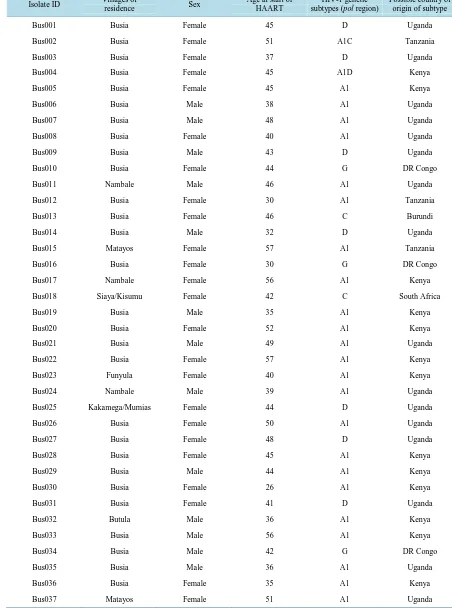
Table 1. Distribution of genetic subtypes of hiv-1 primary isolates from the study villages.
Isolate ID Villages of
residence Sex
Age at start of HAART
HIV-1 genetic subtypes (pol region)
Possible country of origin of subtype
Bus001 Busia Female 45 D Uganda
Bus002 Busia Female 51 A1C Tanzania
Bus003 Busia Female 37 D Uganda
Bus004 Busia Female 45 A1D Kenya
Bus005 Busia Female 45 A1 Kenya
Bus006 Busia Male 38 A1 Uganda
Bus007 Busia Male 48 A1 Uganda
Bus008 Busia Female 40 A1 Uganda
Bus009 Busia Male 43 D Uganda
Bus010 Busia Female 44 G DR Congo
Bus011 Nambale Male 46 A1 Uganda
Bus012 Busia Female 30 A1 Tanzania
Bus013 Busia Female 46 C Burundi
Bus014 Busia Male 32 D Uganda
Bus015 Matayos Female 57 A1 Tanzania
Bus016 Busia Female 30 G DR Congo
Bus017 Nambale Female 56 A1 Kenya
Bus018 Siaya/Kisumu Female 42 C South Africa
Bus019 Busia Male 35 A1 Kenya
Bus020 Busia Female 52 A1 Kenya
Bus021 Busia Male 49 A1 Uganda
Bus022 Busia Female 57 A1 Kenya
Bus023 Funyula Female 40 A1 Kenya
Bus024 Nambale Male 39 A1 Uganda
Bus025 Kakamega/Mumias Female 44 D Uganda
Bus026 Busia Female 50 A1 Uganda
Bus027 Busia Female 48 D Uganda
Bus028 Busia Female 45 A1 Kenya
Bus029 Busia Male 44 A1 Kenya
Bus030 Busia Female 26 A1 Kenya
Bus031 Busia Female 41 D Uganda
Bus032 Butula Male 36 A1 Kenya
Bus033 Busia Male 56 A1 Kenya
Bus034 Busia Male 42 G DR Congo
Bus035 Busia Male 36 A1 Uganda
Bus036 Busia Female 35 A1 Kenya
Continued
Bus038 Butula Female 38 D Kenya
Bus039 Busia Female 44 A1 Kenya
Bus040 Busia Male 41 D Uganda
Bus041 Busia Male 36 D Uganda
Bus042 Busia Female 38 A1 Kenya
Bus043 Bungoma Female 32 A1 Kenya
Bus044 Busia Female 44 A1 Kenya
Bus045 Busia Male 49 A1 Uganda
Bus046 Nambale Female 36 D Uganda
Bus047 Bungoma Female 38 G DR Congo
Bus048 Busia Female 40 D Uganda
Bus049 Busia Female 45 D Uganda
Bus050 Busia Female 34 A1 Kenya
Bus051 Bungoma Male 56 D Uganda
Bus052 Siaya/Kisumu Male 46 A1 Kenya
Bus053 Bungoma Female 44 D Uganda
Bus054 Busia Female 40 A1 Uganda
Bus055 Matayos Female 43 D Uganda
Bus056 Siaya/Kisumu Male 48 A1 Tanzania
Bus057 Busia Female 49 D Uganda
Bus058 Busia Female 43 D Uganda
Bus059 Busia Female 52 A1 Tanzania
Bus060 Bungoma Male 37 A1 Kenya
Bus061 Busia Female 45 G DR Congo
Bus062 Butula Male 33 A2 DR Congo
Bus063 Bungoma Female 43 A1 Kenya
Bus064 Busia Female 55 D Uganda
Bus065 Busia Female 32 C Burundi
Bus066 Siaya/Kisumu Female 41 D Uganda
Bus067 Siaya/Kisumu Female 45 A1 Uganda
Bus068 Busia Female 46 A1 Kenya
Bus069 Bungoma Female 49 C Botswana
Bus070 Busia Male 48 A2 DR Congo
Bus071 Siaya/Kisumu Female 45 A1 Uganda
Bus072 Matayos Female 33 B Brazil
Bus073 Siaya/Kisumu Female 33 D Uganda
Bus074 Budalangi Female 47 C Burundi
Bus075 Matayos Female 34 A1 Uganda
the Genbank with accession numbers HQ176975-HQ177040.
3. Results
The most predominant HIV-1 subtype in this region was A1 (52%) followed by subtype D (28%) both of which are associated with Uganda. Increased HIV-1 genetic diversity is evident as shown by the six genetic subtypes (A1, D, G, C, A2, B) and two intersubtype recombinants (A1/C, and A1/D) identified (Figure 1).
The majority of the HIV-1 subtypes from this study appear to be restricted to those commonly circulating in East Africa. For instance, a total of 35 isolates from this study resembled those previously reported from Uganda in the Genbank, while 23 isolates resembled those from Kenya and only 5 isolates resembled those from Tanza-nia.
4. Discussion
In Kenya, the majority of circulating strains belong to HIV-1 group M. However, there seems to be a lot of variation, which appears to be influenced by neighbour countries. For example, Neilson et al., 1999 [18] has shown that countrywide subtype A predominates (71% - 87%), with significant components of subtype D (7% - 29%) and subtype C (7% - 17%). Carr et al., 2005 [14] only identified subtype G. This scenario is different from that obtaining in the Northern region of Kenya town of Moyale bordering Ethiopia where on the basis of partial
env sequences, Khamadi et al., 2005 [13] reported that subtype A (50%), subtype C (39%) and subtype D (11%) were dominant. He attributed this to the fact that Kenya and Ethiopia share a long common border, resulting in a lot of cross-border migration, due to trade and tourist activities which may be responsible for the dynamic in-troduction of differing HIV subtypes and circulating recombinant forms. It must be noted that Ethiopia is domi-nated by HIV-1 subtype C, which interestingly is also among the dominant subtypes in Northern Kenya.
Lihana et al., 2006 [15] appears to have observed a lot of HIV-1 subtype recombinations among STI patients in Nairobi. He documented that Subtype C and D occur as non-recombinants but to a much lesser extent than subtype A. it is possible that because Nairobi is cosmopolitan, there may exist dually or multiply infected indi-viduals which enhances opportunity for HIV-1 recombinations. These recombinations appear to be occurring recombinants between A1, A2, and D; A2 and D; A1 and D; A1 and G; A1 and C; A1, C and D.
Our study seems to support the observation that HIV-1 diversity in the border regions could be as broad as that reported in major cities. For instance, like other parts of the country the predominant circulating subtype in western Kenya is A1. However, border country influence seems to be in play in that in the Kenya-Uganda bor-der subtype D is predominant, which is also the most dominant subtype in Uganda. Similarly in Moyale (Kenya- Ethiopia border) subtype C is rampant probably due to influence from Ethiopia where subtype C is dominant. Like Lihana et al., 2006 [15] reported, some HIV-1 subtype recombinations were also observed in this study. These were A1C and A1D.
[image:6.595.196.434.531.691.2]It is well known that there is a lot of HIV-1 subtype diversity in the world, albeit with a degree of regional
segregation. Africa has all HIV-1 subtypes but A and C predominate [8]. However, in East Africa, not with-standing the continental predominance of subtypes A, there is also a significant presence of subtype D [9]. To underline the phenomenon of segregation, our study area showed subtypes A1 and D being the most common. The few pockets of subtypes G and C observed could be due to the rampant human travels and migration pat-terns that may lead to introduction of other HIV-1 subtypes. For example the presence of subtype G and C in our study area at the Kenya-Uganda border could probably be due to the fact that this study area is a gateway to Central Africa, notably the Congo where subtype C is predominant. The phenomenon of border country influ-ence could further be supported by phylogenetic similarities of the sequinflu-enced viruses and those they were com-pared to from the Genbank. There was little evidence of HIV-1 subtype B in circulation which is the predomi-nant virus circulating in Europe, and is also found in Indonesia, the Philippines, and Taiwan [19], which coun-tries are far removed from Kenya.
5. Conclusion
Findings from this study reveal the complexity of HIV-1 infection in the Kenya-Uganda border region similar to those documented by Khamadi et al., 2005 [13] at the Kenya-Ethiopia border. There are continuous HIV-1 re-combinations. These could portend future global pandemics, which may be characterised by even more geneti-cally diverse viruses. There is therefore, a need for continuous review of control and treatment programs espe-cially in the border regions. For example, surveillance studies on the extent of inter-subtype recombinations are necessary for continuous improvement of diagnosis, treatment or vaccine development. To understand further the epidemiology of HIV-1 subtype diversity in border areas, analysis of human travels, migration patterns as well as social conditions need to be done, if therapeutic interventions have to be successful.
Acknowledgements
We would like to thank the individual volunteers who made this study possible. We acknowledge the Director, Kenya Medical Research Institute for permission to publish this work.
References
[1] Robertson, D.L., Anderson, J.P., Bradac, J.A., Carr, J.K., Foley, B., Funkhouser, R.K., Gao, F., Hahn, B.H., Kalish, M.L., Kuiken, C., Learn, G.H., Leitner, T., McCutchan, F., Osmanov, S., Peeters, M., Pieniazek, D., Salminen, M., Sharp, P.M., Wolinsky, S. and Korber, B. (2000) HIV-1 Nomenclature Proposal. Science, 288, 55-56.
http://dx.doi.org/10.1126/science.288.5463.55d
[2] Hemelaar, J., Gouws, E., Ghys, P.D. and Osmanov, S. (2006) Global and Regional Distribution of HIV-1 Genetic Subtypes and Recombinants in 2004. AIDS, 20, W13-W23. http://dx.doi.org/10.1097/01.aids.0000247564.73009.bc
[3] Eholie, S. and Anglaret, X. (2006) Commentary: Decline of HIV-2 Prevalence in West Africa: Good News or Bad News? International Journal of Epidemiology, 35, 1329-1330. http://dx.doi.org/10.1093/ije/dyl156
[4] Achkar, J.M., Burda, S.T., Konings, F.A., Urbanski, M.M., Williams, C.A., Seifen, D., Kahirimbanyi, M.N., Vogler, M., Parta, M., Lupatkin, H.C., Zolla-Pazner, S. and Nyambi, P.N. (2004) Infection with HIV Type 1 Group M Non-B Subtypes in Individuals Living in New York City. Journal of Acquired Immune Deficiency Syndromes, 36, 835-844.
http://dx.doi.org/10.1097/00126334-200407010-00011
[5] Triques, K., Bourgeois, A., Vidal, N., Mpoudi-Ngole, E., Mulanga-Kabeya, C., Nzilambi, N., Torimiro, N., Saman, E., Delaporte, E. and Peeters, M. (2000) Near-Full-Length Genome Sequencing of Divergent African HIV Type 1 Subtype F Viruses Leads to the Identification of a New HIV Type 1 Subtype Designated K. AIDS Research and Human Retro-viruses, 16, 139-151. http://dx.doi.org/10.1089/088922200309485
[6] Burke, D.S. (1997) Recombination in HIV: An Important Viral Evolutionary Strategy. Emerging Infectious Diseases, 3, 253-259. http://dx.doi.org/10.3201/eid0303.970301
[7] Peeters, M. and Sharp, P.M. (2000) Genetic Diversity of HIV-1: The Moving Target. AIDS, 14, S129-S140.
[8] Nasioulas, G., Paraskevis, D., Magiorkinis, E., Theodoridou, M. and Hatzakis, A. (1999) Molecular Analysis of the Full-Length Genome of HIV Type 1 Subtype I: Evidence of A/G/I Recombination. AIDS Research and Human Retro-viruses, 15, 745-758. http://dx.doi.org/10.1089/088922299310836
[10] Blackard, J.T., Renjifo, B.R., Mwakagile, D., Montano, M.A., Fawzi, W.W. and Essex, M. (1999) Transmission of Human Immunodeficiency Type 1 Viruses with Intersubtype Recombinant Long Terminal Repeat Sequences. Virology,
254, 220-225. http://dx.doi.org/10.1006/viro.1998.9504
[11] Hoelscher, M., Kim, B., Maboko, L., Mhalu, F., von Sonnenburg, F., Birx, D.L. and McCutchan, F.E. (2001) High Proportion of Unrelated HIV-1 Intersubtype Recombinants in the Mbeya Region of Southwest Tanzania. AIDS, 15, 1461-1470. http://dx.doi.org/10.1097/00002030-200108170-00002
[12] De Baar, M.P., De Ronde, A., Berkhout, B., Cornelissen, M., Van Der Horn, K.H.M., Van Der Schoot, A.M., De Wolf, F., Lukashov, V.V. and Goudsmit, J. (2000) Subtype-Specific Sequence Variation of the HIV Type 1 Long Terminal Repeat and Primer-Binding Site. AIDS Research and Human Retroviruses, 16, 499-504.
http://dx.doi.org/10.1089/088922200309160
[13] Khamadi, S.A., Ochieng, W., Lihana, R.W., Kinyua, J., Muriuki, J., Mwangi, J., Lwembe, R., Kiptoo, M., Osman, S., Lagat, N., Pelle, R., Muigai, A., Carter, J.Y., Oishi, I., Ichimura, H., Mwaniki, D.L., Okoth, F.A., Mpoke, S. and Son-gok, E.M. (2005) HIV Type 1 Subtypes in Circulation in Northern Kenya. AIDS Research and Human Retroviruses, 21, 810-814. http://dx.doi.org/10.1089/aid.2005.21.810
[14] Carr, J.K., Nadai, Y., Eyzaguirre, L., Saad, M.D., Khakimov, M.M., Yakubov, S.K., Birx, D.L., Graham, R.R., Wolfe, N.D., Earhart, K.C. and Sanchez, J.L. (2005) Outbreak of a West African Recombinant of HIV-1 in Tashkent, Uzbe-kistan. Journal of Acquired Immune Deficiency Syndromes, 39, 570-575.
[15] Lihana, R.W., Khamadi, S.A., Kiptoo, M.K., Kinyua, J.G., Lagat, N., Magoma, G.N., Mwau, M.M., Makokha, E.P., Onyango, V., Osman, S., Okoth, F.A. and Songok, E.M. (2006) HIV Type 1 Subtypes among STI Patients in Nairobi: A Genotypic Study Based on Partial pol Gene Sequencing. AIDS Research and Human Retroviruses, 22, 1172-1177.
http://dx.doi.org/10.1089/aid.2006.22.1172
[16] Kumar, S., Tamura, K., Jakobsen, I.B. and Nei, M. (2001) MEGA2: Molecular Evolutionary Genetics Analysis Soft-ware. Bioinformatics, 17, 1244-1245. http://dx.doi.org/10.1093/bioinformatics/17.12.1244
[17] Rhee, S.Y., Gonzales, M.J., Kantor, R., Betts, B.J., Ravela, J. and Shafer, R.W. (2003) Human Immunodeficiency Vi-rus Reverse Transcriptase and Protease Sequence Database. Nucleic Acids Research, 31, 298-303.
http://dx.doi.org/10.1093/nar/gkg100
[18] Neilson, J.R., John, G.C., Carr, J.K., Lewis, P., Kreiss, J.K., Jackson, S., Nduati, R.W., Mbori-Ngacha, D., Panteleeff, D.D., Bodrug, S., Giachetti, C., Bott, M.A., Richardson, B.A., Bwayo, J., Ndinya-Achola, J. and Overbaugh, J. (1999) Subtypes of Human Immunodeficiency Virus Type 1 and Disease Stage among Women in Nairobi, Kenya. Journal of Virology, 73, 4393-4403.