regeneration and delays progression of
Duchenne muscular dystrophy in mice
Ning Liu, … , Rhonda Bassel-Duby, Eric N. Olson
J Clin Invest.
2012;122(6):2054-2065. https://doi.org/10.1172/JCI62656.
Skeletal muscle injury activates adult myogenic stem cells, known as satellite cells, to
initiate proliferation and differentiation to regenerate new muscle fibers. The skeletal
muscle–specific microRNA miR-206 is upregulated in satellite cells following muscle injury,
but its role in muscle regeneration has not been defined. Here, we show that miR-206
promotes skeletal muscle regeneration in response to injury. Genetic deletion of miR-206 in
mice substantially delayed regeneration induced by cardiotoxin injury. Furthermore, loss of
miR-206 accelerated and exacerbated the dystrophic phenotype in a mouse model of
Duchenne muscular dystrophy. We found that miR-206 acts to promote satellite cell
differentiation and fusion into muscle fibers through suppressing a collection of negative
regulators of myogenesis. Our findings reveal an essential role for miR-206 in satellite cell
differentiation during skeletal muscle regeneration and indicate that miR-206 slows
progression of Duchenne muscular dystrophy.
Research Article
Muscle biology
Find the latest version:
microRNA-206 promotes skeletal
muscle regeneration and delays progression
of Duchenne muscular dystrophy in mice
Ning Liu,1 Andrew H. Williams,1 Johanna M. Maxeiner,1 Svetlana Bezprozvannaya,1
John M. Shelton,2 James A. Richardson,1,3 Rhonda Bassel-Duby,1 and Eric N. Olson1
1Department of Molecular Biology, 2Department of Internal Medicine, and 3Department of Pathology, University of Texas Southwestern Medical Center, Dallas, Texas, USA.
Skeletal muscle injury activates adult myogenic stem cells, known as satellite cells, to initiate proliferation
and differentiation to regenerate new muscle fibers. The skeletal muscle–specific microRNA miR-206 is
upregulated in satellite cells following muscle injury, but its role in muscle regeneration has not been defined.
Here, we show that miR-206 promotes skeletal muscle regeneration in response to injury. Genetic deletion
of miR-206 in mice substantially delayed regeneration induced by cardiotoxin injury. Furthermore, loss of
miR-206 accelerated and exacerbated the dystrophic phenotype in a mouse model of Duchenne muscular
dys-trophy. We found that miR-206 acts to promote satellite cell differentiation and fusion into muscle fibers
through suppressing a collection of negative regulators of myogenesis. Our findings reveal an essential role for
miR-206 in satellite cell differentiation during skeletal muscle regeneration and indicate that miR-206 slows
progression of Duchenne muscular dystrophy.
Introduction
Adult skeletal muscle can regenerate in response to exercise, injury, and disease. Skeletal muscle regeneration relies on a small population of stem cells, known as satellite cells (SCs), which reside beneath the basal lamina of myofibers (1, 2). SCs are nor-mally quiescent, but, in response to stress or injury, become acti-vated to proliferate, differentiate, and fuse into multinucleated myotubes (3–6). Activated SCs also undergo asymmetric division, generating progeny that replenish the pool of quiescent SCs (5). Abnormalities in SC specification, proliferation, or differentia-tion result in skeletal muscle dysfuncdifferentia-tion during aging and can promote muscle disease (7).
The paired-box transcription factor Pax7 is a specific marker for quiescent and activated SCs and is downregulated when SCs dif-ferentiate into myotubes (5, 8). Pax7 activates expression of the myogenic regulatory factors Myf5 and MyoD in activated SCs and proliferating myoblasts, which in turn drive the myogenic differ-entiation program (9). Although stem/progenitor cells from other cell origins have also been reported to contribute to regeneration of new myofibers, SC ablation experiments have clearly demon-strated that Pax7-expressing SCs are indispensable for adult skel-etal muscle regeneration (10–13).
Duchenne muscular dystrophy (DMD), the most common and severe form of muscular dystrophy, is caused by mutations in the dystrophin gene on the X chromosome (14, 15). Loss of the subsar-colemmal protein dystrophin in DMD patients causes sensitivity of myofibers to mechanical damage, leading to SC activation and myofiber regeneration (16). However, the unsustainable activation of SCs in DMD patients ultimately results in severe muscle
wast-ing, infiltration of adipocytes, inflammation, and eventual paraly-sis and death (17). Mdx mice, which harbor a premature termina-tion codon in the dystrophin gene, are the most commonly used mouse model of muscular dystrophy (18). Intriguingly, despite sharing the same genetic defects as DMD patients, mdx mice dis-play a relatively mild and slowly progressive dystrophic phenotype, marked by chronic SC activation and regeneration. The relatively mild phenotype of mdx mice has been attributed to the increased regenerative capacity of mouse SCs due to the longer telomeres in mice relative to humans (19). Understanding the mechanisms of DMD pathology remains an important challenge in the quest to develop efficacious therapies for DMD patients.
microRNAs (miRNAs) are a class of small noncoding RNAs that inhibit gene expression via Watson-Crick base pairing between the miRNA “seed” region and sequences located predominantly in the 3′ UTRs of target mRNAs (20). Changes in miRNA expres-sion are associated with various skeletal muscle disorders, includ-ing muscular dystrophies (21, 22). The skeletal muscle–specific miRNA miR-206 is required for efficient regeneration of neuro-muscular synapses after acute nerve injury, and the absence of miR-206 accelerates disease progression of amyotrophic lateral sclerosis (ALS) in mice (23). miR-206 is upregulated during skel-etal muscle regeneration and has been reported to repress pro-liferation and promote differentiation of SCs in vitro (24, 25). However, the functions of miR-206 in skeletal muscle regenera-tion in vivo have not been determined.
In the present study, we show that mice lacking miR-206 have inefficient skeletal muscle regeneration in response to cardiotox-in (CTX) cardiotox-injury. Loss of miR-206 also results cardiotox-in acceleration and exacerbation of muscle dysfunction in mdx mice. The inefficient skeletal muscle regeneration in mice lacking miR-206 results from impaired differentiation of SCs and correlates with the dysregula-tion of a collecdysregula-tion of negative regulators of myogenesis. Our find-ings reveal an important role for miR-206 as a modulator of DMD and a potential target for therapeutic intervention in this disease. Authorship note: Ning Liu and Andrew H. Williams contributed equally to this
work.
Conflict of interest: Eric N. Olson is cofounder of miRagen Therapeutics, a company focused on developing miRNA-based therapies for cardiovascular disease.
Results
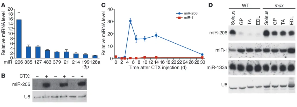
Upregulation of miR-206 during skeletal muscle regeneration. In an effort to identify miRNAs involved in skeletal muscle regenera-tion, we induced skeletal muscle injury and regeneration in mice by injecting CTX in the tibialis anterior (TA) muscle and com-pared the miRNA expression profile 7 days after CTX injection with that of untreated control TA muscle. Microarray analysis identified a number of miRNAs that were dysregulated upon CTX injection (Figure 1A and Supplemental Figure 1, A and B; supple-mental material available online with this article; doi:10.1172/ JCI62656DS1). miR-206 was the most dramatically upregulated miRNA in CTX-injured TA muscle on day 7, as confirmed by real-time RT-PCR and Northern blot analysis (Figure 1, A and B). miR-206 continued to be strongly expressed throughout the course of muscle regeneration (Figure 1C). miR-206 is generated from a bicistronic transcript that also includes miR-133b (23). Two other homologous pairs of muscle-specific miRNAs, miR-1-1/133a-2 and miR-1-2/133a-1, are expressed from separate chromosomes (26). miR-133b was also strongly upregulated following CTX injec-tion, whereas the expression of miR-1, which shares high sequence homology with miR-206, was modestly downregulated (Figure 1C and Supplemental Figure 1C).
We also examined the regulation of miR-206 during the progres-sion of muscular dystrophy in mdx mice. At 2 weeks of age, prior to the onset of muscle damage, miR-206 was expressed in mdx
muscle at levels comparable to those in WT muscle (Supplemen-tal Figure 2A), whereas by 4 weeks of age, after the onset of necro-sis and myofiber degeneration, miR-206 was upregulated in vari-ous muscles of mdx mice (Figure 1D and Supplemental Figure 2). Consistent with these findings, miR-206 expression has been reported to be enriched in newly formed myofibers with central-ized nuclei in mdx TA muscle, but not in intact fibers (24, 27). These data indicate that miR-206 expression is dramatically upregulated in pharmacologic and genetic models of skeletal muscle regeneration and muscular dystrophy.
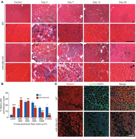
Delayed skeletal muscle regeneration in mice lacking miR-206. In light of the strong upregulation of miR-206 expression during skeletal muscle regeneration and its enrichment in activated SCs (25), we injected WT and miR-206–KO mice with CTX and analyzed myofi-ber regeneration at various time points. miR-206–KO mice do not exhibit apparent histological or functional muscle defects under normal conditions (23). Three days following CTX injection, WT and miR-206–KO mice exhibited extensive myofiber degenera-tion and accumuladegenera-tion of small mononucleated cells at the site of injury, which included proliferating myoblasts, macrophages, and neutrophils (Figure 2A). Seven days following CTX injection, most damaged myofibers in WT mice were cleared and replaced by newly formed myofibers containing centralized nuclei (Figure 2A). In con-trast, in miR-206–KO mice, abundant inflammatory cells and dam-aged myofibers were still present 7 days following CTX injection. On days 14 and 30 following CTX injection, muscle architecture was largely restored in both WT and miR-206–KO mice, indicating that the absence of miR-206 delays but does not completely pre-vent regeneration (Figure 2A). The delayed regeneration response of miR-206–KO muscle was further revealed by extensive miner-alization 14 and 30 days following CTX injection, which resulted from accumulation of damaged muscle fibers (Figure 2A). Oil red O staining also revealed slightly increased fatty infiltration in miR-206–KO mice 30 days following CTX injection (Supplemental Figure 3C). Loss of miR-206 does not affect capillary density of TA muscle 30 days after CTX injection, as assessed by immunostaining for platelet endothelial cell adhesion molecule (PECAM1) staining (Supplemental Figure 3D), indicating that angiogenesis after CTX injury is not affected in miR-206–KO mice.
[image:3.585.43.543.80.261.2]Whereas there was no significant difference in fiber size between WT and miR-206–KO mice under normal conditions (Supple-mental Figure 3, A and B), regenerated myofibers in miR-206–KO mice were significantly smaller than those of WT mice (Figure 2B). Immunostaining for the intermediate filament protein desmin, which is highly expressed in immature muscle fibers during fetal
Figure 1
life and regeneration (28, 29), showed that desmin was strongly expressed in most myofibers in WT mice 7 days following CTX injec-tion (Figure 2C). However, in miR-206–KO mice, desmin expression was absent in many myofibers, and desmin-positive fibers that were present displayed heterogeneity in size, confirming the impairment in regeneration of miR-206–KO muscle (Figure 2C).
Loss of miR-206 exacerbates the dystrophic phenotype in mdx mice. To investigate the potential involvement of miR-206 in muscular dys-trophy, we generated mdx mice lacking expression of miR-206 and
compared the morphology of quadriceps and diaphragm muscles of mdx and miR-206–KO;mdx mice at different ages. Mice used in these studies were of 129SvEv-C57BL/6 mixed background. Absence of miR-206 expression was confirmed by Northern blot analysis (Supplemental Figure 4). At 2 weeks of age, the quadriceps muscle in mdx mice showed a largely normal morphology (Supplemental Figure 5A). Small regenerating fibers and associated mononucle-ated cells were only occasionally observed in a minor subset of miR-206–KO;mdx mice (Supplemental Figure 5A). Strikingly,
how-Figure 2
[image:4.585.65.524.82.553.2]Figure 3
Loss of miR-206 exacerbates the dys-trophic phenotype in mdx mice. (A) H&E staining of quadriceps and dia-phragm muscle of WT, miR-206–KO,
mdx, and miR-206–KO;mdx mice at 4 weeks of age. Upper 2 rows: H&E staining of quadriceps and dia-phragm muscles, respectively. Lower 2 rows: von Kossa staining and Masson’s trichrome staining of dia-phragm muscle showing mineraliza-tion and fibrosis in diaphragm mus-cle fibers. Scale bars: 200 μm. (B) H&E staining of quadriceps and dia-phragm muscle of WT, miR-206–KO,
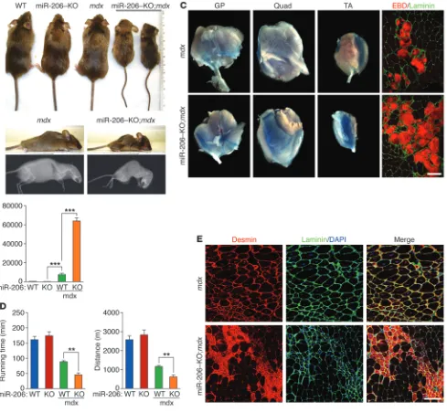
ever, by 4 weeks of age, nearly all miR-206–KO;mdx mice showed severe dystrophic phenotypes compared with mdx mice. We observed massive accumulation of small regenerating fibers and inflam-matory cells in miR-206–KO;mdx quadriceps muscle (Figure 3A). Similarly, the diaphragm of miR-206–KO;mdx mice showed exten-sive fiber damage and degeneration, with calcium deposition, min-eralization, and fibrosis (Figure 3A). The dystrophic phenotype became more apparent at 6 weeks of age, when approximately 17%
of miR-206–KO;mdx mice became severely runted, showed kyphosis, and died (Figure 4A). In contrast, only 5% of mdx mice died at this age (Figure 4A). Histological analysis revealed especially severe mus-cle degeneration, mineralization, and fibrosis in miR-206–KO;mdx
mice compared with mdx mice (Figure 3B).
[image:6.585.53.542.83.534.2]Although miR-206–KO;mdx mice that survived beyond 8 weeks showed a life span similar to that of mdx mice, these mice generally had worse dystrophic phenotypes than mdx mice. At advanced ages
Figure 4
(>17 months), miR-206–KO;mdx mice showed debilitating muscle wasting and infiltration of fat in the diaphragm muscle compared with mdx mice (Figure 3C). In contrast with other muscle groups, the soleus muscle of miR-206–KO;mdx mice was indistinguishable from that of mdx mice (data not shown), which likely reflects the resistance of slow twitch myofibers in the soleus muscle to dystro-phy. There were no apparent cardiac abnormalities in miR-206–KO;
mdx mice at 3 months of age as measured by echocardiography and histological analysis (Supplemental Figure 5, C and D).
To further characterize the dystrophic phenotype of miR-206–KO;
mdx mice, we measured the levels of serum creatine kinase (CK), an indicator of skeletal muscle damage, and diagnostic marker for DMD patients (30). At 4 weeks of age, serum CK levels of
mdx mice were elevated 14-fold compared with those of WT and miR-206–KO mice (Figure 4B). Intriguingly, serum CK levels of miR-206–KO;mdx mice were more than 8-fold higher than those of mdx mice (Figure 4B). At 5 months of age, the difference in serum CK levels between mdx and miR-206–KO;mdx mice became less dramatic. However, miR-206–KO;mdx mice still showed an approximately 1.5-fold increase in serum CK levels compared with
mdx mice (Supplemental Figure 5B). Myofiber membrane perme-ability, monitored by Evans blue dye uptake, was also exacerbated in miR-206–KO;mdx mice (Figure 4C).
To assess muscle functionality and performance, we subjected mice to a downhill treadmill exercise paradigm and measured the running time and distance to exhaustion. At 3 months of age, mdx
mice ran for a significantly shorter time than WT or miR-206–KO mice (Figure 4D). Muscle performance in miR-206–KO;mdx mice was further decreased compared with mdx mice, indicative of severe muscle injury and dysfunction (Figure 4D). These findings support the histological data and further indicate that the absence of miR-206 sensitizes mice to dystrophic abnormalities.
Immunostaining of 6-week-old muscle sections revealed strong accumulation of desmin in small regenerating myofibers that con-tained centralized nuclei in miR-206–KO;mdx mice (Figure 4E). In contrast, regenerating fibers in mdx mice displayed weaker desmin signal and were larger (Figure 4E), indicating that the regenerating
fibers in miR-206–KO;mdx mice were less mature than in mdx mice. Microarray analysis on muscles from mdx and miR-206–KO;mdx
mice also revealed upregulation of embryonic muscle genes as well as inflammatory genes, confirming the delayed regeneration in miR-206–KO;mdx mice (Table 1). Taken together, these data sup-port the conclusion that loss of miR-206 in the mdx background causes a delay in regeneration and maturation of myofibers to replace damaged fibers, which contributes to the more severe dys-trophic phenotype in compound mutant mice.
miR-206 is required for the efficient regeneration of neuro-muscular junctions (NMJs) following nerve injury in mice (23).
mdx mice also have structural abnormalities in NMJs, although the functional significance of this observation is unknown (31). To determine whether the more severe dystrophic phenotype of miR-206–KO;mdx mice was due to innervation defects of NMJs, we compared the innervation of mdx and miR-206–KO;mdx mice using markers for the postsynaptic membrane (α-bungarotoxin [BTX]) and presynaptic nerve terminals (synaptophysin) (32). We observed no differences in the number of synapses or the innerva-tion of NMJs between mdx and miR-206–KO;mdx mice (Supple-mental Figure 5B). Thus, we conclude that the severe dystrophic phenotypes observed in miR-206–KO;mdx mice are due to a func-tion of miR-206 independent of motor innervafunc-tion.
[image:7.585.55.526.113.315.2]miR-206 regulates SC differentiation. To directly examine the influ-ence of miR-206 on SC function in vivo, we isolated activated SCs from WT and miR-206–KO mice by FACS (refs. 5, 7, and Supple-mental Figure 6, A and B). We found no significant difference in the fraction of SCs between WT and miR-206–KO mice 3 days following CTX injury (Supplemental Figure 6A). We confirmed the expression of miR-206 in cultured SCs, but not in the non-SC population by Northern blot analysis (Supplemental Figure 6C). We also analyzed the proliferation of SCs isolated from WT and miR-206–KO mice, but found no differences in the prolifera-tion rates of these cells in culture (Supplemental Figure 7, A and B). These findings contrast with a previous report that miR-206 represses SC proliferation based on overexpression or oligonucle-otide-mediated inhibition of miR-206 in vitro (25).
Table 1
Genes upregulated in miR-206–KO;mdx mice compared with mdx mice
Category Gene symbol Fold change Gene name (miR-206–KO;
mdx/mdx)
Muscle regeneration Myh3 3.2 Mus musculus myosin, heavy polypeptide 3, skeletal muscle, embryonic
Myh8 2.93 Mus musculus myosin, heavy polypeptide 8, skeletal muscle, perinatal
Myl14 2.2 Mus musculus myosin, light polypeptide 4
Tnni1 2.52 Mus musculus troponin T1, skeletal, slow Chemokines, cytokines, Ccl7 3.7 Mus musculus chemokine (C-C motif) ligand 7 interleukins, and receptors Ccl9 3.2 Mus musculus chemokine (C-C motif) ligand 9
Cxcl4 2.33 Mus musculus chemokine (C-X-C motif) ligand 4, mRNA
Igf-2 2.0 Mus musculus insulin-like growth factor 2
Lyzs 1.9 Mus musculus lysozyme, mRNA
Il1rl1 1.7 Mus musculus interleukin 1 receptor-like 1, transcript variant 2, mRNA ECM and ECM processing TIMP-1 1.9 Mus musculus tissue inhibitor of metalloproteinase 1, transcript variant 2, mRNA Angiogenesis and ApoE 1.9 Mus musculus ApoE, mRNA
vascular response
Cytoskeleton Tubb2b 1.8 Mus musculus tubulin, β 2b, mRNA
Coro1a 2.0 Mus musculus coronin, actin binding protein 1A, mRNA
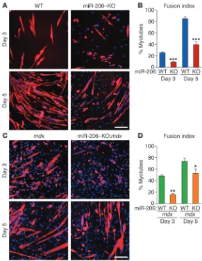
To investigate whether miR-206 modulates SC differentiation, we isolated WT and miR-206–KO SCs by FACS and assessed their abili-ty to differentiate and form myotubes at different time points. Three days after induction of differentiation, most WT SCs became elongat-ed and fuselongat-ed and expresselongat-ed the differentiation marker myosin heavy chain (MHC) (Figure 5, A and B). In contrast, elongation, fusion, and MHC expression were delayed in miR-206–KO SCs (Figure 5, A and B). After 5 days in differentiation medium, most WT SCs had fused to form myotubes, whereas only 40% of miR-206–KO cells underwent fusion (Figure 5, A and B). We also analyzed the dif-ferentiation of SCs from mdx and miR-206–KO;mdx mice. Similar to the miR-206–KO SCs, we observed delayed fusion of miR-206–KO;
mdx SCs compared with mdx SCs (Figure 5, C and D). These results
indicate that the absence of miR-206 causes a cell-autonomous delay in SC differentiation. They are also consistent with the previous finding that loss of miR-206 delayed rather than completely blocked regeneration upon CTX injury (Figure 2A).
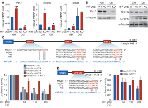
miR-206 targets regulators of SC differentiation. miR-206 has been reported to directly repress Pax7 expression dur-ing SC proliferation and differentiation in vitro (25, 27). While Pax7 mRNA levels were upregulated approxi-mately 1.7-fold, Pax7 protein levels were unchanged in miR-206–KO SCs compared with WT under growth conditions (Figure 6, A and B). Pax7 mRNA and pro-tein are downregulated during differentiation of WT SCs (8). Interestingly, this repression was attenuated in miR-206–KO cells under the same differentiation conditions, such that Pax7 protein was still detectable in miR-206–KO SCs, but not in WT cells 5 days after differentiation (Figure 6, A and B). Using luciferase reporter assays, we found that miR-206 could directly repress Pax7 mRNA translation through interactions with 4 binding sites in the 3′ UTR (Supplemental Fig-ure 8A). These results indicate that miR-206 regulates Pax7 expression during SC differentiation.
[image:8.585.41.332.81.458.2]In addition to Pax7, we identified and confirmed several other miR-206 target genes with func-tions in myoblast and SC differentiation that were upregulated in miR-206–KO SCs. Notch3 mRNA and protein expression was upregulated in miR-206–KO SCs, with the upregulation being more pronounced in miR-206–KO SC–derived myotubes (Figure 6, A and B). Notch3 functions as an inhibitor of MyoD-induced myogenesis (33). Thus, its upregulation would be inhibitory to the differentiation process. We identified 3 miR-206–binding sites within the 3′ UTR of Notch3 mRNA, 2 of which are evolutionarily conserved (Figure 6C). In luciferase assays, miR-206 repressed a luciferase reporter gene linked to the Notch3 3′ UTR, whereas mutation in all 3 predicted miR-206–binding sites abrogated this repression (Figure 6C). In addition, we identified IGF-binding protein 5 (Igfbp5) as a miR-206 target gene. Igfbp5 is a secreted factor that has previously been shown to inhibit skeletal muscle differentiation through IGF-dependent mechanisms (34–36). IGFBP5 protein expression was upregulated in miR-206–KO SCs and miR-206 SC–derived myotubes compared with WT cells (Figure 6, A and B). The 3′ UTR of Igfbp5 mRNA contains one evolutionarily conserved miR-206–binding site (Figure 6D). In luciferase assays, miR-206 repressed a luciferase reporter gene linked to the Igfbp5 3′ UTR, and mutations in the predicted miR-206–binding site abrogated the repression, confirm-ing Igfbp5 as a target of miR-206 (Figure 6D). In addition, we also confirmed upregulation of Gja1 (connexin 43) in miR-206–KO SCs upon differentiation (Supplemental Figure 8B), although the potential function of connexin 43 in myogenesis remains unknown (37). Previously published miR-206 target genes, such as utrophin and myostatin, did not show differences in their expression in miR-206–KO SCs compared with WT cells (Supplemental Figure 8C). In addition, expression of Integrin α7 (Itga7) and VEGF was not changed in miR-206–KO SCs (Supplemental Figure 8C). In
Figure 5
summary, loss of miR-206 results in upregulation of Pax7, Notch3, and Igfbp5 in differentiating SCs compared with WT cells, imply-ing that repression of these inhibitors of myogenesis accounts, at least in part, for the stimulatory influence of miR-206 on SC dif-ferentiation and skeletal muscle regeneration.
Discussion
The results of this study reveal an important role of miR-206 in promoting efficient skeletal muscle regeneration in response to injury or disease. Whereas mice lacking miR-206 do not exhibit overt abnormalities at baseline, the absence of miR-206 in mice results in a delay in skeletal muscle regeneration in response to CTX injury and an exacerbated dystrophic phenotype in mdx mice. These data identify miR-206 as an important modifier of muscu-lar dystrophy disease progression. Our results extend recent find-ings that miRNAs frequently function as “fine-tuners” of cellular phenotypes under conditions of tissue homeostasis, whereas their functions become magnified under conditions of injury or stress.
miR-206 and skeletal muscle regeneration. Skeletal muscle regenera-tion in response to injury involves a highly orchestrated series of events accompanied by necrosis and activation of an inflammatory response, leading to myofiber degeneration and the activation of myogenic cells to proliferate, differentiate, and fuse to generate new myofibers (2). miR-206 is expressed at a low level in adult myo-fibers, but is highly enriched in newly regenerated myofibers dur-ing muscle regeneration. The upregulation of miR-206 followdur-ing injury correlates with the time in which proliferating myoblasts initiate differentiation into mature myotubes and upregulate the myogenic transcription factors MyoD and myogenin, which directly activate miR-206 expression (23, 38). Indeed, miR-206 is strongly expressed in activated SCs, in which MyoD expression is already turned on (Supplemental Figure 6C).
[image:9.585.48.536.80.436.2]It has been reported that injection of a cocktail of muscle-specif-ic miRNAs, including miR-206, can accelerate muscle regeneration in a rat injury model (39), although the specific functions of indi-vidual miRNAs in this setting were not defined. In this study, we
Figure 6
miR-206 regulates expression of target genes involved in SC proliferation and differentiation. (A) Real-time RT-PCR reveals expression of Pax7,
show that the loss of miR-206 in mice delays muscle regeneration upon CTX injury. Taken together, these results identify miR-206 as an important regulator of muscle regeneration. Analysis of SC dif-ferentiation in miR-206–KO mice suggests that miR-206 promotes the differentiation and fusion of progenitor cells into mature myo-tubes. Thus, the delayed regeneration in miR-206–KO mice upon CTX injury can be attributed to the cell-autonomous function of miR-206 in SC differentiation. Although it is possible that delayed regeneration could also result from an impaired inflammatory response, preventing clearance of damaged myofibers, we do not believe this is the case because miR-206 is only expressed in SCs and not in inflammatory cells (Supplemental Figure 6C).
miR-206 and SC proliferation and differentiation. Our results sug-gest that miR-206 promotes SC differentiation and represses a set of negative regulators of muscle differentiation, including Pax7, Notch3, and Igfbp5. In the absence of miR-206, SCs show a delay but not a complete block in differentiation, which is likely due to functional redundancy with the closely related miR-1, which is also upregulated upon SC differentiation (25, 27). Pax7 protein is highly expressed in proliferating SCs. Although Pax7 mRNA lev-els are upregulated in miR-206–KO SCs under growth conditions, Pax7 protein levels are not significantly changed under these con-ditions. These findings suggest that additional mechanisms other than miR-206 are responsible for controlling Pax7 protein levels in proliferating SCs. During differentiation, Pax7 is downregulated, and the loss of miR-206 results in attenuation of this downregu-lation at both mRNA and protein levels. The distinct regudownregu-lation patterns of Pax7 by miR-206 under growth versus differentiation conditions highlight the notion that the functions of miRNAs are important only under certain cellular phenotypes or conditions.
It was recently reported that miR-206 and miR-133b are encoded by a long noncoding RNA (linc-MD1) (40). linc-MD1 is upregulated in mdx mice and promotes muscle differentiation by acting as a competing endogenous RNA (ceRNA) for miR-133 and miR-135 (40). Thus, in addition to regulating SC differentiation through direct binding to the 3′ UTR of target mRNAs, it also possible that miR-206 functions through additional mechanisms to promote the efficient differentiation and fusion of muscle progenitors into myo-tubes. In this regard, there are several reports of small RNAs being capable of directly activating gene transcription and of miRNAs upregulating the translation of target genes (41, 42).
Our results also indicate that miR-206 does not play a major role in the proliferation of SCs. This is in contrast with a previous study demonstrating that the knockdown of miR-1 and miR-206 in vivo results in an increased rate of proliferation of SCs (25). The discrepancy between our results and those published findings could be because of the inhibition of both miR-1 and miR-206 by antisense oligonucleotides in the published studies. Alternatively, transient inhibition of miR-206 expression in adult mice may have different consequences than genetic deletion throughout life. While genetic deletion and oligonucleotide inhibition of miR-206 lead to different conclusions regarding the role of miR-206 in myoblast proliferation, there are numerous reports that demon-strate a function for miR-206 in promoting the differentiation of muscle progenitors and myoblasts into myotubes (37, 43, 44), as supported by our genetic loss-of-function data.
miR-206 as a modifier of DMD. The mdx mouse has provided important insights into the pathological mechanisms of DMD. However, the slow disease progression and mild dystrophic phe-notype of mdx mice relative to DMD patients have limited the
usefulness of this model for therapeutic development. The intro-duction of secondary gene mutations in mdx mice, such as muta-tions in utrophin, α-dystrobrevin, α7-integrin, or cytidine mono-phosphate–sialic acid hydroxylase (Cmah) etc., has highlighted the importance of other cellular components in DMD disease progression (45–49). Similarly, genes involved in SC proliferation and differentiation also play regulatory roles in the pathogenesis of mdx mice. The severe and rapidly progressing phenotype in
mdx mice lacking telomerase activity is caused by the reduced regenerative potential of SCs due to shortened telomere length (19). In addition, loss of MyoD in mdx mice results in impaired differentiation of SCs, leading to a more severe dystrophic dis-ease phenotype (50). In this report, we show that miR-206 mod-ulates the disease progression of DMD by regulating myogenic differentiation in mdx mice.
miR-206 has been shown to be upregulated in mdx mice, but it has been unknown whether miR-206 plays a protective or patho-genic role in the disease (24, 27). Our results clearly demonstrate that miR-206 plays a protective role in the setting of muscular dys-trophy. The strong activation of miR-206 in mdx mice serves as a compensatory mechanism to promote formation of new myofi-bers in response to injury. In the absence of miR-206, the delayed regeneration and myogenic differentiation result in fibrosis and fatty infiltration as well as mineralization of myofibers, which dis-rupt muscle integrity and function.
The dramatic worsening of the dystrophic phenotype of mdx
mice upon genetic deletion of miR-206 is conceptually similar to our prior discovery of the acceleration of skeletal muscle atrophy, paralysis, and death in a mouse model of ALS lacking miR-206 (23). In both cases, strong upregulation of miR-206 expression was seen at disease onset, and miR-206 was found to have a com-pensatory, protective role in both disease contexts. These 2 stud-ies highlight an important function of miR-206 as a stress-induc-ible suppressor of skeletal muscle disease. Although we found no innervation defects in miR-206–KO;mdx mice compared with mdx
mice, perhaps the beneficial effects of miR-206 in the 2 disease models point to an unappreciated relationship between muscle SCs and neuromuscular synapse stability.
Therapeutic implications. Since the identification of the dystrophin gene in 1986, there has been intense effort to develop potential therapies for DMD. Current strategies to treat DMD include deliv-ering functional dystrophin via adenoassociated virus vectors, using antisense oligonucleotides to induce exon skipping, allow-ing production of truncated, but functional, dystrophin protein, or developing stem cell therapies to introduce SCs and myoblasts into muscle (51). Despite advances in these approaches, many chal-lenges remain. The protective role of miR-206 in mdx mice raises the possibility that delivery of miR-206 mimics or other strategies to elevate expression of the endogenous miR-206 gene or modulate its downstream targets could provide therapeutic benefit for DMD or other degenerative skeletal muscle diseases.
Methods
Generation of miR-206–KO;mdx mice. miR-206–/– mice in a 129SvEv-C57BL/6
mixed background were described previously (23). C57BL/10ScSn-Dmdmdx
mice were purchased from the Jackson Laboratory. miR-206–/– male mice
were bred to homozygous Dmdmdx/mdx female mice to generate miR-206+/–;
Dmdmdx/+ female mice or miR-206+/–;Dmdmdx/Y male mice. miR-206+/–;Dmdmdx/Y
male mice were then bred to Dmdmdx/mdx female mice to generate miR-206+/–;
were then intercrossed with miR-206+/–;Dmdmdx/Y male mice to generate
miR-206–/–;Dmdmdx/mdx mice and miR-206–/–;Dmdmdx/Y mice, which are referred
to as miR-206–KO;mdx mice. Mice used in this study were all on a 129SvEv-C57BL/6 mixed background.
CTX injury. CTX from Naja mossambica mossambica (Sigma-Aldrich) was dissolved in sterile saline to a final concentration of 10 μM and aliquoted and stored at –20°C. Mice were anesthetized by intraperitoneal injection of 2.5% Avertin at (15 μl/g). Mouse legs were shaved and cleaned with alcohol. TA muscles were injected with 50 μl of CTX with a 26-gauge needle. After injection, animals were kept under a warming lamp until recovery.
Northern blot analysis. Total RNA was isolated from cultured cells by Trizol
(Invitrogen) or from mouse skeletal muscle tissues by the miRNeasy Mini Kit (QIAGEN). Northern blots to detect miR-206, miR-1, and miR-133 and U6 were performed as described previously (52). 32P-labeled Star-Fire
oligonucleotide probes (IDT) against mature miRNAs and U6 were used for hybridization.
RT-PCR and real-time RT-PCR analysis. RNA was treated with Turbo RNase-free DNase (Ambion Inc.) prior to the reverse transcription step. RT-PCR was performed using random hexamer primers (Invitrogen). Real-time RT-PCR on miRNA was performed using the TaqMan microRNA assay kits (ABI) according to the manufacturer’s protocol. Real-time RT-PCR was performed using TaqMan probes (ABI) or SYBR green probes. SYBR green primers used were as follow: Gja1-F: 5′ -GGACCTTGTCCAGCAGCTT-3′, Gja1-R: 5′-TCCAAGGAGTTCCACCACTT-3′; Notch3-F: 5′ -AAGC-GTCTCCTGGATGCTG-3′, Notch3-R: 5′ -GAATCTGGAAGACACCCTGG-3′; pre-133b-RT-F: 5′CTGGTCAAACGGAACCAAGT-3′, pre-133b-RT-R: 5′-TGATGGCAAAACCAGCATTA-3′.
Microarray analysis. For the microarray, total RNA was isolated from TA muscle of mdx and miR-206–KO;mdx mice at 3 months of age (n = 3 for each). Microarray analysis was performed by the University of Texas Southwestern Microarray Core Facility using the Mouse Genome Illumina Mouse-6 V2 BeadChip. Data have been deposited in NCBI’s Gene Expres-sion Omnibus (GEO GSE36077).
Histological analysis of skeletal muscle. Skeletal muscle groups were harvest-ed and flash frozen in embharvest-edding mharvest-edium containing a 3:1 mixture of Tissue Freezing Medium (Triangle Biomedical Sciences) and gum traga-canth (Sigma-Aldrich) or fixed in 4% paraformaldehyde and processed for routine paraffin histology. Frozen sections were cut on a cryotome and stained with H&E as previously described (53). Masson’s trichrome, von Kossa staining, and oil red O staining were performed using standard pro-cedures. Evans blue dye uptake was performed as described (53).
Immunohistochemistry. Frozen sections were fixed in freshly prepared 4% paraformaldehyde for 20 minutes on ice and then treated with 0.3% Tri-ton X-100/PBS at room temperature for 20 minutes. Sections were incu-bated with mouse IgG-blocking solution from the M.O.M. kit (Vector Lab) diluted in 0.01% Triton X-100/PBS at room temperature for 1 hour. Sections were then incubated with 5% goat serum (Sigma-Aldrich) in M.O.M. protein diluent for 30 minutes. Sections were incubated with pri-mary antibodies diluted in M.O.M. protein diluent at 4°C overnight. The next morning, slides were washed with PBS and incubated with secondary antibodies diluted in M.O.M. protein diluent at room temperature for 45 minutes. Sections were then washed and mounted with VectaShield Mounting Medium with DAPI. Pictures were taken with a Zeiss confo-cal microscope. Primary and secondary antibodies were as follows: Des-min (1:100, Dako), LaDes-minin (1:200, Sigma-Aldrich), MF20 (DSHB), Pax7 (1:100, DSHB), Alexa Fluor 594 goat anti-mouse IgG1 (1:400, Invitrogen), and Alexa Fluor 488 goat anti-rabbit IgG (1:400, Invitrogen). Wheat germ agglutinin (WGA) staining was performed using Alexa Fluor 555–conju-gated WGA (Invitrogen) as previously described (53). Immunostaining of NMJs was done as previously described (23). PECAM1 staining was
per-formed using anti-mouse CD31 antibody (1:40, Dianova) and a biotinylat-ed secondary antibody couplbiotinylat-ed with streptavidin–horseradish peroxidase, followed by DAB chromagen reaction.
Isolation of SCs by FACS. CTX was injected into hind limb muscles of WT and miR-206–KO mice, and activated SCs were isolated 3 days after injection as previously described (7). Briefly, hind limb muscles were pooled, minced, and digested with 0.2% Collagenase II (Gibco; Invitrogen), followed by tritu-ration. SCs were then isolated by further digestion of myofibers with 0.1% Dispase (Gibco; Invitrogen) and 0.05% Collagenase II (Gibco; Invitrogen). Cell suspension was filtered through a 40-μm cell strainer, and SCs were pel-leted after centrifugation.
For FACS, SCs were counted and resuspended in PBS/3% BSA at 1 × 10⁶
cells/50 μl. Cells were then incubated with the following antibodies for 1 hour on ice: Alexa Fluor 488–conjugated rat anti-mouse CD34 (1:50) (AbD Sero-tec), PE-labeled rat anti-mouse CD45 (1:100), PE-labeled rat anti-mouse CD31 (1:100), PE-labeled anti-mouse Sca-1 (1:3000) (all from BD Biosciences — Pharmingen). After incubation, cells were washed twice, filtered through a 40-μm cell strainer, and resuspended in PBS/3% BSA at a concentration of 2 × 10⁷ cells/ml. Cells were separated on a MoFlo Cytometer (Beckman
Coul-ter). Sorting gates were strictly defined by the forward scatter and side scatter patterns of SCs as well as the positive control cells labeled with Alexa Fluor 488–CD34 and the negative control cells labeled with PE-CD45, PE-CD31, and PE–Sca-1. Cells positive for Alexa Fluor 488–CD34 and negative for PE-CD45, PE-CD31, and PE–Sca-1 were sorted to enrich for activated SCs.
SC cultures. SCs isolated by FACS were cultured on Matrigel-coated (BD Biosciences) plates in growth medium consisting of HAM’s F-10 medi-um, 20% bovine calf serum (BCS), and 5 ng/ml bFGF (Gibco; Invitrogen). Medium was changed daily. Cells were passaged at 70% confluency to pre-vent spontaneous differentiation. To induce differentiation, cells at 80% confluency were switched to differentiation medium containing DMEM and 2% horse serum.
For immunostaining, cells were grown on Matrigel-coated cover slips in either growth medium or differentiation medium. Cells were fixed on cover slips with 4% paraformaldehyde for 15 minutes, washed with PBS, and per-meabilized with 0.3% Triton X-100/PBS for 5 minutes. Cells were blocked with 5% goat serum/PBS/0.1% Triton X-100 for 3 minutes, followed by incu-bation with primary antibodies (diluted in 5% goat serum/PBS/0.1% Tri-ton X-100) for 2 hours. Cells were then washed in PBS and incubated with secondary antibodies for 1 hour. After washing with PBS, cover slips were mounted on glass slides with VectoLab Mounting Medium (with DAPI). The following antibodies were used: anti-Pax7 (1:100, DSHB), MF20 (DSHB), Alexa Fluor 594 goat anti-mouse IgG1 (for Pax7) (1:400, Invitrogen), and Alexa Fluor 555 goat anti-mouse IgG (for MF20) (1:400, Invitrogen).
Western blot analysis. Total protein was extracted from cultured cells using RIPA buffer containing protease inhibitor cocktail (Roche) and 1 mM PMSF. Protein concentrations were measured by BCA Protein Assay Kit (Thermo Scientific). Equal amounts of protein from different samples were resolved on 4%–20% SDS-PAGE. Western blotting was performed by stan-dard protocol. The following antibodies were used: Pax7 (1:100, DSHB), Notch3 (1:1000, Abcam), IGFBP5 (1:500, Santa Cruz Biotechnology Inc.), connexin43 (1:500, Cell Signaling), and α-tubulin (1:5000, Sigma-Aldrich). For detection of Pax7, HRP-conjugated mouse IgG1 secondary anti-body (Santa Cruz Biotechnology Inc.) was used.
Transfection and luciferase assays. 3′ UTR fragments of Notch3, Igfbp5, and Pax7 containing miR-206 binding sites were cloned into pMIR-REPORT Vec-tor (Ambion), respectively. Mutagenesis of the miR-206–binding sites, cell culture, and luciferase assay were performed as previously described (52).
treadmill at 5 m/min for 2 minutes, 7 m/min for 2 minutes, 8 m/min for 2 minutes, and 10 m/min for 5 minutes. Afterwards, speed was increased 1 m/min to a final speed of 20 m/min. Exhaustion was defined by the inabil-ity of the animal to remain on the treadmill despite electrical prodding.
Serum CK measurement. Four-week-old and 5-month-old animals were anesthetized by Avertin, and blood was obtained from the periorbital vas-cular plexus directly into microhematocrit tubes (70 μl, Fisher Scientific). Serum was obtained by allowing the blood to clot at room temperature for 30 minutes and then centrifuging at 1,700 ×g for 10 minutes. Serum CK was measured by the University of Texas Southwestern Metabolic Core Facility.
Study approval. All animal experimental procedures were reviewed and approved by the Institutional Animal Care and Use Committees at the Uni-versity of Texas Southwestern Medical Center at Dallas.
Statistics. Data are presented as mean ± SEM. Differences between groups were tested for statistical significance using the unpaired 2-tailed Student’s t test. P < 0.05 was considered significant.
Acknowledgments
We thank the UT Southwestern Metabolic Phenotype Core Facil-ity for serum CK measurements, and the UT Southwestern Flow Cytometry Core for FACS. We are grateful for assistance from the UT Southwestern Small Animal Imaging Resource, which is sup-ported in part by NCI U24 CA126608, the Harold C. Simmons Cancer Center through an NCI Cancer Center Support Grant, 1P30
CA142543, and the Department of Radiology. We are grateful to the members of the Olson lab for discussions and technical help. We thank Thomas Rando (Stanford University) for the protocols of SC isolation and discussion of the project. We thank Cheryl Nolen and Evelyn Tennison for technical help. We thank Jose Cabrera for graphics and Jennifer Brown for editorial assistance. Work in the laboratory of E.N. Olson was supported by grants from the NIH, the Fondation Leducq, and the Robert A. Welch Foundation (grant number I-0025). N. Liu was supported by a Scientist Development Grant from the American Heart Association.
Received for publication December 30, 2011, and accepted in revised form March 14, 2012.
Address correspondence to: Ning Liu or Eric N. Olson, Department of Molecular Biology, University of Texas Southwestern Medical Center, 5323 Harry Hines Boulevard, Dallas, Texas 75390-9148, USA. Phone: 214.648.1187; Fax: 214.648.1196; E-mail: Ning.Liu@ utsouthwestern.edu (N. Liu), Eric.Olson@utsouthwestern.edu (E.N. Olson).
Andrew H. Williams’s present address is: Department of Biochem-istry and Molecular Biophysics, Columbia University Medical Cen-ter, New York, New York, USA.
1. Tedesco FS, Dellavalle A, Diaz-Manera J, Messina G, Cossu G. Repairing skeletal muscle: regenerative potential of skeletal muscle stem cells. J Clin Invest. 2010;120(1):11–19.
2. Charge SB, Rudnicki MA. Cellular and molecu-lar regulation of muscle regeneration. Physiol Rev. 2004;84(1):209–238.
3. Montarras D, et al. Direct isolation of satellite cells for skeletal muscle regeneration. Science. 2005; 309(5743):2064–2067.
4. Collins CA, et al. Stem cell function, self-renewal, and behavioral heterogeneity of cells from the adult mus-cle satellite cell niche. Cell. 2005;122(2):289–301. 5. Kuang S, Kuroda K, Le Grand F, Rudnicki MA.
Asym-metric self-renewal and commitment of satellite stem cells in muscle. Cell. 2007;129(5):999–1010. 6. Sherwood RI, et al. Isolation of adult mouse
myo-genic progenitors: functional heterogeneity of cells within and engrafting skeletal muscle. Cell. 2004; 119(4):543–554.
7. Conboy IM, Conboy MJ, Smythe GM, Rando TA. Notch-mediated restoration of regenerative potential to aged muscle. Science. 2003;302(5650):1575–1577. 8. Seale P, Sabourin LA, Girgis-Gabardo A, Mansouri
A, Gruss P, Rudnicki MA. Pax7 is required for the specification of myogenic satellite cells. Cell. 2000; 102(6):777–786.
9. Kuang S, Gillespie MA, Rudnicki MA. Niche regu-lation of muscle satellite cell self-renewal and dif-ferentiation. Cell Stem Cell. 2008;2(1):22–31. 10. Lepper C, Partridge TA, Fan CM. An absolute
requirement for Pax7-positive satellite cells in acute injury-induced skeletal muscle regeneration.
Development. 2011;138(17):3639–3646.
11. Sambasivan R, et al. Pax7-expressing satellite cells are indispensable for adult skeletal muscle regen-eration. Development. 2011;138(17):3647–3656. 12. Murphy MM, Lawson JA, Mathew SJ, Hutcheson
DA, Kardon G. Satellite cells, connective tis-sue fibroblasts and their interactions are cru-cial for muscle regeneration. Development. 2011; 138(17):3625–3637.
13. McCarthy JJ, et al. Effective fiber hypertrophy in satellite cell-depleted skeletal muscle. Development. 2011;138(17):3657–3666.
14. Hoffman EP, Brown RH Jr, Kunkel LM.
Dystro-phin: the protein product of the Duchenne mus-cular dystrophy locus. Cell. 1987;51(6):919–928. 15. Chamberlain JS, et al. Expression of the murine
Duchenne muscular dystrophy gene in muscle and brain. Science. 1988;239(4846):1416–1418. 16. Wallace GQ, McNally EM. Mechanisms of muscle
degeneration, regeneration, and repair in the mus-cular dystrophies. Annu Rev Physiol. 2009;71:37–57. 17. McNally EM, Pytel P. Muscle diseases: the muscular
dystrophies. Annu Rev Physiol. 2007;2:87–109. 18. Chamberlain JS, Banks GB. The value of mammalian
models for Duchenne muscular dystrophy in devel-oping therapeutic strategies. Curr Top Dev Biol. 2008; 84:431–453.
19. Sacco A, et al. Short telomeres and stem cell exhaustion model Duchenne muscular dystrophy in mdx/mTR mice. Cell. 2010;143(7):1059–1071. 20. Bartel DP. MicroRNAs: genomics, biogenesis,
mech-anism, and function. Cell. 2004;116(2):281–297. 21. Eisenberg I, Alexander MS, Kunkel LM. miRNAS in
normal and diseased skeletal muscle. J Cell Mol Med. 2009;13(1):2–11.
22. Williams AH, Liu N, van Rooij E, Olson EN. MicroRNA control of muscle development and disease. Curr Opin Cell Biol. 2009;21(3):461–469. 23. Williams AH, et al. MicroRNA-206 delays ALS
progression and promotes regeneration of neu-romuscular synapses in mice. Science. 2009; 326(5959):1549–1554.
24. Yuasa K, Hagiwara Y, Ando M, Nakamura A, Takeda S, Hijikata T. MicroRNA-206 is highly expressed in newly formed muscle fibers: implica-tions regarding potential for muscle regeneration and maturation in muscular dystrophy. Cell Struct Funct. 2008;33(2):163–169.
25. Chen JF, et al. microRNA-1 and microRNA-206 reg-ulate skeletal muscle satellite cell proliferation and differentiation by repressing Pax7. J Cell Biol. 2010; 190(5):867–879.
26. Liu N, Olson EN. MicroRNA regulatory networks in cardiovascular development. Dev Cell. 2010; 18(4):510–525.
27. Cacchiarelli D, et al. MicroRNAs involved in molecular circuitries relevant for the Duchenne muscular dystrophy pathogenesis are controlled by the dystrophin/nNOS pathway. Cell Metab. 2010;
12(4):341–351.
28. Goebel HH. Desmin-related neuromuscular disor-ders. Muscle Nerve. 1995;18(11):1306–1320. 29. Helliwell TR. Lectin binding and desmin
stain-ing durstain-ing bupivicaine-induced necrosis and regeneration in rat skeletal muscle. J Pathol. 1988; 155(4):317–326.
30. Zatz M, et al. Serum creatine-kinase (CK) and pyru-vate-kinase (PK) activities in Duchenne (DMD) as compared with Becker (BMD) muscular dystrophy.
J Neurol Sci. 1991;102(2):190–196.
31. Lyons PR, Slater CR. Structure and function of the neuromuscular junction in young adult mdx mice.
J Neurocytol. 1991;20(12):969–981.
32. Fox MA, et al. Distinct target-derived signals orga-nize formation, maturation, and maintenance of motor nerve terminals. Cell. 2007;129(1):179–193. 33. Beres BJ, et al. Numb regulates Notch1, but not
Notch3, during myogenesis. Mech Dev. 2011; 128(5–6):247–257.
34. Cobb LJ, et al. Partitioning of IGFBP-5 actions in myo-genesis: IGF-independent anti-apoptotic function.
J Cell Sci. 2004;117(pt 9):1737–1746.
35. Salih DA, et al. Insulin-like growth factor-binding protein 5 (Igfbp5) compromises survival, growth, muscle development, and fertility in mice. Proc Natl Acad Sci U S A. 2004;101(12):4314–4319. 36. James PL, Stewart CE, Rotwein P. Insulin-like
growth factor binding protein-5 modulates mus-cle differentiation through an insulin-like growth factor-dependent mechanism. J Cell Biol. 1996; 133(3):683–693.
37. Kim HK, Lee YS, Sivaprasad U, Malhotra A, Dutta A. Muscle-specific microRNA miR-206 pro-motes muscle differentiation. J Cell Biol. 2006; 174(5):677–687.
38. Rao PK, Kumar RM, Farkhondeh M, Baskerville S, Lodish HF. Myogenic factors that regulate expres-sion of muscle-specific microRNAs. Proc Natl Acad Sci U S A. 2006;103(23):8721–8726.
39. Nakasa T, Ishikawa M, Shi M, Shibuya H, Adachi N, Ochi M. Acceleration of muscle regeneration by local injection of muscle-specific microRNAs in rat skeletal muscle injury model. J Cell Mol Med. 2010;14(10):2495–2505.
muscle differentiation by functioning as a compet-ing endogenous RNA. Cell. 2011;147(2):358–369. 41. Schwartz JC, et al. Antisense transcripts are
tar-gets for activating small RNAs. Nat Struct Mol Biol. 2008;15(8):842–848.
42. Vasudevan S, Tong Y, Steitz JA. Switching from repression to activation: microRNAs can up-regulate translation. Science. 2007;318(5858):1931–1934. 43. Dey BK, Gagan J, Dutta A. miR-206 and -486
induce myoblast differentiation by downregulat-ing Pax7. Mol Cell Biol. 2011;31(1):203–214. 44. Zhang W, Wang T, Su Y, Li W, Frame LT, Ai G.
Recombinant adenoviral microRNA-206 induces myogenesis in C2C12 cells. Med Sci Monit. 2011; 17(12):BR364–BR371.
45. Deconinck AE, et al. Utrophin-dystrophin-deficient
mice as a model for Duchenne muscular dystrophy.
Cell. 1997;90(4):717–727.
46. Grady RM, Teng H, Nichol MC, Cunningham JC, Wilkinson RS, Sanes JR. Skeletal and cardiac myopathies in mice lacking utrophin and dystro-phin: a model for Duchenne muscular dystrophy.
Cell. 1997;90(4):729–738.
47. Grady RM, et al. Role for alpha-dystrobrevin in the pathogenesis of dystrophin-dependent muscular dystrophies. Nat Cell Biol. 1999;1(4):215–220. 48. Guo C, et al. Absence of alpha 7 integrin in
dystro-phin-deficient mice causes a myopathy similar to Duchenne muscular dystrophy. Hum Mol Genet. 2006; 15(6):989–998.
49. Chandrasekharan K, et al. A human-specific dele-tion in mouse Cmah increases disease severity in
the mdx model of Duchenne muscular dystrophy.
Sci Transl Med. 2010;2(42):42ra54.
50. Megeney LA, Kablar B, Garrett K, Anderson JE, Rudnicki MA. MyoD is required for myogenic stem cell function in adult skeletal muscle. Gene Dev. 1996;10(10):1173–1183.
51. Muntoni F, Wood MJ. Targeting RNA to treat neuromuscular disease. Nat Rev Drug Discov. 2011; 10(8):621–637.
52. Liu N, et al. microRNA-133a regulates cardiomyo-cyte proliferation and suppresses smooth muscle gene expression in the heart. Gene Dev. 2008; 22(23):3242–3254.
53. Liu N, et al. Mice lacking microRNA 133a develop dynamin 2-dependent centronuclear myopathy.
