Characterizing DNA methylation alterations
from The Cancer Genome Atlas
Daniel J. Weisenberger
J Clin Invest.
2014;
124(1)
:17-23.
https://doi.org/10.1172/JCI69740
.
The Cancer Genome Atlas (TCGA) Research Network is an ambitious multi-institutional
consortium effort aimed at characterizing sequence, copy number, gene (mRNA)
expression, microRNA expression, and DNA methylation alterations in 30 cancer types.
TCGA data have become an extraordinary resource for basic, translational, and clinical
researchers and have the potential to shape cancer diagnostic and treatment strategies.
DNA methylation changes are integral to all aspects of cancer genomics and have been
shown to have important associations with gene expression, sequence, and copy number
changes. This Review highlights the knowledge gained from DNA methylation alterations in
human cancers from TCGA.
Review Series
Find the latest version:
Characterizing DNA methylation alterations
from The Cancer Genome Atlas
Daniel J. Weisenberger
USC Epigenome Center, USC Norris Comprehensive Cancer Center, Department of Biochemistry and Molecular Biology, University of Southern California, Keck School of Medicine, Los Angeles, California, USA.
The Cancer Genome Atlas (TCGA) Research Network is an ambitious multi-institutional consortium effort aimed
at characterizing sequence, copy number, gene (mRNA) expression, microRNA expression, and DNA
methyla-tion alteramethyla-tions in 30 cancer types. TCGA data have become an extraordinary resource for basic, translamethyla-tional, and
clinical researchers and have the potential to shape cancer diagnostic and treatment strategies. DNA methylation
changes are integral to all aspects of cancer genomics and have been shown to have important associations with gene
expression, sequence, and copy number changes. This Review highlights the knowledge gained from DNA
methyla-tion alteramethyla-tions in human cancers from TCGA.
The Cancer Genome Atlas overview
The Cancer Genome Atlas (TCGA) Research Network is a multi-institutional consortium aimed at performing comprehensive molecular profiling of 10,000 primary tumors spanning 30 can-cer types (Table 1). Molecular profiling includes whole-exome sequencing for mutation detection, RNA sequencing for gene expression profiling, microRNA sequencing for microRNA expres-sion profiling, single nucleotide polymorphism arrays for deter-mining somatic copy number alterations (SCNAs), and Illumina Infinium BeadArrays for DNA methylation profiling (1–3). The novelty of TCGA stems from large sample numbers, centralized pathological review, selection of samples with a high fraction of tumor nuclei, genomic characterizations using nucleic acids iso-lated from the same tissue samples, and data integration for high-level pathway interpretations. This Review summarizes the TCGA DNA methylation findings to date, focusing on the molecular features of CpG island methylator phenotypes (CIMPs) shared among cancer types, the relationship between DNA methylation profiles and chromatin modifiers, and epigenetic silencing of key driver genes of carcinogenesis.
Overview of cancer epigenetics
The processes of DNA methylation, chromatin modifications, nucleosome positioning, and transcription factor binding shape the epigenome and are implicated in cellular differentiation, imprinting, and X chromosome inactivation (reviewed in refs. 4, 5). In mammalian cells, DNA methylation almost exclusively occurs at the C-5 position of cytosines in the sequence context of 5′-CpG-3′ (reviewed in ref. 6). Most CpGs are methylated but are located in low-density CpG regions. However, there are regions of the genome termed “CpG islands” that contain higher CpG and G:C content, are typically unmethylated in normal somatic tissues, and are frequently located in the promoter/5′ regions of genes (7). Human cancers exhibit DNA hypomethylation in repetitive ele-ments, low-density CpG regions (8–15), and lamin-associated
domains (16–19). This reduction in DNA methylation occurs con-comitant with locus-specific DNA hypermethylation in CpG islands (reviewed in ref. 20) and CpG island shores (21). DNA hypermethyl-ation in gene promoter regions is a frequent event in every human cancer and can inversely correlate with gene expression (reviewed in refs. 4, 5, 22). Epigenetic silencing of tumor suppressor genes has been demonstrated in several cancer types, including the cyclin-dependent kinase inhibitor 2A (CDKN2A, also referred to as p16INK4A) and secreted frizzled-related protein genes in colorectal and lung cancers (23, 24), the breast cancer 1, early onset (BRCA1) gene in breast and ovarian cancers (25), and the von Hippel–Lindau tumor suppressor E3, ubiquitin protein ligase gene in kidney cancers (24).
There is also interest in understanding coordinated cancer-associ-ated DNA methylation events across the genome. For example, poly-comb repressive complex (PRC) gene targets in ES cells also exhibit cancer-specific DNA hypermethylation (26–28). PRCs repress expres-sion of transcription factors and drivers of development and differ-entiation in ES cells (29). This enrichment of DNA hypermethylation at ES PRC targets suggests that crosstalk between PRC and DNA methylation machineries may occur early in tumorigenesis (28). CIMPs in human cancers
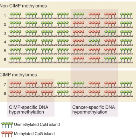
In 1999, Issa and colleagues first described a distinct subset of human colorectal cancers with extensive DNA hypermethylation of a subset of CpG islands that remained unmethylated in the remaining colorectal tumors (30) and are therefore distinguished from general cancer-specific DNA methylation for a specific tumor type (Figure 1). These tumors were classified as positive for a CIMP. TCGA Research Network and others have identified CIMPs in breast, colorectal, and endometrial tumors as well as in glioblastomas and acute myeloid leukemias, but not in serous ovarian, lung squamous, or kidney renal cell cancers. TCGA has unveiled similar and unique characteristics between CIMPs of dif-ferent tumor types that have potential implications for the devel-opment of novel cancer diagnostics and therapeutic agents.
Colorectal CIMP is tightly associated with the BRAFV600E mutation.
Colorectal CIMP has been identified and characterized using can-didate and genome-scale approaches in numerous reports after Issa first identified CIMP in colorectal cancer (30). Colorectal CIMP tightly associates with mutation of the v-raf murine sar-coma viral oncogene homolog B (BRAF) gene correlating with
Conflict of interest: Daniel J. Weisenberger is a consultant for Zymo Research Corporation, which distributes commercially available products for DNA methyla-tion–based experiments. Zymo Research did not fund or in any way contribute to this Review.
review series
the V600 amino acid (BRAFV600E), DNA methylation of the mutL
homolog 1 (MLH1) promoter region, microsatellite instability (MSI), location in the proximal colonic region, and female gender (31–35). Ogino et al. first described a CIMP-low (CIMP-L) sub-group as having an attenuated CIMP phenotype, and showed an association with mutations in the kirsten rat sarcoma viral onco-gene homolog (KRAS) gene rather than BRAF (36). Similarly, Shen et al. identified the CIMP2 subgroup as displaying CIMP-associ-ated DNA methylation, with enrichment in KRAS mutations (32).
KRAS mutations are also enriched in non-CIMP tumors, but are strikingly absent in CIMP-high (CIMP-H) tumors (34–37). TCGA confirmed CIMP-H, CIMP-L, and two non-CIMP subgroups of colorectal cancer (34, 35). CIMP-H was present in approximate-ly 15% of colorectal tumors, the majority of which also showed elevated mutation rates (hypermutated) and showed few SCNAs in contrast to the majority of colorectal tumors, which are non-CIMP, non-hypermutated, and microsatellite stable but which show substantial SCNAs (34).
The molecular mechanisms that explain the tight correlation between CIMP-H and the BRAFV600E mutation are not well
under-stood. TCGA did not identify driver events, such as specific muta-tions or SCNAs in a trans-acting factor in CIMP-H and BRAFV600E
colorectal tumors. While both the BRAFV600E mutation and DNA
methylation changes are thought to occur early in colorectal tumor-igenesis, it is unclear whether CIMP or BRAFV600E mutation is the
initiating event. Hinoue and colleagues did not observe CIMP after introducing exogenous BRAFV600E into a wild-type BRAF, non-CIMP
colorectal cancer cell line (38). However, Hinoue et al. did identi-fy CIMP-specific epigenetic silencing of IGF-binding protein 7 (IGFBP7), which mediates BRAFV600E-induced apoptosis and cellular
senescence. As BRAFV600E has been implicated in oncogene-induced
senescence in melanomas and colorectal cancers (39), CIMP-specific silencing of IGFBP7 may create a favorable context for the genera-tion of the BRAFV600E mutation in CIMP-positive tumors (38).
How-ever, additional experiments are needed to determine the molecular mechanism linking CIMP and BRAFV600E in colorectal cancer.
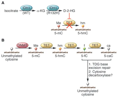
[image:3.585.53.526.111.457.2]Isocitrate dehydrogenase mutations and DNA hypermethylation in glioma and AML. Glioblastoma multiforme (GBM) was the first cancer selected by TCGA Research Network for molecular characteriza-tion (40). The majority of GBM tumors are considered primary or de novo, and approximately 5% are secondary GBMs which progress from lower-grade tumors. Parsons and colleagues used a compre-hensive sequencing strategy and identified specific heterozygous somatic point mutations in the isocitrate dehydrogenase 1 (IDH1) Table 1
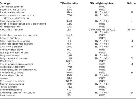
Tumors selected by TCGA for genomic characterization
Tumor type TCGA abbreviation DNA methylation platform Reference
Adrenocortical carcinoma ACC HM450
Bladder urothelial carcinoma BLCA HM450
Breast invasive carcinoma BRCA HM27, HM450 78
Cervical squamous cell carcinoma and CESC HM27, HM450 endocervical adenocarcinoma
Colon adenocarcinoma COAD HM27, HM450 34
Lymphoid neoplasm diffuse large B cell lymphoma DLBC HM450
Esophageal carcinoma ESCA HM450
Glioblastoma multiforme GBM GG OMA-002, GG OMA-003, 40, 44–46
HM27, HM450
Head and neck squamous cell carcinoma HNSC HM450
Kidney chromophobe KICH HM450
Kidney renal clear cell carcinoma KIRC HM27, HM450 86
Kidney renal papillary cell carcinoma KIRP HM27, HM450
Acute myeloid leukemia LAML HM27, HM450 75
Brain lower grade glioma LGG HM450
Liver hepatocellular carcinoma LIHC HM450
Lung adenocarcinoma LUAD HM27, HM450
Lung squamous cell carcinoma LUSC HM27, HM450 83
Mesothelioma MESO HM450
Ovarian serous cystadenocarcinoma OV HM27 82
Pancreatic adenocarcinoma PAAD HM450
Pheochromocytoma and paraganglioma PCPG HM450
Prostate adenocarcinoma PRAD HM450 34
Rectum adenocarcinoma READ HM27, HM450
Sarcoma SARC HM450
Skin cutaneous melanoma SKCM HM450
Stomach adenocarcinoma STAD HM27, HM450
Thyroid carcinoma THCA HM450
Uterine carcinosarcoma UCS HM450
Uterine corpus endometrial carcinoma UCEC HM27, HM450 80
Uveal melanoma UVM HM450
gene (most often at amino acid residue R132) in 12% of GBM patients (41). Somatic mutations in IDH2 (amino acid R172) were also identified in gliomas. These mutations occur at a low frequency and are mutually exclusive with IDH1 mutations (42, 43).
TCGA Research Network sequenced 601 candidate genes for somatic mutations but did not sequence IDH1 or IDH2 in their initial report (40); however, in a recent follow-up report, TCGA identified IDH1 mutations through whole exome and whole genome sequencing approaches (44). In 2010, Noushmehr and TCGA network colleagues (45) convincingly showed an extremely tight correlation between GBM tumors with the IDH1R132H
muta-tion and a glioma CIMP (G-CIMP). All primary GBM tumors with an IDH1 mutation were G-CIMP, but there were also a very small number of WT IDH1 (IDH1WT) G-CIMP tumors. G-CIMP tumors
also showed attenuated SCNAs and TP53 alterations and corre-lated with younger patient age and improved survival. Moreover, G-CIMP is highly prevalent in recurrent and secondary GBM tumors and is inversely correlated with glioma stage (45).
Brennan and TCGA colleagues (44) confirmed the G-CIMP sub-group and its association with IDH1 and TP53 alterations. Includ-ing the G-CIMP subgroup, TCGA identified a total of six DNA methylation subgroups using unsupervised clustering analyses and showed enrichment of some individual DNA methylation groups with gene expression–based subgroups (neural, proneu-ral, mesenchymal, and classical) identified previously (46), with G-CIMP tumors tightly associated with the proneural subgroup. Interestingly, one subgroup (cluster M6) was generally DNA hypo-methylated and enriched for proneural non–G-CIMP tumors with
IDH1WT. G-CIMP patients displayed longer survival times, whereas
non–G-CIMP patients had shorter survival outcomes. Interesting-ly, even though the M6 tumors were proneural, patients with this tumor type did not show survival advantages, which suggests that the aberrant molecular features of G-CIMP may be important in conferring the survival advantage in G-CIMP patients.
Because of the expanded GBM tumor collection and exome sequencing depth, TCGA identified amplifications of MYC (v-myc avian myelocytomatosis viral oncogene homolog) in G-CIMP tumors. MYC is a transcription factor that is frequently altered in cancer and is involved in cell cycle progression, transformation, and apoptosis (reviewed in ref. 47). In addition, TCGA also identi-fied ATRX (α-thalassemia/mental retardation syndrome X-linked) somatic mutations in G-CIMP tumors, which are highly correlated with IDH1 mutations. ATRX belongs to the SWI/SNF family of chromatin remodelers and functions as an ATPase and helicase that facilitates the substitution of variant histone H3.3 into chro-matin at telomeres (48).
ATRX mutations are predominant in the alternative lengthening of telomeres (ALT), a process by which telomere length is main-tained independent of telomerase in cancer cells (48, 49). Interest-ingly, TCGA identified telomerase (TERT) promoter mutations in 21 of 25 GBM tumors sequenced, and these mutations correlated with increased TERT gene expression. Notably, all four tumors with-out TERT mutations did not display elevated TERT gene expression, but instead contained ATRX alterations. Therefore, it is possible that GBM tumors maintain telomere length by either TERT mutations to reactivate TERT gene expression or via ATRX mutations in ALT.
The link between the IDH1R132H mutation and G-CIMP has
gener-ated tremendous attention from basic and translational scientists. Recent studies have shown that introducing exogenous IDH1R132H
into immortalized cell lines with endogenous IDH1WT was sufficient
to drive G-CIMP–based DNA methylation events and increased occu-pancy of histone H3 lysine 9 dimethylation (H3K9me2), histone H3 lysine 27 trimethylation (H3K27me3), and H3K36me3, which cor-relate with methylated DNA regions in the cancer genome (50, 51).
These epigenomic changes may occur as a result of the func-tion of the mutant IDH proteins. IDH1WT functions as a dimer
to catalyze the reduction of nicotinamide adenine dinucleotide phosphate (NADP+) to NADPH by converting of isocitrate to
α-ketoglutarate (α-KG) (reviewed in ref. 52). However, the IDH mutant catalyzes the conversion of α-KG to D-2-hydroxygluta-rate (2-HG) (53–55), resulting in elevated 2-HG levels (53, 56, 57). IDH1 mutant-mutant (IDH1MUT-MUT) homodimers or
mutant-WT (IDH1MUT-WT) heterodimers have been identified in vitro (58).
However, recent in vitro experiments have shown that the presence of the IDH1WT protein is associated with increased 2-HG levels
(54), and that IDH1MUT-WT dimers show more enzymatic activity
toward α-KG than IDH1MUT-MUT alone (59), suggesting that both
forms may be required for 2-HG production.
2-HG inhibits the TET family of enzymes and Jumonji-C domain containing histone lysine demethylases, which normally utilize
[image:4.585.47.284.82.322.2]α-KG as a co-substrate (60, 61). Thus, the production of 2-HG by mutant IDH1-containing enzymes effectively inhibits TET activ-ity (Figure 2A). TETs catalyze the oxidation of 5-methylcytosine (5mC) to 5-hydroxymethylcytosine (5hmC) and ultimately to 5-formylcytosine (5fC) and 5-carboxylcytosine (5caC) (62, 63). 5fC and 5caC are substrates for the thymine DNA glycosylase-mediat-ed base excision repair pathway that ultimately results in replace-ment with an unmethylated cytosine, effectively demethylating the locus (64–67) (Figure 2B).
Figure 1
review series
AML tumors also harbor IDH1 mutations (mostly at the R132 residue), IDH2 mutations (at residues R140 and R172) (68), and
TET mutations. IDH1 and IDH2 mutations are mutually exclu-sive and occur in up to 30% of acute myeloid leukemias (69–74). Moreover, TET2 mutations are also mutually exclusive with IDH
mutations. Figueroa and colleagues showed that AML tumors with
TET2 mutations displayed a DNA hypermethylation signature that is similar to that of AML tumors with IDH mutations (69), suggest-ing that IDH and TET enzymes may have redundant roles in DNA demethylation. TCGA also demonstrated that AML tumors with
IDH somatic mutations showed substantial gains in DNA methyla-tion, and similarly, in TET2-mutated AML tumors (75).
The inhibition of DNA demethylation as a result of the IDH1
and TET mutations is consistent with the epigenomic landscapes identified in G-CIMP and AML tumors, but the basis for the target site specificity of the cancer-associated DNA methylation events observed in IDH1 mutant tumors is unclear. Two recent studies have identified IDAX (inhibition of the Dvl and Axin complex, also known as CXXC4) and early B cell factor 1 (EBF1) as potential TET2-interac-tion partners. IDAX can bind unmethylated promoter CpG islands as well as the TET2 catalytic domain, resulting in decreased 5hmC levels and TET2 degradation via caspase activation. EBF1, by binding to both DNA and TET2, may also regulate DNA demethylation in
IDH1-mutant cancers in a tissue- and sequence-specific manner (76).
Clinical importance of MGMT DNA methylation in GBM. The stan-dard of care chemotherapeutic agent for treating GBM patients is temozolomide (TMZ), which acts as a methyl donor for alkylation of the N-7, O-3, and O-6 positions of nucleotide bases (reviewed in ref. 77). TMZ treatment initiates a DNA repair response, but it is believed that the mismatch repair machinery cannot effec-tively incorporate the correct base opposite to O-6-methylguanine
lesions after the initial DNA strand-nicking step in the repair path-way. The nicks accumulate and are thought to promote an apop-totic response that results in cell death.
O-6-methylguanine methyltransferase (MGMT) removes methyl groups from the O-6 position of guanines, thereby rendering TMZ ineffective. Promoter DNA hypermethylation–based silencing of
MGMT sensitizes GBM tumors to TMZ, and as a result has been used as a diagnostic barometer for selecting TMZ as a treatment option for GBM patients. TCGA recently identified MGMT DNA methyla-tion in nearly 50% of GBM patients, and MGMT DNA methylation was more prevalent in G-CIMP tumors than in non–G-CIMP tumors (44). MGMT DNA methylation correlated with patient response in GBMs belonging to the classical gene expression subgroup only, and not in the proneural, neural, or mesenchymal groups.
Breast cancer CIMP. Breast cancer is a complex and heteroge-neous disease, and breast tumor subgroups have been proposed based on the expression status of estrogen receptor (ER), proges-terone receptor (PR), and human epidermal growth factor recep-tor 2 (HER2; also known as ERBB2). TCGA identified five DNA methylation subgroups of breast tumors (78), with one subgroup exhibiting a CIMP-like DNA methylation signature (B-CIMP), as previously described by Fang and colleagues (79). B-CIMP tumors were positive for ER, PR, and HER2 expression and were enriched for the luminal B gene expression subgroup as well as epigenetic silencing of genes in the Wnt-signaling pathway (78), as has also been described for colorectal tumors (34).
Endometrial carcinoma CIMP. Endometrial tumors are classified into two groups: the type I endometrioid tumors that are hormone receptor positive with good prognosis and the type II serous tumors that mostly occur in older women and correlate with poor out-come. TCGA identified four DNA methylation subgroups of endo-metrial tumors (80), with one subgroup displaying a CIMP-like (E-CIMP) DNA hypermethylation profile. E-CIMP was previously identified by Whitcomb and colleagues (81). Similar to colorec-tal CIMP tumors, the E-CIMP tumors were hypermutated, MSI positive due to MLH1 promoter DNA hypermethylation, and did not contain TP53 somatic mutations or extensive SCNAs (34, 80). However, E-CIMP tumors did not harbor BRAFV600E or IDH1
mutations, as described in colorectal and glioma CIMP tumors, respectively, pointing to an alternative mechanism of CIMP-spe-cific DNA methylation in endometrial tumors.
Similarities and differences among CIMPs of individual human cancers
TCGA has confirmed the presence of CIMP in colorectal, breast, and endometrial cancers, while providing the first comprehensive view of G-CIMP. However, the molecular events that result in CIMP-specific DNA methylation events for each tumor type remain unclear and suggest that universal presentation of CIMP is not apparent across tumor types. For instance, colorectal CIMP is associated with
BRAFV600E mutation, but G-CIMP is tightly linked to IDH1 mutations.
However, B-CIMP and E-CIMP tumors are not associated with these gene mutations. Moreover, colorectal and endometrial CIMPs are associated with MSI via epigenetic silencing of MLH1, but this was not identified in CIMPs from other tumor types. Therefore, CIMP-specific DNA methylation targets may be largely non-overlapping with no consensus CIMP DNA methylation signature across tumor types, suggesting that CIMPs may be manifested by several molecular pathways and diverse sets of genomic alterations. However, formal analyses to determine the extent of common and unique CIMP-spe-Figure 2
DNA demethylation dynamics in human cancers. (A) Role of IDH1 in shaping the cancer methylome. WT IDH1 converts isocitrate to α-KG, but the mutant IDH1R132H enzyme catalyzes the conversion α-KG to
[image:5.585.43.284.82.285.2]cific DNA hypermethylation signatures between tumor types are still needed and are currently in progress by TCGA researchers.
Normal-like DNA methylation subgroups of human cancers
Unsupervised analyses of TCGA breast and endometrial cancers each identified subgroups of tumors with normal-like DNA meth-ylation profiles. In breast cancers, this subgroup showed enrich-ment with the basal-like (triple-negative) gene expression group, in which ER, PR, and HER2 were not expressed. In endometrial tumors, the normal-like DNA methylation subgroup was enriched in the serous-type tumors. Both subgroups displayed TP53 somat-ic mutations and extensive SCNAs and overall were similar to serous ovarian tumors (78, 80, 82). These findings indicate that these subtypes may share a common mechanism of epigenomic changes in tumorigenesis separate from their CIMP counterparts. CIMP-positive tumors generally present with genomic stability and the absence of TP53 mutations, with the exception of G-CIMP tumors, which show enrichment for TP53 mutations.
CDKN2A inactivation in lung squamous cell carcinoma
TCGA Research Network has recently reported the integrated genomic characterization findings of lung squamous cell carcino-ma (LUSC) (83). Overall, LUSCs displayed increased rates of DNA sequence alterations compared with other tumor types. Among the genes that were significantly mutated, CDKN2A alterations are of interest. The CDKN2A locus encodes for two well-characterized cell cycle–regulating proteins, p16INK4A and p14ARF, that are
encod-ed by alternate splicing of an overlapping set of exons in the locus. p16INK4A inactivates cyclin-dependent kinases that phosphorylate
the retinoblastoma protein, resulting in a G1 phase arrest, while p14ARF stabilizes p53 by inducing MDM2 degradation and
block-ing its function (84). TCGA showed that CDKN2A inactivation occurs in over 70% of LUSCs by a mutually exclusive combination of epigenetic silencing, inactivating mutations and deletions, with each contributing to CDKN2A gene expression alterations, sug-gesting that multiple routes of CDKN2A inactivation are impor-tant to LUSC tumorigenesis.
BRCA1 epigenetic silencing in serous ovarian cancers
Integrative analyses of DNA methylation and gene expression data identified 168 epigenetically silenced genes in TCGA serous ovarian tumors (82). These genes display promoter DNA hyper-methylation together with reduced gene expression. Among those is BRCA1, an important driver of carcinogenesis that encodes for a protein involved in repairing DNA double-strand breaks. Interest-ingly, TCGA demonstrated that BRCA1 was inactivated by muta-tion and epigenetic silencing in a mutually exclusive manner in ovarian tumors (82). Although neither BRCA1 mutation nor epi-genetic silencing correlated with prognosis (85), BRCA1 epigenetic silencing was more abundant than somatic mutations in ovarian tumors, and patients with ovarian cancer with BRCA1 epigenetic silencing were younger than those with BRCA1 mutation inactiva-tion, suggesting that epigenetic alterations of BRCA1 are impor-tant early events in ovarian tumorigenesis.
Chromatin modifier gene mutations affect the cancer methylome
Somatic mutations of chromatin-modifier genes have been report-ed in several human cancers (reviewreport-ed in ref. 5). While the effects
of these mutations on the cancer methylome have not been com-pletely explored, TCGA AML tumors displayed substantial levels of mutations in chromatin-modifier genes, with 30% of AML tumors harboring somatic mutations in chromatin modifier genes and 44% of tumors with mutations in DNA methylation–related genes. AML tumors with fusion events involving the myeloid/ lymphoid or mixed-lineage leukemia (MLL) gene family or concor-dant mutations in nucleophosmin (NPM1), fms-related tyrosine kinase (FLT3), and DNA (cytosine-5-)-methyltransferase 3 alpha (DNMT3A) displayed DNA hypomethylation (75). The MLL gene family encodes histone lysine methyltransferases, and DNMT3A
codes for a de novo DNA methyltransferase. These findings point to a strong link between cancer genetics and epigenetics that will be better understood by forthcoming mechanistic studies.
TCGA also identified somatic mutations of SET domain con-taining 2 (SETD2) in 11.5% of KIRC tumors (86). SETD2 is a H3K36 methyltransferase, and H3K36me3 marks are associated with DNA methylation at actively transcribed gene regions (87). Recent work from Chantalat and colleagues has also identified H3K36me3 marks in facultative and constitutive heterochromatin (88). TCGA demonstrated that KIRC tumors with SETD2 muta-tions displayed DNA hypomethylation at non-promoter regions that are marked by H3K36me3 and DNA hypermethylation at non-H3K36me3 occupied regions in normal kidney cells, suggest-ing a crosstalk between DNA methylation and H3K36 methylation in shaping the epigenome.
Future directions
Several interesting applications utilizing TCGA DNA methyla-tion data are possible. For instance, TCGA data can be analyzed to delineate between driver and passenger DNA methylation events in carcinogenesis. Also of interest is determining an understanding of DNA methylation changes across several cancer types, not only in relation to CIMP, but also in identifying specific cancer-associated DNA methylation alterations for potential diagnostic purposes. Such DNA methylation–based biomarkers of disease can be used to identify primary tumors as belonging to specific subgroups and may have utility in personalized medicine. In addition, DNA meth-ylation biomarkers can be used as tools for early detection of cancer in patient blood and a mechanism to track response to therapy.
TCGA has been and will continue to be a widely used discovery and validation resource for other genomics-based projects. TCGA will also continue to inspire the development of novel informatics approaches for integrative, high-level summaries of a wide range of molecular data types. With the tremendous volume of genomic data generated by TCGA efforts, functional experiments are needed to fully characterize cancer genomes. Currently, TCGA Research Net-work is completing genomic characterizations across several cancers to identify epigenomic signatures that will provide a powerful view into the molecular events that shape human carcinogenesis. Acknowledgments
I thank Peter A. Jones and Peter W. Laird for helpful discussions and constructive comments.
review series
1. Bibikova M, et al. High density DNA methylation array with single CpG site resolution. Genomics. 2011;98(4):288–295.
2. Bibikova M, Fan JB. GoldenGate assay for DNA methylation profiling. Methods Mol Biol. 2009; 507:149–163.
3. Bibikova M, et al. Genome-wide DNA methylation profiling using Infinium(R) assay. Epigenomics. 2009; 1(1):177–200.
4. Jones PA, Baylin SB. The epigenomics of cancer.
Cell. 2007;128(4):683–692.
5. Shen H, Laird PW. Interplay between the cancer genome and epigenome. Cell. 2013;153(1):38–55. 6. Jones PA. Functions of DNA methylation: islands,
start sites, gene bodies and beyond. Nat Rev Genet. 2012;13(7):484–492.
7. Bird A, Taggart M, Frommer M, Miller OJ, Macleod D. A fraction of the mouse genome that is derived from islands of nonmethylated, CpG-rich DNA.
Cell. 1985;40(1):91–99.
8. Ehrlich M, Wang RY. 5-Methylcytosine in eukary-otic DNA. Science. 1981;212(4501):1350–1357. 9. Diala ES, Hoffman RM. Hypomethylation of
HeLa cell DNA and the absence of 5-methylcyto-sine in SV40 and adenovirus (type 2) DNA: analy-sis by HPLC. Biochem Biophys Res Commun. 1982; 107(1):19–26.
10. Diala ES, Hoffman RM. DNA methylation levels in normal and chemically-transformed mouse 3T3 cells. Biochem Biophys Res Commun. 1982; 104(4):1489–1494.
11. Ehrlich M, et al. Amount and distribution of 5-methylcytosine in human DNA from different types of tissues of cells. Nucleic Acids Res. 1982; 10(8):2709–2721.
12. Feinberg AP, Vogelstein B. Hypomethylation distin-guishes genes of some human cancers from their normal counterparts. Nature. 1983;301(5895):89–92. 13. Gama-Sosa MA, et al. The 5-methylcytosine content
of DNA from human tumors. Nucleic Acids Res. 1983; 11(19):6883–6894.
14. Feinberg AP, Gehrke CW, Kuo KC, Ehrlich M. Reduced genomic 5-methylcytosine content in human colonic neoplasia. Cancer Res. 1988; 48(5):1159–1161.
15. Weisenberger DJ, et al. Analysis of repetitive ele-ment DNA methylation by MethyLight. Nucleic Acids Res. 2005;33(21):6823–6836.
16. Berman BP, et al. Regions of focal DNA hyper-methylation and long-range hypohyper-methylation in colorectal cancer coincide with nuclear lamina-associated domains. Nat Genet. 2012;44(1):40–46. 17. Hansen KD, et al. Increased methylation variation
in epigenetic domains across cancer types. Nat Genet. 2011;43(8):768–775.
18. Hon GC, et al. Global DNA hypomethylation cou-pled to repressive chromatin domain formation and gene silencing in breast cancer. Genome Res. 2012; 22(2):246–258.
19. Lister R, et al. Human DNA methylomes at base resolution show widespread epigenomic differ-ences. Nature. 2009;462(7271):315–322. 20. Jones PA, Baylin SB. The fundamental role of
epigenetic events in cancer. Nat Rev Genet. 2002; 3(6):415–428.
21. Irizarry RA, et al. The human colon cancer methy-lome shows similar hypo- and hypermethylation at conserved tissue-specific CpG island shores. Nat Genet. 2009;41(2):178–186.
22. Baylin SB. DNA methylation and gene silencing in cancer. Nat Clin Pract Oncol. 2005;2(suppl 1):S4–S11. 23. Suzuki H, et al. Epigenetic inactivation of SFRP
genes allows constitutive WNT signaling in colorectal cancer. Nat Genet. 2004;36(4):417–422. 24. Herman JG, Graff JR, Myohanen S, Nelkin BD,
Baylin SB. Methylation-specific PCR: a novel PCR assay for methylation status of CpG islands. Proc Natl Acad Sci U S A. 1996;93(18):9821–9826.
25. Hilton JL, Geisler JP, Rathe JA, Hattermann-Zogg MA, DeYoung B, Buller RE. Inactivation of BRCA1 and BRCA2 in ovarian cancer. J Natl Cancer Inst. 2002;94(18):1396–1406.
26. Ohm JE, et al. A stem cell-like chromatin pattern may predispose tumor suppressor genes to DNA hypermethylation and heritable silencing. Nat Genet. 2007;39(2):237–242.
27. Schlesinger Y, et al. Polycomb-mediated methyla-tion on Lys27 of histone H3 pre-marks genes for de novo methylation in cancer. Nat Genet. 2007; 39(2):232–236.
28. Widschwendter M, et al. Epigenetic stem cell signa-ture in cancer. Nat Genet. 2007;39(2):157–158. 29. Lee TI, et al. Control of developmental regulators
by Polycomb in human embryonic stem cells. Cell. 2006;125(2):301–313.
30. Toyota M, Ahuja N, Ohe-Toyota M, Herman JG, Baylin SB, Issa JP. CpG island methylator phenotype in colorectal cancer. Proc Natl Acad Sci U S A. 1999; 96(15):8681–8686.
31. Ogino S, Kawasaki T, Ogawa A, Kirkner GJ, Loda M, Fuchs CS. Fatty acid synthase overexpression in colorectal cancer is associated with microsatellite instability, independent of CpG island methylator phenotype. Hum Pathol. 2007;38(6):842–849. 32. Shen L, et al. Integrated genetic and
epigen-etic analysis identifies three different subclasses of colon cancer. Proc Natl Acad Sci U S A. 2007; 104(47):18654–18659.
33. Yagi K, et al. Three DNA methylation epigenotypes in human colorectal cancer. Clin Cancer Res. 2010; 16(1):21–33.
34. Cancer Genome Atlas Network. Comprehensive molecular characterization of human colon rectal cancer. Nature. 2012;487(7407):330–337. 35. Hinoue T, et al. Genome-scale analysis of aberrant
DNA methylation in colorectal cancer. Genome Res. 2012;22(2):271–282.
36. Ogino S, Kawasaki T, Kirkner GJ, Loda M, Fuchs CS. CpG island methylator phenotype-low (CIMP-low) in colorectal cancer: possible associations with male sex and KRAS mutations. J Mol Diagn. 2006; 8(5):582–588.
37. Weisenberger DJ, et al. CpG island methylator phe-notype underlies sporadic microsatellite instability and is tightly associated with BRAF mutation in colorectal cancer. Nat Genet. 2006;38(7):787–793. 38. Hinoue T, et al. Analysis of the association between
CIMP and BRAF in colorectal cancer by DNA methylation profiling. PLoS One. 2009;4(12):e8357. 39. Wajapeyee N, Serra RW, Zhu X, Mahalingam M,
Green MR. Oncogenic BRAF induces senescence and apoptosis through pathways mediated by the secreted protein IGFBP7. Cell. 2008;132(3):363–374. 40. Cancer Genome Atlas Research Network. Compre-hensive genomic characterization defines human glioblastoma genes core pathways. Nature. 2008; 455(7216):1061–1068.
41. Parsons DW, et al. An integrated genomic analysis of human glioblastoma multiforme. Science. 2008; 321(5897):1807–1812.
42. Hartmann C, et al. Type and frequency of IDH1 and IDH2 mutations are related to astrocytic and oligodendroglial differentiation and age: a study of 1,010 diffuse gliomas. Acta Neuropathol. 2009; 118(4):469–474.
43. Yan H, et al. IDH1 and IDH2 mutations in gliomas.
N Engl J Med. 2009;360(8):765–773.
44. Brennan CW, et al. The somatic genomic landscape of glioblastoma. Cell. 2013;155(2):462–477. 45. Noushmehr H, et al. Identification of a CpG island
methylator phenotype that defines a distinct sub-group of glioma. Cancer Cell. 2010;17(5):510–522. 46. Verhaak RG, et al. Integrated genomic analysis
iden-tifies clinically relevant subtypes of glioblastoma characterized by abnormalities in PDGFRA, IDH1, EGFR, and NF1. Cancer Cell. 2010;17(1):98–110.
47. Hanahan D, Weinberg RA. Hallmarks of cancer: the next generation. Cell. 2011;144(5):646–674. 48. Goldberg AD, et al. Distinct factors control histone
variant H3.3 localization at specific genomic regions. Cell. 2010;140(5):678–691.
49. Lovejoy CA, et al. Loss of ATRX, genome instabil-ity, and an altered DNA damage response are hall-marks of the alternative lengthening of telomeres pathway. PLoS Genet. 2012;8(7):e1002772. 50. Turcan S, et al. IDH1 mutation is sufficient to
establish the glioma hypermethylator phenotype.
Nature. 2012;483(7390):479–483.
51. Duncan CG, et al. A heterozygous IDH1R132H/WT mutation induces genome-wide alterations in DNA methylation. Genome Res. 2012;22(12):2339–2355. 52. Cohen AL, Holmen SL, Colman H. IDH1 and
IDH2 mutations in gliomas. Curr Neurol Neurosci Rep. 2013;13(5):345.
53. Dang L, et al. Cancer-associated IDH1 muta-tions produce 2-hydroxyglutarate. Nature. 2009; 462(7274):739–744.
54. Jin G, et al. Disruption of wild-type IDH1 sup-presses D-2-hydroxyglutarate production in IDH1-mutated gliomas. Cancer Res. 2013;73(2):496–501. 55. Ward PS, et al. The common feature of leukemia-associated IDH1 and IDH2 mutations is a neo-morphic enzyme activity converting alpha-keto-glutarate to 2-hydroxyalpha-keto-glutarate. Cancer Cell. 2010; 17(3):225–234.
56. Gross S, et al. Cancer-associated metabolite 2-hydroxyglutarate accumulates in acute myelog-enous leukemia with isocitrate dehydrogenase 1 and 2 mutations. J Exp Med. 2010;207(2):339–344. 57. Sellner L, et al. Increased levels of 2-hydroxyglu-tarate in AML patients with IDH1-R132H and IDH2-R140Q mutations. Eur J Haematol. 2010; 85(5):457–459.
58. Jin G, et al. 2-hydroxyglutarate production, but not dominant negative function, is conferred by glioma-derived NADP-dependent isocitrate dehy-drogenase mutations. PLoS One. 2011;6(2):e16812. 59. Bralten LB, et al. IDH1 R132H decreases prolifera-tion of glioma cell lines in vitro and in vivo. Ann Neurol. 2011;69(3):455–463.
60. Lu C, et al. IDH mutation impairs histone demeth-ylation and results in a block to cell differentiation.
Nature. 2012;483(7390):474–478.
61. Xu W, et al. Oncometabolite 2-hydroxyglutarate is a competitive inhibitor of alpha-ketogluta-rate-dependent dioxygenases. Cancer Cell. 2011; 19(1):17–30.
62. Ito S, et al. Tet proteins can convert 5-methylcyto-sine to 5-formylcyto5-methylcyto-sine and 5-carboxylcyto5-methylcyto-sine.
Science. 2011;333(6047):1300–1303.
63. Booth MJ, et al. Quantitative sequencing of 5-meth-ylcytosine and 5-hydroxymeth5-meth-ylcytosine at single-base resolution. Science. 2012;336(6083):934–937. 64. Song CX, et al. Genome-wide Profiling of
5-formyl-cytosine reveals its roles in epigenetic priming. Cell. 2013;153(3):678–691.
65. Shen L, et al. Genome-wide analysis reveals TET- and TDG-dependent 5-methylcytosine oxidation dynamics. Cell. 2013;153(3):692–706.
66. Raiber EA, et al. Genome-wide distribution of 5-formylcytosine in embryonic stem cells is associ-ated with transcription and depends on thymine DNA glycosylase. Genome Biol. 2012;13(8):R69. 67. Hashimoto H, Hong S, Bhagwat AS, Zhang X,
Cheng X. Excision of 5-hydroxymethyluracil and 5-carboxylcytosine by the thymine DNA glycosylase domain: its structural basis and implications for active DNA demethylation. Nucleic Acids Res. 2012; 40(20):10203–10214.
68. Yang H, Ye D, Guan KL, Xiong Y. IDH1 and IDH2 mutations in tumorigenesis: mechanistic insights and clinical perspectives. Clin Cancer Res. 2012; 18(20):5562–5571.
mutations result in a hypermethylation phenotype, disrupt TET2 function, and impair hematopoietic differentiation. Cancer Cell. 2010;18(6):553–567. 70. Marcucci G, et al. IDH1 and IDH2 gene mutations
identify novel molecular subsets within de novo cytogenetically normal acute myeloid leukemia: a Cancer and Leukemia Group B study. J Clin Oncol. 2010;28(14):2348–2355.
71. Mardis ER, et al. Recurring mutations found by sequencing an acute myeloid leukemia genome.
N Engl J Med. 2009;361(11):1058–1066.
72. Paschka P, et al. IDH1 and IDH2 mutations are fre-quent genetic alterations in acute myeloid leuke-mia and confer adverse prognosis in cytogenetically normal acute myeloid leukemia with NPM1 muta-tion without FLT3 internal tandem duplicamuta-tion.
J Clin Oncol. 2010;28(22):3636–3643.
73. Tefferi A, et al. IDH1 and IDH2 mutation studies in 1473 patients with chronic-, fibrotic- or blast-phase essential thrombocythemia, polycythemia vera or myelofibrosis. Leukemia. 2010;24(7):1302–1309. 74. Wagner K, et al. Impact of IDH1 R132 mutations
and an IDH1 single nucleotide polymorphism in cytogenetically normal acute myeloid leukemia:
SNP rs11554137 is an adverse prognostic factor.
J Clin Oncol. 2010;28(14):2356–2364.
75. Cancer Genome Atlas Research Network. Genom-ic and epigenomGenom-ic landscapes of adult de novo acute myeloid leukemia. N Engl J Med. 2013; 368(22):2059–2074.
76. Guilhamon P, et al. Meta-analysis of IDH-mutant cancers identifies EBF1 as an interaction partner for TET2. Nat Commun. 2013;4:2166.
77. Friedman HS, Kerby T, Calvert H. Temozolomide and treatment of malignant glioma. Clin Cancer Res. 2000;6(7):2585–2597.
78. Cancer Genome Atlas Network. Comprehensive molecular portraits of human breast tumours.
Nature. 2012;490(7418):61–70.
79. Fang F, et al. Breast cancer methylomes establish an epigenomic foundation for metastasis. Sci Transl Med. 2011;3(75):75ra25.
80. Cancer Genome Atlas Research Network. Inte-grated genomic characterization of endometrial carcinoma. Nature. 2013;497(7447):67–73. 81. Whitcomb BP, Mutch DG, Herzog TJ, Rader JS,
Gibb RK, Goodfellow PJ. Frequent HOXA11 and THBS2 promoter methylation, and a methylator
phenotype in endometrial adenocarcinoma. Clin Cancer Res. 2003;9(6):2277–2287.
82. Cancer Genome Atlas Research Network. Integrat-ed genomic analyses of ovarian carcinoma. Nature. 2011;474(7353):609–615.
83. Cancer Genome Atlas Research Network. Compre-hensive genomic characterization of squamous cell lung cancers. Nature. 2012;489(7417):519–525. 84. Haber DA. Splicing into senescence: the curious
case of p16 and p19ARF. Cell. 1997;91(5):555–558. 85. Yang D, et al. Association of BRCA1 and BRCA2
mutations with survival, chemotherapy sensitivity, and gene mutator phenotype in patients with ovar-ian cancer. JAMA. 2011;306(14):1557–1565. 86. Cancer Genome Atlas Research Network.
Com-prehensive molecular characterization of clear cell renal cell carcinoma. Nature. 2013;499(7456):43–49. 87. Bannister AJ, Schneider R, Myers FA, Thorne AW,
Crane-Robinson C, Kouzarides T. Spatial distribu-tion of di- and tri-methyl lysine 36 of histone H3 at active genes. J Biol Chem. 2005;280(18):17732–17736. 88. Chantalat S, et al. Histone H3 trimethylation at lysine