listening to human biology and medicine
Don Ganem
J Clin Invest.
2010;
120(4)
:939-949.
https://doi.org/10.1172/JCI40567
.
The linkage of Kaposi sarcoma (KS) to infection by a novel human herpesvirus (Kaposi
sarcoma–associated herpesvirus [KSHV]) is one of the great successes of contemporary
biomedical research and was achieved by using advanced genomic technologies in a
manner informed by a nuanced understanding of epidemiology and clinical investigation.
Ongoing efforts to understand the molecular mechanisms by which KSHV infection
predisposes to KS continue to be powerfully influenced by insights emanating from the
clinic. Here, recent developments in KS pathogenesis are reviewed, with particular
emphasis on clinical, pathologic, and molecular observations that highlight the many
differences between this process and tumorigenesis by other oncogenic viruses.
Science in Medicine
Find the latest version:
It is now 15 years since the discovery by Yuan Chang, Patrick Moore, and their colleagues (1) of DNA from a novel herpesvirus in biopsy specimens of human Kaposi sarcoma (KS). That virus, now called KS-associated herpesvirus (KSHV) or human herpes-virus 8 (HHV-8), has since been cloned (2–4) and sequenced (2, 5), grown in culture (6), and extensively studied in vitro. Epidemio-logic studies (7, 8) provide strong evidence that infection by KSHV is required for KS tumorigenesis and further link the viral genome to at least two rare lymphoproliferative disorders: primary effusion lymphoma (PEL) and multicentric Castleman disease (MCD) (9). This review discusses the proposed mechanisms underlying the association of KSHV with KS, with particular emphasis on how they relate to the distinctive clinical and pathologic features of this unique human neoplasm.
The pathobiology of KS
It is a great misfortune that the term sarcoma was applied to the disease by Moritz Kaposi in the 19th century (10). The name implies a similarity of this entity to traditional mesenchymal tumors, but in fact the differences between KS and classical cancers outnumber their similarities. Such differences begin at the light microscope level: unlike most cancers, which are histologically monotonous clonal outgrowths of a single cell type, KS lesions display a remarkable diversity of cell types (11–14) whose propor-tions vary with the stage of the disease. The earliest recognizable foci of KS are the so-called patch lesions — these are not masses, but flat lesions in the dermis that display prominent numbers of inflammatory cells (T and B cells, monocytes) and abundant neovascularity, features as characteristic of granulation tissue as of cancer. Already at this stage, angiogenesis is so profound that the gross lesions are red to the naked eye. This is an important fact: neovascularity in KS begins prior to establishment of a mass, in contrast to classical cancers, in which angiogenesis only begins after proliferation results in outgrowing the antecedent vascular supply (leading to selection for upregulation of proangiogenesis
genes, termed the “angiogenic switch”) (15). Patch lesions do con- tain the elongated, spindle-shaped cells that will come to domi-nate the lesion at its later stages, but these so-called spindle cells are only one of many elements at this stage. With time, dermal KS progresses to the plaque stage — in which the lesion is more indu-rated, often edematous, and more intensely red or even violaceous in color. As spindle cell proliferation continues, the lesions prog-ress to the nodular stage, characterized by visible masses dominated by spindle cells but again accompanied by inflammatory cells and the continued elaboration of slit-like neovascular spaces (Figure 1). These new vessels, one of the histologic signatures of KS, are very abnormal and prone to leakage of fluid and extravasation of rbc, whose degeneration leads to phagocytosis and accumulation of hemosiderin-laden histiocytes. It is this extravasation of blood that gives the lesions their bruise-like purplish discoloration.
Because their proliferation leads to nodule formation, attention has long been focused on the spindle cell as the driver of KS patho-genesis — a notion that is in keeping with the fact that these cells are the principal target of KSHV infection in the lesion (16, 17) Spindle cells are clearly of endothelial origin, as they bear many markers of the endothelial lineage, including CD31, CD34, CD36, and factor XIII, and reactivity with the endothelial cell–specific mAb PAL-E (18, 19). But the pattern of marker expression by spindle cells is also somewhat heterogeneous. A minority of spindle cells, for example, bear markers typical of smooth muscle cells — prompting some to suggest that spindle cells may derive from primitive mesenchymal precursors of vascular elements (11, 12). Some years ago, Beckstead et al. (20) proposed that spindle cells derive from lymphatic rather than vascular endothelium, a view supported by the fact that KS never arises in compartments lacking lymphatics (e.g., the central nervous system). This view gained currency when spindle cells were found to express lymphatic-specific markers (e.g., podoplanin and the lymphatic vessel hyaluronan receptor LYVE-1), as well as the signaling machinery involved in lymphangiogenesis (VEGF-C and its receptor VEGFR3) (21–23). However, transcriptional profil-ing studies (24–26) indicate that KSHV infection of endothelium alters the pattern of endothelial marker expression in a way that confounds lineage assignment. When vascular endothelial cells are infected in culture, they upregulate several markers of the lym-phatic lineage (25, 26); conversely, infected lymphatic endothelial cells shift toward a more vascular-like transcript profile (24). While this reprogramming likely explains at least some of the pleiotropy
KSHV and the pathogenesis of Kaposi sarcoma:
listening to human biology and medicine
Don Ganem
Howard Hughes Medical Institute, G.W. Hooper Foundation, and Departments of Medicine and Microbiology, University of California, San Francisco.
The linkage of Kaposi sarcoma (KS) to infection by a novel human herpesvirus (Kaposi sarcoma–
associated herpesvirus [KSHV]) is one of the great successes of contemporary biomedical research
and was achieved by using advanced genomic technologies in a manner informed by a nuanced
understanding of epidemiology and clinical investigation. Ongoing efforts to understand the
molec-ular mechanisms by which KSHV infection predisposes to KS continue to be powerfully influenced
by insights emanating from the clinic. Here, recent developments in KS pathogenesis are reviewed,
with particular emphasis on clinical, pathologic, and molecular observations that highlight the
many differences between this process and tumorigenesis by other oncogenic viruses.
Conflict of interest: The author receives funds in return for service on the Scientific Advisory Boards for the Novartis Institute for Biomedical Research and 3V Biosci-ences Inc.
of marker expression in spindle cells, it also suggests that we may never be able to assign the exact endothelial lineage of spindle cells by marker studies alone.
Spindle cells are often referred to as the “malignant” cells of KS. But this designation is not strictly correct, and its continuing use largely reflects the absence of an alternative word to denote their centrality to KS pathogenesis. In fact, spindle cells have few proper-ties in common with malignantly transformed cells — they usually (27, 28) (though not invariably; ref. 29) lack clonality, even in well-developed lesions. They are typically diploid — a sharp contrast to classical cancers, which are usually strikingly aneuploid. When put into culture, most spindle cells fail to display another malignant phenotype: reduced dependence on extracellular growth factors. In fact, KS spindle cells display the opposite phenotype — exaggerated dependence on such factors. To date, the only reproducible way to grow such cells in culture has been to incubate them in condi-tioned medium from activated T cells (18, 19, 30), an environment laden with cytokines and growth factors. Moreover, cells prepared in this fashion do not display other experimental signatures of malignant transformation: they do not grow in soft agar and do not produce tumors in nude mice (18, 19, 31).
Everything we know about the clinical behavior of KS also sup-ports the distinction from traditional malignancy. KS occurs in two major forms — classical KS, which is unaffiliated with HIV infection, and AIDS-related KS. The two forms are histologically identical, and both are etiologically linked to antecedent KSHV infection (32). Clas-sical KS is typically an indolent disorder that is generally confined to the skin, especially that of the legs. The lesions progress very slowly, such that many patients require no therapy — indeed, cases of spon-taneous remission, though uncommon, have been well documented (33). When these lesions do progress, most such progression is local, with widespread dissemination being distinctly uncommon. Thus, classical KS is rarely life threatening (34). Although some traditional cancers can also be indolent, this relatively benign natural history is certainly compatible with the observation that spindle cells are not fully malignant by classical criteria.
AIDS-KS presents a more malign face — on the skin it can be widespread, involving large areas of the body surface, sometimes
in a symmetrical fashion (35). Local nodularity and edema can be marked and can be profoundly disfiguring. Life-threatening compli-cations arise from its propensity for visceral involvement, especially in the lungs (leading to respiratory failure) or the gastrointestinal tract (resulting in gastrointestinal bleeding). Interestingly, in AIDS-KS, the temporal pattern of occurrence of multifocal lesions is often not consistent with spread from a primary lesion, but rather sug-gests independent occurrences (multicentricity) (33). This inference has been supported by molecular analysis of KSHV genomes from KS lesions, which has affirmed that different lesions from the same patient often harbor genomes of differing terminal structure, sug-gesting they arose from independent infection events (28).
One other clinical aspect of KS is worth noting here — namely, its relationship to inflammatory states, both systemic and local. In the era prior to effective antiretroviral therapy, it was often noted that AIDS patients with stable KS who experienced a severe systemic infection would sometimes develop a florid worsening of KS during or after the infection. More telling still is the pro-pensity of KS to occur at local sites of inflammation (e.g., surgical incisions or sites of prior zoster eruptions) (36–38). These obser-vations suggest that an inflammatory microenvironment, which is always a part of KS histology, actually promotes the establish-ment or development of KS lesions — an idea that is strikingly concordant with the need for cytokine-rich media for the propa-gation of spindle cells in vitro.
From a pathogenetic viewpoint, it is helpful to think of KS as being composed of three parallel processes: proliferation (prin-cipally affecting spindle cells), inflammation, and angiogenesis. Unlike traditional cancer, which is predominantly a proliferative state driven by tumor cells that have achieved substantial autono-my and only later trigger inflammatory and angiogenic responses, KS is a disease in which all three processes participate simultane-ously from its inception and are continuously necessary for the lesion to progress. A useful synthesis envisions that spindle cells produce proinflammatory and proangiogenic factors that recruit inflammatory cells and neovascular elements; these in turn provide growth factors and other substances necessary for spindle cell sur-vival and proliferation (19, 39, 40). Unlike in traditional cancers, no one component of this triad is truly autonomous. Although this is an attractive formulation, it has been difficult to test experimen-tally, in no small measure owing to the absence of suitable animal models of KS. Nonetheless, this paradigm rationalizes most of the cardinal clinical and experimental observations made to date.
The etiologic role of KSHV
In the 1980s, the widespread occurrence of KS in AIDS patients initially suggested that HIV might be its proximate cause. But two facts soon put this idea to rest: (a) HIV proviral DNA was not pres-ent in the tumor; and (b) not all HIV-positive subjects were equally at risk of KS. KS risk was much greater in homosexual men with AIDS than in any other AIDS risk group (41). It soon became clear that sexual transmission of HIV was linked to much higher risk of KS than parenteral transmission of the virus, even though recipi- ents in both cases became equally immunodeficient. This sug-gested that, in addition to HIV infection, a second agent linked to sexual activity must be involved — the search for which led Chang, Moore, et al. to KSHV (1).
[image:3.585.71.258.87.216.2]The discovery of the KSHV genome allowed rapid development of both PCR tests for viral DNA and serologic tests for antiviral antibodies. This in turn made possible population-based studies
Figure 1
that soon delineated the key facts of KSHV epidemiology — all of which supported a central role for KSHV infection in KS devel-opment. The major pillars of this association have been reviewed elsewhere (8) and can be summarized as follows: (a) all KS lesions, whether HIV-positive or -negative, harbor KSHV DNA (42–44); (b) in KS tumors, KSHV infection specifically localizes to the spindle cells, the cell type whose proliferation is thought to drive KS histogenesis (16); (c) in any given locale, KSHV seroprevalence is high (30%–60%) in AIDS risk groups in which KS is frequent and low (2%–4%) in groups in which it is rare (45–47); (d) globally, KSHV prevalence mirrors the distribution of classical KS — high (15%–60%) in regions where classical KS is common (Southern Mediterranean and Africa) and low (1%–5%) in regions where clas-sical KS is rare (e.g., the United States) (7, 46); (e) KSHV infection precedes KS development (48) and prospectively predicts elevated KS risk (49); and (f) consonant with KS epidemiology, KSHV is sexually transmitted, with male homosexuals at especially high risk (49, 50). Taken together, these facts strongly imply that KSHV is the agent predicted by KS epidemiology and is necessary for KS development — KS is never observed in the absence of KSHV.
However, these facts also imply something equally important: that while necessary for KS development, KSHV infection is not sufficient for it. For example, although 1%–5% of the U.S. popula-tion is KSHV-seropositive, most of these individuals never develop overt KS. Population-based estimates suggest that even in endemic zones, only about 1 in 10,000 infected subjects will develop classi-cal KS (51–54). Clearly, additional events are necessary to trigger KS development. The identity of those additional events in classi- cal KS is unknown and represents one of the great unsolved prob-lems of KS research. In AIDS-KS, however, the second hit is clearly HIV. The magnitude of HIV’s contribution to KS risk is immense — 50% of dually infected men who receive no effective treatment for either HIV or KSHV will develop KS in a ten-year period (49). Further evidence that HIV infection is a central cofactor comes from the greater than 90% decline in incident KS in the United States and Europe in the era of highly active antiretroviral therapy (HAART) (55, 56). Moreover, established KS frequently goes into remission when AIDS-KS patients are treated with HAART (57–59). Given this, it seems a bit diminutive to refer to HIV as a mere “cofactor” — but formally speaking, that is its role.
The exact mechanisms by which HIV amplifies KS risk during
KSHV infection remain contentious. Certainly the immune defi-ciency state of advanced HIV disease is a major contributor to risk — a fact supported by the existence of transplant-associated KS, which arises from KSHV infection in the context of iatrogenic immune suppression (60). But there are other, more direct pos-sibilities. Laboratory experiments indicate that in certain settings, HIV infection can augment KSHV replication, in both cell-autono-mous (61, 62) and paracrine (63) fashions. However, the frequency of dual infection of cells in vivo by HIV and KSHV is very low (17), making a major contribution from cell-autonomous pathways unlikely. Paracrine pathways provide a more attractive mechanis-tic connection. Mercader et al. (63) found that cytokines, especially oncostatin M and IFN-γ , produced by HIV-infected cells can trig-ger lytic KSHV reactivation, which could foster dissemination of KSHV infection, thereby predisposing to KS. Barillari and Ensoli have proposed a different connection, pointing out that soluble HIV Tat protein can serve as a growth factor for cultured KS spin-dle cells in vitro (64). Sadly, absent a reliable animal model of KS, decisive in vivo tests of any of these ideas will be difficult.
KSHV virology: a primer
As found in the virion, KSHV DNA is a linear duplex of approxi-mately 165 kb; 140 kb of this DNA contains coding information, flanked on either side by tandem (terminal) repeats of 1.4 kb of highly GC-rich noncoding sequences (65, 66). In infected cell nuclei, the genome circularizes to form a covalently closed circular episome. The complete nucleotide sequence (2) of KSHV predicts the existence of at least 87 ORFs.
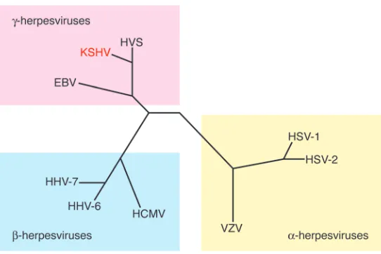
[image:4.585.46.317.81.262.2]Tropism. Phylogenetic analysis reveals that KSHV is a member of the lymphotropic (or γ) herpesvirus subfamily, whose prototype human member is EBV (Figure 2). Like EBV, KSHV’s primary tar-get cell is the B cell, and in healthy seropositive hosts viral DNA is principally found in this compartment (67). Evidence that KSHV also infects endothelium in vivo derives chiefly from the presence of viral DNA in KS spindle cells, and also in some cells lining the aberrant slit-like vessels of KS lesions (68, 69). One study has also sighted KSHV markers within monocytes infiltrating KS lesions (70). But overall, it bears emphasis that KSHV’s tropism in vivo is quite restricted. This is in sharp contrast to its behavior in vitro: KSHV will efficiently infect a wide variety of adherent human cells in culture, including epithelial cells, fibroblasts, and keratinocytes, as well as endothelial cells (71–73). Paradoxically, most established human B cell lines are not infectible, though recent studies show
Figure 2
that primary peripheral blood B cells can be infected in vitro if they are first activated by CD40 ligand and IL-4 (74). Why cell types that can be so readily infected in vitro are refractory in vivo is unknown, but this fact needs to be kept in mind when evaluating claims about KSHV tropism based solely on studies in culture.
Replication cycles. Like all herpesviruses, KSHV can express its genes in one of two alternative genetic programs, depending upon the circumstances of infection. The first of these, latency, is a state in which viral gene expression is sharply restricted, with only a handful of viral genes being stably expressed. In latent infection, the incoming linear viral genome circularizes in the nucleus, and the resulting large episome is autonomously maintained there, usually at low-moderate copy number. Because most viral genes are not expressed, there is no cytotoxicity and no virus is released. In KSHV, as in EBV, latency is the default pathway, at least in cul-ture. In most established cell lines in culture, the latent infection that follows KSHV exposure displays no evident phenotype (73). Similarly, primary B cells infected with KSHV become neither immortalized nor transformed (74, 75). This is in striking contrast to infection by EBV, whose latency program is powerfully immor-talizing in B cells (76). However, there is one cell type whose in vitro infection by KSHV does yield phenotypic consequences — pri-mary endothelial cells (Figure 3). When such cells are exposed to KSHV, striking morphologic changes occur, dominated by a rear-rangement of the actin cytoskeleton that produces an elongated morphology strongly reminiscent of that of the spindle cell (77, 78). However, even these cells are not immortalized, do not grow in soft agar, and do not form tumors in nude mice — although they do display heightened resistance to induction of apoptosis by growth factor withdrawal (79).
Importantly, latency is not irreversible. Because the full comple-ment of viral DNA is retained in the nucleus, under the appropriate circumstances the second program of viral gene expression, lytic replication, can be activated. In this program, expression of virtu-ally all viral genes is activated, in a temporally regulated cascade; infectious viral progeny are produced, and the infected cell is killed (6, 80). The physiologic signals that trigger lytic KSHV reactivation in vivo are unknown. We do know that periodic “spontaneous” reactivation from latency occurs regularly, both in cell culture (6)
and in vivo (72, 81, 82). In the human host, the principal site of lytic virus replication is the oropharynx, most likely in B cells of tonsillar or other pharyngeal lymphoid tissue, though growth in pharyngeal epithelium is another possibility (83). Careful clinical studies show that shedding of KSHV virions, reflecting periodic bouts of lytic reactivation, is intermittent and generally asymp-tomatic (81, 82). This biology underlies much of the epidemiology of KSHV, which is presumed to be driven by mucosal exposure to salivary virus, both in sexual transmission among adults (49) and in horizontal spread of virus among prepubertal children in the endemic zones of Africa and the Mediterranean basin (7).
How does KSHV infection predispose to KS?
For the virologist, this question reduces to: what KSHV genes are expressed in the tumor, and how do they act? Although this may seem a straightforward question, it is complicated by the fact that herpesviral genomes can be expressed via either latent or lytic pro-grams. Both programs are on display in KS tumors (17), especially those of patients with advanced HIV-induced immune deficiency. Most spindle cells in an advanced KS tumor are latently infected, but a small minority of cells express lytic markers (17, 68, 69, 84).
The bimodal expression program of the KSHV genome requires that, when considering the pathogenetic roles of viral genes, careful attention be paid to which program of viral gene expression governs transcription of that gene; failure to do so can invite misinterpreta-tion. For example, early findings that viral genes such as ORF-K9 (encoding an IRF-1 homolog; ref. 85) or ORF-74 (encoding a viral GPCR; ref. 86) could immortalize transfected mouse fibroblasts led to suggestions that they might drive proliferation in KS (85–88). However, the subsequent discovery that these genes appear to be expressed only during lytic growth (39, 89–91) rendered such roles implausible, since lytically infected cells invariably die. This is not to say that lytic genes can play no role in KS pathogenesis — in fact, considerable evidence is accumulating that they do (see Lytic genes and KS development, below). But because of the cell death induced by lytic replication, any contribution of lytic products to KS must be non–cell autonomous. Because of considerations such as these, in the following sections, the potential oncogenic contributions of latent- and lytic-cycle genes are discussed separately.
[image:5.585.62.259.83.224.2]Latent viral genes. The best characterized latent genes constitute a major latency locus that is transcribed in all latently infected cells (Figure 4). This region includes several ORFs, encoding the proteins latency-associated nuclear antigen (LANA), viral cyclin (v-cyclin), v–Flice-inhibitory protein (v-FLIP), and kaposins A, B, and C. The first three genes are under the control of a single promoter (the LANA promoter, or LTc), which generates a series of coterminal mRNAs via differential splicing (92–94). A second promoter (the kaposin promoter, or LTd) encodes a spliced tran- script encoding the kaposins (95–97) and can also generate a bicis-tronic RNA for v-cyclin and v-FLIP. This promoter also governs the expression of 12 pre-miRNAs (Figure 4), which can be processed to yield a total of 18 mature miRNAs (98–102). All of these latent products have been found to be expressed in KS spindle cells as well as PEL cells (103–105). A second locus, expressed in latent PEL cells, encodes the v-IRF3 (or LANA-2) protein, a member of the IRF superfamily that dominantly inhibits IFN induction (106). This gene is expressed only in PEL cells and not in KS cells and for this reason will not be further considered here. In this section, the functions of selected latent products with known or suspected links to KS pathogenesis are highlighted.
Figure 3
The best understood of the latency proteins is LANA, whose principal role in viral replication is to promote replication of the latent viral episome — a property mediated by its ability to bind specifically to sequences within the terminal repeats of the viral genome (107–112). Its ability to also bind cellular histones H2A and H2B (113) (and possibly other chromosomal proteins; ref. 114) also allows it to tether viral genomes to mitotic chromo-somes, assuring their segregation to daughter cells in mitosis (107, 109, 113, 115). Thus, LANA expression is necessary for persistent infection, without which KS will not develop. However, it is to be emphasized that LANA’s support of genomic maintenance is inefficient — rapidly dividing cultured cells often lose the KSHV episome within 5–10 cell doublings (unless there is a genetic selec-tion for episome maintenance) (116). This explains why most spindle cell lines derived from KS tumors lack the viral genome after outgrowth from the primary tumor (117–119). Interestingly, some viral genomes can undergo epigenetic changes that stabilize the latent genome, allowing persistence in the absence of selec-tion (116) — such changes appear to be the rule in PEL but are uncommon in KS. The relative instability of KSHV genomes in KS has important implications for tumorigenesis. If KS lesion devel-opment requires viral persistence, and if episome maintenance is inefficient in spindle cells, then it might be predicted that progres-sion of a KS lesion would require concomitant lytic replication to allow de novo infection of cells to replace infected cells lost via episome segregation. In fact, strong clinical evidence (120) sup-ports the idea that lytic KSHV replication is also important for KS progression (see Lytic genes and KS development, below).
LANA very likely makes additional, more direct biochemical con-tributions to tumorigenesis, since it has also been shown to bind and (partially) inhibit the cellular tumor suppressor genes p53 (121) and Rb (122). It also can posttranslationally upregulate expression of host β-catenin (123), which activates a proliferative gene expression program that includes the protooncogenes c-myc, c-jun, and cyclin D. Together, these activities could inhibit apoptosis and thus extend spindle cell survival, and also stimulate spindle cell proliferation.
The discovery that KSHV encodes a functional cyclin D homolog (termed v-cyclin) (124) in latency provoked great interest, given
the known roles of this family of proteins in the regulation of the cell cycle (125) and the fact that v-cyclin makes cyclin-dependent kinase 6 (cdk6) more refractory to the inhibitory effects of cdk inhibitors such as p27 (126–128). However, it has been extremely difficult to rigorously identify its pathogenetic role in KS, since isolated expression of v-cyclin in cells tends to promote replicative stress and DNA damage responses, leading to growth arrest and, in some contexts, apoptosis (129, 130).
The role of the adjacent v-FLIP gene, which encodes a homolog of known cellular FLIPs, is much better understood. Cellular FLIPs are known to inhibit Fas-mediated caspase activation, promoting resistance to Fas-mediated apoptosis (131). Although it has been alleged that the KSHV v-FLIP protein shares this activity (132, 133), most current evidence suggests it does not (134). But there is no question that KSHV v-FLIP is a potent antiapoptotic effec-tor; for example, siRNA-mediated inactivation of v-FLIP provokes apoptosis in PEL cells (135, 136). v-FLIP’s prosurvival activity is linked to its ability to activate the transcription factor NF-κB (137, 138). NF-κB is maintained in cells in an inactive cytoplasmic form, bound to the inhibitor IκB. v-FLIP binds and activates the γ sub-unit of IκB kinase (IKK) (139–141). The resulting Iκ B phosphory-lation displaces IκB from NF-κB, releasing the active transcription factor to the nucleus, where it activates a large panel of antiapop-totic and proinflammatory genes. Expression of v-FLIP in spindle cells thus not only can extend their lifespan (142, 143) but also can explain, at least in part, the inflammatory phenotype of KS lesions. NF-κB activation by v-FLIP expression in endothelial cells has also been linked to a third phenotype relevant to KS — the dramatic rearrangement of the cytoskeleton that gives the cells their charac-teristic spindle shape (78, 144). Finally, in many (145) (but not all; ref. 146) cell contexts, activation of NF-κB by v-FLIP opposes lytic reactivation, thereby stabilizing latency.
[image:6.585.84.520.82.240.2]A few kilobases away from the v-FLIP gene is the kaposin locus, a complex and poorly understood region that encodes at least 3 proteins, kaposins A, B, and C (147). Kaposin A is a tiny (60-aa) transmembrane protein whose overexpression in fibroblasts can lead (albeit inefficiently) to their transformation in vitro, suggest-ing that the molecule can stimulate signaling pathways linked
Figure 4
Structure of transcripts from the major latency locus of KSHV. Top panel: Disposition of ORFs in the latency cluster. ORF-73 encodes LANA;
to growth deregulation (148). How it does so is unclear but may relate to its ability to bind cytohesin-1 (149), an exchange factor for ADP ribosylation factor (ARF) family GTPases, key regulators of vesicular trafficking and of the dynamics of the actin cytoskeleton (150). Kaposin B is a scaffolding protein, one of whose functions is to activate the p38 MAPK signaling pathway, via direct interac-tion with the kinase MK2, a key p38 substrate (151). An important consequence of this is the stabilization of cytokine and growth factor mRNAs, by inhibition of a degradative pathway that targets AU-rich elements (AREs) in their 3ʹ untranslated regions (UTRs) (151). Thus, kaposin B is a second latent gene product that pro-motes the proinflammatory microenvironment so characteristic of KS lesions — and upon which they appear to depend.
The kaposin transcription unit also encodes 12 pre-miRNAs (refs. 98–101; Figure 4). As noted above, these pre-miRNAs can engender 18 mature miRNAs at last count (102). Some of these miRNAs appear to function as modulators of the latent-lytic switch, which they can influence in either a negative (152) or posi-tive (153) direction. Both host and viral mRNAs are targeted by the KSHV miRNAs, but identification of these targets is still is in its infancy. One miRNA targets the expression of the viral RTA protein (152), the master regulator of lytic induction. As to host targets, several KSHV miRNAs have been found to downregulate thrombospondin, a known antagonist of angiogenesis — thus, they could contribute to the neovascular phenotype of KS (154). One KSHV miRNA, miRK11, shares seed sequence identity with a lymphoid-specific host miRNA (miR155) whose targets affect B cell differentiation (155, 156); this miRNA may play roles in B cell infection and possibly in PEL development. Its participation in KS pathogenesis is unknown, but very recent data indicate it could play a role in the regulation of endothelial differentiation via regulation of the MAF transcription factor (157).
Finally, very recent evidence suggests that a small number of genes outside the major latency locus may also be expressed in latency (158). Chief among these is ORF-K1, a transmembrane sig- naling molecule whose activity mimics signaling via the B cell anti- gen receptor (159). These findings may be important in KS patho-genesis, since it has been shown that K1 overexpression in primary endothelial cells can substantially extend their lifespan (160).
Lytic genes and KS development. In other oncogenic herpesviruses, the lytic cycle has not generally been considered to play a promi-nent role in tumorigenesis, since it causes cell death. In most formulations, its only (imputed) role is to allow spread of virus throughout the body early in infection, putting many cells into latency. From that point on, according to this view, the enhanced survival and proliferation engendered by latency promotes muta-tions, replicative errors, and chromosomal rearrangements that put the cells on their long mutational march to cancer. But does this model apply to KSHV and KS? Certainly the weak phenotype of KSHV latency provides ample grounds for wondering whether latency in this virus has the capacity to do all the heavy lifting of tumorigenesis. But much stronger evidence implicating the lytic cycle in KS has come from clinical study of the role of ganciclovir in AIDS patients (who were being treated with the drug for CMV retinitis). This revealed that patients receiving systemic ganciclo-vir had a 5- to 8-fold decline in incident KS during the follow-up period (120). Since these end-stage AIDS patients had certainly carried HIV and KSHV for many years, and since ganciclovir spe-cifically blocks lytic replication and has no effect on latency, the
results strongly suggest that lytic KSHV replication is continu-ously necessary throughout the long natural history of infection in order for KS to develop.
Why might this be so? There could be three (non–mutually exclusive) reasons. First, if KSHV latency is not immortalizing (73–75, 77), then even if a latently infected cell’s lifespan is extend-ed (79), sooner or later that cell will die, and the only way it can be replaced is for another, uninfected cell to be recruited to latency by infection with a virus produced by lytic replication. Second, if latency is unstable (116), then proliferating infected cells will ulti-mately segregate uninfected daughter cells, and optimal growth of an infected lesion will be impaired without de novo infection of more cells by lytically produced virions. A third model harkens back to the notion that KS tumors comprise a triad of prolifera-tion, inflammation, and angiogenesis. It was noted above that lytic infection of proliferating cells will kill them. However, the majority of the inflammatory and neovascular elements in a KS tumor are composed of uninfected cells. If a small percentage of KS cells are in the lytic cycle, and if these cells can secrete proinflamma-tory or proangiogenic factors , then they could influence the inflam-matory and vascular components of the lesion even though their survival is only transient.
Many genes in the lytic cycle have been proposed as candidates for contributing to such paracrine signaling. First, the virus itself encodes several secreted proteins that are active signaling mole-cules, some of which are clearly proinflammatory. Chief among these is v–IL-6, a homolog of cellular IL-6 that can signal by bind-ing directly to gp130 without interaction with the high-affinity IL-6 receptor (161). v–IL-6 signaling upregulates an angiogenic program (162), especially the production of angiopoietin 2 (163) — a protein that is known to promote vascular remodeling and enhance vascular permeability. The latter feature is of great inter-est, since local edema is a prominent clinical feature of KS lesions. Three CC chemokine homologs are also encoded by KSHV (164). These chemokines have important effects on lymphocytes — some are chemotactic for Th2 cells (165, 166), contributing to immune evasion by promoting Th2 polarization of the microenvironment (167). But viral chemokines can also affect the microvasculature by promoting endothelial cell migration (168), upregulating VEGF expression (169), and stimulating angiogenesis (164, 170). Recent studies (171) reveal that the viral chemokine ligands v-CCL1 and v-CCL2 can promote the survival of primary endothelial cells by inhibiting Bim expression and thereby blocking apoptosis.
polytopic transmembrane protein whose cytosolic tail can under-go tyrosine phosphorylation, leading to TRAF3 recruitment and NF-κB activation. The net result is activation of a large program of cytokine and chemokine expression (179, 180).
All of the above data were generated by transfection of indi-vidual lytic genes into uninfected cells in culture. However, a caveat must be made before these results can be extrapolated to the setting of viral infection: during authentic infection, start-ing midway through the lytic cycle (roughly contemporaneously with the expression of v-GPCR and the ORF-K15 protein), KSHV imposes a strong block on host gene expression. This block is due to a global acceleration of host mRNA decay, mediated by a single KSHV gene called SOX (181). About 80% of host genes are subject to SOX-mediated turnover and display variable but often profound decreases in mRNA abundance (175) The steady-state levels of another 15%–18% of mRNAs are unchanged after infec-tion, while 2% of the host transcriptome is actually upregulated during lytic growth, reflecting escape from SOX-mediated regu-lation (175). (Interestingly, this 2% includes gene products such as IL-6 and angiopoietin 2.) Notably, VEGF mRNA is subject to SOX-mediated decay. However, modest increases in VEGF mRNA are observed early in the lytic cycle (175), before SOX-mediated shutoff is established, so translation of this message could lead to modest rises in VEGF in the microenvironment of lytically infect-ed cells. The same is true of other SOX-regulated host transcripts — expression in the brief window of time prior to SOX accumu-lation could allow for modest accumulation of their translation products. Thus, although transfection of individual KSHV sig-naling genes in cultured cells usually overstates the magnitude of the induction they can achieve in vivo, it does still provide useful qualitative hints about this process.
Recently, an interesting small animal model of KS pathogenesis has been presented by Mesri and colleagues that has further sup-ported the likely role of lytic cycle genes in KS generation (182). In this system, murine bone marrow–derived endothelial cells were transfected with a bacmid bearing the entire KSHV genome. After selection for a linked drug-resistance marker, cell lines emerged in vitro that harbored the complete KSHV genome. These cells had several properties reminiscent of KS spindle cells, including elon-gated morphology; inability to grow in soft agar; upregulation of VEGF, VEGFR2, and angiopoietin 2; and instability of the viral episome (i.e., when drug selection was removed, the episome was rapidly lost). Implantation of these cells into nude mice produced vascularized, sarcoma-like lesions populated by spindle-shaped cells. Importantly, these lesions depended upon the presence of the viral genome: segregants that had lost the genome were incapable of producing the tumors upon implantation. This is an appeal-ing set of properties and clearly reinforces the connection between viral gene expression and spindle cell shape, survival, and induc-tion of neoangiogenesis. It is the first animal model that clearly reproduces the selective advantage conferred by the presence of the viral genome in vivo in the absence of overt transformation in vitro — a central feature of the human biology of KS.
However, there are indications that the model does not faith- fully reproduce all the features of authentic KS. Expression pro-filing reveals that a large number of lytic KSHV genes are being expressed at elevated levels in these cells after implantation in the animal host (182). Even though many lytic genes are turned on (and are on in a large number of cells), no cytopathic effect is present and no viral progeny is being produced. It appears that a large population of these mouse cells is undergoing a kind of
abortive infection not typical of what is seen in human KS. Given the rather promiscuous nature of gene expression in this model, and the large number of lytic genes that can trigger proangiogenic or proinflammatory changes, there are legitimate questions as to whether the model will be an accurate guide to the subset of viral genes that is actually responsible for KS development in human cells. Despite these caveats, the system is a substantial advance and is highly consistent with the idea that lytic gene expression con-tributes to the KS phenotype.
KS clinical investigation: the gift that keeps on giving
While progress in the last decade on the molecular and cellular biology of KSHV infection has been explosive, clinical investigation has not stood still either. In fact, it has continued to provide pro-vocative new observations that reshape our thinking about KSHV pathogenesis — and even suggest new directions for laboratory investigation. For example, examination of patients with AIDS-KS who undergo successful antiretroviral therapy reveals that as CD4+ T cell counts rise and immune function is restored, preexisting KS lesions often transiently get worse, with more swelling, induration, and vascularity (183, 184). With time, most such lesions will either stabilize or resolve, but on occasion the process is severe enough to warrant chemotherapy (185, 186). This transient worsening of KS during the period of immunologic recovery (termed immune reconstitution inflammatory syndrome [IRIS]) is powerful testa-ment to the role of inflammation in KS pathogenesis. Studies of cytokine and chemokine expression in KS-IRIS lesions are currently underway in an effort to understand the nature of the signals that might promote KS; other efforts center on determining the nature of innate and adaptive immune responses that ultimately lead to resolution of KS in this context. It will also be of interest to see whether different patterns of viral gene expression are induced during the transient worsening.
The study of transplantation-related KS is also advancing our understanding of this disease. Traditionally, post-transplantation KS is treated with withdrawal or reduction of immunosuppressive drugs; generally, KS can be controlled by this maneuver, but injury to (or loss of) the transplanted organ is often the result (60). In an effort to improve the outcome of post-transplantation KS, clini-cians have recently tried substituting rapamycin (sirolimus) for the calcineurin inhibitors cyclosporine or tacrolimus after the appear-ance of a KS lesion (187). Rapamycin is not a calcineurin inhibitor; it binds the 12-kDa protein FK506-binding protein 1A (FKBP12), and the resulting complex binds the mammalian target of rapamy-cin (mTOR), thereby disrupting the function of one of the known mTOR signaling complexes, mTOR complex 1 (mTORC1). Signal- ing via mTORC1 promotes enhanced translation and cell prolif-eration (188). (Note: Rapamycin likely also affects signaling via a second mTOR complex, mTORC2, at higher drug concentrations.) Rapamycin is also an immunosuppressant, acting to impair T cell proliferation in response to IL-2, but is less potent than the calcineu-rin inhibitors, which block IL-2 production by activated T cells.
lessened immunosuppression, or does inhibition of mTOR signal-ing have direct antitumor effects? There is reason to believe that the latter might play a role: studies of both PEL cells and KSHV-infected endothelial cells have shown activation of PI3K and Akt phosphor-ylation (191, 192), both of which are events that occur upstream of mTORC1, the principal target of rapamycin action. Thus, viral infection may turn on the mTORC1 pathway. Rapamycin has been shown to induce growth arrest in many PEL cell lines (and apop-tosis in a subset of them) (191), suggesting that this activation is functionally important; experiments are ongoing to determine the drug’s effects in KSHV-infected endothelial cells. But perhaps the most direct and accurate way to gauge rapamycin’s intrinsic anti-KS activity will be a clinical trial of the drug in classical (HIV-negative) KS patients, since they have no overt immune deficiency. Coda The pace of discovery in KS pathogenesis in the 15 years since the first sighting of the KSHV viral genome can only be described as breathtaking. Our understanding of the molecular basis of viral replication and gene expression has generated new hypotheses about the pathogenetic events that lead to this remarkable neo- plasm. Everything we learn about replication from experimen-tal systems, however, should be interpreted in the context of the human biology of KSHV. Most of the phenotypes of KSHV infec-tion in cultured cells are subtle and not obviously informative about how KS evolves in vivo. Conversely, many of the phenotypes observed for individual KSHV genes overexpressed in transfected cells are more dramatic than those of KS itself and therefore invite misinterpretation. Such interpretive errors can be avoided only by rigorous comparison of each result with what is known of the pathology and clinical behavior of KS. Continued close attention to how experimental results square with the picture emerging from ongoing clinical investigations promises to make the next 15 years even more illuminating than the last.
Address correspondence to: Don Ganem, University of Cali-fornia, 513 Parnassus Ave., San Francisco, CA, 91413. Phone: 415.476.2826; Fax: 415.476.0939; E-mail: ganem@cgl.ucsf.edu.
1. Chang Y, et al. Identification of herpesvirus-like DNA sequences in AIDS-associated Kaposi’s sar-coma. Science. 1994;266(5192):1865–1869. 2. Russo JJ, et al. Nucleotide sequence of the Kaposi
sarcoma-associated herpesvirus (HHV8). Proc Natl Acad Sci U S A. 1996;93(25):14862–14867. 3. Zhong W, Wang H, Herndier B, Ganem D. Restricted
expression of Kaposi sarcoma-associated herpesvi-rus (human herpesvirus 8) genes in Kaposi sarcoma.
Proc Natl Acad Sci U S A. 1996;93(13):6641–6646. 4. Nicholas J, et al. Kaposi’s sarcoma-associated
human herpesvirus-8 encodes homologues of mac-rophage inflammatory protein-1 and interleukin-6.
Nat Med. 1997; 3(3):287–292.
5. Neipel F, Albrecht JC, Fleckenstein B. Cell-homolo- gous genes in the Kaposi’s sarcoma-associated rha-dinovirus human herpesvirus 8: determinants of its pathogenicity? J Virol. 1997;71(6):4187–4192.
6. Renne R, et al. Lytic growth of Kaposi’s sarcoma-associated herpesvirus (human herpesvirus 8) in culture. Nat Med. 1996;2(3):342–346.
7. Schulz TF. Epidemiology of Kaposi’s sarcoma-associated herpesvirus/human herpesvirus 8. Adv Cancer Res. 1999;76:121–160.
8. Cohen A, Wolf DG, Guttman-Yassky E, Sarid R. Kaposi’s sarcoma-associated herpesvirus: clinical, diagnostic, and epidemiological aspects. Crit Rev Clin Lab Sci. 2005;42(2):101–153.
9. Malnati MS, Dagna L, Ponzoni M, Lusso P. Human herpesvirus 8 (HHV-8/KSHV) and hematologic malig-nancies. Rev Clin Exp Hematol. 2003;7(4):375–405. 10. Kaposi M. Idiopathisches multiples pigmentsarkom
her haut. Arch Dermat Shypilol. 1872;4:265–273. 11. Regezi JA, et al. Oral Kaposi’s sarcoma: a 10-year
retrospective histopathologic study. J Oral Pathol Med. 1993;22(7):292–297.
12. Regezi JA, MacPhail LA, Daniels TE, DeSouza YG, Greenspan JS, Greenspan D. Human immunode-ficiency virus-associated oral Kaposi’s sarcoma. A heterogeneous cell population dominated by spindle-shaped endothelial cells. Am J Pathol. 1993; 143(1):240–249.
13. Herndier B, Ganem D. The biology of Kaposi’s sar-coma. Cancer Treat Res. 2001;104:89–126. 14. Grayson W, Pantanowitz L. Histological
vari-ants of cutaneous Kaposi sarcoma. Diagn Pathol. 2008;3:31.
15. Hanahan D, Folkman J. Patterns and emerging mechanisms of the angiogenic switch during tumor-igenesis. Cell. 1996;86(3):353–364.
16. Boshoff C, et al. Kaposi’s sarcoma-associated her-pesvirus infects endothelial and spindle cells. Nat
Med. 1995;1(12):1274–1278.
17. Staskus KA, et al. Kaposi’s sarcoma-associated her-pesvirus gene expression in endothelial (spindle) tumor cells. J Virol. 1997;71(1):715–719. 18. Ensoli B, Sturzl M. Kaposi’s sarcoma: a result of the
interplay among inflammatory cytokines, angio-genic factors and viral agents. Cytokine Growth Fac-tor Rev. 1998;9(1):63–83.
19. Ensoli B, Sgadari C, Barillari G, Sirianni MC, Sturzl M, Monini P. Biology of Kaposi’s sarcoma. Eur J Cancer. 2001;37(10):1251–1269.
20. Beckstead JH, Wood GS, Fletcher V. Evidence for the origin of Kaposi’s sarcoma from lymphatic endothelium. Am J Pathol. 1985;119(2):294–300. 21. Weninger W, et al. Expression of vascular endothelial
growth factor receptor-3 and podoplanin suggests a lymphatic endothelial cell origin of Kaposi’s sar-coma tumor cells. Lab Invest. 1999;79(2):243–251. 22. Skobe M, et al. Vascular endothelial growth factor-C
(VEGF-C) and its receptors KDR and flt-4 are expressed in AIDS-associated Kaposi’s sarcoma.
J Invest Dermatol. 1999;113(6):1047–1053. 23. Marchio S, et al. Vascular endothelial growth
factor-C stimulates the migration and prolifera-tion of Kaposi’s sarcoma cells. J Biol Chem. 1999; 274(39):27617–27622.
24. Wang HW, et al. Kaposi sarcoma herpesvirus-induced cellular reprogramming contributes to the lymphatic endothelial gene expression in Kaposi sarcoma. Nat Genet. 2004;36(7):687–693. 25. Hong YK, et al. Lymphatic reprogramming of blood
vascular endothelium by Kaposi sarcoma-associ-ated herpesvirus. Nat Genet. 2004;36(7):683–685. 26. Carroll PA, Brazeau E, Lagunoff M. Kaposi’s sar-coma-associated herpesvirus infection of blood endothelial cells induces lymphatic differentiation. Virology. 2004;328(1):7–18. 27. Judde JG, et al. Monoclonality or oligoclonality of human herpesvirus 8 terminal repeat sequences in Kaposi’s sarcoma and other diseases. J Natl Cancer Inst. 2000;92(9):729–736.
28. Duprez R, et al. Evidence for a multiclonal origin of multicentric advanced lesions of Kaposi sarcoma.
J Natl Cancer Inst. 2007;99(14):1086–1094. 29. Rabkin C, et al.
Monoclonal origin of multicen-tric Kaposi’s sarcoma lesions. N Engl J Med. 1997; 336(14):988–993.
30. Ensoli B, et al. AIDS-Kaposi’s sarcoma-derived cells express cytokines with autocrine and paracrine growth effects. Science 1989;243(4888):223–226. 31. Salahuddin SZ, et al. Angiogenic properties of
Kaposi’s sarcoma-derived cells after long-term cul-ture in vitro. Science. 1988;242(4877):430–433. 32. Moore PS, Chang Y. Detection of herpesvirus-like
DNA sequences in Kaposi’s sarcoma in patients with and without HIV infection. N Engl J Med. 1995;332(18):1181–1185.
33. Brooks JJ. Kaposi’s sarcoma: a reversible hyperpla-sia. Lancet. 1986;2(8519):1309–1311.
34. Safai B. Kaposi’s sarcoma: a review of the classi-cal and epidemic forms. Ann N Y Acad Sci. 1984; 437:373–382.
35. Dezube BJ. Clinical presentation and natural his-tory of AIDS--related Kaposi’s sarcoma. Hematol Oncol Clin North Am. 1996;10(5):1023–1029. 36. Niedt GW, Prioleau PG. Kaposi’s sarcoma occurring
in a dermatome previously involved by herpes zos-ter. J Am Acad Dermatol. 1988;18(2 Pt 2):448–451. 37. Potouridou I, Katsambas A, Pantazi V, Armenaka
M, Vareltzidis A, Stratigos J. Koebner phenomenon in classic Kaposi’s sarcoma. Acta Derm Venereol. 1997;77(6):481.
38. French PD, Harris JR, Mercey DE. The Koebner phenomenon and AIDS-related Kaposi’s sarcoma.
Br J Dermatol. 1994;131(5):746–747.
39. Kirshner J, Staskus K, Haase A, Lagunoff M, Ganem D. The expression of the ORF 74 (G-protein cou-pled receptor) gene of Kaposi’s sarcoma-associated herpesvirus: implications for KS pathogenesis.
J Virol. 1999;73(7):6006–6014.
40. Cesarman E, Mesri EA, Gershengorn MC. Viral G protein-coupled receptor and Kaposi’s sarcoma: a model of paracrine neoplasia? J Exp Med. 2000; 191(3):417–422.
41. Beral V, Peterman TA, Berkelman RL, Jaffe HW. Kaposi’s sarcoma among persons with AIDS: a sexually transmitted infection? Lancet. 1990; 335(8682):123–128.
42. Dupin N, et al. Herpesvirus-like DNA sequences in patients with Mediterranean Kaposi’s sarcoma.
Lancet. 1995;345(8952):761–762.
43. Boshoff C, et al. Kaposi’s-sarcoma-associated her-pesvirus in HIV-negative Kaposi’s sarcoma. Lancet. 1995;345(8956):1043–1044.
44. Collandre H, Ferris S, Grau O, Montagnier L, Blanchard A. Kaposi’s sarcoma and new herpesvirus. Lancet. 1995;345(8956):1043. 45. Kedes DH, Operskalski E, Busch M, Kohn R, Flood J, Ganem D. The seroepidemiology of human herpesvi- rus 8 (Kaposi’s sarcoma-associated herpesvirus): dis-tribution of infection in KS risk groups and evidence for sexual transmission. Nat Med. 1996;2(8):918–924. 46. Gao SJ, et al. KSHV antibodies among Americans,