Molecular genetics of B-precursor acute
lymphoblastic leukemia
Charles G. Mullighan
J Clin Invest. 2012;
122(10)
:3407-3415.
https://doi.org/10.1172/JCI61203
.
B-precursor acute lymphoblastic leukemia (B-ALL) is the most common childhood tumor
and the leading cause of cancer-related death in children and young adults. The majority of
B-ALL cases are aneuploid or harbor recurring structural chromosomal rearrangements that
are important initiating events in leukemogenesis but are insufficient to explain the biology
and heterogeneity of disease. Recent studies have used microarrays and sequencing to
comprehensively identify all somatic genetic alterations in acute lymphoblastic leukemia
(ALL). These studies have identified cryptic or submicroscopic genetic alterations that
define new ALL subtypes, cooperate with known chromosomal rearrangements, and
influence prognosis. This article reviews these advances, discusses results from ongoing
second-generation sequencing studies of ALL, and highlights challenges and opportunities
for future genetic profiling approaches.
Review Series
Find the latest version:
Molecular genetics of B-precursor acute
lymphoblastic leukemia
Charles G. Mullighan
Department of Pathology, St. Jude Children’s Research Hospital, Memphis, Tennessee, USA.
B-precursor acute lymphoblastic leukemia (B-ALL) is the most common childhood tumor and the leading cause
of cancer-related death in children and young adults. The majority of B-ALL cases are aneuploid or harbor
recur-ring structural chromosomal rearrangements that are important initiating events in leukemogenesis but are
insuf-ficient to explain the biology and heterogeneity of disease. Recent studies have used microarrays and sequencing
to comprehensively identify all somatic genetic alterations in acute lymphoblastic leukemia (ALL). These studies
have identified cryptic or submicroscopic genetic alterations that define new ALL subtypes, cooperate with known
chromosomal rearrangements, and influence prognosis. This article reviews these advances, discusses results from
ongoing second-generation sequencing studies of ALL, and highlights challenges and opportunities for future
genetic profiling approaches.
Introduction
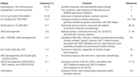
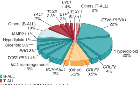
Acute lymphoblastic leukemia (ALL) is a neoplasm of imma-ture lymphoid progenitors that is most commonly of B cell lin-eage. Most childhood cases of B-precursor ALL (B-ALL) may be subclassified by the presence of either gross or submicroscopic genetic alteration. Approximately 75% of B-ALL cases exhibit aneuploidy or a recurring gross chromosomal rearrangement (1, 2) (Table 1 and Figure 1). Hyperdiploidy, with gain of at least 5 chromosomes, is one of the most frequent alterations in child-hood ALL and is associated with favorable outcome (3). However, the biologic basis of the acquisition of multiple whole chromo-somal gains is poorly understood. Conversely, hypodiploidy, with fewer than 44 chromosomes, is associated with dismal prog-nosis (4). A number of recurring chromosomal rearrangements are common in B-ALL and are critical events in leukemogenesis. These rearrangements commonly perturb genes encoding regula-tors of hematopoiesis, tumor suppressors, oncogenes, or tyrosine kinases but commonly require additional genetic hits to estab-lish the full leukemic phenotype.
In the last five years, genome-wide profiling using microarrays, candidate gene, and second-generation sequencing have provided a number of key insights into the genetic basis of ALL. These stud-ies have identified new subtypes of ALL and have uncovered recur-ring submicroscopic genetic alterations in known ALL subtypes. These include loss-of-function mutations involving genes regulat-ing lymphoid development that contribute to the arrest in matu-ration characteristic of B-ALL, mutations that inactivate tumor suppressor and cell cycle regulatory proteins, and mutations that drive cytokine receptor and/or kinase signaling. Thus, as in AML, concomitant lesions disrupting hematopoietic development and tumor suppression as well as driving signaling and proliferation are hallmarks of many ALL subtypes. Importantly, several of these alterations are associated with specific subtypes of ALL defined by recurring chromosomal alterations.
This Review describes the genetic landscape of ALL, focusing on important subtypes and recently discovered genetic alterations of biologic and therapeutic relevance.
ETV6-RUNX1 (TEL-AML1) B-ALL
The most common rearrangement in B-ALL is the t(12;21) (p13;q22) rearrangement that encodes ETV6-RUNX1 (5). This rearrangement is usually cryptic on cytogenetic analysis but is readily detected by fluorescent in situ hybridization and molecu-lar techniques. ETV6 is a member of the ETS family of transcrip-tion factors that are frequently targeted by rearrangements and mutations in leukemia and other malignancies (6). With CBFB, RUNX1 forms the core binding factor transcription complex and is commonly rearranged in acute myeloid leukemia (7), and harbors sequence mutations in myeloid and lymphoid dis-orders (8–10). Both ETV6 and RUNX1 are required for normal definitive hematopoiesis (7, 11), and the ETV6-RUNX1 protein may perturb expression of RUNX1-regulated genes, converting RUNX1 to a transcriptional repressor (12). ETV6-RUNX1 also causes overexpression of the erythropoietin receptor (EPOR) and activation of downstream JAK-STAT signaling (13). Expres-sion of ETV6-RUNX1 promotes self-renewal in B cell progeni-tors but alone does not induce leukemia (14, 15). Furthermore, ETV6-RUNX1 is commonly detectable at birth (16), years prior to the onset of leukemia, suggesting that secondary genetic events are required to induce leukemia. This model is supported by genome-wide profiling studies of genetic alterations in ALL that have identified additional recurring submicroscopic genetic alterations in ETV6-RUNX1 ALL, including deletions of the B cell transcription factors PAX5 and EBF1 and deletion of the second copy of ETV6 (17–19). Moreover, monozygotic twins concordant for ETV6-RUNX1 ALL exhibit distinct submicroscopic DNA copy number alterations (CNAs), indicating that acquisition of the
ETV6-RUNX1 is an early event in leukemogenesis (20). Submicro-scopic genetic alterations are more common in ETV6-RUNX1 ALL than in many other ALL subtypes and include deletions target-ing putative lymphoid signaltarget-ing molecules (BTLA, TOX), tran-scriptional coactivators (TBL1XR1), the glucocorticoid receptor gene NR3C1, and the putative apoptosis regulatory gene BTG1. Although these alterations may directly contribute to leukemo-genesis, the genomic breakpoints of many of these deletions bear the hallmark of aberrant activity of the recombinase activation genes. Thus, further work directly examining these alterations in leukemogenesis is required.
Conflict of interest: The author has declared that no conflict of interest exists.
review series
TCF3-PBX1 (E2A-PBX1) and TCF3-HLF (E2A-HLF) B-ALL The t(1;19)(q23;p13) translocation is present in up to 6% of child-hood B-ALL cases (21). The rearrangement is commonly unbal-anced, with duplication of 1q distal to PBX1.
This translocation fuses the transactivation domains of TCF3
with the C-terminal region of the homeobox gene PBX1. TCF3
encodes the basic helix-loop-helix (bHLH) transcription factors E12 and E47 that are required for early lymphoid development, and loss of E12/E47 promotes the development of T cell lympho-ma (22, 23). PBX1, which encodes a member of the three–amino acid loop extension family of homeodomain proteins, is required for the development of lymphoid precursors (24). TCF3-PBX1
binds HOX proteins and likely interferes with hematopoietic dif-ferentiation by disrupting HOX-regulated gene expression (25).
The t(17;19)(q22;p13) translocation is a rare rearrangement that fuses the aminoterminal transactivation domains of TCF3 to the C-terminal DNA-binding and dimerization domains of HLF, a member of the PAR family of basic leucine zipper transcription factors (26). TCF3-HLF aberrantly regulates genes that control cell death in lymphoid progenitors, including LMO2 and BCL2
(27). TCF3-HLF–positive leukemia is associated with older age and very poor outcome (28).
BCR–ABL1 ALL
Breakpoint cluster region–ABL1 (BCR-ABL1) is generated by the der(22) of the t(9;22)(q34;q11) translocation, or Philadelphia (Ph) chromosome, which is present in over 95% of chronic myeloge-nous leukemia (CML) cases, 25% of adult ALL cases, and 3% to 5% of pediatric ALL cases (29). The t(9;22) fuses 5′ sequences of breakpoint cluster region (BCR) to 3′ sequences from ABL1, which encodes a tyrosine kinase. The breakpoints on chromosome 9 are
scattered over a nearly 200-kb region within the first intron of ABL, whereas the BCR breakpoints on chromosome 22 are clustered in two areas: a 5.8-kb major BCR (M-bcr) in CML and a minor BCR (m-bcr) in most cases of childhood Ph-positive ALL (30). Fusion genes created by breaks in M-bcr (CML-type break) encode a 210-kDa fusion protein (p210), whereas fusions that occur in m-bcr (ALL-type break) encode p190.
Both forms of BCR-ABL1 can transform hematopoietic cells in vitro and induce a syndrome similar to CML in mice (31). BCR-ABL1 activates multiple signaling pathways, increases cell pro-liferation, and deregulates differentiation and adhesion. Treat-ment of BCR-ABL1–positive leukemic cells with tyrosine kinase inhibitors results in the activation of the transcription factor BCL6, which may directly influence responsiveness to treatment with these agents (32).
Additional genetic alterations are critical determinants of the lineage and progression of BCR-ABL1 leukemia. Deletion of the early lymphoid transcription factor gene IKZF1 (IKAROS) is common in BCR-ABL1 lymphoid leukemia (CML at progression to lymphoid blast crisis) but is rarely present in CML at chron-ic phase (33, 34). IKAROS is a zinc finger transcription factor required for the development of all lymphoid lineages (35, 36). The IKZF1 deletions result either in haploinsufficiency or expres-sion of dominant-negative isoforms. These alterations cooperate with BCR-ABL1 in the induction of lymphoblastic leukemia and promote resistance to therapy in recent experimental models of
BCR-ABL1 ALL (37).
[image:3.585.52.529.111.376.2]In childhood ALL, the presence of BCR-ABL1 is associated with older age, higher leukocyte count, and more frequent CNS leukemia at diagnosis (38). Historically, BCR-ABL1 ALL has been associated with extremely poor outcome (29, 39). Tyrosine kinase inhibitors Table 1
Key cytogenetic alterations and genetic subtypes in childhood B-ALL
Subtype Frequency (%) Comment References
Hyperdiploidy (>50 chromosomes) 20–30 Excellent prognosis with antimetabolite-based therapy 3 Hypodiploidy (<44 chromosomes) 1–2 Poor prognosis, high frequency of RAS pathway and 4
IKAROS gene family mutations
t(12;21)(p13;q22) ETV6-RUNX1 15–25 Expression of myeloid antigens; excellent outcome 5 t(1;19)(q23;p13) TCF3-PBX1 2–6 Increased incidence in African Americans; 121–123
generally excellent prognosis; association with CNS relapse
t(9;22)(q34;q11.2) BCR-ABL1 2–4 Historically dismal outcome, improved with addition 39, 42 of imatinib to intensive chemotherapy
PAX5 rearrangement ~2% Multiple partners, commonly from dic(7;9), dic/t(9;12) 17, 94 and dic(9;20); outcome unknown
ABL1, PDGFRB, JAK2 rearrangements 2–5 Identified in BCR-ABL1–like ALL; multiple rearrangements encoding 84 chimeric proteins, fusing 5′ partners with 3′ kinase domains;
associated with IKZF1 alteration and very high leukocyte count; potentially amenable to tyrosine kinase inhibitor therapy
t(4;11)(q21;q23) MLL-AF4 1–2 Common in infant ALL (especially <6 months of age); 45 poor prognosis
MYC rearrangement [t(8;14)(q24;q32), 2 Favorable prognosis with short-term, high-dose chemotherapy 70 t(2;8)(q12;q24)]
CRLF2 rearrangement (IGH@-CRLF2, 5–7 Extremely common in DS ALL (55%); association with 71–75 PAR1 deletion, and P2RY8-CRLF2) IKZF1 deletion/mutation and JAK1/2 mutation;
poor prognosis in non-DS ALL
such as imatinib mesylate (Gleevec) have transformed the treatment and outcome of patients with BCR-ABL1–positive leukemia (40, 41). The addition of imatinib to intensive chemotherapy in childhood BCR-ABL1–positive ALL results in a 4-year event-free survival rate of 84%, more than double that of historical controls (42). A propor-tion of patients become resistant to these agents, typically through acquired mutations in the ABL1 kinase domain (43).
MLL–rearranged B-ALL
Mixed-lineage leukemia–rearranged (MLL-rearranged) leukemia is a unique entity notable for initiation in utero, for myeloid and lymphoid features, and for poor responsiveness to therapy (44, 45). Rearrangement of the MLL gene at 11q23 occurs in at least two-thirds of infants with ALL, 5% of AML, and 85% of second-ary leukemias that occur in patients treated with topoisomerase II inhibitors (46, 47) but is less frequent in older individuals (48, 49). Over 80 partners of MLL rearrangement have been identified (50).
The most common MLL rearrangements are t(4;11)(q21;q23)/
MLL-AFF1(AF4), found in approximately 50% of cases, followed by t(9;11)(p22;q23)/MLL-MLLT3(AF9), MLL-ENL, and t(10;11) (p13-15;q14-21)/MLL-MLLT10(AF10) (51, 52). The transloca-tion breakpoints are located in an 8.5-kb cluster between exons 5 and 11 and juxtapose the A-T hook and methyltransferase domains to partner proteins. MLL regulates hematopoiesis through maintenance of normal homeotic gene expression (53), in part through transcriptional activation of HOX genes, medi-ated though the histone H3 lysine 4 (H3K4) methyltransferase activity of the SET domain (54). In contrast to other subtypes of B-ALL, additional genetic alterations are uncommon in MLL -rearranged leukemia (17, 55, 56).
MLL-rearranged ALL exhibits a distinct gene expression profile characterized by the upregulation of class I HOX genes (HOXA5, -A7, and -A9 and the HOX cofactor gene MEIS1) (57). Co-expres-sion of HOXA genes and MEIS1 cooperates with MLL fusions in induction of leukemia and maintenance of a “stem cell–like” state of maturation (58). fms-related tyrosine kinase 3 (FLT3) is overexpressed in MLL-rearranged ALL and harbors point muta-tions in 10%–20% of cases (59). In animal models, FLT3 kinase inhibitors have shown potential as therapeutic agents for MLL -rearranged leukemias, and FLT3 inhibitors are currently being tested in clinical trials (59, 60). MLL-rearranged leukemias also exhibit a distinct epigenetic profile, with signatures of cytosine (61), microRNA (62) and H3K79 methylation (63) that differ from non-MLL leukemias.
Many of the MLL fusion proteins are located in protein com-plexes that regulate transcriptional elongation, which may be in part responsible for transcriptional dysregulation and leuke-mogenesis. These include polymerase-associated factor complex, which associates with the N terminus of MLL and regulates RNA polymerase II (Pol II), the pTEFb (CDK9/cyclin T) complex, which associates with the fusion partners MLLT1 (ENL), ELL, and AFF1 (AF4) to release stalled Pol II and stimulate transcriptional elon-gation, and the H3K79 histone methyltransferase DOT1L, which binds MLL fusion partners including AFF1, AF9, AF10, and ENL (64). H3K79 methylation is a mark of actively transcribed genes, and suppression or inactivation of DOT1L in human or murine leukemia cells leads to suppression of the MLL fusion protein– induced gene expression program (63), differentiation and/or apoptosis of leukemic cell lines, and suppression of leukemogen-esis (65). DOT1L inhibition is thus being pursued as a therapeutic approach in MLL-rearranged ALL (66).
These findings highlight the potential importance of epigenetic dysregulation in other subtypes of ALL. Several studies profiling cytosine methylation have shown that different ALL subgroups exhibit distinct methylation profiles and that promoter meth-ylation is associated with gene expression and outcome (67, 68). Additional studies are needed to comprehensively determine the relative importance of genetic and epigenetic alteration in the pathogenesis of ALL as well as the potential therapeutic role of epigenetic modifying drugs.
Rearrangement of CRLF2 and other immunoglobulin heavy locus rearrangements in B-ALL
Rearrangements of the immunoglobulin heavy locus (IGH@) at 14q32.33, resulting in juxtaposition of IGH@ enhancer elements with transcription factor and cytokine receptor genes, are observed in both B-ALL and T-lineage ALL (69). B-lineage leukemia/lym-phoma with rearrangement of MYC is reviewed elsewhere (70).
Cytokine receptor-like factor 2 (CRLF2; also known as thymic stromal-derived lymphopoietin [TSLP] receptor) is located at the pseudoautosomal region (PAR1) at Xp22.3/Yp11.3 and is rear-ranged or mutated in up to 7% of B-ALL cases (71, 72). With the IL-7Rα chain, CRLF2 forms a heterodimeric receptor for TSLP. The rearrangements are either rearrangement into IGH@-CRLF2
[image:4.585.46.285.84.231.2]or focal deletion immediately upstream of CRLF2, resulting in a chimeric fusion of the purinergic receptor gene P2RY8 to the entire coding region of CRLF2. Both rearrangements result in overexpression of CRLF2 on the cell surface (71). Less commonly, Figure 1
review series
a p.Phe232Cys mutation in the transmembrane domain of CRLF2 results in receptor dimerization and overexpression (73).
CRLF2 rearrangements are present in at least 50% of Down syn-drome–associated (DS-associated) B-ALL cases (71, 72) and are also present in up to 50% of a novel subtype of high-risk B-ALL, BCR-ABL1–like ALL (see below) (74). CRLF2 alterations are associ-ated with the presence of activating mutations in the Janus kinase genes JAK1 and JAK2 (71–73, 75), which are otherwise uncommon in B-ALL. The JAK mutations in B-ALL most commonly disrupt p.Arg683 in the pseudokinase domain of JAK2, in contrast to the JAK2 p.Val617Phe mutations that are a hallmark of myeloprolif-erative diseases. Like the JAK2 p.Val617Phe mutation, the JAK1/2 mutant alleles observed in ALL confer cytokine-independent proliferation in cultured cells, particularly in the presence of a cytokine receptor such as the EPOR or CRLF2, suggesting that a receptor scaffold is required for mutant JAK alleles to drive pro-liferation and lymphoid transformation (76, 77). In non-DS ALL,
CRLF2 rearrangements are associated with IKZF1 alteration, JAK
mutation, and poor outcome (74, 78). Recently, activating muta-tions in IL-7R have been identified in both B-ALL and T-lineage ALL, including cases of CRLF2 rearrangement (10, 79, 80). Thus, the use of JAK inhibitors is actively being pursued to target leu-kemic cells harboring CRLF2/JAK alterations and leukemic cells with IL-7R mutations that also activate JAK/STAT signaling. Sev-eral other IGH@ translocation partners have been identified in B-ALL, including EPOR, the CEBP transcription factor gene fam-ily, the inhibitor of DNA binding 4 (ID4) gene, the LIM domain– containing protein LHX4, and rarely, IL-3 (69).
BCR-ABL1–like ALL
Up to 15% of childhood B-ALL cases exhibit a gene expression profile similar to that of BCR-ABL1–positive ALL, often have deletion/mutation of IZKF1, and have a very poor outcome (81–83). CRLF2 is rearranged in up to 50% of BCR-ABL1–like ALL cases, but until recently the lesions responsible for kinase activation in the remaining cases were unknown. Second-gener-ation mRNA sequencing and whole genome sequencing of 15 BCR-ABL1–like ALL cases identified rearrangements, sequence mutations, and DNA CNAs activating kinase signaling in all cases (84), including rearrangements of PDGFRB, ABL1, JAK2, and EPOR as well as deletion/mutation of SH2B3 and IL7R. Sev-eral of these alterations have been shown to result in activation of downstream (e.g., JAK/STAT) signaling pathways, and cellu-lar transformation was attenuated with JAK or ABL1/PDGFRB inhibitors. Thus, defining kinase-activating alterations in this subtype of ALL is critical to identifying and treating patients with this subtype of leukemia.
Intrachromosomal amplification of chromosome 21 Intrachromosomal amplification of chromosome 21 (iAMP21) occurs in up to 2% of B-progenitor ALL cases (85). The region of amplification is typically large but variable, but includes gain of at least three copies of RUNX1, commonly with deletion of the sub-telomeric regions of chromosome 21. iAMP21 is characterized by commonly complex patterns of chromosomal rearrangement sug-gestive of a breakage-fusion-bridge cycle (86). iAMP21 frequently occurs in older children or adolescents and has been reported to be associated with poor outcome (87), although this association may not be independent of other established prognostic variables such as levels of minimal residual disease (88).
Hypodiploid ALL
Hypodiploidy, with fewer than 44 chromosomes, is present in up to 3% of childhood ALL cases and is associated with a high risk of treatment failure (89). Hypodiploid ALL may be subclassified by severity of aneuploidy into near-haploid (NH; 24–31 chromo-somes) and low-hypodiploid (LH; 32–44 chromochromo-somes) cases, but until recently the concomitant submicroscopic genetic alterations were poorly characterized. NH ALL has a very high frequency of deletions and sequence mutations that activate RAS signaling, and NH and LH ALL have a high frequency of inactivating alterations of IKAROS genes IKZF2 (HELIOS) and IKZF3 (AIOLOS) that are otherwise rare in ALL (90). Moreover, LH cases have a very high frequency of loss-of-function mutations in TP53 (encoding p53) and RB1, which are otherwise uncommon at diagnosis in B-ALL.
Submicroscopic genetic alterations in B-ALL
The observation that up to 25% of children with ALL lack a recur-ring chromosomal alteration (2) and the identification of trans-location-encoded fusions at birth or in years prior to the onset of leukemia (16) indicates that additional genetic alterations cooper-ate in leukemogenesis. The advent of array-comparative genomic hybridization and SNP microarrays has enabled interrogation of structural genetic alterations at very high resolution (91). These approaches, coupled with candidate gene sequencing and inte-grated gene expression profiling (92), have identified a number of critical new targets of mutation in B-ALL that often involve genes that regulate lymphoid development, cell cycle, tumor suppres-sion, and a variety of other key cellular pathways (Table 2). The mean number of DNA CNAs is lower than in many solid tumors (an average of 6–8 lesions per case) (17, 19, 33), but over 50 recur-ring CNAs have been identified. The nature and frequency of these CNAs are significantly associated with ALL subtype, with few lesions in MLL-rearranged ALL (17, 55) but 6–8 lesions per case in
BCR-ABL1 and ETV6-RUNX1 ALL (33, 34).
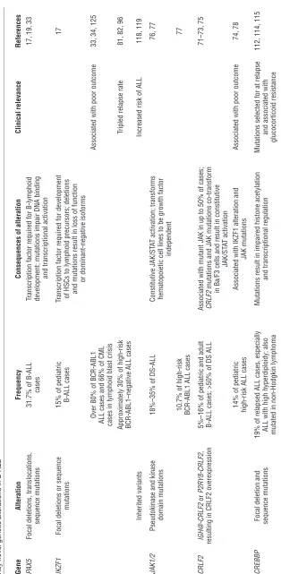
Most notably, genes encoding transcriptional regulators of B-lineage commitment and differentiation are mutated in over two-thirds of B-ALL. These include PAX5 (encoding paired box 5, mutated in over 30% of cases), EBF1 (early B cell factor 1), IKZF1–3, and lymphoid enhancer factor 1. PAX5 harbors broad deletions predicted to result in haploinsufficiency, focal internal dele-tions that result in the expression of truncated PAX5 transcripts, sequence mutations that disrupt DNA binding or truncate the transactivating domain (17, 93), and translocations that create chimeric fusion proteins (17, 93, 94). These lesions usually only involve a single copy of PAX5, and expression of key PAX5 targets, such as CD19, in ALL cells is frequently normal, suggesting that PAX5 haploinsufficiency contributes to leukemogenesis.
Despite their high frequency of occurrence, PAX5 alterations are not associated with outcome in ALL (81, 95). In contrast, IKZF1
func-Tab
le 2
Key novel genetic alterations in B-ALL Gene
Alteration
Frequency
Consequences of alteration
Clinical relevance
References
PAX5
Focal deletions, translocations,
31.7% of B-ALL
Transcription factor required
for B-lymphoid
17, 19, 33
sequence mutations
cases
development; mutations impair DNA binding
and transcriptional activation
IKZF1
Focal deletions or sequence
15% of pediatric
Transcription factor required for development
17
mutations
B-ALL cases
of HSCs to lymphoid precursors; deletions and mutations result in loss of function
or dominant-negative isoforms
Over 80% of BCR-ABL1
Associated with poor outcome
33, 34, 125
ALL cases and 66% of CML cases in lymphoid blast crisis
Approximately 30% of high-risk
Tripled relapse rate
81, 82, 96
BCR-ABL1–negative ALL cases
Inherited variants
Increased risk of ALL
118, 119
JAK1/2
Pseudokinase and kinase
18%–35% of DS-ALL
Constitutive JAK/ST
AT activation; transforms
76, 77
domain mutations
hematopoietic cell lines to be growth factor
independent
10.7% of high-risk
77
BCR-ABL1 ALL cases
CRLF2
IGH@-CRLF2
or
P2R
Y8-CRLF2,
5%–16% of pediatric and adult
Associated with mutant JAK in up to 50% of cases;
71–73, 75
resulting in CRLF2 overexpression
B-ALL cases; >50% of DS ALL
CRLF2
mutations and JAK mutations co-transform in Ba/F3 cells and result in constitutive
JAK/ST
AT activation
14% of pediatric
Associated with IKZF1 alteration and
Associated with poor outcome
74, 78
high-risk ALL cases
JAK mutations
CREBBP
Focal deletion and
19% of relapsed ALL cases, especially
Mutations result in impaired histone acetylation
Mutations selected for at relapse
112, 114, 115
sequence mutations
ALL with high hyperdiploidy; also
and transcriptional regulation
and associated with
mutated in non-Hodgkin lymphoma
glucocorticoid resistance
[image:6.585.60.374.88.729.2]review series
tion, focal internal deletions of exons 4–7, resulting in expression of the dominant-negative isoform IK6, and less commonly, loss-of-function and dominant-negative sequence mutations. How these IKZF1 alterations contribute to leukemogenesis remains poorly understood. IKZF1 inactivation likely contributes to the arrest in maturation characteristic of ALL, and loss of Ikzf1, like that of
Pax5, accelerates BCR-ABL1 lymphoid leukemia development in murine models (37). However, the striking association of IKZF1
alterations with poor outcome suggests that other mechanisms may be involved. Detection of IKZF1 alterations at diagnosis to aid risk stratification and treatment assignment is consequently under investigation in ongoing prospective clinical trials.
Additional recurring genetic alterations in B-ALL target cell cycle regulation, transcriptional activation, tumor suppression, lymphoid signaling, apoptosis, and drug responsiveness. These include CDKN2A/CDKN2B, RB1, CD200, BTLA/TOX, BTG1, the glucocorticoid receptor NR3C1, TBL1XR1, ETV6, and ERG (17–19, 97). Candidate gene sequencing approaches have identified addi-tional sequence mutations in B-ALL, most commonly mutations resulting in activation in RAS signaling, which are enriched in
MLL and hyperdiploid ALL (98, 99). Additional pathways targeted include JAK/STAT signaling, the TP53/RB1 pathway, and lym-phoid development and additional mutations have been found in transcriptional coactivators, including ERG, TBL1XR1, CTCF,
CREBBP, and EP300 (100). Genetics of relapse in ALL
ALL genomes are not static, frequently acquiring secondary karyotypic and genetic alterations during disease progression (101–103). Genome-wide profiling has enabled key questions in the genetic basis of treatment failure to be investigated, including the nature of genetic alterations that are acquired with disease progression, the genetic relationship between the diagnosis and relapsed leukemic clones, and the degree of clonal heterogeneity present at the time of diagnosis.
Microarray profiling has shown that relapse samples exhibit striking differences in the patterns of genomic alterations from the matched diagnosis samples (104–108), with the majority of relapse samples exhibiting the loss of CNAs present at diagnosis and the acquisition of new genetic alterations. Importantly, most
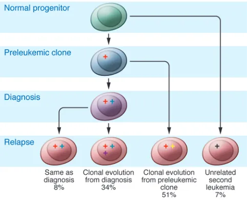
paired diagnosis-relapse samples share a common clonal origin. Moreover, lesions detected at relapse are often detectable in diag-nostic samples, indicating that the predominant relapse clone(s) are present at low levels at diagnosis. Genetically unrelated sec-ond leukemias (at least at the level of DNA CNA analysis by SNP array) are infrequent but may be more common in late relapse (109). Approximately one-third of cases show a pattern of linear clonal evolution (with the acquisition of new genetic changes by the relapse clone in addition to those seen at relapse), and over 50% show a complex picture with both loss of diagnosis CNAs and the acquisition of new lesions, as well as evidence of low levels of relapse clones at diagnosis (Figure 2).
Together, these data suggest that a pre-diagnosis “ancestral” clone harboring one or more genetic alterations (such as a founding trans-location) undergoes divergent evolution into multiple clones that acquire different genetic alterations and emerge as the predomi-nant clones at diagnosis and relapse. This is supported by studies of monozygotic twins concordant for ALL, showing different sec-ondary CNAs in each twin (20), and recent xenotransplantation studies that have modeled clonal evolution and heterogeneity by engrafting ETV6-RUNX1 and BCR-ABL1 samples into immunocom-promised mice. These studies have confirmed clonal heterogeneity in the majority of cases at the time of diagnosis and have shown that specific genetic alterations (e.g., deletion of CDKN2A/CDKN2B) influence the efficiency and tempo of engraftment (110, 111).
[image:7.585.44.285.84.279.2]Genome-wide sequencing is required to comprehensively iden-tify all sequence and structural variants contributing to the patho-genesis of ALL, and a number of sequencing efforts are ongoing. However, a recent study performed Sanger sequencing of 300 genes in 23 matched diagnosis-relapse samples, with recurrence testing of over 300 paired and unpaired ALL samples (112). This study identified a number of uncharacterized mutations in ALL, most notably deleterious mutations in CREBBP (encoding CREB-binding protein) in over 20% of relapse ALL samples (112). The mutations were present at relapse and were detected in either the predominant clone or a subclone at diagnosis. CREBBP is a tran-scriptional coactivator, histone and non-histone acetylase, and ubiquitin ligase (113). Mutations were common in the histone ace-tyl transferase (HAT) domain and resulted in impaired aceace-tylation of histone targets (112). CREBBP mutations are highly enriched Figure 2
in hyperdiploid ALL (114) and are common at diagnosis in B cell lymphoma samples (115). Importantly, the HAT mutations impair the normal CREBBP-mediated transcriptional response to gluco-corticoids, which are widely used in ALL therapy. Thus, CREBBP mutations may impair the response of leukemic cells to glucocor-ticoids and influence treatment responsiveness, and therapeutic approaches modifying acetylation may reverse glucocorticoid resistance. Mutations in TP53, which are otherwise infrequent in ALL, are also enriched in relapsed ALL (116).
Inherited genetic variation and risk of ALL
Several candidate gene studies have implicated inherited genetic variation, for example in the folate metabolic pathway, in the risk of developing leukemia (117). Multiple genome-wide association studies have reproducibly identified variations at several genomic loci that are associated with ALL risk, including IKZF1, ARID5B
(encoding the AT-rich interactive domain 5B transcription factor), and CEBPE (encoding the transcription factor CCAAT/enhancer-binding protein, epsilon) (118, 119). All three genes encode pro-teins considered important for normal lymphoid development and/or lymphoid leukemogenesis. CEBPE is a target of translo-cations in ALL, and Arid5b knockout mice exhibit defects in the B lymphoid compartment (120). Although the functional effects of these inherited variants are poorly understood, the IKZF1 vari-ants may influence level of IKZF1 gene expression (119), suggest-ing a direct role for these variants in disease susceptibility.
Conclusions
The use of contemporary technologies to identify genetic alterations in B-ALL has been tremendously informative, but clearly much work
remains to be done. Microarray and candidate gene sequencing approaches are incapable of identifying mutations at nucleotide-level resolution, and initial second-generation sequencing of ALL genomes has identified multiple new genes targeted in ALL. An additional high-priority area of investigation is ALL in older chil-dren, adolescents, and adults, who typically have a substantially inferior prognosis. Profiling of ALL in these age groups, as well as in additional uncommon but high-risk ALL subtypes, is ongoing and likely to yield additional insights and therapeutic approaches.
Acknowledgments
The author thanks members of his laboratory and colleagues at St. Jude Children’s Research Hospital, the Children’s Oncology Group, and the National Cancer Institute Therapeutically Appli-cable Research to Generate Effective Treatments (TARGET) Initia-tive, who have contributed to the studies described in this Review, and apologizes to the many investigators whose important studies could not be cited due to space restrictions. The author is sup-ported by the American Lebanese Syrian Associated Charities of St. Jude Children’s Research Hospital and by funding from the National Cancer Institute, the American Society of Hematology, the American Association of Cancer Research, and Stand Up to Cancer. The author is a Pew Scholar in the Biomedical Sciences and a St. Baldrick’s Scholar.
Address correspondence to: Charles G. Mullighan, Department of Pathology, St. Jude Children’s Research Hospital, 262 Danny Thomas Place, Mail Stop 342, Room 4047G, Memphis, Tennes-see 38105, USA. Phone: 901.595.3387; Fax: 901.595.5947; E-mail: charles.mullighan@stjude.org.
1. Pui CH, Robison LL, Look AT. Acute lymphoblas-tic leukaemia. Lancet. 2008;371(9617):1030–1043. 2. Harrison CJ. Cytogenetics of paediatric and
adoles-cent acute lymphoblastic leukaemia. Br J Haematol.
2009;144(2):147–156.
3. Harrison CJ, Foroni L. Cytogenetics and molecular genetics of acute lymphoblastic leukemia. Rev Clin
Exp Hematol. 2002;6(2):91–113.
4. Nachman JB, et al. Outcome of treatment in chil-dren with hypodiploid acute lymphoblastic leuke-mia. Blood. 2007;110(4):1112–1115.
5. Shurtleff SA, et al. TEL/AML1 fusion resulting from a cryptic t(12;21) is the most common genet-ic lesion in pediatrgenet-ic ALL and defines a subgroup of patients with an excellent prognosis. Leukemia.
1995;9(12):1985–1989.
6. Bohlander SK. ETV6: a versatile player in leukemo-genesis. Semin Cancer Biol. 2005;15(3):162–174. 7. Okuda T, van Deursen J, Hiebert SW, Grosveld G,
Downing JR. AML1, the target of multiple chro-mosomal translocations in human leukemia, is essential for normal fetal liver hematopoiesis. Cell.
1996;84(2):321–330.
8. Song WJ, et al. Haploinsufficiency of CBFA2 causes familial thrombocytopenia with propensity to develop acute myelogenous leukaemia. Nat Genet.
1999;23(2):166–175.
9. Roumier C, Fenaux P, Lafage M, Imbert M, Eclache V, Preudhomme C. New mechanisms of AML1 gene alteration in hematological malignancies.
Leukemia. 2003;17(1):9–16.
10. Zhang J, et al. The genetic basis of early T-cell precursor acute lymphoblastic leukaemia. Nature.
2012;481(7380):157–163.
11. Wang LC, Kuo F, Fujiwara Y, Gilliland DG, Golub TR, Orkin SH. Yolk sac angiogenic defect and intra-embryonic apoptosis in mice lacking the Ets-related factor TEL. EMBO J. 1997;16(14):4374–4383.
12. Hiebert SW, et al. The t(12;21) translocation con-verts AML-1B from an activator to a repressor of transcription. Mol Cell Biol. 1996;16(4):1349–1355. 13. Torrano V, Procter J, Cardus P, Greaves M, Ford
AM. ETV6-RUNX1 promotes survival of early B lineage progenitor cells via a dysregulated erythro-poietin receptor. Blood. 2011;118(18):4910–4918. 14. Andreasson P, Schwaller J, Anastasiadou E, Aster
J, Gilliland DG. The expression of ETV6/CBFA2 (TEL/AML1) is not sufficient for the transfor-mation of hematopoietic cell lines in vitro or the induction of hematologic disease in vivo. Cancer
Genet Cytogenet. 2001;130(2):93–104.
15. Morrow M, Horton S, Kioussis D, Brady HJ, Wil-liams O. TEL-AML1 promotes development of spe-cific hematopoietic lineages consistent with pre-leukemic activity. Blood. 2004;103(10):3890–3896. 16. Wiemels JL, et al. Prenatal origin of acute
lym-phoblastic leukaemia in children. Lancet. 1999; 354(9189):1499–1503.
17. Mullighan CG, et al. Genome-wide analysis of genetic alterations in acute lymphoblastic leukae-mia. Nature. 2007;446(7137):758–764.
18. Parker H, et al. The complex genomic profile of ETV6-RUNX1 positive acute lymphoblastic leuke-mia highlights a recurrent deletion of TBL1XR1.
Genes Chromosomes Cancer. 2008;47(12):1118–1125.
19. Kuiper RP, et al. High-resolution genomic pro-filing of childhood ALL reveals novel recurrent genetic lesions affecting pathways involved in lym-phocyte differentiation and cell cycle progression.
Leukemia. 2007;21(6):1258–1266.
20. Bateman CM, et al. Acquisition of genome-wide copy number alterations in monozygotic twins with acute lymphoblastic leukemia. Blood. 2010; 115(17):3553–3558.
21. Hunger SP. Chromosomal translocations involv-ing the E2A gene in acute lymphoblastic leukemia:
clinical features and molecular pathogenesis. Blood.
1996;87(4):1211–1224.
22. Zhuang Y, Soriano P, Weintraub H. The helix-loop-helix gene E2A is required for B cell formation. Cell.
1994;79(5):875–884.
23. Bain G, et al. E2A deficiency leads to abnormalities in alphabeta T-cell development and to rapid devel-opment of T-cell lymphomas. Mol Cell Biol. 1997; 17(8):4782–4791.
24. Sanyal M, et al. B-cell development fails in the absence of the Pbx1 proto-oncogene. Blood. 2007; 109(10):4191–4199.
25. Lu Q, Kamps MP. Heterodimerization of Hox pro-teins with Pbx1 and oncoprotein E2a-Pbx1 gener-ates unique DNA-binding specifities at nucleo-tides predicted to contact the N-terminal arm of the Hox homeodomain--demonstration of Hox-dependent targeting of E2a-Pbx1 in vivo. Oncogene.
1997;14(1):75–83.
26. Inaba T, et al. Fusion of the leucine zipper gene HLF to the E2A gene in human acute B-lineage leukemia. Science. 1992;257(5069):531–534. 27. de Boer J, et al. The E2A-HLF oncogenic fusion
protein acts through Lmo2 and Bcl-2 to immor-talize hematopoietic progenitors. Leukemia. 2011; 25(2):321–330.
28. Heltemes-Harris LM, et al. Ebf1 or Pax5 haploin-sufficiency synergizes with STAT5 activation to initiate acute lymphoblastic leukemia. J Exp Med.
2011;208(6):1135–1149.
29. Ribeiro RC, Abromowitch M, Raimondi SC, Mur-phy SB, Behm F, Williams DL. Clinical and bio-logic hallmarks of the Philadelphia chromosome in childhood acute lymphoblastic leukemia. Blood.
1987;70(4):948–953.
30. Melo JV. The diversity of BCR-ABL fusion proteins and their relationship to leukemia phenotype.
review series
31. Van Etten RA. Studying the pathogenesis of BCR-ABL+ leukemia in mice. Oncogene. 2002; 21(56):8643–8651.
32. Duy C, et al. BCL6 enables Ph+ acute lymphoblastic leukaemia cells to survive BCR-ABL1 kinase inhibi-tion. Nature. 2011;473(7347):384–388.
33. Mullighan CG, et al. BCR-ABL1 lymphoblastic leu-kaemia is characterized by the deletion of Ikaros.
Nature. 2008;453(7191):110–114.
34. Iacobucci I, et al. Identification and molecular char-acterization of recurrent genomic deletions on 7p12 in the IKZF1 gene in a large cohort of BCR-ABL1-positive acute lymphoblastic leukemia patients: on behalf of Gruppo Italiano Malattie Ematologiche dell’Adulto Acute Leukemia Working Party (GIME-MA AL WP). Blood. 2009;114(10):2159–2167. 35. Georgopoulos K, et al. The Ikaros gene is required
for the development of all lymphoid lineages. Cell.
1994;79(1):143–156.
36. Molnar A, Georgopoulos K. The Ikaros gene encodes a family of functionally diverse zinc finger DNA-bind-ing proteins. Mol Cell Biol. 1994;14(12):8292–8303. 37. Virely C, et al. Haploinsufficiency of the IKZF1
(IKAROS) tumor suppressor gene cooperates with BCR-ABL in a transgenic model of acute lympho-blastic leukemia. Leukemia. 2010;24(6):1200–1204. 38. Crist W, et al. Philadelphia chromosome positive
childhood acute lymphoblastic leukemia: clinical and cytogenetic characteristics and treatment out-come. A Pediatric Oncology Group study. Blood.
1990;76(3):489–494.
39. Arico M, et al. Clinical outcome of children with newly diagnosed Philadelphia chromosome-positive acute lymphoblastic leukemia treated between 1995 and 2005. J Clin Oncol. 2010;28(31):4755–4761. 40. Druker BJ, et al. Activity of a specific inhibitor of
the BCR-ABL tyrosine kinase in the blast crisis of chronic myeloid leukemia and acute lymphoblas-tic leukemia with the Philadelphia chromosome.
N Engl J Med. 2001;344(14):1038–1042.
41. Druker BJ, et al. Efficacy and safety of a spe-cific inhibitor of the BCR-ABL tyrosine kinase in chronic myeloid leukemia. N Engl J Med. 2001; 344(14):1031–1037.
42. Schultz KR, et al. Improved early event-free sur-vival with imatinib in Philadelphia chromosome-positive acute lymphoblastic leukemia: a chil-dren’s oncology group study. J Clin Oncol. 2009; 27(31):5175–5181.
43. Pfeifer H, et al. Prevalence and dynamics of bcr-abl kinase domain mutations during imatinib treat-ment differ in patients with newly diagnosed and recurrent bcr-abl positive acute lymphoblastic leu-kemia. Leukemia. 2012;26(7):1475–1481. 44. Chen CS, et al. Molecular rearrangements on
chro-mosome 11q23 predominate in infant acute lym-phoblastic leukemia and are associated with specif-ic biologspecif-ic variables and poor outcome. Blood. 1993; 81(9):2386–2393.
45. Pui CH, et al. 11q23/MLL rearrangement confers a poor prognosis in infants with acute lymphoblastic leukemia. J Clin Oncol. 1994;12(5):909–915. 46. Pui CH, Kane JR, Crist WM. Biology and treatment
of infant leukemias. Leukemia. 1995;9(5):762–769. 47. Pui CH, et al. Secondary acute myeloid leukemia
in children treated for acute lymphoid leukemia.
N Engl J Med. 1989;321(3):136–142.
48. Chessels JM, Swansbury GJ, Reeves B, Bailey CC, Richards SM. Cytogenetics and prognosis in child-hood lymphoblastic leukaemia: results of MRC UKALL X. Medical Research Council Working Party in Childhood Leukaemia. Br J Haematol. 1997; 99(1):93–100.
49. Moorman AV, et al. Karyotype is an independent prognostic factor in adult acute lymphoblas-tic leukemia (ALL): analysis of cytogenelymphoblas-tic data from patients treated on the Medical Research Council (MRC) UKALLXII/Eastern Cooperative
Oncology Group (ECOG) 2993 trial. Blood. 2007; 109(8):3189–3197.
50. Meyer C, et al. The MLL recombinome of acute leu-kemias. Leukemia. 2006;20(5):777–784.
51. Chessells JM, et al. Clinical features, cytogenetics and outcome in acute lymphoblastic and myeloid leukaemia of infancy: report from the MRC Child-hood Leukaemia working party. Leukemia. 2002; 16(5):776–784.
52. Moorman AV, et al. No prognostic effect of addi-tional chromosomal abnormalities in children with acute lymphoblastic leukemia and 11q23 abnormalities. Leukemia. 2005;19(4):557–563. 53. Ziemin-van der Poel S, McCabe NR, Gill HJ,
Espi-nosa R. Identification of a gene, MLL, that spans the breakpoint in 11q23 translocations associated with human leukemias. Proc Natl Acad Sci U S A.
1991;88(23):10735–10739.
54. Milne TA, et al. MLL targets SET domain meth-yltransferase activity to Hox gene promoters. Mol
Cell. 2002;10(5):1107–1117.
55. Bardini M, et al. DNA copy-number abnormalities do not occur in infant ALL with t(4;11)/MLL-AF4.
Leukemia. 2010;24(1):169–176.
56. Andersson AK, et al. Whole genome sequence anal-ysis of 22 MLL rearranged infant acute lympho-blastic leukemias reveals remarkably few somatic mutations: a report from the St. Jude Children’s Research Hospital — Washington University Pedi-atric Cancer Genome Project. ASH Annual Meeting Abstracts. 2011;118:69.
57. Armstrong SA, et al. MLL translocations specify a distinct gene expression profile that distinguishes a unique leukemia. Nat Genet. 2002;30(1):41–47. 58. Faber J, et al. HOXA9 is required for survival in
human MLL-rearranged acute leukemias. Blood.
2009;113(11):2375–2385.
59. Armstrong SA, et al. Inhibition of FLT3 in MLL. Validation of a therapeutic target identified by gene expression based classification. Cancer Cell. 2003; 3(2):173–183.
60. Stubbs MC, et al. MLL-AF9 and FLT3 cooperation in acute myelogenous leukemia: development of a model for rapid therapeutic assessment. Leukemia.
2008;22(1):66–77.
61. Stumpel DJ, et al. Specific promoter methylation identifies different subgroups of MLL-rearranged infant acute lymphoblastic leukemia, influ-ences clinical outcome, and provides therapeutic options. Blood. 2009;114(27):5490–5498. 62. Stumpel DJ, et al. Hypermethylation of specific
microRNA genes in MLL-rearranged infant acute lymphoblastic leukemia: major matters at a micro scale. Leukemia. 2011;25(3):429–439.
63. Krivtsov AV, et al. H3K79 methylation profiles define murine and human MLL-AF4 leukemias.
Cancer Cell. 2008;14(5):355–368.
64. Yan L, et al. Clinical, immunophenotypic, cytoge-netic, and molecular genetic features in 117 adult patients with mixed–phenotype acute leukemia defined by WHO–2008 classification [published online ahead of print May 11, 2012]. Haematologica.
doi:10.3324/haematol.2012.064485.
65. Jo SY, Granowicz EM, Maillard I, Thomas D, Hess JL. Requirement for Dot1l in murine postnatal hematopoiesis and leukemogenesis by MLL trans-location. Blood. 2011;117(18):4759–4768. 66. Daigle SR, et al. Selective killing of mixed lineage
leukemia cells by a potent small-molecule DOT1L inhibitor. Cancer Cell. 2011;20(1):53–65. 67. Schafer E, et al. Promoter hypermethylation in MLL-r
infant acute lymphoblastic leukemia: biology and therapeutic targeting. Blood. 2010;115(23):4798–4809. 68. Milani L, et al. DNA methylation for subtype clas-sification and prediction of treatment outcome in patients with childhood acute lymphoblastic leu-kemia. Blood. 2010;115(6):1214–1225.
69. Dyer MJ, et al. Immunoglobulin heavy chain (IGH)
locus chromosomal translocations in B-cell pre-cursor acute lymphoblastic leukemia (BCP-ALL): rare clinical curios or potent genetic drivers? Blood.
2010;115(8):1490–1499.
70. Klapproth K, Wirth T. Advances in the understand-ing of MYC-induced lymphomagenesis. Br J Haema-tol. 2010;149(4):484–497.
71. Russell LJ, et al. Deregulated expression of cytokine receptor gene, CRLF2, is involved in lymphoid transformation in B-cell precursor acute lympho-blastic leukemia. Blood. 2009;114(13):2688–2698. 72. Mullighan CG, et al. Rearrangement of CRLF2
in B-progenitor- and Down syndrome-associated acute lymphoblastic leukemia. Nat Genet. 2009; 41(11):1243–1246.
73. Yoda A, et al. Functional screening identifies CRLF2 in precursor B-cell acute lymphoblastic leukemia.
Proc Natl Acad Sci U S A. 2010;107(1):252–257.
74. Harvey RC, et al. Rearrangement of CRLF2 is asso-ciated with mutation of JAK kinases, alteration of IKZF1, Hispanic/Latino ethnicity, and a poor out-come in pediatric B-progenitor acute lymphoblas-tic leukemia. Blood. 2010;115(26):5312–5321. 75. Hertzberg L, et al. Down syndrome acute
lympho-blastic leukemia: a highly heterogeneous disease in which aberrant expression of CRLF2 is associated with mutated JAK2: a report from the iBFM Study Group. Blood. 2010;115(5):1006–1017.
76. Bercovich D, et al. Mutations of JAK2 in acute lymphoblastic leukaemias associated with Down’s syndrome. Lancet. 2008;372(9648):1484–1492. 77. Mullighan CG, et al. JAK mutations in high-risk
childhood acute lymphoblastic leukemia. Proc Natl
Acad Sci U S A. 2009;106(23):9414–9418.
78. Cario G, et al. Presence of the P2RY8-CRLF2 rear-rangement is associated with a poor prognosis in non-high-risk precursor B-cell acute lymphoblastic leukemia in children treated according to the ALL-BFM 2000 protocol. Blood. 2010;115(26):5393–5397. 79. Shochat C, et al. Gain-of-function mutations in
interleukin-7 receptor-alpha (IL7R) in childhood acute lymphoblastic leukemias. J Exp Med. 2011; 208(5):901–908.
80. Zenatti PP, et al. Oncogenic IL7R gain-of-function mutations in childhood T-cell acute lymphoblastic leukemia. Nat Genet. 2011;43(10):932–939. 81. Mullighan CG, et al. Deletion of IKZF1 and
prog-nosis in acute lymphoblastic leukemia. N Engl J Med. 2009;360(5):470–480.
82. Den Boer ML, et al. A subtype of childhood acute lymphoblastic leukaemia with poor treatment out-come: a genome-wide classification study. Lancet
Oncol. 2009;10(2):125–134.
83. Loh ML, et al. A BCR–ABL1–like gene expression profile confers a poor prognosis in patients with high–risk acute lymphoblastic leukemia (HR– ALL): a report from Children’s Oncology Group (COG) AALL0232. ASH Annual Meeting Abstracts. 2011;118:743.
84. Roberts KG, et al. Genetic alterations activating kinase and cytokine receptor signaling in high rish acute lymphoblastic leukemia. Cancer Cell. 2012; 22(2):153–166.
85. Harewood L, et al. Amplification of AML1 on a duplicated chromosome 21 in acute lymphoblas-tic leukemia: a study of 20 cases. Leukemia. 2003; 17(3):547–553.
86. Robinson HM, Harrison CJ, Moorman AV, Chu-doba I, Strefford JC. Intrachromosomal amplifica-tion of chromosome 21 (iAMP21) may arise from a breakage-fusion-bridge cycle. Genes Chromosomes
Cancer. 2007;46(4):318–326.
87. Inukai T, et al. Clinical significance of early T-cell precursor acute lymphoblastic leukaemia: results of the Tokyo Children’s Cancer Study Group Study L99-15. Br J Haematol. 2012;156(3):358–365. 88. Attarbaschi A, et al. Minimal residual disease
in children with B-cell precursor acute lymphoblas-tic leukemia and an intrachromosomal amplifica-tion of chromosome 21: the Austrian and German acute lympho blastic leukemia Berlin-Frankfurt-Munster (ALL-BFM) trials. J Clin Oncol. 2008; 26(18):3046–3050.
89. Harrison CJ, et al. Three distinct subgroups of hypodiploidy in acute lymphoblastic leukaemia.
Br J Haematol. 2004;125(5):552–559.
90. Holmfeldt L, et al. Genome-wide analysis of genetic alterations in hypodiploid acute lymphoblastic leu-kemia identifies a high frequency of mutations tar-geting the IKAROS gene family and Ras signaling. ASH Annual Meeting Abstracts. 2010;116:411. 91. Mullighan CG. Single nucleotide polymorphism
microarray analysis of genetic alterations in cancer.
Methods Mol Biol. 2011;730:235–258.
92. Yeoh EJ, et al. Classification, subtype discovery, and prediction of outcome in pediatric acute lym-phoblastic leukemia by gene expression profiling.
Cancer Cell. 2002;1(2):133–143.
93. Bousquet M, et al. A novel PAX5-ELN fusion pro-tein identified in B-cell acute lymphoblastic leu-kemia acts as a dominant negative on wild-type PAX5. Blood. 2007;109(8):3417–3423.
94. Nebral K, et al. Incidence and diversity of PAX5 fusion genes in childhood acute lymphoblastic leukemia. Leukemia. 2009;23(1):134–143. 95. Iacobucci I, et al. The PAX5 gene is frequently
rear-ranged in BCR-ABL1-positive acute lymphoblastic leukemia but is not associated with outcome. A report on behalf of the GIMEMA Acute Leukemia Working Party. Haematologica. 2010;95(10):1683–1690. 96. Kuiper RP, et al. IKZF1 deletions predict relapse in
uniformly treated pediatric precursor B-ALL. Leuke-mia. 2010;24(7):1258–1264.
97. Kawamata N, et al. Molecular allelokaryotyping of pediatric acute lymphoblastic leukemias by high-resolution single nucleotide polymorphism oligonucleotide genomic microarray. Blood. 2008; 111(2):776–784.
98. Yamamoto T, et al. PTPN11, RAS and FLT3 muta-tions in childhood acute lymphoblastic leukemia.
Leuk Res. 2006;30(9):1085–1089.
99. Paulsson K, et al. Mutations of FLT3, NRAS, KRAS, and PTPN11 are frequent and possibly mutually exclusive in high hyperdiploid childhood acute lymphoblastic leukemia. Genes Chromosomes Cancer.
2008;47(1):26–33.
100. Zhang J, et al. Key pathways are frequently mutated in high-risk childhood acute lymphoblastic leuke-mia: a report from the Children’s Oncology Group.
Blood. 2011;118(11):3080–3087.
101. Raimondi SC, Pui CH, Head DR, Rivera GK, Behm FG. Cytogenetically different leukemic clones at relapse of childhood acute lymphoblastic leuke-mia. Blood. 1993;82(2):576–580.
102. Maloney KW, McGavran L, Odom LF, Hunger SP. Acquisition of p16(INK4A) and p15(INK4B) gene abnormalities between initial diagnosis and relapse in children with acute lymphoblastic leukemia.
Blood. 1999;93(7):2380–2385.
103. Irving JA, Minto L, Bailey S, Hall AG. Loss of het-erozygosity and somatic mutations of the gluco-corticoid receptor gene are rarely found at relapse in pediatric acute lymphoblastic leukemia but may occur in a subpopulation early in the disease course. Cancer Res. 2005;65(21):9712–9718. 104. Mullighan CG, et al. Genomic analysis of the clonal
origins of relapsed acute lymphoblastic leukemia.
Science. 2008;322(5906):1377–1380.
105. Yang JJ, et al. Genome-wide copy number profil-ing reveals molecular evolution from diagnosis to relapse in childhood acute lymphoblastic leuke-mia. Blood. 2008;112(10):4178–4183.
106. van Delft FW, et al. Clonal origins of relapse in ETV6-RUNX1 acute lymphoblastic leukemia.
Blood. 2011;117(23):6247–6254.
107. Kawamata N, et al. Molecular allelokaryotyping of relapsed pediatric acute lymphoblastic leukemia.
Int J Oncol. 2009;34(6):1603–1612.
108. Kuster L, et al. ETV6/RUNX1-positive relapses evolve from an ancestral clone and frequently acquire deletions of genes implicated in glucocor-ticoid signaling. Blood. 2011;117(9):2658–2667. 109. Szczepanski T, et al. Late recurrence of childhood
T-cell acute lymphoblastic leukemia frequently represents a second leukemia rather than a relapse: first evidence for genetic predisposition. J Clin
Oncol. 2011;29(12):1643–1649.
110. Notta F, et al. Evolution of human BCR-ABL1 lym-phoblastic leukaemia-initiating cells. Nature. 2011; 469(7330):362–367.
111. Anderson K, et al. Genetic variegation of clonal architecture and propagating cells in leukaemia.
Nature. 2011;469(7330):356–361.
112. Mullighan CG, et al. CREBBP mutations in relapsed acute lymphoblastic leukaemia. Nature. 2011;
471(7337):235–239.
113. Goodman RH, Smolik S. CBP/p300 in cell growth, transformation, and development. Genes Dev. 2000;14(13):1553–1577.
114. Inthal A, et al. CREBBP HAT domain mutations prevail in relapse cases of high hyperdiploid child-hood acute lymphoblastic leukemia [published online ahead of print March 5, 2012]. Leukemia.
doi:10.1038/leu.2012.60.
115. Pasqualucci L, et al. Inactivating mutations of acetyltransferase genes in B-cell lymphoma. Nature.
2011;471(7337):189–195.
116. Hof J, et al. Mutations and deletions of the TP53 gene predict nonresponse to treatment and poor outcome in first relapse of childhood acute lymphoblastic leu-kemia. J Clin Oncol. 2011;29(23):3185–3193. 117. Koppen IJ, Hermans FJ, Kaspers GJ. Folate related
gene polymorphisms and susceptibility to develop childhood acute lymphoblastic leukaemia. Br J
Hae-matol. 2010;148(1):3–14.
118. Trevino LR, et al. Germline genomic variants asso-ciated with childhood acute lymphoblastic leuke-mia. Nat Genet. 2009;41(9):1001–1005.
119. Papaemmanuil E, et al. Loci on 7p12.2, 10q21.2 and 14q11.2 are associated with risk of childhood acute lymphoblastic leukemia. Nat Genet. 2009; 41(9):1006–1010.
120. Lahoud MH, et al. Gene targeting of Desrt, a novel ARID class DNA-binding protein, causes growth retardation and abnormal development of repro-ductive organs. Genome Res. 2001;11(8):1327–1334. 121. Pui CH, et al. Treating childhood acute lympho-blastic leukemia without cranial irradiation. N Engl
J Med. 2009;360(26):2730–2741.
122. Pui CH, et al. Results of therapy for acute lympho-blastic leukemia in black and white children. JAMA. 2003;290(15):2001–2007.
123. Jeha S, et al. Increased risk for CNS relapse in pre-B cell leukemia with the t(1;19)/TCF3-PBX1. Leuke-mia. 2009;23(8):1406–1409.
124. Mullighan CG, et al. ERG deletions define a novel subtype of B-progenitor acute lymphoblas-tic leukemia. ASH Annual Meeting Abstracts. 2007;110:691.