protein STXBP5
Shaojing Ye, … , Yoshimi Takai, Sidney W. Whiteheart
J Clin Invest. 2014;124(10):4517-4528. https://doi.org/10.1172/JCI75572.
Genome-wide association studies (GWAS) have linked genes encoding several soluble NSF attachment protein receptor (SNARE) regulators to cardiovascular disease risk factors. Because these regulatory proteins may directly affect platelet secretion, we used SNARE-containing complexes to affinity purify potential regulators from human platelet extracts. Syntaxin-binding protein 5 (STXBP5; also known as tomosyn-1) was identified by mass spectrometry, and its expression in isolated platelets was confirmed by RT-PCR analysis. Coimmunoprecipitation studies showed that STXBP5 interacts with core secretion machinery complexes, such as syntaxin-11/SNAP23 heterodimers, and fractionation studies suggested that STXBP5 also interacts with the platelet cytoskeleton. Platelets from Stxbp5 KO mice had normal expression of other key secretory components; however, stimulation-dependent secretion from each of the 3 granule types was markedly defective. Secretion defects in STXBP5-deficient platelets were confirmed via lumi-aggregometry and FACS analysis for P-selectin and LAMP-1 exposure. Interestingly, STXBP5-deficient platelets had altered granule cargo levels, despite having normal morphology and granule numbers. Consistent with secretion and cargo deficiencies, Stxbp5 KO mice showed dramatic bleeding in the tail transection model and defective hemostasis in the FeCl3-induced carotid injury model. Transplantation experiments indicated that these defects were due to loss of STXBP5 in BM-derived cells. Our data demonstrate that STXBP5 is required for normal arterial hemostasis, due to its contributions to platelet granule cargo packaging and secretion.
Research Article
Find the latest version:
Introduction
Cardiovascular diseases, such as myocardial infarction, stroke, and deep vein thrombosis, are leading causes of death and dis-ability. These potentially occlusive processes are influenced in part by platelet secretion (1, 2). As a physiological response to vascular damage, platelets are activated and secrete components that pro-mote thrombus formation and initiate its sequelae, e.g., wound healing and angiogenesis (1, 3, 4). Platelet secretion is mediated by highly conserved soluble N-ethylmaleimide–sensitive factor (NSF) attachment protein receptor (SNARE) proteins: vesicle/granule SNAREs (v-SNAREs) consist of the vesicle-associated membrane proteins (VAMPs), and target membrane SNAREs (t-SNAREs) consist of 2 classes, syntaxins and members of the synaptosomal-associated protein 23/25 (SNAP23/25) family. A trans-membrane complex of these 3 proteins mediates bilayer fusion and thus granule cargo release (5). In human and mouse platelets, this core fusion complex includes VAMP8, SNAP23, and syntaxin-11 (STX11) (6–9). These core elements are acted on by regulatory proteins, which affect when and where SNARE interactions occur. This assures physiologically relevant secretion (10, 11). In platelets, syntaxin regulators such as Munc18b and tethering proteins such
as Munc13-4 and Rab27 are required for granule release (1, 12, 13). Genome-wide association studies (GWAS) have linked other secre-tory regulators to cardiovascular diseases (14–20); thus, to under-stand the significance of these associations, it is important to deter-mine which types of SNARE regulators control platelet secretion.
In our continuing effort to identify new secretion regulators, we used t-SNARE–containing complexes to capture SNARE-binding proteins from platelet extracts. We identified syntaxin-binding pro-tein 5 (STXBP5; also known as tomosyn-1 [i.e., “friend of syntax-in”]) as a novel candidate for affecting platelet secretion. STXBP5 is a 130-kDa protein that was originally identified as a STX1-binding partner in neuronal tissue (21). Subsequent studies have shown that STXBP5 interacts with other syntaxin isoforms and is widely expressed (22). STXBP5 belongs to a family of WD40 repeat–con-taining proteins associated with exocytosis and with the actin cyto-skeleton: Sro7/Sro77 in yeast, Tom1 in C. elegans, and STXBP5 in mammals (23–28). In mammals, 2 genes are present, Stxbp5 and
Stxbp5l (also known as Tomosyn-2), and alternative splicing
pro-duces 7 protein isoforms (29, 30). STXBP5 has 3 isoforms, based on molecular weight: big, middle, and small (b-STXBP5, m-STXBP5, and s-STXBP5, respectively). Structurally, STXBP5 is composed of an N-terminal WD40 repeat (~90% of the total protein sequence), a highly variable linker region, and a C-terminal v-SNARE (or R-SNARE) motif (30). The v-SNARE motif is proposed to exert a negative effect on fusogenic SNARE complex assembly by blocking v-SNARE binding to t-SNAREs (31). Consistently, overexpression of STXBP5 in neurons and neuroendocrine secretory cells inhib-its exocytosis, and gene mutations in C. elegans or genetic deple-Genome-wide association studies (GWAS) have linked genes encoding several soluble NSF attachment protein receptor
(SNARE) regulators to cardiovascular disease risk factors. Because these regulatory proteins may directly affect platelet secretion, we used SNARE-containing complexes to affinity purify potential regulators from human platelet extracts. Syntaxin-binding protein 5 (STXBP5; also known as tomosyn-1) was identified by mass spectrometry, and its expression in isolated platelets was confirmed by RT-PCR analysis. Coimmunoprecipitation studies showed that STXBP5 interacts with core secretion machinery complexes, such as syntaxin-11/SNAP23 heterodimers, and fractionation studies suggested that STXBP5 also interacts with the platelet cytoskeleton. Platelets from Stxbp5 KO mice had normal expression of other key secretory components; however, stimulation-dependent secretion from each of the 3 granule types was markedly defective. Secretion defects in STXBP5-deficient platelets were confirmed via lumi-aggregometry and FACS analysis for P-selectin and LAMP-1 exposure. Interestingly, STXBP5-deficient platelets had altered granule cargo levels, despite having normal morphology and granule numbers. Consistent with secretion and cargo deficiencies, Stxbp5 KO mice showed dramatic bleeding in the tail transection model and defective hemostasis in the FeCl3-induced carotid injury model. Transplantation experiments indicated that these defects were due to loss of STXBP5 in BM-derived cells. Our data demonstrate that STXBP5 is required for normal arterial hemostasis, due to its contributions to platelet granule cargo packaging and secretion.
Platelet secretion and hemostasis require
syntaxin-binding protein STXBP5
Shaojing Ye,1 Yunjie Huang,1 Smita Joshi,1 Jinchao Zhang,1 Fanmuyi Yang,2 Guoying Zhang,2 Susan S. Smyth,2 Zhenyu Li,2
Yoshimi Takai,3 and Sidney W. Whiteheart1
1Department of Molecular and Cellular Biochemistry and 2Department of Internal Medicine, University of Kentucky College of Medicine, Lexington, Kentucky, USA. 3Department of Biochemistry
and Molecular Biology, Kobe University Graduate School of Medicine, Kobe, Japan.
Related Commentary: p. 4231
Conflict of interest: Yoshimi Takai serves on scientific advisory boards for, and has
received consulting fees from, Kan Research Institute Inc. and Asubio Pharma Co.
Submitted: February 5, 2014; Accepted: July 24, 2014.
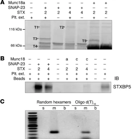
STX4 were used as surrogates to create syntaxin-SNAP23 and syntaxin-Munc18 complexes for pulldown assays. Using human platelet extracts and various syntaxin-containing complexes as bait, we recovered 5 bands that represented proteins specifically bound to 1 or more of the syntaxin-containing baits used (Figure 1A). Mass spectrometry analysis showed that bands T1 and T2 were STXBP5, band T3 was phosphofructokinase C, band T4 was Munc18b, and band T5 was granuphilin (also known as SLP4) (Fig-ure 1 and Supplemental Table 1; supplemental material available online with this article; doi:10.1172/JCI75572DS1). Munc18b is known to be required for platelet secretion and to bind to t-SNARE complexes (13, 37). Granuphilin is a known Rab27 effector and is important for dense granule secretion (12, 38). The role of phos-phofructokinase is unclear at present. STXBP5 has been shown to be involved in neuronal and neuroendocrine exocytosis (21, 24, 39). Platelet STXBP5 (bands T1 and T2) was specifically associ-ated with t-SNARE heterodimers, consistent with previous reports showing that STXBP5 forms ternary complexes with STX4/ SNAP23 and STX1/SNAP25 (22, 31). To verify STXBP5 expression in human platelets and our mass spectrometry results, we probed platelet extracts and affinity-purified complexes by IB using an anti-STXBP5 peptide mAb (whose epitope is a region shared by all 3 isoforms). A specific immunoreactive band was only seen in samples bound to the t-SNARE heterodimers, not in those bound to Munc18a- or Munc18c-containing complexes (Figure 1B). To determine which STXBP5 isoforms were present in human platelets, RT-PCR analysis was performed using random hexam-ers and oligo d(T) to prime the revhexam-erse transcription step, then using isoform-specific primers for PCR (29, 40). Only primers for m-STXBP5 produced a product (Figure 1C), which suggests that m-STXBP5 is the most abundant isoform in platelets. Expression of STXBP5 has been consistently reported previously, in the mass spectrometry analysis of the human platelet proteome by Burkhart et al. (41) and in a genome-wide RNA sequence analysis of human and mouse platelets by Rowley et al. (42).
tion of Stxbp5 in mice enhances synaptic transmission (24, 32, 33). However, the N-terminal domain of STXBP5, lacking the syntaxin- binding v-SNARE motif, can also inhibit secretion from PC-12 cells, which suggests that other interactions with STXBP5 are important (33). Conversely, knockdown of STXBP5 in superior cervical gan-glion neurons (34) and in the rat β cell line INS-1E (35), and genetic depletion of the homologs Sro7 and Sro77 in yeast (25), negatively affects exocytosis. Thus, although STXBP5 may be a negative regu-lator of secretion in some cells, it may play a positive role in others (24). To date, the role of this potential SNARE regulator in platelets has not been addressed.
Various studies have linked STXBP5 with neuropsychological and cardiovascular diseases in humans. Deletions in the Stxbp5 gene are linked to autism (36). GWAS show genetic variations in
Stxbp5 are linked with increased plasma levels of vWF (14–19),
alterations in tissue plasminogen activator (tPA) levels (20), venous thrombosis (16), and arterial thrombosis (19). Specifically, 1 SNP that produces a nonsynonymous mutation (N436S) was associated with increased bleeding (18). These associations sug-gest a role for STXBP5 in both endothelial cell (EC) and platelet secretion and point to a role for the protein in normal hemostasis. In the present study, we examined the platelet phenotype of mice lacking STXBP5 to understand how this t-SNARE regulator affects platelet exocytosis, granule biogenesis, and hemostasis.
Results
STXBP5 is present in human platelets. The critical SNAREs in
[image:3.585.45.274.58.297.2]plate-lets have been identified: STX11 and SNAP23 as the t-SNAREs, and VAMP8 as the primary v-SNARE (6, 8, 9). Of these 3 SNARE types, syntaxins and their binding proteins have dominated the ranks of potential secretion regulators, which suggests that syn-taxins or syntaxin-containing complexes might serve as useful “bait” to identify additional secretion regulators. Because of our problems with the insolubility of STX11 when expressed in bac-teria (S. Ye and J. Zhang, unpublished observation), STX2 and
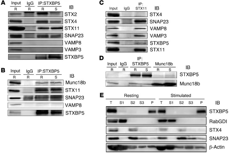
less, these data showed that STXBP5 associated with the critical secretory components SNAP23, STX11, and Munc18b in platelets and that its association was exclusive of the VAMPs.
STXBP5 localization in platelets. We next sought to
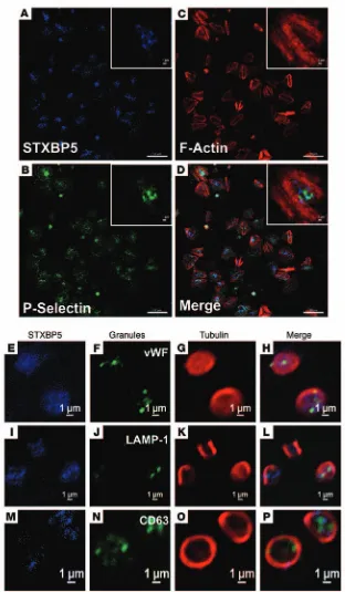
deter-mine the distribution of STXBP5 in platelets using biochemical fractionation and immunofluorescence microscopy. Resting and thrombin-activated platelets were disrupted by repeated freeze-thaw and separated into cytosolic and pelleted fractions by cen-trifugation (Figure 2E). The pelleted fractions were extracted sequentially with Triton X-100 and n-octyl-β-d-glucopyranoside to solubilize membrane proteins and lipid raft components, respectively. The cytosolic marker RabGDI fractionated into the first supernatant, and the membrane protein marker STX4 was found in the Triton X-100–solubilized fraction. The raft marker SNAP23 was present both in the Triton X-100–solubilized frac-tion and in the n-octyl-β-d-glucopyranoside–solubilized fraction. STXBP5 was found in the final cytoskeletal pellet, consistent with actin’s presence (43). The distribution of STXBP5 in plate-lets (spread on fibrinogen-coated coverslips) was also sugges-tive of interaction with the cytoskeleton. STXBP5 was concen-trated in the central region of the platelets and was surrounded by phalloidin-stained actin filaments (Figure 3, A–D). Because these platelets were surface-activated by spreading on coated glass, few granules remained, but P-selectin staining did appear in the central area of some platelets (Figure 3, A–D), consistent
STXBP5 is associated with platelet SNARE machinery. To
dem-onstrate that STXBP5 does interact with the functionally rel-evant syntaxin isoform in platelets, STX11 (as well as SNAP23 and VAMP8), we immunoprecipitated endogenous STXBP5 from detergent-solubilized extracts and probed the precipitate with antibodies against known elements of the platelet secretory machinery. Both resting and thrombin-stimulated human platelets were lysed with Triton X-100, and immunoprecipitation was per-formed using nonspecific IgG or an anti-STXBP5 mAb. It should be noted that much of the platelets STXBP5 was in the pellet frac-tion under these condifrac-tions; however, we chose to focus only on the soluble pool, so as to lessen the chance of detecting artifac-tual interactions due to incomplete solubilization. IB showed that the t-SNAREs STX11 and SNAP23 were recovered in the STXBP5 immunoprecipitates (Figure 2, A and B). Reciprocal immunopre-cipitation with anti-STX11 antibodies confirmed this interaction (Figure 2C). In agreement with the in vitro pulldown results (Fig-ure 1A), endogenous STXBP5 could also bind STX2 and STX4. No VAMP3 or VAMP8 was detected, consistent with the notion that STXBP5 competes with v-SNAREs to bind t-SNARE complexes (31). Another platelet secretory component, Munc18b, was coim-munoprecipitated with STXBP5 (Figure 2B). Reciprocal immuno-precipitation confirmed the STXBP5/Munc18b interaction (Figure 2D). At this stage, it was not mechanistically clear why the com-plex composition was unaffected by platelet activation.
Regard-Figure 2. STXBP5 is associated with platelet SNARE complexes and cytoskeleton. (A–D) Platelet extracts (Input) from resting (R) or thrombin-stimulated (S) platelets were prepared by solubilization with 1% Triton X-100. After clarification, platelet extracts were incubated with anti-STXBP5 mAb (A) or rabbit polyclonal Ab (B and D), anti-STX11 rabbit polyclonal Ab (C), anti-Munc18b goat polyclonal Ab (D), or IgG control for 3 hours at 4°C. Immune complexes were recovered with protein A and G sepharose. The bound proteins were eluted and separated by SDS-PAGE, followed by IB with the indicated antibodies. (E) Washed platelets (2.5 × 10⁸) were resuspended in HEPES/Tyrode buffer and incubated with (stimulated) or without (resting) 0.1 U/ml thrombin for
5 minutes. After disruption by freeze-thaw, the unbroken cells were removed by centrifugation at 700 g for 5 minutes, and supernatants were subjected to ultracentrifugation (100,000 g for 1 hour at 4°C). The cytosolic fractions (S1) were collected. The remaining pellets were solubilized sequentially with Triton X-100 (S2), then n-octyl-β-d-glucopyranoside (S3). The supernatants and insoluble pellet (P) were analyzed by IB with the indicated antibodies. T, total
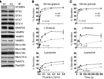
[image:4.585.107.478.58.307.2]and subjected to IB analysis. The protein levels of 14 known secre-tory machinery components were examined; other than STXBP5, none appeared to be significantly altered (Figure 4A). To under-stand the role of STXBP5, we examined stimulation-dependent release of cargo from all 3 platelet granules: dense granules, α granules, and lysosomes. Secretion of granule cargo from Stxbp5 KO and WT platelets showed the expected thrombin dose depen-dence (Figure 4B); however, the percentage release from α gran-ules and lysosome was significantly affected by the loss of STXBP5. The release from dense granules was significantly affected, but to a lesser extent, in this assay system. The kinetics of secretion from with this area representing the centralized granulomere of an
activated platelet. Immunofluorescence staining of STXBP5 in resting platelets, with circumferential tubulin rings, did not show any obvious colocalization with markers for the 3 specific platelet granule types (Figure 3, E–P).
STXBP5 is important for platelet secretion. To explore the
[image:5.585.33.345.51.586.2]func-tional role of STXBP5, we analyzed Stxbp5 KO mice. As might be predicted from GWAS analysis (14–19), plasma vWF levels were increased in these mice, although plasma IgG was unaffected, as indicated by light chain levels (Supplemental Figure 1). Platelet extracts from Stxbp5 KO and littermate WT mice were prepared
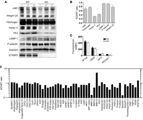
decrease in platelet factor 4 (PF4) in Stxbp5 KO versus WT platelets (Table 1). Serotonin and β-hexosaminidase levels were not signifi-cantly reduced; in fact, total serotonin levels and uptake of [3
H]-serotonin were increased by approximately 60% (Table 1). These data suggest that STXBP5 contributes to granule biogenesis as well as to secretion. To characterize the effect of STXBP5 loss on plate-let cargo, we probed extracts with granule-specific antibodies (Fig-ure 7, A and B). IB data confirmed the reduction of PF4 and showed reduced levels of factor V. Platelet fibrinogen and vWF levels were modestly affected. The total levels of granule membrane proteins (e.g., LAMP-1 and P-selectin) were only slightly affected, while the surface expression of several platelet markers (e.g., GP1bβ, GPVI, and PECAM-1) was largely unaffected (Figure 7, B and C). Consis-tent with the levels of LAMP-1 and P-selectin, EM analysis showed no significant difference in the number of granules present per platelet section when dense and α granules were counted (Table 1). These data argue that granules are made, but may not be normally loaded with cargo. Further analysis of soluble cargo levels yielded a complex phenotype. Antibody array analysis showed that several cargo proteins were reduced in Stxbp5 KO platelets. Others were unchanged, and a few, including MMP-9, showed robust increases (Figure 7D). All cargo proteins tested were detectable at some level (Supplemental Figure 4). These data indicate that STXBP5 contrib-utes to the efficacy — and perhaps the selectivity — of granule cargo packaging, but is not essential for the process.
STXBP5 is important for hemostasis. The primary function of
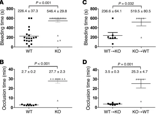
platelet secretion is in hemostasis. To assess whether STXBP5 contributes to hemostasis, we used 2 in vivo assays. First, a tail bleeding time assay was performed on 5- to 6-week-old mice. Whereas the 19 WT littermates showed an average bleeding time of 226.4 ± 37.3 s, of the 24 Stxbp5 KO mice, only 3 had normal bleeding times; the remainder bled excessively until the study was ended at 10 minutes (Figure 8A). The prolonged tail
bleed-Stxbp5 KO platelets was also significantly defective upon
activa-tion with 0.1 U/ml thrombin (Figure 4C).
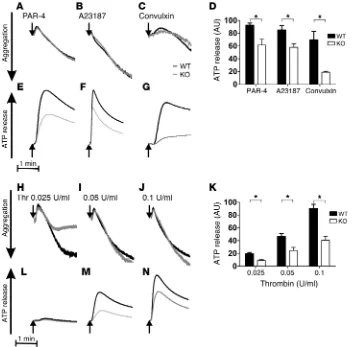
Because dense granule release is such a rapid process, measur-ing [3H]-serotonin release in our assay configuration at room
tem-perature (Figure 4, B and C) may not be sufficient to fully appre-ciate a subtle, albeit significant, kinetic defect. To rectify this, we applied more sensitive lumi-aggregometry to analyze ATP/ADP release from dense granules. ATP/ADP release in response to 100 μM PAR-4, 10 nM A23187, 0.1 μg/ml convulxin, and 0.025, 0.05, and 0.1 U/ml thrombin was substantially inhibited, whereas aggregation was not (Figure 5). Quantification of ATP/ADP release from 3 independent experiments showed significant reductions in
Stxbp5 KO platelets (Figure 5, D and K). To further confirm that
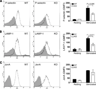
STXBP5 is required for α granule and lysosome release, exposure of the respective markers P-selectin and LAMP-1 was analyzed by flow cytometry. Consistently, activation-dependent exposure of both granule membrane markers was reduced approximately 2-fold in Stxbp5 KO versus WT platelets (Figure 6, A and B).
In agreement with the aggregation data, there was no sig-nificant difference in PE-conjugated JonA staining (Figure 6C), which suggests that depletion of STXBP5 did not affect integrin αIIbβ3 activation (15, 44, 45). Electron microscopy (EM) analysis detected no overt morphology differences in Stxbp5 KO platelets (Supplemental Figure 2). Upon thrombin stimulation, there was similar filopodia formation in both WT and Stxbp5 KO platelets, which suggests that deficiency of STXBP5 did not affect platelet cytoskeletal rearrangements or platelet activation, but did affect secretion. Additionally, adhesion to fibrinogen was not affected in
Stxbp5 KO platelets, although there was a clear defect in spreading
on fibrinogen (Supplemental Figure 3). This is consistent with the role of granule secretion in platelet spreading (46).
STXBP5 is important for platelet granule cargo packaging. As we
[image:6.585.44.380.57.313.2]measured cargo release as a percentage of total, we also detected a
Figure 4. STXBP5-deficient mouse platelets have a secretion defect. (A) Platelet extracts (5 × 10⁷ platelets/lane) were prepared from
Stxbp5 KO and WT mice, and the indicated proteins were analyzed by IB. (B and C) [3
H]-Serotonin–labeled platelets from Stxbp5 KO and WT mice were prepared as described in Methods. Release of [3H]-serotonin from
ing of the Stxbp5 KO mice indicates that STXBP5 is required for hemostasis in this transection injury model.
In agreement with the tail bleeding, there was a robust throm-bus formation defect observed in Stxbp5 KO mice in response to FeCl3 injury of the carotid artery. The average time required to form a stable thrombus in WT littermates was 2.7 ± 0.2 minutes, in contrast to 27.7 ± 2.3 minutes for Stxbp5 KO mice (Figure 8B), which indicates that STXBP5 is critical for hemosta-sis in this arterial injury model.
Platelet counts were not significantly different in Stxbp5 KO and WT littermate mice, although the former exhibited an increase in mean platelet volume (Table 1); therefore, throm-bocytopenia is unlikely to be the cause of the bleeding diathe-sis. The combination of cargo and secretion deficiencies in the
Stxbp5 KO platelets could account for the robust bleeding, but
since the mouse strain is a global deletion, other processes could precipitate the hemostatic defects. To address whether the loss of STXBP5 in BM-derived platelets accounted for the hemo-static defect, BM chimeras were constructed via transplantation:
Stxbp5 KO BM was grafted into WT recipients, and vice versa.
When Stxbp5 KO BM was grafted into WT recipients, there was a significant increase in bleeding times in the resulting chimeras compared with WT BM grafted into Stxbp5 KO recipients (Figure 8C). Similarly, occlusion times in the carotid injury model were significantly prolonged in Stxbp5 KO BM–grafted WT chimeric mice (Figure 8D). These results suggest that loss of STXBP5 in
BM-derived hematopoietic cells (e.g., platelets) contributes to the robust bleeding defect observed (Figure 8, A and B). Since the
Stxbp5 KO animals grafted with WT BM had a normal bleeding
profile, it seems unlikely that loss of STXBP5 affects coagulation factors, although this was not directly measured. In summary, these results indicate a role for STXBP5 in platelet granule pack-aging and secretion and in arterial hemostasis.
Discussion
The novel findings herein were that STXBP5 was required for nor-mal platelet secretion, granule cargo packaging, and hemostasis. This requirement of STXBP5 for platelet exocytosis diverges from the protein’s proposed role as a negative regulator in neuronal cell, neuroendocrine cell, and EC secretion (21, 24, 39, 47). Our pres-ent data indicate that STXBP5 may be important for cardiovascu-lar health, as echoed in recent GWAS in which STXBP5 has been linked to thrombosis (14–20).
[image:7.585.35.392.56.403.2]Platelet granule release requires the secretory machinery components that are universally used for regulated secretion: SNAREs, Munc18, Rabs, and Munc13 (48). Much of the pub-lished data suggest that STXBP5 is a negative regulator of secre-tion, competing with v-SNAREs for t-SNARE binding. As in those prior studies, we showed here that STXBP5 interacted with t-SNARE–containing complexes and that its presence was exclu-sive of at least 2 platelet v-SNAREs (Figure 2). However, deletion of STXBP5, as well as inclusion of an anti-STXBP5 antibody into Figure 5. Depletion of STXBP5 in platelets affects ATP/ADP release, but not aggrega-tion. Aggregation (A–C and H–J) and ATP/ ADP release (D–G and K–N) were moni-tored simultaneously in a lumi-aggregom-eter. Washed platelets (2.0 × 10⁵/μl) from
a permeabilized platelet secretion assay, inhibited rather than enhanced platelet secretion (Figures 4–6 and Supplemental Fig-ure 5). This result raises the question of how STXBP5 could have a positive effect on exocytosis. Studies in Drosophila show that loss of STXBP5 prolongs the excitatory junctional currents (EJCs) at neuromuscular junctions (NMJs) (49). This could be due to ecto-pic vesicle priming (adjacent to active zones) or prolonged fusion pore opening, which suggests that the role of STXBP5 is to spatial-ly restrict vesicle priming and/or fusion. In platelets, spatial gran-ule priming for exocytosis may be critical for efficacious secre-tion. While our in-suspension secretion assays (Figures 4–6) may be less influenced by a loss of polarization, secretion in a form-ing thrombus almost assuredly needs to be polarized (50). Thus, STXBP5 may be important for normal thrombosis, and its dele-tion would cause the hemostasis defects we observed (Figure 8). At this stage, it is unclear why STXBP5 is important for secretion from yeast (25), INS-1E cells (35), or platelets (Figures 4–6); how-ever, its interactions with cytoskeleton (Figure 2E and Figure 3), especially myosin Va (28, 42, 51), may hint at a role in spatially coordinating SNARE complexes for membrane fusion and sub-sequent secretion. STXBP5 interactions with cytoskeleton could contribute to granule biogenesis by directing the trafficking of cargo proteins to α granules forming in megakaryocytes. The heterogeneity in cargo levels we observed herein (Figure 7 and Supplemental Figure 4) may be indicative of multiple pathways for granule cargo sorting and packaging. Further work will be required to address these potential functions of STXBP5.
GWAS have linked SNPs in STXBP5 to alterations in vWF levels (14–19) and tPA levels (18). Loss of STXBP5 increases vWF release from ECs (47), but decreases tPA release (20). These
results highlight potentially conflicting roles for STXBP5 in ECs, which may relate to which granules are being released. vWF and tPA are enriched in different granules in ECs (52). Combining these 2 effects, heightened vWF and decreased tPA, might be expected to increase thrombotic risk, consistent with the premise of the GWAS. Conversely, an SNP encoding a nonsynonymous substitution in STXBP5 (N436S) has been linked to increased bleeding in a female population homozygous for the allele (18). The dramatically prolonged bleeding times in the tail transection model and the hemostasis defects in the FeCl3-induced carotid artery injury model we observed in Stxbp5 KO mice and in Stxbp5 KO BM–grafted WT mice are consistent with this prior report and support a connection between STXBP5 and bleeding risk. At this Figure 6. Deletion of STXBP5 inhibits P-selectin and LAMP-1 exposure, but not integrin activa-tion. Washed platelets (2 × 10⁶) from Stxbp5 KO
[image:8.585.43.366.58.357.2]and WT mice were stimulated with or without 0.1 U/ml thrombin for 1 minute and then incu-bated with FITC-conjugated anti–P-selectin (A), FITC-conjugated LAMP-1 (B), or PE-conjugated JonA (C) Abs for 15 minutes at room tempera-ture. Fluorescence intensities were measured by flow cytometry. Shown are representative data (black lines, resting; gray lines, stimulated) and geometric MFI (mean ± SEM) of 3 indepen-dent experiments. Significant P values (2-way ANOVA) are indicated.
Table 1. Properties of WT and Stxbp5 KO mouse platelets
WT Stxbp5 KO P
Platelet count (×1,000/μl) 824 ± 123 881 ± 101 0.561 Mean platelet volume (fl) 4.20 ± 0.23 6.28 ± 0.10 0.001 Serotonin (μM/108 platelets)A 6.31 ± 0.5 10.64 ± 1.4 0.006 3[H]-Serotonin uptake (cpm/108 platelets) 2,515 ± 224 4,101 ± 545 0.036
PF4 (pg/106 platelets) 1,690 ± 120 545.5 ± 15 <0.0001 β-Hexosaminidase (nMh–1/106 platelets) 54 ± 10 57 ± 11 0.878
Dense granules/plateletB 0.46 ± 0.57 0.52 ± 0.57 0.320 α Granules/plateletB 7.97 ± 1.80 7.84 ±1.22 0.453
P values were calculated using Student’s t test. ADetermined using the
fluorescence o-phthalaldehyde assay (see Supplemental Methods).
BGranules in each platelet in an EM section were counted (n = 200 platelets
In summary, our data provide a potential explanation for the GWAS (14–19) linking STXBP5 with cardiovascular disease risk. Our findings showed that STXBP5 was present in platelets and was important for cargo release from dense granules, α granules, and lysosomes as well as for packaging of some cargo into granules. Mice with platelets deficient in STXBP5 displayed robust defects in thrombus formation in 2 arterial hemostasis models, consis-tent with the granule biogenesis and release defects. Our data add another component to the pathway of protein-protein interac-tions that culminates in platelet secretion. They also suggest that platelets may be a useful system for understanding how STXBP5 positively affects exocytosis in other systems, perhaps through its associations with cytoskeleton. Moreover, platelet STXBP5 may be a useful diagnostic marker for cardiovascular diseases. stage, it is unclear whether the N436S substitution has any effect
on STXBP5 stability or function.
[image:9.585.53.532.55.455.2]Zhu et al. have shown that loss of STXBP5 enhances EC secre-tion of vWF and increases platelet binding to activated ECs (47). Consistent with our results (Supplemental Figure 1), Zhu et al. showed significantly increased plasma vWF levels in Stxbp5 KO mice (47). Clearly, the increased vWF is not sufficient to offset the platelet secretion defect described herein, since we observed bleed-ing tendencies in the 2 arterial hemostasis models tested (Figure 8). However, venous thrombi are mainly composed of fibrin and red blood cells, whereas arterial thrombi are dominated by platelets and fibrin; therefore, it is possible that STXBP5-mediated platelet secretion is more important for arterial hemostasis. It remains to be determined how the loss of STXBP5 affects venous thrombosis.
Figure 7. STXBP5-deficient mouse platelets have a complex granule cargo defect. (A and B) Platelet extracts (5 × 10⁷ platelets/lane) were prepared
from 3 individual Stxbp5 KO or WT mice. The indicated proteins were analyzed by IB (A) and quantified with Image Lab 4.0.1 or ImageQuant TL. After normalization using RabGDI as loading control, the Stxbp5 KO/WT ratio was calculated (B). (C) Washed platelets (2 × 10⁶) from 3 individual Stxbp5 KO or
specific for the b-STXBP5 (5′-CTCCGACTTCCGGTTCTTCCTC-3′
and 5′-TTCAGCGTGATGACAAAGGC-3′), m-STXBP5 (5′
-CTCCG-ACTTCCGCAAAGATGTC-3′ and 5′
-TTCAGCGTGATGACAAAG-GC-3′), and s-STXBP5 (5′-CTCCGACTTCCGATGTGAAAG-3′ and
5′-TTCAGCGTGATGACAAAGGC-3′) isoforms (29, 40).
Immunoprecipitation of STXBP5-interacting proteins. Washed
platelet suspensions (109 platelets) were lysed with equal volumes
of 2× lysis buffer (100 mM HEPES/KOH, pH 7.4; 2% Triton X-100; 2 mM EGTA; 2 mM EDTA; 150 mM NaCl; and 2× protease inhibitor cocktail) on ice for 30 minutes. The lysates were clarified by cen-trifugation, and the supernatants were precleared with protein G/A sepharose (GE Healthcare). The interacting proteins were immuno-precipitated with anti-STXBP5, anti-Munc18b, or anti–Munc13-4 Abs, followed by incubation with protein G sepharose. The immunoprecip-itates were recovered by centrifugation, washed with lysis buffer, and analyzed by SDS-PAGE and IB.
Genotyping of Stxbp5 KO mice. Stxbp5 KO mice on a 50% 129Sv, 25% C57BL/6, and 25% DBA/2 background were generated as previously described (32). Heterozygous embryos of Stxbp5 KO mice were recov-ered into live mice by the Jackson Laboratory. Genotype was determined by PCR using DNA isolated from tail tip biopsies. The primers used were
as follows: Stxbp5 KO forward, 5′-GGGCGCCCGGTTCTTTTTGTC-3′;
Stxbp5 KO reverse, 5′-GCCATGATGGATACTTTCTCG-3′; WT
for-ward, 5′-TTCTGCTCCCCGCTGCTCCTT-3′; WT reverse, 5′
-TCCCC-GCTCCCTTCACCTTGC-3′. PCR products were 224 bp for the Stxbp5
KO allele and 300 bp for the WT allele.
Measurement of secretion from intact platelets. The secretion assay was carried out as described previously (13); see Supplemental
Meth-ods for details. Briefly, washed platelets were labeled with [3
H]-sero-tonin (0.4 μCi/ml) for 1 hour at 37°C. After washing with HEPES/
Tyrode buffer (10 mM HEPES/NaOH, pH 7.4; 5.56 mM glucose;
137 mM NaCl; 12 mM NaHCO3; 2.7 mM KCl; 0.36 mM KH2PO4; and
1 mM MgCl2) in the presence of 0.2 U/ml apyrase, the platelets were
resuspended and adjusted to 2.5 × 10⁵/μl. For titration experiments,
the indicated concentrations of thrombin were added, and the reac-tions were stopped at 1 minute with a 2-fold excess of hirudin. For time course experiments, 0.1 U/ml thrombin was added for the indicated times, and reactions were stopped with hirudin. Superna-tants and pellets were recovered after centrifugation at 13,000 g for
Methods
Antibodies and reagents. Anti-STXBP5 mAb (611296), anti-Rab27a mAb (clone 20.1), anti-PECAM, FITC-conjugated anti-mouse P-selectin, and unconjugated anti-mouse P-selectin were from BD Biosciences. A rabbit anti-STXBP5 polyclonal Ab (HPA039991) was from Atlas Anti-bodies. The anti–hLAMP-1 (H4A3) and anti-hCD63 (H5C6) mAbs were from the University of Iowa Developmental Studies Hybridoma Bank. Munc18b (sc-14563) polyclonal Ab was from Santa Cruz. Anti-bodies against STX2, STX4, STX7, SNAP23, VAMP8, Munc18c, Rab-GDI, VAMP2 (synaptobrevin), VAMP3 (cellubrevin), VAMP7, VAMP8 (endobrevin), and Munc13-4 were as described previously (6, 8, 53). Anti-STX11 polyclonal Ab and anti-Munc18a mAb (clone 131.1) were purchased from Synaptic System GmbH. PE-conjugated JonA mAb,
anti-GP1bβ, and anti GPVI Abs were purchased from Emfret Analytics.
Anti-human fibrinogen and anti-vWF polyclonal Abs were from Dako.
The anti–integrin β3 antibody and the Alexa Fluor 647–labeled rabbit
anti–α-tubulin antibody (11H10) were from Cell Signaling. The anti–
factor V antibody was provided by B. Bouchard (University of Vermont, Burlington, Vermont, USA). A STXBP5 expression vector (provided by U. Ashery, Tel Aviv University, Tel Aviv, Israel) was used to make anti-gens for the production and purification of a polyclonal anti-STXBP5
antibody (see Supplemental Methods). Anti–β-actin antibody and
alkaline phosphatase–conjugated anti-IgGs were from Sigma-Aldrich. Protein A–conjugated sepharose, 2-methyl-2-butanol, 2,2,2
tribromo-ethanol (Avertin), and FeCl3 were from Sigma-Aldrich. The rest of the
materials used in this study were of reagent grade.
Protein production. Expression plasmids to create complexes of full-length human SNAP23, Munc18a, and Munc18c and the cyto-plasmic domains of human STX2 (aa 1–251) and STX4 (aa 1–265) were constructed using pRSFDuet-1 (Novagen) and confirmed by DNA sequencing. All syntaxin-containing complexes were expressed
in E. coli and purified using Ni2+-NTA (6) and S-protein (Novagen)
affinity chromatography.
[image:10.585.44.303.58.256.2]RT-PCR. Identification of STXBP5 isoforms was performed as described previously (29, 40), with slight modification. Human plate-lets were isolated from banked, platelet-rich plasma (13), total RNA was isolated using QIAamp RNA Blood Mini Kit (QIAGEN), and cDNA was synthesized using TaqMan Reverse Transcription Reagents Kit (Applied Biosystems). PCR amplification was performed using primers
Inc.) was placed on the artery to monitor blood flow. Thrombus for-mation was induced by placing a small piece of filter paper, saturated
with 5% FeCl3 solution, on the vessel for 3 minutes. Time from
appli-cation to cessation of flow was measured. Experiments were termi-nated at 30 minutes. Statistical analysis was performed using the log-rank test (GraphPad Prism 5).
BM transplantation mouse model. BM transplantation was per-formed as previously described (58), with some modification. Briefly, BM harvested from femurs and tibias of 6-week-old WT or Stxbp5 KO mice was resuspended with 10 mM HEPES, pH 7.4 buffer containing
25 U/ml heparin and 5% FBS and adjusted to 5 × 10⁷ cells/ml. The cell
suspension (0.1 ml) was injected into the retroorbital sinus of lethally
irradiated mice. After 6 weeks of recovery, tail bleeding and FeCl3
-induced hemostasis assays were performed. Statistical analysis was performed using the log-rank test (GraphPad Prism 5).
Subcellular fractionation of platelets. Human platelets were resus-pended in HEPES/Tyrode buffer (pH 7.4) and treated with or without 0.1 U/ml thrombin for 5 minutes. The resting and activated platelets were disrupted by 5 freeze-thaw cycles. After disruption, the unbroken cells were removed by centrifugation at 700 g for 5 minutes, and the supernatants were fractionated by ultracentrifugation at 100,000 g for 1 hour. The supernatant (cytosol) was collected, and the result-ing pellets were solublized with 1% Triton X-100, incubated on ice for 15 minutes, and then subjected to centrifugation at 100,000 g for 1 hour. The supernatant (Triton X-100–soluble fraction) was
col-lected, and the resulting pellets were solublized with 1% n-octyl-β
-d-glucopyranoside, incubated on ice for 15 minutes, and then subjected to centrifugation at 100,000 g for 1 hour. The supernatant
(n-octyl-β-d-glucopyranoside–soluble fraction) was collected. The
detergent-insoluble pellets were solublized in SDS-PAGE loading buffer. The supernatants (soluble fractions) and pellets (detergent-insoluble frac-tions) were probed by IB using anti-STXBP5, anti-RabGDI, anti-STX4,
anti-SNAP23, and anti–β-actin antibodies.
Angiogenesis antibody array analysis of platelet granule cargo.
Pro-teome Profiler Mouse Angiogenesis Antibody Array (ARY 015; R&D
Systems) was used according to the manufacturer’s instructions. Briefly, the nitrocellulose membranes were first incubated in blocking
buffer for 1 hour. Platelet extracts (from 108 platelets) were prepared in
1 ml lysis buffer (1% NP-40; 20 mM Tris-HCl, pH 8.0; 137 mM NaCl; 10% glycerol; 2 mM EDTA; and 10× protease inhibitor cocktail) and mixed with a cocktail of biotin-labeled detection antibodies against different individual angiogenesis-related proteins. After incubation for 1 hour, the mixture of the platelet sample and detection antibod-ies was then incubated with the nitrocellulose membranes O/N at 4°C. The membranes were washed 3 times and incubated with HRP-con-jugated streptavidin for 30 minutes. After washing the membranes 3 times, the signals for each array spot were detected with Supersignal ELISA Femto Maximum Sensitivity Substrate (Thermo-Scientific) using a ChemiDoc MP System (BioRad). The array experiments were run in duplicate. Fluorescence intensities for each spot were mea-sured, background was subtracted, and the Stxbp5 KO/WT ratio was calculated and plotted using SigmaPlot (version 12.0; Systat Software Inc.). Under these conditions, spots for 3 proteins — SDF-1, PF4, and angiopoietin 1 — were saturated, and thus failed to yield quantitative data (Supplemental Figure 4). These were excluded from analysis.
Statistics. Data from bleeding time and occlusion assays were ana-lyzed by log-rank test. Data from secretion assays and FACS-based 1 minute, and the pellets were lysed with an equal volume of lysis
buffer (PBS, pH 7.4, and 1% Triton X-100) for 1 hour on ice. Both supernatants and pellets were assayed for 3 granule cargo markers:
[3H]-serotonin for dense granules by scintillation counting, platelet
factor 4 (PF4) for α granules by ELISA, and β-hexosaminidase for
lysosomes by colorimetric assay using p-nitrophenyl-N-acetyl-β-
d-glucosaminide, as previously described (54). Secretion was then calculated as (supernatant/[supernatant + pellet]) and expressed as a percentage; the analysis yields a measurement of total cargo con-tent (supernatant plus pellet) and allows for assessment of secretion efficacy independent of cargo content.
Immunofluorescence microscopy. Platelet preparation and immuno-staining were performed as previously described (55), with slight modification. Briefly, sterile glass coverslips were coated with 50
μg/ml human fibrinogen for 24 hours at 4°C. Washed platelets were
placed onto fibrinogen-coated coverslips for 1 hour at 37°C. Platelets were fixed with 4% paraformaldehyde for 15 minutes and quenched
with 50 mM NH4Cl. Cells were rinsed with 10% FBS/PBS and
incu-bated with the indicated primary antibodies in 10% FBS/PBS contain-ing 0.2% saponin O/N at 4°C. After washcontain-ing with 10% FBS/PBS, cells were incubated with the appropriate fluorophore-conjugated anti-IgG secondary antibody for 2 hours. After washing, some samples were incubated with TRITC-conjugated phalloidin in 10% FBS/PBS with 0.2% saponin. Coverslips were mounted and examined with a LSM 780 confocal microscope (Carl Zeiss), and images were processed using ZEN blue software (Carl Zeiss). Additional imaging was done using a Nikon A1R confocal microscope, and images were processed using NIS-Elements AR 3.2 software (Nikon).
Platelet aggregation and ATP/ADP release. Murine platelets (2.5 × 10⁵/
μl) were prepared (13), recalcified with 0.7 mM CaCl2, placed into
silicon-ized cuvettes, and stirred for 3 minutes at 37°C at 1,200 rpm. Luciferin-luciferase substrate was added, followed by the indicated agonists, and the percent change in light transmission was measured. Aggregation and ATP/ADP secretion were monitored using a Model 460VS Lumi-Dual-aggregometer, and traces were acquired using a Model 810 Aggro/Link interface with Aggro/Link software (all from Chrono-Log).
Flow cytometry analysis. Washed murine platelets (2 × 10⁶) were kept as resting or were stimulated with thrombin (0.1 U/ml) for 1 min-ute, after which the reaction was stopped with hirudin. After
incuba-tion with antibodies (5 μl) for 15 minutes at room temperature, the
reactions were diluted 10-fold with HEPES/Tyrode buffer (pH 6.5). The samples were transferred to tubes, and fluorescence intensities were measured using a FACScan flow cytometer and analyzed using CellQuest (BD Biosciences).
Tail bleeding assay. Tail vein bleeding times were determined as previously described (56). Briefly, 5- to 6-week-old mice were anes-thetized with ketamine (75 mg/kg i.p.). Tails were transected 3 mm from the tip and immersed in saline at 37°C. The time from incision to bleeding cessation was recorded. Animals were observed for an addi-tional minute to assess rebleeding. Prolonged bleeding was stopped manually after 10 minutes. Statistical analysis was performed using the log-rank test (GraphPad Prism 5).
(University of California, Davis, California, USA) for insightful dis-cussions, the staff of the Kentucky Blood Center for support, and the members of the Whiteheart laboratory for comments and care-ful proofreading of the manuscript. The mass spectrometry analy-sis was performed by Carol Beach at the University of Kentucky Center for Structural Biology Protein Core Facility; this core and the imaging facility are supported in part by funds from NIH National Center for Research Resources (NCRR) grant P20 RR020171. This work is supported by NIH grants HL56652 and HL082193 to S.W. Whiteheart and NIH grant T32HL091812 to S. Ye.
Address correspondence to: Sidney W. Whiteheart, Department of Molecular and Cellular Biochemistry, University of Kentucky College of Medicine, 741 South Limestone, Biomedical Biological Sciences Research Building, Lexington, Kentucky 40536-0509, USA. Phone: 859.257.4882; E-mail: whitehe@uky.edu.
experiments were analyzed by 2-way ANOVA. A 1-tailed Student’s t test was used to analyze the properties of platelets from Stxbp5 KO and WT mice. A P value less than 0.05 was considered significant.
Study approval. Animal procedures were approved by the IACUC of University of Kentucky. Human samples were from anonymized units; thus, no IRB approval or informed consent was required.
Acknowledgments
We thank Uri Ashery for providing a STXBP5 expression construct, Beth Bouchard for anti–factor V antibody, Mary Gail Engle and Jim Begley (Imaging Facility of University of Kentucky) for help with EM analysis, Greg Bauman (Flow Cytometry Core Facility of Uni-versity of Kentucky) for help with FACS analysis, the staff of the Department of Laboratory Animal Resources at University of Ken-tucky for assistance, Richard Charnigo (Department of Statistics, University of Kentucky) for help with data analysis, Leighton Izu
1. Michelson AD. Antiplatelet therapies for the treatment of cardiovascular disease. Nat Rev Drug Discov. 2010;9(2):154–169.
2. Lopes RD. Antiplatelet agents in cardiovascular disease. J Thromb Thrombolysis. 2011; 31(3):306–309.
3. Michelson AD. Platelets. San Diego, California, USA: Elservier Science; 2002.
4. Graham GJ, Ren Q, Dilks JR, Blair P, Whiteheart SW, Flaumenhaft R. Endobrevin/VAMP-8-depen-dent dense granule release mediates thrombus formation in vivo. Blood. 2009;114(5):1083–1090. 5. Weber T, et al. SNAREpins: minimal machinery
for membrane fusion. Cell. 1998;92(6):759–772. 6. Chen D, Bernstein AM, Lemons PP, Whiteheart SW. Molecular mechanisms of platelet exocyto-sis: role of SNAP-23 and syntaxin 2 in dense core granule release. Blood. 2000;95(3):921–929. 7. Chen D, Lemons PP, Schraw T, Whiteheart SW.
Molecular mechanisms of platelet exocytosis: role of SNAP-23 and syntaxin 2 and 4 in lysosome release. Blood. 2000;96(5):1782–1788.
8. Bryceson YT, et al. Defective cytotoxic lym-phocyte degranulation in syntaxin-11 deficient familial hemophagocytic lymphohistiocytosis 4 (FHL4) patients. Blood. 2007;110(6):1906–1915. 9. Ye S, Karim ZA, Al Hawas R, Pessin JE,
Filipo-vich AH, Whiteheart SW. Syntaxin-11, but not syntaxin-2 or syntaxin-4, is required for platelet secretion. Blood. 2012;120(12):2484–2492. 10. Sudhof TC, Rothman JE. Membrane fusion:
grappling with SNARE and SM proteins. Science. 2009;323(5913):474–477.
11. Paumet F, Rahimian V, Rothman JE. The speci-ficity of SNARE-dependent fusion is encoded in the SNARE motif. Proc Natl Acad Sci U S A. 2004;101(10):3376–3380.
12. Tolmachova T, Abrink M, Futter CE, Authi KS, Seabra MC. Rab27b regulates number and secre-tion of platelet dense granules. Proc Natl Acad Sci U S A. 2007;104(14):5872–5877.
13. Al Hawas R, Ren Q, Ye S, Karim ZA, Filipov-ich AH, Whiteheart SW. Munc18b/STXBP2 is required for platelet secretion. Blood. 2012;120(12):2493–2500.
14. Antoni G, et al. Combined analysis of three genome-wide association studies on vWF
and FVIII plasma levels. BMC Med Genet. 2011;12:102.
15. Campos M, et al. Genetic determinants of plasma von Willebrand factor antigen levels: a target gene SNP and haplotype analysis of ARIC cohort. Blood. 2011;117(19):5224–5230.
16. Smith NL, et al. Genetic variation associated with plasma von Willebrand factor levels and the risk of incident venous thrombosis. Blood. 2011;117(22):6007–6011.
17. Smith NL, et al. Novel associations of multiple genetic loci with plasma levels of factor VII, factor VIII, and von Willebrand factor: The CHARGE (Cohorts for Heart and Aging Research in Genome Epidemiology) Consortium. Circula-tion. 2010;121(12):1382–1392.
18. van Loon JE, Sanders YV, de Wee EM, Kruip MJ, de Maat MP, Leebeek FW. Effect of genetic variation in STXBP5 and STX2 on von Wille-brand factor and bleeding phenotype in type 1 von Willebrand disease patients. PLoS One. 2012;7(7):e40624.
19. van Loon JE, et al. Effect of genetic variations in syntaxin-binding protein-5 and syntaxin-2 on von Willebrand factor concentration and cardiovascu-lar risk. Circ Cardiovasc Genet. 2010;3(6):507–512. 20. Huang J, et al. Genome-wide association study
for circulating tissue plasminogen activator levels and functional follow-up implicates endothelial STXBP5 and STX2. Arterioscler Thromb Vasc Biol. 2014;34(5):1093–1101.
21. Fujita Y, et al. Tomosyn: a syntaxin-1-binding protein that forms a novel complex in the neurotransmitter release process. Neuron. 1998;20(5):905–915.
22. Widberg CH, Bryant NJ, Girotti M, Rea S, James DE. Tomosyn interacts with the t-SNAREs syn-taxin4 and SNAP23 and plays a role in insulin-stimulated GLUT4 translocation. J Biol Chem. 2003;278(37):35093–35101.
23. Scales SJ, Hesser BA, Masuda ES, Scheller RH. Amisyn, a novel syntaxin-binding protein that may regulate SNARE complex assembly. J Biol Chem. 2002;277(31):28271–28279.
24. Ashery U, Bielopolski N, Barak B, Yizhar O. Friends and foes in synaptic transmission: the role of tomosyn in vesicle priming. Trends
Neuro-sci. 2009;32(5):275–282.
25. Lehman K, Rossi G, Adamo JE, Brennwald P. Yeast homologues of tomosyn and lethal giant larvae function in exocytosis and are associated with the plasma membrane SNARE, Sec9. J Cell Biol. 1999;146(1):125–140.
26. Gracheva EO, Burdina AO, Touroutine D, Berthel-ot-Grosjean M, Parekh H, Richmond JE. Tomosyn negatively regulates both synaptic transmitter and neuropeptide release at the C. elegans neuromus-cular junction. J Physiol. 2007;585(pt 3):705–709. 27. Strand D, et al. The Drosophila lethal(2)giant
lar-vae tumor suppressor protein forms homo-oligo-mers and is associated with nonmuscle myosin II heavy chain. J Cell Biol. 1994;127(5):1361–1373. 28. Pruyne DW, Schott DH, Bretscher A.
Tropo-myosin-containing actin cables direct the Myo2p-dependent polarized delivery of secretory vesicles in budding yeast. J Cell Biol. 1998;143(7):1931–1945.
29. Yokoyama S, Shirataki H, Sakisaka T, Takai Y. Three splicing variants of tomosyn and identifi-cation of their syntaxin-binding region. Biochem Biophys Res Commun. 1999;256(1):218–222. 30. Groffen AJ, Jacobsen L, Schut D, Verhage M. Two
distinct genes drive expression of seven tomosyn isoforms in the mammalian brain, sharing a con-served structure with a unique variable domain. J Neurochem. 2005;92(3):554–568.
31. Hatsuzawa K, Lang T, Fasshauer D, Bruns D, Jahn R. The R-SNARE motif of tomosyn forms SNARE core complexes with syntaxin 1 and SNAP-25 and down-regulates exocytosis. J Biol Chem. 2003;278(33):31159–31166.
32. Sakisaka T, et al. Dual inhibition of SNARE complex formation by tomosyn ensures con-trolled neurotransmitter release. J Cell Biol. 2008;183(2):323–337.
33. Yizhar O, et al. Multiple functional domains are involved in tomosyn regulation of exocytosis. J Neurochem. 2007;103(2):604–616.
34. Baba T, Sakisaka T, Mochida S, Takai Y. PKA-cata-lyzed phosphorylation of tomosyn and its implica-tion in Ca2+-dependent exocytosis of neurotrans-mitter. J Cell Biol. 2005;170(7):1113–1125. 35. Cheviet S, et al. Tomosyn-1 is involved in a
exocytosis. J Cell Sci. 2006;119(pt 14):2912–2920. 36. Davis LK, et al. Novel copy number variants in
chil-dren with autism and additional developmental anomalies. J Neurodev Disord. 2009;1(4):292–301. 37. Sandrock K, Nakamura L, Vraetz T, Beutel K, Ehl S, Zieger B. Platelet secretion defect in patients with familial hemophagocytic lymphohistiocytosis type 5 (FHL-5). Blood. 2010;116(26):6148–6150. 38. Hampson A, O’Connor A, Smolenski A.
Synap-totagmin-like protein 4 and Rab8 interact and increase dense granule release in platelets. J Thromb Haemost. 2013;11(1):161–168. 39. Yizhar O, et al. Tomosyn inhibits priming
of large dense-core vesicles in a calcium-dependent manner. Proc Natl Acad Sci U S A. 2004;101(8):2578–2583.
40. Zhang W, et al. Tomosyn is expressed in beta-cells and negatively regulates insulin exocytosis. Diabetes. 2006;55(3):574–581.
41. Burkhart JM, Schumbrutzki C, Wortelkamp S, Sickmann A, Zahedi RP. Systematic and quantitative comparison of digest efficiency and specificity reveals the impact of trypsin quality on MS-based proteomics. J Proteomics. 2012;75(4):1454–1462.
42. Rowley JW, et al. Genome-wide RNA-seq analysis of human and mouse platelet transcriptomes. Blood. 2011;118(14):e101–e111.
43. Falet H, Hoffmeister KM, Neujahr R, Hartwig JH. Normal Arp2/3 complex activation in platelets lacking WASp. Blood. 2002;100(6):2113–2122. 44. Bergmeier W, Stefanini L. Novel molecules in
calcium signaling in platelets. J Thromb Haemost. 2009;7(suppl 1):187–190.
45. Buensuceso CS, Arias-Salgado EG, Shattil SJ. Protein-protein interactions in platelet alphaI-Ibbeta3 signaling. Semin Thromb Hemost. 2004;30(4):427–439.
46. Peters CG, Michelson AD, Flaumenhaft R. Gran-ule exocytosis is required for platelet spreading: differential sorting of α-granules expressing VAMP-7. Blood. 2012;120(1):199–206. 47. Zhu Q, et al. Syntaxin-binding protein STXBP5
inhibits endothelial exocytosis and promotes plate-let secretion.J Clin Invest. 2014;124(10):4503–4516. 48. Sudhof TC, Rizo J. Synaptic vesicle
exo-cytosis. Cold Spring Harb Perspect Biol. 2011;3(12):a005637.
49. Chen K, Richlitzki A, Featherstone DE, Schwar-zel M, Richmond JE. Tomosyn-dependent regu-lation of synaptic transmission is required for a late phase of associative odor memory. Proc Natl Acad Sci U S A. 2011;108(45):18482–18487. 50. Brass LF, Tomaiuolo M, Stalker TJ. Harnessing
the platelet signaling network to produce an optimal hemostatic response. Hematol Oncol Clin
North Am. 2013;27(3):381–409. 51. Pastural E, et al. Griscelli disease maps to
chromosome 15q21 and is associated with mutations in the myosin-Va gene. Nat Genet. 1997;16(3):289–292.
52. Knipe L, et al. A revised model for the secretion of tPA and cytokines from cultured endothelial cells. Blood. 2010;116(12):2183–2191. 53. Schraw TD, Lemons PP, Dean WL, Whiteheart
SW. A role for Sec1/Munc18 proteins in platelet exocytosis. Biochem J. 2003;374(pt 1):207–217. 54. Schraw TD, et al. Granule stores from
cel-lubrevin/VAMP-3 null mouse platelets exhibit normal stimulus-induced release. Blood. 2003;102(5):1716–1722.
55. Choi W, Karim ZA, Whiteheart SW. Arf6 plays an early role in platelet activation by collagen and convulxin. Blood. 2006;107(8):3145–3152. 56. Broze GJ Jr, Yin ZF, Lasky N. A tail vein bleeding
time model and delayed bleeding in hemophiliac mice. Thromb Haemost. 2001;85(4):747–748. 57. Zhang G, et al. Biphasic roles for soluble
guany-lyl cyclase (sGC) in platelet activation. Blood. 2011;118(13):3670–3679.