Development 139, 1956-1964 (2012) doi:10.1242/dev.079772 © 2012. Published by The Company of Biologists Ltd
INTRODUCTION
A recurring theme in development is the use of morphogen gradients, where different concentrations of a regulatory molecule specify different cell fates (reviewed by Ashe and Briscoe, 2006; Rogers and Schier, 2011). For example, patterning of the dorsoventral (DV) axis of the Drosophila blastoderm embryo utilizes two morphogen gradients. A ventral-to-dorsal gradient of nuclear-localized Dorsal (Dl) divides the blastoderm embryo into three basic domains: mesoderm, neuroectoderm and dorsal ectoderm; and a dorsal-to-ventral gradient of Dpp further subdivides the dorsal ectoderm into the dorsal-most amnioserosa, and dorsomedial and dorsolateral epidermis.
Dl is a transcription factor related to mammalian NFB, whereas Dpp is an extracellular ligand of the TGFfamily and is most related to vertebrate BMP2. Dpp interaction with transmembrane receptors results in the phosphorylation of cytoplasmic Smad (Mad-P), which in combination with Smad4 (Medea) enters the nucleus and functions as a transcription factor. Hence, both the Dl and Dpp gradients are interpreted by downstream target genes. Several candidate target genes for Dl and Dpp have been identified and classified into categories depending on the level of morphogen that they respond to: high, intermediate and low, also referred to as Type I, II and III, respectively (Ashe et al., 2000; Stathopoulos and Levine, 2004).
How do target genes interpret and respond to morphogen gradients? One widely accepted mechanism is the affinity threshold model, whereby target enhancers respond to low levels of morphogen if they contain high-affinity binding sites. For example, the sogenhancer contains high-affinity Dl binding sites and is thereby activated in regions of low-level Dl protein (the lateral neuroectoderm). By contrast, the twienhancer contains low-affinity Dl binding sites and requires high levels of Dl for activation. Conversion of the low-affinity sites in the twienhancer to high-affinity sites prompted reporter expression in regions containing lower levels of Dl (Jiang and Levine, 1993).
The mechanisms utilized by Dpp target genes in the early embryo are not well understood. Peak levels of Dpp and Mad-P are present along the dorsal midline in a 5- to 6-cell wide domain, the presumptive amnioserosa, while lower levels are found in the 3-4 cells to either side; beyond this, Dpp and Mad-Pdrop precipitously to very low levels (Rushlow et al., 2001; Dorfman and Shilo, 2001; Sutherland et al., 2003; Wang and Ferguson, 2005). Using the same approach of changing binding site affinities it was shown that the target gene Race (Ance – FlyBase), which is expressed in the presumptive amnioserosa, could respond to lower levels of Dpp if high-affinity sites were introduced into its enhancer (Wharton et al., 2004), indicating that differential binding site affinity could underlie threshold responses to the Dpp gradient.
Additional studies on Raceregulation by Xu et al. (Xu et al., 2005) revealed that Raceactivation relies on a feed-forward loop, whereby peak levels of Dpp/Smads first activate zenexpression in the dorsal-most region, and then both Smads and Zen bind and activate Race. The Dpp/Zen feed-forward motif provided a mechanism by which all amnioserosa-specific genes might be activated (Xu et al., 2005).
1Department of Biology, Center for Developmental Genetics, New York University, New York, NY 10003, USA. 2Division of Biological Sciences Pritzker School of Medicine, The University of Chicago, Chicago, IL 60637, USA. 3College of Life Science, National Taiwan University, Taipei, Taiwan 106.
*Author for correspondence ([email protected])
Accepted 26 March 2012
SUMMARY
Pattern formation in the developing embryo relies on key regulatory molecules, many of which are distributed in concentration gradients. For example, a gradient of BMP specifies cell fates along the dorsoventral axis in species ranging from flies to mammals. In Drosophila, a gradient of the BMP molecule Dpp gives rise to nested domains of target gene expression in the dorsal region of the embryo; however, the mechanisms underlying the differential response are not well understood, partly owing to an insufficient number of well-studied targets. Here we analyze how the Dpp gradient regulates expression of pannier (pnr), a candidate low-level Dpp target gene. We predicted that the pnrenhancer would contain high-affinity binding sites for the Dpp effector Smad transcription factors, which would be occupied in the presence of low-level Dpp. Unexpectedly, the affinity of Smad sites in the pnrenhancer was similar to those in the Raceenhancer, a high-level Dpp target gene, suggesting that the affinity threshold mechanism plays a minimal role in the regulation of pnr. Our results indicate that a mechanism involving a conserved bipartite motif that is predicted to bind a homeodomain factor in addition to Smads and the Brinker repressor, establishes the pnrexpression domain. Furthermore, the pnrenhancer has a highly complex structure that integrates cues not only from the dorsoventral axis, but also from the anteroposterior and terminal patterning systems in the blastoderm embryo.
KEY WORDS: Morphogen, BMP gradient, Brk repressor
Response to the BMP gradient requires highly combinatorial
inputs from multiple patterning systems in the
Drosophila
embryo
Hsiao-Lan Liang1, Mu Xu2, Yi-Chun Chuang3and Christine Rushlow1,*
D
E
V
E
LO
P
M
E
N
Analysis of a second Dpp target gene, C15(Lin et al., 2006), which is expressed in a wider domain (8-10 cells) than Racethat extends into the region of intermediate Dpp levels, revealed a different mechanism for threshold responses. The C15enhancer contains multiple clusters of Smad binding sites that contribute cumulatively to the C15expression domain, and, importantly, although it contains higher affinity Smad sites than those in Race, abolishing the highest affinity Mad sites had little effect on the width of the expression domain. By contrast, decreasing the number of Smad sites resulted in a narrow and sporadic expression domain (Lin et al., 2006). Using multiple Smad sites to ensure robust and precise spatial expression might be a distinguishing feature of enhancers that respond to intermediate levels of Dpp.
It remains unclear how target genes respond to low levels of Dpp in the dorsolateral region. Do they also contain multiple clusters of Smad binding sites and/or do they use the affinity threshold mechanism? Here we analyze the regulation of the pannier(pnr) gene, which is expressed in a broad dorsal domain of ~32-35 cells (Jazwinska et al., 1999a; Ashe et al., 2000). To our surprise, the pnr enhancer structure resembled that of Race, containing a similar number of Smad sites with the same relative affinity as those in the Raceenhancer, suggesting that the affinity threshold mechanism plays a minimal role, if any, in the response of pnrto low levels of Dpp. Instead, we found that sequences that lie adjacent to a crucial Smad site are required for proper pnractivation, and that the extent of pnrexpression is limited by Brinker (Brk)-mediated repression. We also found that the pnr expression pattern is influenced by anteroposterior (AP) genes. Thus, pnrgene regulation relies on a complex combinatorial mechanism that integrates spatial cues from diverse patterning systems.
MATERIALS AND METHODS
Fly strains
y1w67c23embryos were used as wild type. dppH46is a null allele balanced
over CyO23, P[dpp+] (Wharton et al., 1993). Homozygous dppH46
embryos were identified by lack of Racestaining. The null alleleszenw36,
brkM68, kni301, tll1, gtx11, hb12and Kr1were balanced over lacZ-marked
balancer chromosomes to distinguish hemizygous or homozygous mutant embryos from their heterozygous and homozygous balancer siblings. bcdE1
is a null allele.
In vitro mutagenesis and transgenic analysis
DNA fragments (summarized in Fig. 2A) were prepared by PCR using genomic DNA as template (Clontech) and the Expand High Fidelity PCR System (Roche Molecular Biochemicals). Nucleotide changes in the Mad binding sites and the HD site were introduced by PCR mutagenesis (see Figs 3 and 5 for DNA sequences). Amplified DNA fragments were first cloned into the pCRII vector using the TOPO cloning system (Invitrogen) and then subcloned into the EcoRI site of the pCasPeRhs43--gal transformation vector (Thummel and Pirrotta, 1992). Plasmid constructs were mixed with helper ‘turbo’ DNA and injected into ywembryos to make transgenic flies. At least three transformant lines for each construct were analyzed.
Bacterial expression of Zen, Mad, Medea and Brk
Expression plasmids encoding GST-Mad and GST-Medea fusion proteins containing the N-terminal MH1 domains were obtained from A. Laughon (Kim et al., 1997) and M. Frasch (Xu et al., 1998), respectively. GST-BrkN
contains the N-terminal 266 amino acids (Rushlow et al., 2001). GST-Zen contained the full-length protein (Xu et al., 2005). The expression and purification of the recombinant proteins were carried out as described (Kirov et al., 1993). The concentration of the isolated proteins was
In vitro DNA binding assays
DNase I footprint analyses and electrophoretic mobility shift (gel shift) assays were carried out as described (Kirov et al., 1993; Rushlow et al., 2001). All DNA fragments were generated from the pnr P3 and P4 enhancers by restriction digestion or PCR (summarized in Fig. 2B) or prepared as oligonucleotides (Integrated DNA Technologies). In each gel shift reaction, 10 ng or 30 ng of GST-Mad, GST-Medea or 5 ng Brk recombinant protein was used; about ten times more was used for footprinting reactions.
In situ hybridization and antibody staining
Wild-type, mutant and transgenic embryos were fixed, hybridized with pnr or lacZ antisense RNA probes labeled with digoxigenin (Roche Molecular Biochemicals) and mounted in Aqua Polymount (Polysciences) as described (Rushlow et al., 2001).
RESULTS
pnris regulated by Dpp and Brk
pnr encodes a GATA factor that is required for several developmental processes including proper subdivision of the embryonic dorsal ectoderm (Ramain et al., 1993; Winick et al., 1993; Heitzler et al., 1996; Herranz and Morata, 2001). In addition, pnrmutant embryos do not undergo proper dorsal closure, leaving holes in the dorsal cuticle (Heitzler et al., 1996). The pnr expression pattern is consistent with these embryonic phenotypes. pnr transcripts are first detected in the dorsal region of the cellularizing embryo from future parasegment 3 (labial) to 14 (A9) (Fig. 1A) (Winick et al., 1993; Heitzler et al., 1996; Herranz and Morata, 2001). In older embryos, expression becomes segmentally repeated and is lost from the amnioserosa, but the AP limits of the pattern remain the same (Herranz and Morata, 2001). Thus, pnr appears to be highly regulated on both the DV and AP axes.
Fig. 1.pnris regulated by Dpp, Zen and Brk.Wild-type (wt) (A) and mutant (B-D) Drosophilaembryos hybridized with pnrRNA probes. Dorsal views, except D in lateral view, with anterior to the left. (A)The
pnrexpression domain at the late blastoderm stage comprises ~30% of the DV circumference and extends from 20-60% egg length. Note that six stripes of different width can be distinguished in the pattern. (B,C)Stripes become more visible in dppH46(B) and zenw36(C) embryos, although there is a much greater effect on the overall pattern in the
V
E
LO
P
M
E
N
To determine how pnrresponds to DV cues, we examined pnr in different mutant backgrounds. pnrexpression was reduced to a few faint stripes in dppmutants (Fig. 1B), but was only slightly affected in zenmutants (Fig. 1C). pnrexpression expanded into the ventrolateral region in brkmutant embryos (Fig. 1D). Interestingly, the ‘stripey’ nature of pnrexpression that can be seen in wild type (Fig. 1A) becomes more apparent in the absence of Brk. These results suggest that pnris activated by Dpp, but that additional activators are also required, and that Brk establishes the pnr border ventrally. Snail (Sna) is thought to repress pnrin the mesoderm, as pnrexpands into the entire ventral region in the absence of both Brk and Sna (Jazwinska et al., 1999a).
Thepnrexpression domain comprises two patterns
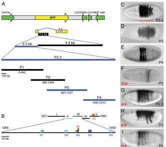
To study the cis-regulation of pnr, we isolated a 2.3 kb fragment from its first intron that recapitulated the wild-type blastoderm pnr pattern in transgenic reporter assays (Fig. 2A,C). We dissected the 2.3 kb enhancer into four overlapping subfragments termed P1-P4 (summarized in Fig. 2A), and two of these, P3 and P4, were able to drive blastoderm lacZ reporter expression (Fig. 2D,E), but gave different patterns. P3 gave rise to a dorsal expression domain similar to the wild-type pnrdomain, and two posteriorly located stripes. P4 drove four anterior stripes that could be clearly distinguished. These results indicated that: (1) the stripe pattern can be separated from the broad dorsal pattern, which we now refer to as the dorsal patch; and (2) the stripes can be separated from each other, suggesting that they are regulated by discrete enhancers, as in the case of pair-rule genes such as hairy(h) and even skipped (eve) (Howard et al., 1988; Harding et al., 1989), although the pnr stripes are more closely spaced than pair-rule stripes.
The observation that the pnr pattern is a composite of two patterns was further substantiated by assaying the reporter genes in dpp and brkmutant backgrounds (Fig. 2F-I). In dppmutants, the
P3-lacZ dorsal patch was abolished, but the posterior stripes remained (Fig. 2F). P4-lacZ was unaffected (data not shown), indicating that only the dorsal patch in the P3 enhancer is Dpp responsive, whereas the stripes are regulated by other factors. In brk mutants, the pattern expanded ventrally (Fig. 2G-I) in a similar manner to the expansion of endogenous pnrin brkmutants (Fig. 1D), but the patch expanded to a lesser degree than the stripes to only ~40% of the circumference, whereas the stripes expanded through the ventral neuroectoderm (Fig. 2H,I). This explains why, in the brkmutant, the expanded pnrstripes become more evident. These results indicate that the pnr pattern comprises two subpatterns of a dorsal patch and six stripes that are regulated by different factors. Dpp activates the patch, whereas AP factors are likely to direct the precise location of the stripes (see below). Interestingly, Brk represses both patterns in the ventrolateral region.
[image:3.612.49.382.437.733.2]Smad sites are crucial for pnrpatch expression In order to identify Smad binding sites in the P3 and P4 enhancers, we performed DNA binding assays (gel shifts) with five overlapping DNA fragments (I-V; Fig. 2B) and recombinant GST-fused Mad (GST-Mad) and GST-GST-fused Medea (GST-Medea) proteins. Two fragments (II and III) spanning 350 bp in the P3 region were able to bind Mad and Medea, whereas the fragments in P4 showed no binding (data not shown). This is consistent with the genetic analysis of P3 and P4 showing that only P3 is Dpp responsive. To search for specific sequences responsible for Smad binding, we performed gel shift assays with ten overlapping oligonucleotides (a-j) covering most of fragments II and III within P3 (Fig. 3A; binding summarized in Fig. 2B as blue ovals). Strong Mad binding was observed with oligonucleotide f (Fig. 3A), which contains the sequence GGCGCC (Fig. 3B, labeled as M3), resembling the GC-rich DrosophilaMad consensus sequence (DSC in Fig. 3B) GCCGnCG (Kim et al., 1997; Xu et al., 1998). Strong
Fig. 2. Dissection of the pnrenhancer reveals a composite expression pattern comprising a dorsal patch and six AP stripes.
(A)A segment of Drosophilachromosome 3R including pnr(yellow) and surrounding genes (green). DNA fragments mapping to the first intron of the pnr-atranscript were tested in transgenic reporter (lacZ) assays. Fragments that drove blastoderm-specific expression are represented in blue. Numbering is with respect to nucleotide position within the 2.3 kb fragment. (B) Map of pnrfragments (I-V) used in DNA binding assays. Blue ovals and lines denote Smad binding sites (strong, dark blue; weak, light blue); red ovals, Brk binding sites; green ovals, Zen binding sites. (C-I)Dorsal (C-F) or lateral (G-I) views of wild-type (C-E) and mutant (F-I) blastoderm embryos hybridized withlacZ
probe. The 2.3 kb fragment (P2.3; genome coordinates 11852900-11855219, dm3) was capable of activating lacZexpression in a pattern identical to the endogenous pnrpattern in blastoderm embryos (C). Fragments P3 (D) and P4 (E) drove subsets of the pnr pattern. Expression driven by P3 is Dpp dependent (F). The stripes (G-I) and dorsal patch (G,H) expand ventrally in brk–.
D
E
V
E
LO
P
M
E
N
Medea binding was observed with oligonucleotide h, which contains a sequence matching the Smad binding element (SBE in Fig. 3B) CAGAC (Zawel et al., 1998), as well as a GC-rich sequence (GC in Fig. 3B).
Two Brk binding sites, B1 and B2, located in the P3 and P4 enhancers, respectively, were identified by DNase I protection (footprinting) assays (supplementary material Fig. S1; summarized in Fig. 2B as red ovals) and confirmed by gel shift (see Fig. 5F). Both sites contain the Brk recognition core GGCGC (Kirkpatrick et al., 2001; Rushlow et al., 2001; Zhang et al., 2001). B1 is a stronger Brk site than B2, probably because it contains two overlapping Brk core sites in opposite orientation (GGCGCC). Importantly, the B1 site overlaps with the Smad M3 site (Fig. 3B), suggesting that Mad-Pemanating from the dorsal side and Brk from the ventral region (Jazwinska et al., 1999a) set the border of the pnrpatch by competing for binding to the pnrenhancer, as has been shown for other Dpp targets (Sivasankaran et al., 2000; Kirkpatrick et al., 2001; Rushlow et al., 2001; Zhang et al., 2001). To determine whether the Smad binding sites are important for pnractivation, mutations were introduced into the two strongest Smad sites M3 and M4. We first tested oligonucleotides containing mutated Smad sites in gel shift assays. Mutation of M3 (Fig. 3B, M3m) abolished Smad binding (Fig. 3C). For M4, we tested oligonucleotides that altered the SBE site (M4m1), the GC-rich site (M4m2) or both (M4m). M4m1 greatly reduced Smad binding,
Since only the patch is Dpp responsive, we tested the in vivo relevance of the Smad site mutations in the context of the P3-lacZ reporter gene. Surprisingly, eliminating the two strong Smad sites M3 and M4 caused complete loss of the patch pattern (Fig. 4B), similar to what happens to P3 in dpp– (Fig. 3F), but the two
posterior stripes remained, further verifying that the stripes are regulated by different cues. The ventral expansion of these two stripes can be explained by a lack of direct Brk repression, as mutation of the M3 site also abolished Brk binding.
[image:4.612.51.297.59.316.2]Mutation of the M3 and M4 sites individually caused different Fig. 3. Smads bind to the pnrenhancer. (A)Oligonucleotides (a-j)
comprising the P3 fragments II and III were tested for binding with GST-Mad and GST-Medea in gel shift assays. Bound oligonucleotides were further designated M1-M5. (B)DNA sequence of M1-M5 with putative Smad binding sites labeled in blue. DSC, DrosophilaSmad consensus site; SBE, Smad binding element; GC, GC-rich. Oligonucleotides with nucleotide changes (red) in the strongest Smad binding sites (M3m, M4m1, M4m2 and M4m) were tested in gel shift assays. (C)M3m and M4m were not capable of binding Smads. +, with protein as
indicated; –, no-protein control. Fig. 4. Two Smad sites and an adjacent HD site are crucial for patch expression.Dorsal views of transgenic Drosophilaembryos pnr hybridized with lacZprobes carrying wild-type (A) and altered (B-H)
P3-lacZtransgenes. Summaries of binding site alterations are given beneath (sequences as in Fig. 3B). (A)The wild-type P3 enhancer drove a lacZexpression domain that was 30-35 cells wide. (B)Mutating both the M3 and M4 Smad binding sites abolished expression.
(C,D)Mutation of the M3 site caused a dramatic narrowing of the patch domain width to 5-10 cells (D), whereas mutation of M4 caused a moderate narrowing to 25 cells (C). The stripes expanded only when the M3 site was altered (B,D), as M3 contains a Brk site, which was also altered. (E,F)Expression of P3M3 disappeared in dppH46heterozygous embryos (E) and zenw36embryos (F) indicating that P3 without the M3 site behaves like a high-level Dpp target. (G,H)Replacement of the M3 site with a Smad site from the Raceenhancer had little effect on the
lacZpattern (G, see sequence in Fig. 5B), whereas mutation of the HD site that lies adjacent to the M3 Smad site caused a severe reduction and narrowing of lacZexpression (H, see sequence in Fig. 5G).
V
E
LO
P
M
E
N
[image:4.612.319.561.63.399.2]in a slightly narrower patch (Fig. 4C), whereas the M3m mutation resulted in a very narrow patch domain (Fig. 4D) that resembled the pattern of Race. Hence, by abolishing a single Mad site, we converted a low-level target into a high-level target. We further examined this idea by assaying P3M3m-lacZin different mutant backgrounds. Expression of the patch pattern was absent in dppH46
heterozygotes (Fig. 4E) and zenw36homozygotes (Fig. 4F), indicating
that P3M3m-lacZ responds only to high levels of Dpp [Race expression disappears in such mutants (Xu et al., 2005)]. These results suggested that Smad sites collectively contribute to the broad domain of pnr, but that the M3 site is crucial.
The structure of the pnrenhancer resembles that
of the high-level Dpp target Race
The dramatic effect of mutating the M3 site suggested that a single Smad site might mediate the response to low levels of Dpp. One possibility is that M3 is a high-affinity Smad site that is capable of binding Smads when present in low concentrations. To address this, we first compared the enhancer structure of pnrwith those of Race and C15, all of which give different expression boundaries. To our surprise, the Raceandpnrenhancers had a similar number of Smad sites (Fig. 5A,B), whereas the C15enhancer contained many more Smad sites spread over a broad region (Fig. 5A). We next performed gel shift assays with DNA fragments of the Race,C15 and pnr enhancers (Fig. 5C) and oligonucleotides containing
individual Smad sites (Fig. 5D). Unexpectedly, the strongest Smad sites in the pnrenhancer, M3 and M4, showed a similar degree of binding to Smads as sites in the Raceenhancer (Fig. 5D).
To further compare Smad sites, we replaced the Smad site in the M3 oligonucleotide with that from the Race enhancer (M3R; sequences shown in Fig. 5B). Competitive gel shift analysis showed that M3 and M3R had similar Smad binding affinities (Fig. 5E). Importantly, swapping the M3R site for M3 in the P3-lacZtransgene had little effect on patch expression (Fig. 4G); however, the posterior stripes expanded, a likely result of the observed decreased binding of Brk to M3R (Fig. 5F). Based on these results, the affinity threshold model is not sufficient to explain the broad expression domain of pnr, as there is no correlation between Smad binding affinity and the extent of the Raceand pnrexpression domains.
A conserved DNA sequence is required for broad
pnrexpression
The fact that responsiveness to the Dpp gradient does not depend on differential Smad binding raised the possibility that other factors might be involved; for instance, a co-factor might interact with Mad to enhance Smad binding to M3 in vivo. A study of the BMP-responsive enhancer of Msx2, a mammalian homolog of tinman, revealed that the sequence AGCAATTAA, which includes a consensus site (AATTAA) for Antennapedia (Antp) homeodomain (HD) factors, lies adjacent to a Smad site (CGGCTCCGGC), and
Fig. 5. The pnrenhancer structure is similar to that of
Race.(A)Summary of Smad and Zen sites in the Race [R1 (Wharton et al., 2004); R2 and R3 (Xu et al., 2005)], C15(Lin et al., 2006) and pnr enhancers. The bar indicates the 100 bp fragment used in gel shift assays in C. (B) Comparison of DNA sequences and organization of functional Smad sites in the
Raceand pnrenhancers. Arrow designates the site from Race
that was swapped into P3 (P3M3R). (C-F)Gel shift analysis of 100 bp fragments (C) and oligonucleotides (D-F) containing Smad sites from the different enhancers as indicated. (C,D)The Smad binding affinities of the pnrand Race
fragments are similar. (E)Unlabeled M3 or M3R can compete with labeled M3 for Mad binding, suggesting that M3R has the same binding capability as M3. B2, an oligonucleotide that contains the Brk site from the P4 enhancer, was used as a negative control and was unable to compete with M3. (F)Brk binding to M3R was weaker than to M3. (G)The HD site, which has been shown to be essential for the BMP responsiveness of the mouse Msx2enhancer, is conserved between mouse and Drosophilaspecies.
D
E
V
E
LO
P
M
E
N
[image:5.612.53.326.345.727.2]that both parts of this ‘bipartite’ motif are required for BMP responsiveness in mouse embryos (Brugger et al., 2004). Intriguingly, the exact same sequence AGCAATTAA lies adjacent to the M3 site (Fig. 5G). Comparison of this bipartite motif in the pnr gene among Drosophila species showed that these nine nucleotides are highly conserved (Fig. 5G) and, importantly, are not present in the enhancers of Raceor C15(Fig. 5B; data not shown). Introducing a mutation into the bipartite site (Fig. 5G, P3HDm) in the P3-lacZreporter gene resulted in a dramatically altered expression pattern that was sporadic and narrow (Fig. 4H). Thus, as in the case of the mouse Msx2enhancer, both the Smad site and the HD site of the pnr bipartite motif are essential for proper response to Dpp.
AP factors regulate thepnrstripe pattern
The six stripes of pnr comprise 20-60% egg length (Fig. 1C) (Winick et al., 1993). The anterior-most stripe overlaps with eve stripe 2 and the posterior-most stripe abutsfushi tarazu(ftz) stripe 7; thus, pnrextends from the labial head segment through the entire abdomen (Herranz and Morata, 2001). Since pair-rule stripe borders are established by gap repressor proteins, which are localized in specific domains across the AP axis, we analyzed pnr expression in bicoid(bcd) and gap gene mutants (Fig. 6). The pnr pattern was affected in all of the mutants that we tested [bcd, hunchback(hb), knirps(kni), Kruppel(Kr), giant(gt) and tailless (tll)] and the changes correlated with what is known about the function of these genes and their cross-regulatory interactions (Fig. 6H). More specifically, pnrexpression expanded anteriorly in bcd, hband gt mutants (Fig. 6B-D), which was likely to be due to a lack of anterior Hb and Gt repression (Clyde et al., 2003). Likewise, pnr expanded posteriorly in tll–(Fig. 6G) due to a lack of posterior Tll
repressor activity (Moran and Jimenez, 2006). Note that weak derepression of pnralso occurs in the head region in tll–. The lack
of mid-body region repressors in the Kr–and kni–mutants caused
shifting and/or expansion of gap domains (Kosman and Small, 1997); thus, both direct and indirect effects alter the middle pnr stripes in these mutants (Fig. 6E,F).
Results from these genetic experiments, together with genome-wide binding data in which all AP factors that we tested except Bcd were found to bind to the pnrenhancer (Li et al., 2008; MacArthur et al., 2009), and with binding site predictions using ClusterDrawII (Papatsenko, 2007) across P3 and P4 (not shown), strongly suggest that the gap genes establish the AP limits of the pnrdomain and the stripes within the domain.
DISCUSSION
Our studies on the low-level Dpp target gene pnrrevealed that: (1) the pnrexpression pattern is a composite of a dorsal patch and six AP stripes; (2) the pnrpatch depends on Dpp, although we predict that additional co-factors that bind adjacent to Smads enable the pnrenhancer to respond to low levels of Dpp, leading to the unique broad domain of pnr; (3) Brk repression establishes the ventral border of both the patch and stripe expression domains and thus Brk shapes the pnrexpression domain on the DV axis; (4) pnr expression is precisely positioned along the AP axis by the segmentation gap genes.
The pnrenhancer integrates spatial cues from
diverse patterning systems
Most blastoderm genes are regulated primarily on either the DV or
[image:6.612.348.493.54.488.2]them may exhibit regulation along the DV axis, they are nonetheless considered AP genes. pnrrepresents an interesting case because although it was originally reported as a DV gene (Jazwinska et al., 1999a; Ashe et al., 2000), closer inspection of its expression pattern in wild-type and mutant embryos and detailed dissection of its cis-regulatory enhancers revealed that pnris highly regulated by both AP and DV genes (summarized in Fig. 7). Its Fig. 6. Gap genes regulate the expression pattern of pnr. Dorsal view of embryos in wild-type (A) and different mutant (B-G) backgrounds hybridized with pnrprobe. Red dotted lines delimit the anterior (left) and posterior (right) borders of the pnr expression domain. (A-D)Compared with wild type (A), in bcd, hband gtmutants the anterior border extends anteriorly. (E)In the Kr mutant,pnrwas slightly compressed in the DV axis and became less stripey, probably owing to expansion of Gt and Kni in the Krmutant. (F)In the kni
mutant, stripes 3 and/or 4 were expanded. (G)In the tllmutant, the posterior domain of pnrextended toward the posterior end. The arrow indicates anterior pnrexpression. (H) Representation of the expression of gap genes along the AP axis. Purple rectangles represent the pnr
stripe domains.
V
E
LO
P
M
E
N
stripes, which are limited to the dorsal 30% of the embryo (Fig. 1A). The patch domain, but not the stripes, disappeared in dpp mutants (Fig. 1B), whereas both the patch and stripes expand ventrally in the absence of Brk (Fig. 1D). The stripes are more sensitive to Brk repression because activation of the patch domain is limited to the region where Dpp is present dorsally, whereas the stripes can be activated along the entire DV axis. Brk in the ventrolateral region and Sna in the ventral-most region repress stripe expression (Fig. 1F) (Jazwinska et al., 1999a). Since pnr specifies dorsomedial fates, restricting its expression to the dorsal 30% of the circumference is crucial. Ectopic expression of pnr ventrally causes transformation of ventral epidermis into dorsomedial epidermis (Herranz and Morata, 2001).
Competition between Brk and Smads for binding to overlapping DNA sequences is likely to set the border of the patch domain. Two Smad sites are particularly important for patch expression (Fig. 4B), and one of these, the M3 site, is a composite site that binds both Brk and Smads (see Fig. 2B and supplementary material Fig. S1), raising the possibility that the patch border is established by competition between activating inputs from Smads in the dorsal region and repressive inputs from Brk emanating from the ventral region. Competition between Brk and Smads for overlapping binding sites has been observed for several Dpp target enhancers (Sivasankaran et al., 2000; Kirkpatrick et al., 2001; Rushlow et al., 2001; Zhang et al., 2001).
Repression of the AP stripes ventrally requires both Brk sites B1 and B2 (Fig. 2B, red ovals). The two posterior stripes driven by P3 expand to a lesser degree (Fig. 2D) than the four anterior stripes driven by P4 (Fig. 2E). This can be explained by the fact that P4 lacks Brk site B1, which is a stronger Brk site. Loss of both Brk sites would likely result in expansion to the edge of the mesoderm, as seen in embryos that lack Brk protein (Fig. 1D; Fig. 2G-I). Repression by Sna is likely to involve the Sna binding sites in the pnrenhancer, as genome-wide binding studies have shown that the pnrenhancer is bound by Sna (summarized in Fig. 6G) (Zeitlinger et al., 2007).
The positioning of the stripes, as well as of the patch, along the AP axis is regulated by the gap genes. Our results suggest that Hb, Gt and Tll set the anterior edge of the pnrdomain, whereas Tll sets
the posterior, and that direct and indirect interactions among the gap proteins establish the stripe borders relative to one another, as has been observed for eve(Clyde et al., 2003). For example, the broad central stripe seen in kni–(Fig. 6F) could be explained by the
lack of direct Kni repression. However, owing to the complex cross-regulatory interactions among the gap genes, it is difficult to predict which gap proteins regulate the pnr stripes directly, although genome-wide binding data of the gap factors (Li et al., 2008; MacArthur et al., 2009) support their direct binding to the pnr enhancer. Although Bcd does not appear to bind directly to the pnrenhancer, its effects are mediated through its targets Gt and Hb.
Dpp target genes utilize multiple mechanisms to set their domains of expression
In depth studies of three genes with different boundary positions in the dorsal region, Race(Xu et al., 2005), C15(Lin et al., 2006) and pnr(this study), indicate that complex combinatorial mechanisms are employed to establish their expression domains, with each gene having a unique regulatory network of its own. Although they all respond to Dpp signaling, their borders of expression are not set by a simple threshold response to the Dpp gradient that depends on differential binding site affinity.
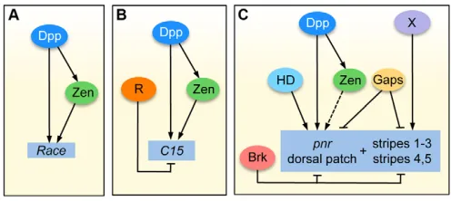
The feature that has been shown to be important for high-level Dpp target expression is the feed-forward motif involving Dpp and Zen (Xu et al., 2005). High levels of Dpp/Smads first activate zen expression in the dorsal-most region, the presumptive amnioserosa, and then both Zen and Smads bind and activate the Raceenhancer (Fig. 7) (Xu et al., 2005). The intermediate-level target C15has a different enhancer structure than high-level targets, containing many Smad sites that act in a cumulative manner to drive expression in regions of intermediate Dpp levels. Mutation analysis has shown that the number of intact Smad binding sites, rather than their affinity, is important for the C15response (Lin et al., 2006). Nevertheless, the enhancer structure of C15might promote high levels of Smad binding in vivo, and this may increase the response to Dpp. Do all intermediate-level Dpp targets have a similar enhancer structure? We examined the enhancer that drives expression of the intermediate-level Dpp target gene tup(Zeitlinger et al., 2007) for putative Smad binding sites (SBEs and GC-rich regions), and observed multiple Smad sites across the enhancer (data not shown), similar to that seen with C15. Thus, the multiple Smad site signature might be necessary for response to lower than peak levels of Dpp. In addition, intermediate-level targets may utilize repression mechanisms to help establish their borders of expression, as was shown for C15(Fig. 7) (Lin et al., 2006).
[image:7.612.50.298.60.171.2]Our studies here revealed that the pnrenhancer resembles that of a high-level target in Smad site organization and Smad binding site affinity (Fig. 5A,B). In fact, it was surprisingly easy to convert the low-level target enhancer into a high-level target by mutating a single Smad site (Fig. 4D). This result could be easily explained if the M3 site had a higher affinity for Smads than those in Race; however, comparison of the binding sites by gel shift showed they have similar affinities (Fig. 5D). Furthermore, replacing the M3 Smad site with a RaceSmad site had little effect on the expression pattern (Fig. 4G). These results suggest that activation of pnrin its broad domain has little to do with Smad binding affinity. How then does pnrrespond to low levels of Dpp? One possible mechanism involves the highly conserved AGCAATTAA site that lies adjacent to the Smad sites (Fig. 5G). In the absence of this site, the P3 enhancer could not respond to low-level Dpp (Fig. 4H). It is possible that this site, when bound, leads to greater Smad binding, which would then promote pnr activation.
Fig. 7. Comparison of the gene networks that regulate the Dpp target genes Race, C15and pnr.(A,B)Dpp first activates zen
expression, then Smads and Zen bind directly to the Race(A) and C15
(B) enhancers to activate their transcription in regions of high-level and intermediate-level Dpp, respectively. A repressor (R) is required to set the C15border of expression (B). (C)Low levels of Dpp in the dorsolateral region function with other activators, including a putative homeodomain factor, to activate pnrin a patch domain, while Brk repression establishes the ventral border of pnrexpression. The pnr
stripes are also repressed ventrally by Brk, but the activators (X) remain unknown. Gap genes (Gaps) establish the pnrpatch and stripe domains along the AP axis. Dashed line indicates possible activation by Zen.
D
E
V
E
LO
P
M
E
N
What factor(s) might bind to the AGCAATTAA site? ATTA is the core binding site for Antp class HD proteins. Although Zen binds to the ATTA site in vitro (supplementary material Fig. S1), neither the endogenous pnr pattern (Fig. 1C) nor P3-lacZ expression (data not shown) is significantly affected in zenmutants. To identify candidate factors, we used the TOMTOM tool (Gupta et al., 2007) at FlyFactor Survey (Zhu et al., 2011), and the best match was to the HD protein Hmx, which binds CAATTAA. However, DrosophilaHmx is expressed only in an anterior region that does not overlap with pnr(see FlyBase). Likewise, although several Antp class HD proteins were predicted to bind to the ATTA core sequence, their timing or domains of expression do not overlap ideally with those of pnr.
Brugger et al. (Brugger et al., 2004) proposed that the AGCAATTAA site in the Msx2enhancer might bind a factor in addition to an HD protein via the 5⬘half of the site, perhaps a transcriptional partner such as FAST1, which was previously shown to function with Smads (Chen et al., 1996). Although our search did not reveal any candidates, if this is the case for pnrthen the bipartite motif could potentially bind four proteins: Smads, Brk, HD and ‘partner X’. The combination of these proteins in a given cell along the DV axis would determine pnrtranscriptional activity. The fact that the bipartite motif is not present in the enhancers of Race or C15, or in the other pnr enhancers we identified, demonstrates the versatility of how Dpp uses different partners to establish multiple target gene domains.
Lessons from studying Dpp target gene regulation Is the structure of the pnr enhancer typical for low-level Dpp targets? This is difficult to address owing to the lack of candidate low-level Dpp targets. Brk is considered a low-level Dpp target in imaginal disc development; however, Dpp represses brk, giving rise to a reciprocal gradient of the Brk repressor (Jazwinska et al., 1999b; Pyrowolakis et al., 2004). Target gene borders are thus established by competition between Smad and Brk for overlapping binding sites, as mentioned above for pnr. The brk enhancer contains multiple enhancer/silencer modules consisting of activator and repressor (Mad/Medea/Schnurri) binding sites, which contribute to threshold responses to the Dpp gradient (Yao et al., 2008), and thus it does not resemble the pnrenhancer. Although we have made good progress in understanding how pnr is expressed in regions with low levels of Dpp, learning the general rules that control broad dorsal patterns will require the analysis of more enhancer elements.
What rules do target genes for other morphogens follow? Long before the ‘feed-forward’ term was coined (Shen-Orr et al., 2002; Lee et al., 2002) it was shown that both the Dl and Bcd morphogens interact with their high-level targets, Twi and Hb, respectively, to activate downstream targets (Jiang et al., 1991; Ip et al., 1992; Small et al., 1992); thus, combinatorial motifs are generally utilized. Moreover, as more target genes of Dl and Bcd were identified and studied, it became apparent that the affinity threshold model could not explain all cases of differential response to the gradient. For example, analysis of several enhancers that drive Bcd-dependent expression in anterior regions of the embryo revealed a poor correlation between Bcd binding site affinity and the AP limits of the pattern (Ochoa-Espinosa et al., 2005). Also, although Dl targets remain archetypal examples of genes that utilize the affinity threshold mechanism, it was found that genes expressed in the lateral region also require input from the Zelda
in target enhancers, and it was proposed that Zelda boosts Dl binding to help activate the neuroectodermal genes (Nien et al., 2011).
Downstream target gene interactions also shape domains of expression, in particular cross-repression among the targets. In both the Drosophila neuroectoderm and the vertebrate neural tube, morphogen targets are expressed in discrete domains rather than nested overlapping domains due to the repression of one target by another (reviewed by Dessaud et al., 2008; Ashe and Briscoe, 2006). This mechanism establishes sharp boundaries among the target genes.
Thus, it is clear that additional factors help morphogens set threshold responses. Given that the pnrenhancer could potentially interact with four different factors along the DV axis and at least four factors along the AP axis, several combinations of inputs could regulate other Dpp target genes. More generally, depending on the number of different factors that interact with the cis-regulatory regions of target genes, morphogen gradients could elicit multiple threshold responses, as has been seen for morphogens such as Dl in Drosophila, Activin in the Xenopusblastula (Green et al., 1992) and Shh in the vertebrate neural tube (Ericson et al., 1997), where up to seven threshold responses have been described. Only by dissecting enhancers can we fully understand how target genes integrate diverse inputs.
Acknowledgements
We thank Nikolai Kirov and Jeongsook Park for help in preparing Smad and Brk proteins; Lilly Sehgal and Jenna DeRovira for help with transgenic constructs and flies; Zhe Xu for helpful discussions; and Steve Small for insightful suggestions on the manuscript.
Funding
This work was supported from a grant from the National Institutes of Health [GM63024]. Deposited in PMC for release after 12 months.
Competing interests statement
The authors declare no competing financial interests.
Supplementary material
Supplementary material available online at
http://dev.biologists.org/lookup/suppl/doi:10.1242/dev.079772/-/DC1
References
Ashe, H. L. and Briscoe, J.(2006). The interpretation of morphogen gradients.
Development133, 385-394.
Ashe, H. L., Mannervik, M. and Levine, M.(2000). Dpp signaling thresholds in the dorsal ectoderm of the Drosophila embryo.Development 127, 3305-3312. Brugger, S. M., Merrill, A. E., Torres-Vazquez, J., Wu, N., Ting, M. C., Cho, J.
Y., Dobias, S. L., Yi, S. E., Lyons, K., Bell, J. R. et al.(2004). A phylogenetically conserved cis-regulatory module in the Msx2 promoter is sufficient for BMP-dependent transcription in murine and Drosophila embryos.
Development 131, 5153-5165.
Chen, X., Rubock, M. J. and Whitman, M.(1996). A transcriptional partner for MAD proteins in TGF-beta signalling. Nature383, 691-696.
Clyde, D. E., Corado, M. S., Wu, X., Pare, A., Papatsenko, D. and Small, S. (2003). A self-organizing system of repressor gradients establishes segmental complexity in Drosophila. Nature 426, 849-853.
Dessaud, E., McMahon, A. P. and Briscoe, J.(2008). Pattern formation in the vertebrate neural tube: a sonic hedgehog morphogen-regulated transcriptional network. Development 135, 2489-2503.
Dorfman, R. and Shilo, B. Z.(2001). Biphasic activation of the BMP pathway patterns the Drosophila embryonic dorsal region. Development128, 965-972. Ericson, J., Briscoe, J., Rashbass, P., van Heyningen, V. and Jessell, T. M.
(1997). Graded sonic hedgehog signaling and the specification of cell fate in the ventral neural tube. Cold Spring Harb. Symp. Quant. Biol. 62, 451-466. Gallo, S. M., Li, L., Hu, Z. and Halfon, M. S.(2006). REDfly: A regulatory
element database for Drosophila. Bioinformatics22, 381-383. Green, J. B., New, H. V. and Smith, J. C.(1992). Responses of embryonic
Xenopus cells to activin and FGF are separated by multiple dose thresholds and
V
E
LO
P
M
E
N
Harding, K., Hoey, T, Warrior, R. and Levine, M.(1989). Autoregulatory and gap gene response elements of the even-skipped promoter of Drosophila.
EMBO J. 8, 1205-1212.
Heitzler, P., Haenlin, M., Ramain, P., Calleja, M. and Simpson, P.(1996). A genetic analysis of pannier, a gene necessary for viability of dorsal tissues and bristle positioning in Drosophila. Genetics143, 1271-1286.
Herranz, H. and Morata, G.(2001). The functions of pannier during Drosophila embryogenesis. Development128, 4837-4846.
Howard, K., Ingham, P. and Rushlow. C.(1988). Region-specific alleles of the Drosophila segmentation gene hairy.Genes Dev. 2, 1037-1046.
Ip, Y. T., Park, R. E., Kosman, D., Bier, E. and Levine, M.(1992). The dorsal gradient morphogen regulates stripes of rhomboid expression in the presumptive neuroectoderm of the Drosophila embryo. Genes Dev. 6, 1728-1739.
Jaz´win´ska, A., Rushlow, C. and Roth, S.(1999a). The role of brinker in mediating the graded response to Dpp in early Drosophila embryos.
Development126, 3323-3334.
Jaz´win´ska, A., Kirov, N., Wieschaus, E., Roth, S. and Rushlow, C.(1999b). The Drosophila gene brinker reveals a novel mechanism of Dpp target gene regulation. Cell96, 563-573.
Jiang, J. and Levine, M.(1993). Binding affinities and cooperative interactions with bHLH activators delimit threshold responses to the dorsal gradient morphogen. Cell72, 741-752.
Jiang, J., Kosman, D., Ip, Y. T. and Levine, M.(1991). The dorsal morphogen gradient regulates the mesoderm determinant twist in early Drosophila embryos.
Genes Dev. 5, 1881-1891.
Kim, J., Johnson, K., Chen, H. J., Carroll, S. and Laughon, A.(1997). Drosophila Mad binds to DNA and directly mediates activation of vestigial by Decapentaplegic. Nature388, 304-308.
Kirkpatrick, H., Johnson, K. and Laughon, A.(2001). Repression of dpp targets by binding of brinker to mad sites. J. Biol. Chem. 276, 18216-18222. Kirov, N., Zhelnin, L., Shah, J. and Rushlow, C.(1993). Conversion of a silencer
into an enhancer: evidence for a co-repressor in dorsal-mediated repression in Drosophila.EMBO J. 12, 3193-3199.
Kosman, D. and Small, S.(1997). Concentration-dependent patterning by an ectopic expression domain of the Drosophilagap gene knirps. Development
124, 1343-1354.
Lee, T. I., Rinaldi, N. J., Robert, F., Odom, D. T., Bar-Joseph, Z., Gerber, G. K., Hannett, N. M., Harbison, C. T., Thompson, C. M., Simon, I. et al.(2002). Transcriptional regulatory networks in Saccharomyces cerevisiae. Science298, 799-804.
Li, X. Y., MacArthur, S., Bourgon, R., Nix, D., Pollard, D. A., Iyer, V. N., Hechmer, A., Simirenko, L., Stapleton, M., Luengo Hendriks, C. L. et al. (2008). Transcription factors bind thousands of active and inactive regions in the Drosophila blastoderm. PLoS Biol. 6, e27.
Lin, M. C., Park, J., Kirov, N. and Rushlow, C.(2006). Threshold response of C15 to the Dpp gradient in Drosophila is established by the cumulative effect of Smad and Zen activators and negative cues. Development133, 4805-4813. MacArthur, S., Li, X. Y., Li, J., Brown, J. B., Chu, H. C., Zeng, L., Grondona, B.
P., Hechmer, A., Simirenko, L., Keränen, S. V. et al.(2009). Developmental roles of 21 Drosophila transcription factors are determined by quantitative differences in binding to an overlapping set of thousands of genomic regions.
Genome Biol. 10, R80.
Mauhin, V., Lutz, Y., Dennefeld, C. and Alberga, A.(1993). Definition of the DNA-binding site repertoire for the Drosophila transcription factor SNAIL.
Nucleic Acids Res. 21, 3951-3957.
Moran, E. and Jimenez, G.(2006). The tailless nuclear receptor acts as a dedicated repressor in the early Drosophila embryo. Mol. Cell. Biol. 26, 3446-3454.
Nien, C. Y., Liang, H. L., Butcher, S., Sun, Y., Fu, S., Gocha, T., Kirov, N., Manak, J. R. and Rushlow, C.(2011). Temporal coordination of gene networks by Zelda in the early Drosophila embryo. PLoS Genet. 7, e1002339. Ochoa-Espinosa, A., Yucel, G., Kaplan, L., Pare, A., Pura, N., Oberstein, A., Papatsenko, D. and Small, S.(2005). The role of binding site cluster strength in Bicoid-dependent patterning in Drosophila. Proc. Natl. Acad. Sci. USA102, 4960-4965.
Papatsenko, D.(2007). ClusterDraw web server: A tool to identify and visualize clusters of binding motifs for transcription factors.Bioinformatics 23, 1032-1034.
Pyrowolakis, G., Hartmann, B., Müller, B., Basler, K. and Affolter, M.(2004). A simple molecular complex mediates widespread BMP-induced repression during Drosophila development. Dev. Cell7, 229-240.
Ramain, P., Heitzler, P., Haenlin, M. and Simpson, P.(1993). pannier, a negative regulator of achaete and scute in Drosophila, encodes a zinc finger protein with homology to the vertebrate transcription factor GATA-1. Development119, 1277-1291.
Rogers, K. W. and Schier, A. F.(2011). Morphogen gradients: from generation to interpretation. Annu. Rev. Cell Dev. Biol. 27, 377-407.
Rushlow, C., Colosimo, P. F., Lin, M. C., Xu, M. and Kirov, N.(2001). Transcriptional regulation of the Drosophila gene zen by competing Smad and Brinker inputs. Genes Dev. 15, 340-351.
Shen-Orr, S. S., Milo, R., Mangan, S. and Alon, U.(2002). Network motifs in the transcriptional regulation network of escherichia coli. Nat. Genet. 31, 64-68. Sivasankaran, R., Vigano, M. A., Müller, B., Affolter, M. and Basler, K.(2000). Direct transcriptional control of the Dpp target omb by the DNA binding protein Brinker. EMBO J. 19, 6162-6172.
Small, S., Blair, A. and Levine, M.(1992). Regulation of even-skipped stripe 2 in the Drosophila embryo. EMBO J. 11, 4047-4057.
Stathopoulos, A. and Levine, M.(2004). Whole-genome analysis of Drosophila gastrulation. Curr. Opin. Genet. Dev. 14, 477-484.
Sutherland, D. J., Li, M., Liu, X. Q., Stefancsik, R. and Raftery, L. A.(2003). Stepwise formation of a SMAD activity gradient during dorsal-ventral patterning of the Drosophila embryo. Development130, 5705-5716.
Thummel, C. S. and Pirrotta, V.(1992). Technical notes: new pCasper P-element vectors. Drosophila Information Service 71, 150.
Wang, Y. C. and Ferguson, E. L.(2005). Spatial bistability of Dpp-receptor interactions during Drosophila dorsal-ventral patterning. Nature434, 229-234. Wharton, K. A., Ray, R. P. and Gelbart, W. M.(1993). An activity gradient of
decapentaplegic is necessary for the specification of dorsal pattern elements in the Drosophila embryo. Development117, 807-822.
Wharton, S. J., Basu, S. P. and Ashe, H. L.(2004). Smad affinity can direct distinct readouts of the embryonic extracellular Dpp gradient in Drosophila. Curr. Biol. 14, 1550-1558.
Winick, J., Abel, T., Leonard, M. W., Michelson, A. M., Chardon-Loriaux, I., Holmgren, R. A., Maniatis, T. and Engel, J. D.(1993). A GATA family transcription factor is expressed along the embryonic dorsoventral axis in Drosophila melanogaster. Development119, 1055-1065.
Xu, M., Kirov, N. and Rushlow, C.(2005). Peak levels of BMP in the Drosophila embryo control target genes by a feed-forward mechanism. Development 132, 1637-1647.
Xu, X., Yin, Z., Hudson, J. B., Ferguson, E. L. and Frasch, M.(1998). Smad proteins act in combination with synergistic and antagonistic regulators to target Dpp responses to the Drosophila mesoderm. Genes Dev. 12, 2354-2370. Yao, L. C., Phin, S., Cho, J., Rushlow, C., Arora, K. and Warrior, R.(2008).
Multiple modular promoter elements drive graded brinker expression in response to the Dpp morphogen gradient. Development135, 2183-2192. Zawel, L., Dai, J. L., Buckhaults, P., Zhou, S., Kinzler, K. W., Vogelstein, B.
and Kern, S. E.(1998). Human Smad3 and Smad4 are sequence-specific transcription activators. Mol. Cell 1, 611-617.
Zeitlinger, J., Zinzen, R. P., Stark, A., Kellis, M., Zhang, H., Young, R. A. and Levine, M. (2007). Whole-genome ChIP-chip analysis of Dorsal, Twist, and Snail suggests integration of diverse patterning processes in the Drosophila embryo.
Genes Dev. 21, 385-390.
Zhang, H., Levine, M. and Ashe, H. L.(2001). Brinker is a sequence-specific transcriptional repressor in the Drosophila embryo. Genes Dev. 15, 261-266. Zhu, L. J., Christensen, R. G., Kazemian, M., Hull, C. J., Enuameh, M. S.,
Basciotta, M. D., Brasefield, J. A., Zhu, C., Asriyan, Y., Lapointe, D. S. et al. (2011). FlyFactorSurvey: a database of Drosophila transcription factor binding specificities determined using the bacterial one-hybrid system.Nucleic Acids Res.
39, D111-D117.