How to cite this paper: Shrestha, D., Liang, F. and Gyanwali, B. (2015) Olfactory Groove Meningioma Extension to Paranasal Sinus and Nasal Cavity: Combined Approach by Unilateral Subfrontal and Endoscopic Endonasal. Open Access Library Jour-nal, 2: e2218. http://dx.doi.org/10.4236/oalib.1102218
Olfactory Groove Meningioma Extension to
Paranasal Sinus and Nasal Cavity: Combined
Approach by Unilateral Subfrontal and
Endoscopic Endonasal
David Shrestha
1, Feng Liang
1, Bibek Gyanwali
2*1Department of Neurosurgery, First Affiliated Hospital of Liaoning Medical University, Jinzhou, China
2Department of Otolaryngology Head and Neck Surgery, First Affiliated Hospital of Guangxi Medical University,
Nanning, China
Email: *bibekgyanwali@gmail.com
Received 25 November 2015; accepted 10 December 2015; published 15 December 2015
Copyright © 2015 by authors and OALib.
This work is licensed under the Creative Commons Attribution International License (CC BY).
http://creativecommons.org/licenses/by/4.0/
Abstract
The local extension of olfactory groove meningioma into the paranasal sinuses and nasal cavity eroding underlying bone in young patients is considered as an unusual and extremely rare event. We report the case of a 25-year-old female of primary growth of olfactory groove meningioma ex-tension to left ethmoidal sinus and nasal cavity, presented with sudden onset of seizure after de-livering a baby. Although olfactory groove meningioma can grow to extreme size with unnoticed symptoms, frequent nasal obstruction, unilateral headache and gradual loss of smell are described. Head image revealed large mass into the anterior cranial fossa extending into the paranasal sinus and left nasal cavity containing cancellous bone. Considering the possibility of postoperative mor-bidity and mortality, complete removal of large tumor and the dura tail that extends beyond its attachment at skull base becomes challenging. The combined unilateral subfrontal and endoscopic endonasal approach is applied to achieve the Simpson grade-I removal.
Keywords
Bony Defect, Cancellous Bone, Nasal Extension, Olfactory Groove Meningioma
Subject Areas: Neurology
1. Introduction
The incidence of olfactory groove meningioma is approximately 8% - 14% of all intracranial meningiomas [1].
OALibJ | DOI:10.4236/oalib.1102218 2 December 2015 | Volume 2 | e2218 It originates near the anterior cranial base, most commonly, at the cribiform plate, planum sphenoidale, and the frontosphenoidal suture. They are often bilateral rather than unilateral but asymmetrical [1]. It grows slowly and usually remains clinically undetectable during the early stage, and is often of considerable size [2][3]. The ex-tension of an olfactory groove meningioma into the paranasal sinuses and nasal cavities in young patients is considered as an extremely rare event [4]. Considering the operative and post-operative morbidity and mortality encountered, a combined multidisciplinary approach is necessary.
2. Case Report
A postpartum 25-year-old female was referred from maternity hospital six hours after the delivery, having two episodes of seizure: convulsive status epilepticus, within two hours of interval. History obtained from the patient revealed frequent congestion of nose and unilateral frontal headache accompanied by gradual loss of smell since one and half year ago. Usually headache was described as having intensity of up to seven out of ten without ra-diating. Patient denied of any nasal discharge, epistaxis, visual impairment and any neurologic abnormalities. Family history was unremarkable. On neurological examination, patient was found to be complete anosmic, with no other signs of cranial nerve deficit.
The computed tomography (CT) of head revealed large hyper dense mass on the left frontal lobe extended to the left nasal cavity with calcification. Further magnetic resonance imaging (MRI) with gadolinium enhance highlighted irregular mass measuring 7.8 * 5.2 cm in anterior cranial fossa with large peri-lesion edema, ex-tended from right medial orbital roof to left lateral and an irregular bulky mass in left nasal cavity involving ethmoidal sinus (Figure 1).
Due to the size of tumor, its extension, and adjacent to anterior and left middle cerebral artery, we performed unilateral subfrontal and endoscopic approach for the removal of mass. Initially, unilateral endoscopic endonasal approach was performed. Tumor was pale-pink in color; consistency was hard with smooth surface. Tumor was excised from its margin including entire middle turbinate and involved superior turbinate. Then existing tumor in the ethmoid sinus was removed along its roof and approximately 0.1 * 0.5 cm2 bone defect was visible. Later, left unilateral subfrontal craniotomy was performed. Tumor was irregular in shape, firm in consistency with clear border. Microscopically, tumor was removed in piecemeal manner exposing anterior cerebral artery. Bony defect of about 0.1 * 0.5 cm2 was noted in the base of anterior cranial fossa. The involved dura was resected and the involved bone was drilled down with diamond drill. The defect was slightly extended to minimize the further recurrence and multilayer reconstruction of the skull base dural defect was done.
[image:2.595.110.518.462.684.2](a) (b)
OALibJ | DOI:10.4236/oalib.1102218 3 December 2015 | Volume 2 | e2218 Postoperatively, no lumbar drain was used. Patient was treated with intravenous antibiotics, antiepileptic drug, dexamethasone, and mannitol. Patient responded normally within 24 hours after the surgery. Nasal pledgets were removed on 3rd postoperative day and patient was discharged on 16th day without neurological or systemic complication.
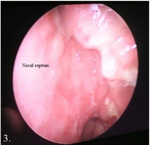
Postoperative image showed total removal of tumor and pathology confirmed the “Transitional meningioma”, and also noted tumor found in nasal cavity contained cancellous bone. Five months later, re-examination of MRI confirmed the Simpson grade I removal (Figure 2) and nasal endoscopy demonstrate excellent re-epthelializa- tion of anterior skull base repair (Figure 3).
[image:3.595.116.513.191.405.2](a) (b)
Figure 2.(a) Postoperative image obtained five months after surgery; gadolinium enhanced T1 weighted MRI coronal view; (b) Postoperative T2 MRI axial view demonstrate total resection.
[image:3.595.185.443.443.692.2]OALibJ | DOI:10.4236/oalib.1102218 4 December 2015 | Volume 2 | e2218
3. Discussion
Meningioma is thought to be originated from arachnoid cap cells: the outer lining of the arachnoid membrane, protruding into the venous sinuses. The incidence rate increases with age, and the ratio of female is twice or higher than that in male. The prevalence of extracranial extension in patients presenting with intracranial menin-giomas has been reported by D. Simpson and H. W. Farr, et al. to be 8.13% and 17.53% respectively. And the extension of olfactory groove meningioma into the paranasal sinuses and nasal cavity was reported to be 2.03% by D. Simpson, 3% by H. W. Farr, et al., 8.9% by H. Bassiouni, et al. and 19.5% by Nakamura, et al. respectively. Extracranial extension results as direct extension of intracranial meningiomas through pressure necrosis of the underlying bone or natural opening. The pathologic features of extracranial meningiomas are identical to those of more frequent intracranial lesions [5]. Bony erosion was reported to be 8% - 16% [3][6][7], and L. Bakay and H. L. Cares mentioned in their literature that entire cribriform plate was disappeared in a patient due to the erosion.
Various approaches have been introduced to remove large, extended olfactory groove meningioma and still today it remains debatable. Not only the size of tumor but also the level of resection is important in planning the surgical approach and very few literatures are available to-date [8].We believe to be the first to report the com-bine approach by unilateral subfrontal and endoscopic endonasal. Comcom-bined approach provides two separate accesses to deal with the tumor from above and below at the same time, and can be easily removed with very minimal invasion to paranasal sinuses and damage to the surrounding structures. Intraoperative vascular injuries are unpredictable, in which quick controlling of fatal consequences is crucial. Combined approach will offers better restoration to vascular injuries and air-tight multilayer reconstruction of the skull base dural defect. In this approach, contralateral side will be left untouched and superior sagittal sinuses can be preserved to reduce some degree of postoperative complication. We have few experience of being encountered with venous infraction and cerebral edema postoperatively due to ligation of the sagittal sinus. During the reconstruction of skull base, temporal muscle was placed as an inlay graft from above, then few surgical and pericranial flaps were placed above using fibrin glue. Direct visualization of multilayer reconstruction was achieved with the nasal endoscopy and no leakage of cerebrospinal fluid was confirmed at the same time. Surgical were introduced in the nasal roof and antiseptic soaked nasal pledgets were placed bilaterally to hold the repairment. To prevent the recurrence, infiltrated underlying bone needs to be removed but some surgeons prefer a conservative approach to prevent CSF rhinorrhea and adjacent vascular injury [3][4], or sometimes if there is not enough surgical field, surgeons may only remove the visible tumor mass and coagulate the dura or remove the superficial hyperostosis bone. Simpson grade-I resection of meningioma is critical in preventing future recurrence. The recurrence of olfactory groove meningioma in Simpson grade-I resection is about 4% to 9% but the tumor extension to paranasal is higher [1][6]. In the year of 2010 we had operated a forty-year-old male patient with the similar condition using the similar combine approach and till date, there is no sign of recurrence. Though endoscopic endonasal approach provides direct and adequate exposure, it alone cannot address the radical resection of large olfactory groove meningioma where dural attachment extends laterally over the orbital roof [7]. Overall, unilateral subfrontal and endoscopic endonasal approach is effective providing easy access to radical removal and early devascularization of the tumor, early confirmation of multilayer reconstruction of skull base and good cosmetic outcome.
Consent
Written informed consent was obtained from the patient for publication of this case report and any accompany-ing images.
Competing Interests
None.No any financial support received.
Authors’ Contributions
All authors equally contributed in writing the case and giving care to the patient.
Acknowledgements
OALibJ | DOI:10.4236/oalib.1102218 5 December 2015 | Volume 2 | e2218
References
[1] Nakamura, M., Struck, M., Roser, F., Vorkapic, P. and Samii, M. (2007) Olfactory Groove Meningiomas: Clinical Outcome and Recurrence Rates after Tumor Removal through the Frontolateral and Bifrontal Approach. Neurosurgery,
60, 844-852. http://dx.doi.org/10.1227/01.NEU.0000255453.20602.80
[2] Babu, R., Barton, A. and Kasoff, S.S. (1995) Resection of Olfactory Groove Meningiomas: Technical Note Revisited.
Surgical Neurology, 44, 567-572. http://dx.doi.org/10.1016/0090-3019(95)00196-4
[3] Bakay, L. and Cares, H.L. (1972) Olfactory Meningiomas: Report on a Series of Twenty-Five Cases. Acta Neurochi-rurgica, 26, 1-12. http://dx.doi.org/10.1007/BF01413528
[4] Francesco, M., Francesco, A.S., Sergio, M., Giuseppe, C. and Luigi, S. (1998) Olfactory Groove Meningioma with pa-ranasal Sinus and Nasal Cavity Extension: Removal by Combined Subfrontal and Nasal Approach. Journal of Cranio- Maxillo-Facial surgery, 26, 314-317. http://dx.doi.org/10.1016/S1010-5182(98)80060-7
[5] Farr, H.W., Gray, G.F., Vrana, M. and Panio, M. (1973) Extracranial Meningioma. Journal of Surgical Oncology, 5, 411-420. http://dx.doi.org/10.1002/jso.2930050503
[6] Simpson, D. (1957) The Recurrence of Intracranial Meningiomas after Surgical Treatment. Journal of Neurology,
Neurosurgery & Psychiatry, 20, 22-39. http://dx.doi.org/10.1136/jnnp.20.1.22
[7] James, K.L., Lana, D.C., Smruti, K.P., Shane, T.R. and Jean, A.E. (2011) Surgical Nuances for Removal of Olfactory Groove Meningiomas Using the Endoscopic Endonasal Transcribriform Approach. Neurosurg Focus, 30, E3.