C h a r a c t e r iz a ti o n of c ell uloly tic
a c tivity i n t h e g u t of t h e
t e r r e s t ri al l a n d sl u g Ario n a t e r :
Bio c h e m i c al id e n tific a ti o n of
t a r g e t s fo r i n t e n siv e s t u d y
Joy n s o n , R E, S w a my, AM, Bo u, PA, C h a p u i s, A a n d F e r ry, N
h t t p :// dx. d oi.o r g / 1 0 . 1 0 1 6 /j. c b p b . 2 0 1 4 . 0 8 . 0 0 3
T i t l e
C h a r a c t e r iz a ti o n of c ell uloly tic a c tivi ty in t h e g u t of t h e
t e r r e s t ri al l a n d sl u g Ario n a t e r : Bio c h e m i c al id e n tific a ti o n
of t a r g e t s fo r i n t e n siv e s t u d y
A u t h o r s
Joy n s o n , R E, S w a my, AM, Bo u , PA, C h a p u i s, A a n d F e r ry, N
Typ e
Ar ticl e
U RL
T hi s v e r si o n is a v ail a bl e a t :
h t t p :// u sir. s alfo r d . a c . u k /i d/ e p ri n t/ 3 5 1 4 0 /
P u b l i s h e d D a t e
2 0 1 4
U S IR is a d i gi t al c oll e c ti o n of t h e r e s e a r c h o u t p u t of t h e U n iv e r si ty of S alfo r d .
W h e r e c o p y ri g h t p e r m i t s , f ull t e x t m a t e r i al h el d i n t h e r e p o si t o r y is m a d e
f r e ely a v ail a bl e o nli n e a n d c a n b e r e a d , d o w nl o a d e d a n d c o pi e d fo r n o
n-c o m m e r n-ci al p r iv a t e s t u d y o r r e s e a r n-c h p u r p o s e s . Pl e a s e n-c h e n-c k t h e m a n u s n-c ri p t
fo r a n y f u r t h e r c o p y ri g h t r e s t r i c ti o n s .
Characterization of cellulolytic activity in the gut of the terrestrial land slug
1
Arion ater
2Ryan Joynson
a, Arvind Swamy
a, Paz Aranega Bou
a, Ambre Chapuis
a, Natalie
3
Ferry
a4
5
aSchool of environment and life science, University of Salford, United Kingdom M5 4WT 6
7
ms. has 18 pages, 5 figures, 1 table
8
*Corresponding author
9
Dr Natalie Ferry
10
School of Environment and Life Science, University of Salford
11
Cockcroft building, 43 The Crescent
12
Salford, Greater Manchester M5 4WT
13
T: +44 (0)161 295 3886
14
n.ferry@salford.ac.uk
15
16
17
18
Abstract:
20
The level of cellulolytic activity in different areas of the gut of the terrestrial slug Arion ater 21
was assayed at different temperatures and pH values. To do this, crude gut proteins were 22
isolated and assayed using modified dinitrosalicylic acid reducing sugar assay. Crude 23
proteins sample were also separated and cellulolytic activity identified using in gel CMC 24
zymography and esculin hydrate activity gel assays. pH and temperature profiling revealed 25
optimum cellulolytic activity between pH 5.0 and 6.0 for different gut regions and retention 26
of up to 90% of activity at temperatures up to 50 °C . Zymograms and activity gels revealed 27
multiple endoglucanase and β-glucosidase enzymes. To further investigate the source of this 28
cellulolytic activity bacterial isolates from the gut were tested for carboxymethylcellulase 29
and β-glucosidase activity using growth plate assays. 12 cellulolytic microbes were identified 30
using 16s rDNA gene sequencing. These include members of the genera Buttiauxella, 31
Enterobacter, Citrobacter, Serratia, Klebsiella. Gut metagenomic DNA was then subjected to 32
PCR, targeting a 400 bp region of the 16srDNA gene which was subsequently separated and 33
individuals identified using DGGE. This identified members of the genera Citrobacter, 34
Serratia, Pectobacterium, Acinetobacter, Mycoplasma, Pantoea and Erwinia. In summary 35
multiple glycoside hydrolase enzymes active over a broad range of temperature and pH 36
values in a relatively under studied organism were detected, Indicating that the gut of Arion 37
ater is a viable target for intensive study to identify novel carbohydrate active enzymes that 38
may be used in the biofuel industry. 39
Key words:
40
Glycoside hydrolase, Slug, Cellulose degradation, Digestive fluids, Cellulolytic activity, 41
1. Introduction
43
Lignocellulose derived from plant cell walls is one of the most abundant organic materials 44
on the planet. The most abundant carbohydrate component it contains is cellulose, made 45
solely of 1 β(1→4) linked D-glucose units. Three enzymes act sequentially to degrade 46
cellulose into simple sugars, endo-β-1,4-glucanases (endocellulases; EC. 3.2.1.4), exo-β-1,4-47
cellobiohydrolases (exocellulases; EC. 3.2.1.91), and β-glucosidases (EC.3.2.1.21). The 48
glucose monosaccharides produced can then be fermented to produce bioethanol. Use of 49
lignocellulose as a bioethanol feedstock has the potential to overcome many of the 50
economic and environmental consequences of using food crops but lignocellose has an 51
inherent resistance to degradation due to the complexity of the plant cell wall 52
superstructure; current methods require expensive pre-treatments making its use 53
economically unattractive (Cao et al., 2012; Ibrahim et al., 2011). The most promising 54
method for production of bioethanol from lignocellose is the simultaneous sacchirification 55
and fermentation (SSR) method. This method incorporates lignocellulose degrading enzyme 56
cocktails and fermenting microorganisms or fermenting bacteria metabolically engineered 57
to produce high numbers of lignocellulose degrading enzymes, which are used to produce 58
ethanol from lignocellulose feedstocks. These enzyme cocktails produce monosaccharides 59
which are fermented into ethanol by bacteria such as Escherichia coli recombinant strains 60
(Cotta, 2012). Many of these modified strains have been engineered to express highly active 61
cellulase enzymes found in other species. A study by Edwards et al. (2011) showed the 62
benefits of introducing a highly active cellobiase enzyme found in Klebsiella oxytoca to 63
Furthermore, cellulase enzymes are also of great importance in the textile industry, in the 65
food industry and as components of detergents, resulting in a high global demand. 66
To that end there is considerable interest in the potential for microbial enzymes (cellulases, 67
hemicellulases and lignases) to bring about the biological breakdown of lignocellulose. Of 68
particular interest is the scope for degradation by the symbiont microbiota in wood/plant 69
feeding invertebrates. Mutualisms between microbes and insects have been widely studied 70
and are found in almost every case, they facilitate exploitation of many different food 71
sources by host insects, including plant cell walls which are difficult and sometimes 72
impossible for most animals to digest (Watanabe and Tokuda, 2010).However the of the 73
enzymatic contributions of microbes to insect hebivory is still unclear. Some herbivorous 74
insects possess genes encoding plant cell wall degrading enzymes including a termite which 75
produces its own cellulase (Watanabe et al., 1998), but the overall structural complexity of 76
the plant cell wall superstructure requires a multitude of enzyme classes which gut microbes 77
contribute to. It is therefore thought that the interactions of host and microbe has had a 78
direct impact on the evolutionary transitions in diet in many herbivorous eukaryotes , 79
including insects (Hansen and Moran, 2014). Enzymatic activity has been studied 80
extensively in the digestive fluid of various insects including members of the orders Isoptera 81
(Konig et al., 2013), Coleoptera (Dojnov et al., 2013) and Othoptera (Shi et al., 2011), all of 82
which have a high lignoceullose diet. However, this focus on arthropods has been at the 83
expense of other groups such as gastropods. Specifically, there has not yet been a definitive 84
characterisation of the origin of cellulolytic activity in the gut of the common garden slug, 85
Arion ater, a significant pest throughout Europe. The diet of the slug is extremely varied 86
stems along with dead plant material with a preference for young leaf/stem plants. A. ater 88
uses its barbed tongue like appenditure called the radula, which contains up to 27,000 89
teeth, to shred its food. This increases the surface area of its food for enzymatic 90
degradation. The radula also allows the slug to eat even the toughest plant material in times 91
where food is scarce. Due to the large portion of plant material in its diet, it is logical that 92
the gut contains multiple enzymes which allow it to digest plant cell wall material into 93
utilizable simple sugars. The A.Ater gut is particularly interesting as a potential source of 94
active enzymes given the variation in pH along its digestive tract and its ability to eat twice 95
its body weight in vegetation per day. This efficiency in crop degradation has led to more 96
than £30 million pounds a year being spent on slug pellets in the UK alone and a ~70 fold 97
increase in utilization of molluscicides over 3 decades (Agular and Wink, 2005). 98
Consequently, we have carried out in-depth analysis of the cellulolytic activity and 99
associated microbial community of the terrestrial gastropod A. ater. 100
2. Materials and methods
101
2.1 Slug collection and dissection 102
Slugs were collected from a suburban area in North Cheshire (53.391463 N, 2.211214 W) 2 103
hours after last light. Individuals were allowed to feed on celery/lettuce cores for 12 hours. 104
Individuals were cooled to 4 °C prior to dissection to reduce metabolism and spontaneous 105
mucus production during dissection. Whole gut tracts were removed, avoiding rupture that 106
would result in loss or contamination of gut juices. Mucus that might interfere with the 107
assays was removed by blotting. Total guts were further separated into ‘crop’ which 108
denotes the region from the mouth up to and including the digestive gland and the ‘gut’ 109
2.2 Initial detection of total cellulolytic activity 111
Gut samples were cut up using a scalpel in a petri dish and then homogenised with a sterile 112
glass rod in a 1.5 mL tube containing 200 µL of 0.2 M sodium acetate buffer (pH 5.2) 113
followed by vigorous vortexing,. To clear cell debris and food matter, samples were 114
centrifuged at 13.3 Krpm for 5 minutes. Supernatants were extracted, pooled (subsequently 115
referred to as ‘crude protein samples’) and stored at -80 °C. Protein content of the crude 116
samples was estimated using a standard Bradford assay (Bradford, 1976) using BSA to 117
construct the standard curve. Total cellulase activity was measured using the dinitro salysilic 118
acid (DNSA) cellulase assay of (Ghose, 1987) with slight adjustments. This assay allows the 119
detection of cellulolytic enzymes which hydrolyse cellulose internally or externally along 120
with the breakdown of cellobiose, each of these actions produces reducing sugar free 121
carbonyl groups which are measured in this assay. The cellulolytic activity of 50µl of crop 122
and gut samples were tested by mixing 1% carboxymethyl cellulose (CMC) (Sigma Aldrich) in 123
a 100mM sodium citrate buffer (pH 4.5). Samples were incubated at 50 °C for 30 minutes. 124
Reactions were terminated by placing samples on ice, adding DNS reagent and heating to 95 125
°C for 10 minutes to allow colour development. All samples were tested and boiled 126
simultaneously. Samples were cooled to room temperature and absorbance read at 540 nm 127
using a CMC control sample as a blank. Correction for background sugars in the sample was 128
undertaken by subtracting a time 0 duplicate sample absorbance from the final result. All 129
activities in this paper are given in enzyme units, where 1 U is equal to 1 µM glucose 130
released per minute per mg of protein. 131
The cellulase detection assay previously described was modified to measure the pH profile 133
of the crude protein cellulolytic activity against CMC, replacing the pH 4.5 buffer with 134
100mM sodium citrate buffers ranging between pH 4-9 while all other conditions remained 135
the same. To determine the temperature profile of the crude protein sample, the assay was 136
modified by varying incubation temperature between 20 °C and 70 °C. 137
2.4 Identification of endocellulases using CMC SDS PAGE zymography 138
CMC Zymography was carried out following the procedure of Schwatz (1987) and Willis et al 139
(2010). Samples were ran using a 12% acrylamide SDS gel containing 0.2% CMC as a 140
substrate for activity staining. Before polymerisation was induced, solutions were heated to 141
30 °C and CMC was added slowly to the resolving gel mixture. Gels were allowed to 142
polymerize for 2 hours and used the same day. Crop and gut crude protein samples were 143
thawed on ice followed by addition of a modified Laemmli loading buffer (minus 144
denaturants). Samples were then heated to 80 °C for 10 minutes followed by pulse 145
centrifugation to denature proteins and prevent substrate digestion during electrophoresis. 146
Size determination and separation was conducted by using 50 µg of each crude extract 147
along with 15 µL of SeeBlue® Plus2 Pre-Stained Standard (Invitrogen). Gels were run at a 148
constant 100 V for 4 hours 30 minutes. For size estimation, the distances travelled by the 149
pre-stained standard bands were measured prior to incubation/staining steps which cause 150
the standards to become difficult to visualise, estimated Mw of bands is indicated on gels by 151
an arrow at appropriate position. The CMC gel was washed in a 5% tritron X-100 solution for 152
30 minutes (repeated 5 times) to remove SDS. The gel was then rinsed with distilled water, 153
placed in sodium phosphate buffer (50 mM, pH 6.5) and incubated for 2 hours at 4 °C to 154
exchange the buffer system and allow renaturation of proteins in the gel. Phosphate buffer 155
was refreshed and the gel was then incubated at 37 °C overnight. Following incubation, the 156
gels were stained with 0.1% (w/v) Congo red for 1 hour, and then destained with a 1 M 157
sodium chloride solution for 3 hours. To enhance visualisation of clear zones acetic acid was 158
added drop wise to the NaCl solution containing the gel, turning the Congo Red from red to 159
a deep purple. 160
2.5 Identification of β-glucosidase enzymes using esculin hydrate – ferric ammonium citrate 161
Native PAGE activity gel 162
A 12% native tris-glycine PAGE gel was created using a standard protocol. Native loading 163
buffer was added to crop and gut crude protein extracts and 50 µg of each was loaded on 164
the gel. Gels were run at 100 V for 4 hours. The gel was then placed in a 0.2 M sodium 165
acetate buffer (pH 5.5) for 10 minutes to exchange the buffer system, then the gel was 166
placed in a 0.2 M sodium acetate buffer (pH 5.5) containing 0.1% (w/v) esculin hydrate 167
(Sigma) and 0.03% (w/v) ferric ammonium citrate (Sigma) and incubated for 3 hours at 37 °C 168
to allow in gel hydrolytic activity. Where β-glucosidase enzymes are present esculin is 169
cleaved producing esculitin which goes onto react with ferric iron to produce a black 170
precipitate. To stop the reaction, the gel was placed into a 10% glucose solution. 171
2.6 Identification of culturable cellulolytic microbes using esculin and CMC LB agar plate 172
assays 173
Whole guts were extracted as previously described and homogenised in 500 µL of 1 quarter 174
strength Ringer solution. A range of dilutions was placed on LB agar plates containing 0.5% 175
CMC and grown overnight at 25 °C. Replica plates were created and incubated for a further 176
caused by extracellular endoglucanase enzymes in the plated gut fluid. Replica plates were 178
stained with a 0.1% Congo red solution for 1 hour, followed by destaining with 1 M NaCl for 179
a further hour. Colonies corresponding with zones of clearance were isolated from replica 180
plates, grown overnight in lb broth. Isolates were then plated onto lb agar containing 0.1% 181
esculin and 0.03% ferric ammonium citrate and incubated at 25 °C for 3 hours to confirm β-182
glucosidase activity. Isolates were identified using 16s rDNA PCR using primers 8F (5’-183
AGAGTTTGATCCTGGCTC-3’) and 1512R (5’-ACGGCTACCTTGTTACGA-3’). Each amplified PCR 184
product was sequenced using Sanger sequencing system big dye v3.1. Sequences were 185
searched using BLASTn for matches in the 16s rDNA database. 186
2.7 Culture independent microbe identification using DGGE analysis 187
Other members of the A. ater gut community were identified using denaturing gradient gel 188
electrophoresis (DGGE). Metagenomic DNA was extracted from a whole gut using a 189
modified version of the Meta-G-nome DNA isolation kit protocol (Epicentre) and extracted 190
DNA was subjected to PCR targeting a 400 bp region of the 16s rDNA using primers F984GC 191
and R1378 according to Heuer et al (18). PCR products were separated by sequence 192
variation using a 30-60% gradient of urea and formamide in a polyacrylamide gel, using the 193
protean 2 system run at a constant 100 V for 16 hours at 60 °C. Gels were stained with Gel 194
Red™ (Biotium, Inc.) and individual bands were excised and placed into wells of a 1% 195
agarose gel and electrophoresed into agarose. Bands were then extracted using the Wizard 196
gel extraction kit (Promega) and sequenced using big dye v3.1. Sequences were submitted 197
to BLASTn for bacterial identification against the 16s rDNA database. 198
3. Results
3.1 Measurement of cellulolytic activity in A. ater gut samples 200
Total cellulase activity in the crop and gut regions (Fig. 1) of A. ater were assayed (Fig. 2A). 201
Cellulase activity was observed in both the gut and crop with the crop portion showing the 202
highest activity at 1.57 U/mg of protein and the gut showing 1.11U/mg of protein. 203
3.2 Temperature and pH profiling of total gut cellulolytic activity 204
Both gut and crop samples showed resilience to heat up to around 50 °C at which point 205
activity begins to decline, with both crude samples showing greatest activity at 30-35 °C (Fig 206
3A). The pH profiles for the two samples were however quite distinct, with the crop samples 207
showing greatest activity at pH 5 and gut at pH 6 (Fig. 3B). At pH values higher than 6.5 the 208
activity of both samples begins to decline up to pH 9 at which point activity is ~4 fold lower 209
than at optimum pH for each sample. 210
3.3 CMC zymography and esculin hydrate activity gel assays 211
Due to the differences seen in the crop and gut cellulolytic activity profiles, CMC 212
zymography (Fig. 2B) and esculin hydrate activity gel assays (Fig 2C) were carried out in 213
order to identify whether or not similar enzyme systems were being incorporated in the 214
crop and gut digestive juices. In CMC zymograms we observed almost identical cellulose 215
activity patterns. We observed 3 main bands in both crude samples, corresponding to 216
proteins of approximately 103, 58 and 22 kDa in size. The β-glucosidase activity gels showed 217
three bands at positions 1, 2 and 3 (indicated with black arrows) which appear to be at 218
identical locations in the gel for both the gut and crop samples. 219
To gain an understanding of the origin of at least a portion of the cellulolytic activity seen in 221
this study, gut microorganisms were isolated and tested for cellulolytic activity. Microbial 222
isolates were grown on agar containing CMC and on agar containing ferric ammonium 223
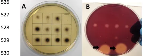
citrate and esculin hydrate to identify endoglucanase (Fig 4B) and β-glucosidase (Fig. 4A) 224
respectively. 12 isolates showed both endoglucanase and β-glucosidase activity, including 225
members of Aeromonas, Acinetobacter, Buttiauxella, Citrobacter, Enterobacter, Klebsiella, 226
Kluyvera, Salmonella and Serratia (Table 1). Only 4 of these microbes could be identified to 227
within 97% similarity of bacterial 16s rDNA genes in the NBCI 16s rDNA and NR databases 228
while the remaining 8 were seen to have between 96-79% similarity to database entries. 229
Subsequently, a DGGE study was carried out to identify microbes that might be present but 230
which may be less easy to culture, using metagenomic DNA samples as templates for 16s 231
rDNA targeted PCR (Fig. 5). This revealed multiple bands from which DNA was extracted and 232
sequenced. Nine further microbes were identified, from the genera Citrobacter, Serratia, 233
Pectobacterium, Acinetobacter, Mycoplasma, Pantoea and Erwina (Table 1). Sequences for 234
cultured and uncultured 16s rDNA studies can be seen in supplementary file 1. 235
Discussion:
236
This study has further characterized the cellulolytic activity in the gut of A. ater through 237
biochemical testing of different portions of the gut, along with identification of multiple 238
cellulolytic microorganisms and thus we begin to characterize the A. ater gut microbiome. 239
Cellulase activity assays showed the overall cellulolytic activity in the gut of A. ater found in 240
the North of England to be greater than that of many insects (Oppert et al., 2010), including 241
members of the genera Coleoptera, Isoptera, Orthoptera and Diptera. We also demonstrate 242
that would be feasible for use in modern industrial lignocellulose degradation methods. A 244
separate investigation of the cellulolytic activity of A. ater of North American origin by 245
James et al. (1997) showed higher overall cellulolytic activity than in this study, but with an 246
optimal pH of 7 as opposed to the crop optimum of pH 5 observed here. A possible reason 247
for this observed difference in optimal pH is the native environment from which individuals 248
were taken, with the average soil pH for the area of North Cheshire being <5.0, whereas in 249
Bellingham WA, the soil is at a pH of between 6-6.6, each correlating with the optimal pH 250
values observed. Acidic environments have been observed in multiple land Pulmonates such 251
as Helix aspersa, (6.1-7.4) Helix pomatia (5.5-6.4), Elona quimperiana (5.3-6.6) (Charrier and 252
Brune, 2003) and Pomacea canaliculata (6.0-7.4) (Godoy et al., 2013) which would suggests 253
that members of this class harbour dietary enzymes that can function in acidic 254
environments, including A. ater, as we have observed. Also, the cellulolytic systems appear 255
to have varying temperature profiles, with our study showing crop and gut samples 256
retaining 90% and 85% activity respectively at 50 °C while the study of the North American 257
species shows practically no activity against CMC in the same conditions. It is also important 258
to note that the gut microbiome is a very dynamic environment which can be heavily altered 259
by living in a different habitat, this has been demonstrated not only in humans 260
(Huttenhower et al., 2012), but also in insects (Dillon and Dillon, 2004). The temperature 261
profile we observed shows the crude enzyme extracts retain much of their activity even at 262
50 °C and demonstrates no clear optimum temperature. However this is not surprising 263
when the complexity of the crude mixture is taken into account, as having multiple enzymes 264
of different microbial origin would cause there to be variation in optimum temperatures for 265
activity for cellulase enzymes of different glycoside hydrolase groups and, furthermore even 266
Using modified cellulase zymograms and esculin hydrate activity gel assays we have also 268
identified three highly abundant individual endocellulase and β-glucosidase enzymes 269
present in both the crop and gut juices, thereby demonstrating that a very similar 270
cellulolytic system throughout the gut and therefore suggesting little activity 271
compartmentalization throughout the gut regions. It is also important to take into 272
consideration that the minimum detectable amount of active enzyme in the esculin hydrate 273
activity gel assay is relatively low at >10 ng (Kwon et al., 1994). Our discovery of multiple 274
endoglucanase and β-glucosidase producing bacteria suggests that there are much greater 275
number of individual cellulolytic enzymes present than we observed in our gel methods. The 276
individual microbes isolated may not make up a high enough proportion of the gut 277
microbiome to produce their enzymes in sufficient abundances to be detectable using in gel 278
separation methods. 279
Our study also confirmed that at least a portion the cellulolytic activity seen in the gut of A. 280
ater is due to symbiotic activity of gut microbes and, for the first time, isolated and 281
identified individual cellulolytic microbes. Many studies have carried out growth plate 282
assays successfully, quickly and accurately isolating gut cellulolytic microbes from 283
gastropods (Antonio et al., 2010), insects (Huang et al., 2012) and mammals (Ruijssenaars 284
and Hartmans, 2001). CMC and esculin hydrate activity growth plate assays allowed us to 285
identify 12 cellulolytic gut microbes, only 4 of which could be identified with great 286
confidence (>97% similarity). This strongly suggests that the A. ater gut microbiome contains 287
uncharacterized microbes with uncharacterized cellulolytic systems that we have shown to 288
have robust pH and temperature activity profiles. In the non-culture based DGGE study we 289
Erwinia tasmaniensis species all have cellulolytic enzymes linked to their species in the NCBI 291
database (http://www.ncbi.nlm.nih.gov/). In this study we have identified a high number of 292
memebers of the gut belong to the Gammaproteobacteria class, with only two Mycoplasma 293
species being from outside that class. The microbes Klebsiella pneumonia, Citrobacter 294
freundii and Serratia liquefaciens have also been identified in the gut of the Bombyx mori 295
larvae (silk worm) and their cellulolytic activity was also observed (Anand et al., 2010). 296
Multiple Enterobacter species, the species Salmonella enterica and serratia marcescens 297
have also been identified in the gut of beetle larvae during their development (Azambuja et 298
al., 2004; Butera et al., 2012). Further to this, a metagenomic study into the gut microbiome 299
of the giant African Snail interestingly shares all but one of the microbial species identified 300
here (Cardoso et al., 2012), this suggests that there may be a set of gut microbes on which 301
multiple land gastropods rely to aid their digestion of lignocellulose. This also indicates that 302
the gut microbe host interaction could have played an important role in the evolutionary 303
dietary transitions of land gastropods as it is thought to have in insects (Hansen and Moran, 304
2014). 305
Gastropods have not been the main focus of recent cellulase prospecting using modern 306
methods due to the initial successes with the insect families, specifically in termites (Tokuda 307
and Watanabe, 2007) but also in beetles (Wei et al., 2006b) (Wei et al., 2006a) and 308
grasshoppers (Oppert et al., 2010) (Willis et al., 2010). However the recent study into the 309
microbiome of the giant African snail has identified thousands of glycoside hydrolase 310
enzymes and carbohydrate binding modules of microbial origin (Cardoso et al., 2012). Our 311
findings and these promising results from related species give a strong indication that the 312
wall degrading enzymes which may be key to improving contemporary biochemical methods 314
in the biofuel industry. In addition, further understanding of the essential biochemical 315
pathways involved in slug feeding could be used to develop more target-specific pest 316
control measures for slugs. Here for example, the identification of these different classes of 317
enzymes demonstrates that the slug gut has the capability to digest the cellulose portion of 318
its diet from long polymer cellulose to individual, utilizable, glucose monosaccharides. This 319
therefore confirms that the slug has the ability to efficiently utlilize the cellulose portion of 320
plant matter it consumes as a source of carbon and we have also identified that gut 321
microbes play a significant role in making this glucose accessible. Increases in physiological 322
understanding are especially important given the detection of high levels of the generic slug 323
pellet poison metaldehyde in water in the UK (Kay and Grayson, 2013) and the recent 324
European Union regulation , which imposes a complete ban on sales of traditional slug 325
pellets by 19th September 2014 (Commission Implementing Regulation 187/2014). 326
327
328
Acknowledgments 329
The authors would like to thank Lucie De Longprez, Sherif Elkhadem and Cassie-Jo Gormley 330
and for their technical assistance. 331
References 332
Agular, R., Wink, M., 2005. How do slugs cope with toxic alkaloids? Chemoecology 15, 167-177. 333
Anand, A.A.P., Vennison, S.J., Sankar, S.G., Prabhu, D.I.G., Vasan, P.T., Raghuraman, T., Geoffrey, C.J., 334
Vendan, S.E., 2010. Isolation and characterization of bacteria from the gut of Bombyx mori that 335
Antonio, E.S., Kasai, A., Ueno, M., Kurikawa, Y., Tsuchiya, K., Toyohara, H., Ishihi, Y., Yokoyama, H., 337
Yamashita, Y., 2010. Consumption of terrestrial organic matter by estuarine molluscs determined by 338
analysis of their stable isotopes and cellulase activity. Est Coast Shelf Sci 86, 401-407. 339
Azambuja, P., Feder, D., Garcia, E.S., 2004. Isolation of Serratia marcescens in the midgut of 340
Rhodnius prolixus: impact on the establishment of the parasite Trypanosoma cruzi in the vector. Exp 341
Parasitol 107, 89-96. 342
Bradford, M.M., 1976. A rapid and sensitive method for the quantitation of microgram quantities of 343
protein utilizing the principle of protein-dye binding. Anal Biochem 72, 248-254. 344
Butera, G., Ferraro, C., Colazza, S., Alonzo, G., Quatrini, P., 2012. The culturable bacterial community 345
of frass produced by larvae of Rhynchophorus ferrugineus Olivier (Coleoptera: Curculionidae) in the 346
Canary island date palm. Lett Appl Microbiol 54, 530-536. 347
Cao, W.X., Sun, C., Liu, R.H., Yin, R.Z., Wu, X.W., 2012. Comparison of the effects of five pretreatment 348
methods on enhancing the enzymatic digestibility and ethanol production from sweet sorghum 349
bagasse. Bioresource Technol 111, 215-221. 350
Cardoso, A.M., Cavalcante, J.J., Cantao, M.E., Thompson, C.E., Flatschart, R.B., Glogauer, A., Scapin, 351
S.M., Sade, Y.B., Beltrao, P.J., Gerber, A.L., Martins, O.B., Garcia, E.S., de Souza, W., Vasconcelos, 352
A.T., 2012. Metagenomic analysis of the microbiota from the crop of an invasive snail reveals a rich 353
reservoir of novel genes. PloS one 7, e48505. 354
Charrier, M., Brune, A., 2003. The gut microenvironment of helicid snails (Gastropoda : Pulmonata): 355
in-situ profiles of pH, oxygen, and hydrogen determined by microsensors. Can J Zool 81, 928-935. 356
Cotta, M.A., 2012. Ethanol production from lignocellulosic biomass by recombinant Escherichia coli 357
strain FBR5. Bioengineered 3, 197-202. 358
Dillon, R.J., Dillon, V.M., 2004. The gut bacteria of insects: Nonpathogenic interactions. Annu Rev 359
Entomol 49, 71-92. 360
Dojnov, B., Pavlovic, R., Bozic, N., Margetic, A., Nenadovic, V., Ivanovic, J., Vujcic, Z., 2013. Expression 361
and distribution of cellulase, amylase and peptidase isoforms along the midgut of Morimus funereus 362
L. (Coleoptera: Cerambycidae) larvae is dependent on nutrient substrate composition. Comp 363
Biochem Physiol B Biochem Mol Biol 164, 259-267. 364
Edwards, M.C., Henriksen, E.D., Yomano, L.P., Gardner, B.C., Sharma, L.N., Ingram, L.O., Peterson, 365
J.D., 2011. Addition of Genes for Cellobiase and Pectinolytic Activity in Escherichia coli for Fuel 366
Ethanol Production from Pectin-Rich Lignocellulosic Biomass. Applied and environmental 367
microbiology 77, 5184-5191. 368
Ghose, T.K., 1987. Measurement of Cellulase Activities. Pure Appl Chem 59, 257-268. 369
Godoy, M.S., Castro-Vasquez, A., Vega, I.A., 2013. Endosymbiotic and host proteases in the digestive 370
tract of the invasive snail Pomacea canaliculata: diversity, origin and characterization. PloS one 8, 371
e66689. 372
Hansen, A.K., Moran, N.A., 2014. The impact of microbial symbionts on host plant utilization by 373
herbivorous insects. Mol Ecol 23, 1473-1496. 374
Huang, S.W., Sheng, P., Zhang, H.Y., 2012. Isolation and Identification of Cellulolytic Bacteria from 375
the Gut of Holotrichia parallela Larvae (Coleoptera: Scarabaeidae). Int J Mol Sci 13, 2563-2577. 376
Huttenhower, C., Gevers, D., Knight, R., Abubucker, S., Badger, J.H., Chinwalla, A.T., Creasy, H.H., 377
Earl, A.M., FitzGerald, M.G., Fulton, R.S., Giglio, M.G., Hallsworth-Pepin, K., Lobos, E.A., Madupu, R., 378
Magrini, V., Martin, J.C., Mitreva, M., Muzny, D.M., Sodergren, E.J., Versalovic, J., Wollam, A.M., 379
Worley, K.C., Wortman, J.R., Young, S.K., Zeng, Q.D., Aagaard, K.M., Abolude, O.O., Allen-Vercoe, E., 380
Alm, E.J., Alvarado, L., Andersen, G.L., Anderson, S., Appelbaum, E., Arachchi, H.M., Armitage, G., 381
Arze, C.A., Ayvaz, T., Baker, C.C., Begg, L., Belachew, T., Bhonagiri, V., Bihan, M., Blaser, M.J., Bloom, 382
T., Bonazzi, V., Brooks, J.P., Buck, G.A., Buhay, C.J., Busam, D.A., Campbell, J.L., Canon, S.R., Cantarel, 383
B.L., Chain, P.S.G., Chen, I.M.A., Chen, L., Chhibba, S., Chu, K., Ciulla, D.M., Clemente, J.C., Clifton, 384
S.W., Conlan, S., Crabtree, J., Cutting, M.A., Davidovics, N.J., Davis, C.C., DeSantis, T.Z., Deal, C., 385
Delehaunty, K.D., Dewhirst, F.E., Deych, E., Ding, Y., Dooling, D.J., Dugan, S.P., Dunne, W.M., Durkin, 386
S., Fodor, A.A., Forney, L.J., Foster, L., Di Francesco, V., Friedman, J., Friedrich, D.C., Fronick, C.C., 388
Fulton, L.L., Gao, H.Y., Garcia, N., Giannoukos, G., Giblin, C., Giovanni, M.Y., Goldberg, J.M., Goll, J., 389
Gonzalez, A., Griggs, A., Gujja, S., Haake, S.K., Haas, B.J., Hamilton, H.A., Harris, E.L., Hepburn, T.A., 390
Herter, B., Hoffmann, D.E., Holder, M.E., Howarth, C., Huang, K.H., Huse, S.M., Izard, J., Jansson, J.K., 391
Jiang, H.Y., Jordan, C., Joshi, V., Katancik, J.A., Keitel, W.A., Kelley, S.T., Kells, C., King, N.B., Knights, 392
D., Kong, H.D.H., Koren, O., Koren, S., Kota, K.C., Kovar, C.L., Kyrpides, N.C., La Rosa, P.S., Lee, S.L., 393
Lemon, K.P., Lennon, N., Lewis, C.M., Lewis, L., Ley, R.E., Li, K., Liolios, K., Liu, B., Liu, Y., Lo, C.C., 394
Lozupone, C.A., Lunsford, R.D., Madden, T., Mahurkar, A.A., Mannon, P.J., Mardis, E.R., Markowitz, 395
V.M., Mavromatis, K., McCorrison, J.M., McDonald, D., McEwen, J., McGuire, A.L., McInnes, P., 396
Mehta, T., Mihindukulasuriya, K.A., Miller, J.R., Minx, P.J., Newsham, I., Nusbaum, C., O'Laughlin, M., 397
Orvis, J., Pagani, I., Palaniappan, K., Patel, S.M., Pearson, M., Peterson, J., Podar, M., Pohl, C., Pollard, 398
K.S., Pop, M., Priest, M.E., Proctor, L.M., Qin, X., Raes, J., Ravel, J., Reid, J.G., Rho, M., Rhodes, R., 399
Riehle, K.P., Rivera, M.C., Rodriguez-Mueller, B., Rogers, Y.H., Ross, M.C., Russ, C., Sanka, R.K., 400
Sankar, P., Sathirapongsasuti, J.F., Schloss, J.A., Schloss, P.D., Schmidt, T.M., Scholz, M., Schriml, L., 401
Schubert, A.M., Segata, N., Segre, J.A., Shannon, W.D., Sharp, R.R., Sharpton, T.J., Shenoy, N., Sheth, 402
N.U., Simone, G.A., Singh, I., Smillie, C.S., Sobel, J.D., Sommer, D.D., Spicer, P., Sutton, G.G., Sykes, 403
S.M., Tabbaa, D.G., Thiagarajan, M., Tomlinson, C.M., Torralba, M., Treangen, T.J., Truty, R.M., 404
Vishnivetskaya, T.A., Walker, J., Wang, L., Wang, Z.Y., Ward, D.V., Warren, W., Watson, M.A., 405
Wellington, C., Wetterstrand, K.A., White, J.R., Wilczek-Boney, K., Wu, Y.Q., Wylie, K.M., Wylie, T., 406
Yandava, C., Ye, L., Ye, Y.Z., Yooseph, S., Youmans, B.P., Zhang, L., Zhou, Y.J., Zhu, Y.M., Zoloth, L., 407
Zucker, J.D., Birren, B.W., Gibbs, R.A., Highlander, S.K., Methe, B.A., Nelson, K.E., Petrosino, J.F., 408
Weinstock, G.M., Wilson, R.K., White, O., Consortiu, H.M.P., 2012. Structure, function and diversity 409
of the healthy human microbiome. Nature 486, 207-214. 410
Ibrahim, M.M., El-Zawawy, W.K., Abdel-Fattah, Y.R., Soliman, N.A., Agblevor, F.A., 2011. Comparison 411
of alkaline pulping with steam explosion for glucose production from rice straw. Carbohyd Polym 83, 412
720-726. 413
James, R., Nguyen, T., Arthur, W., Levine, K., Williams, D.C., 1997. Hydrolase (beta-glucanase, alpha-414
glucanase, and protease) activity in Ariolimax columbianus (banana slug) and Arion ater (garden 415
slug). Comp Biochem Physiol B Biochem Mol Biol 416
118, 275-283. 417
Kay, P., Grayson, R., 2013. Using water industry data to assess the metaldehyde pollution problem. 418
Water and Environ J, n/a-n/a. 419
Konig, H., Li, L., Frohlich, J., 2013. The cellulolytic system of the termite gut. Appl Microbiol 420
Biotechnol 97, 7943-7962. 421
Kwon, K.S., Lee, J., Kang, H.G., Hah, Y.C., 1994. Detection of Beta-Glucosidase Activity in 422
Polyacrylamide Gels with Esculin as Substrate. Appl Environ Microbiol 60, 4584-4586. 423
Oppert, C., Klingeman, W.E., Willis, J.D., Oppert, B., Jurat-Fuentes, J.L., 2010. Prospecting for 424
cellulolytic activity in insect digestive fluids. Comp Biochem Physiol B Biochem Mol Biol 155, 145-425
154. 426
Ruijssenaars, H.J., Hartmans, S., 2001. Plate screening methods for the detection of polysaccharase-427
producing microorganisms. Appl Microbiol Biotechnol 55, 143-149. 428
Shi, W.B., Ding, S.Y., Yuan, J.S., 2011. Comparison of Insect Gut Cellulase and Xylanase Activity Across 429
Different Insect Species with Distinct Food Sources. Bioenerg Res 4, 1-10. 430
Tokuda, G., Watanabe, H., 2007. Hidden cellulases in termites: revision of an old hypothesis. Biol Lett 431
3, 336-339. 432
Watanabe, H., Noda, H., Tokuda, G., Lo, N., 1998. A cellulase gene of termite origin. Nature 394, 330-433
331. 434
Watanabe, H., Tokuda, G., 2010. Cellulolytic systems in insects. Annu Rev Entomol 55, 609-632. 435
Wei, Y.D., Lee, K.S., Gui, Z.Z., Yoon, H.J., Kim, I., Je, Y.H., Lee, S.M., Zhang, G.Z., Guo, X., Sohn, H.D., 436
Jin, B.R., 2006a. N-linked glycosylation of a beetle (Apriona germari) cellulase Ag-EGase II is 437
Wei, Y.D., Lee, K.S., Gui, Z.Z., Yoon, H.J., Kim, I., Zhang, G.Z., Guo, X., Sohn, H.D., Jin, B.R., 2006b. 439
Molecular cloning, expression, and enzymatic activity of a novel endogenous cellulase from the 440
mulberry longicorn beetle, Apriona germari. Comp Biochem Physiol B Biochem Mol Biol 145, 220-441
229. 442
Willis, J.D., Klingeman, W.E., Oppert, C., Oppert, B., Jurat-Fuentes, J.L., 2010. Characterization of 443
cellulolytic activity from digestive fluids of Dissosteira carolina (Orthoptera: Acrididae). Comp 444
Biochem Physiol B Biochem Mol Biol 157, 267-272. 445
446
447
448
449
450
451
452
453
454
455
456
457
458
459
460
461
462
463
464
465
466
0 0.5 1 1.5 2
Crop Gut
Sp
e
ci
fi
c
A
ci
ti
vt
y
U
/mg o
f p
ro
te
in
s
Crop
[image:20.595.31.303.137.620.2]Gut
Figures and tables
468
469
470
471
472
473
474
475
Figure 1 Dissected whole gut tract of Arion ater
476
477
478
479
480
481
482
483
484
485
486
487
488
489
490
491
Figure 2 (A) Total specific cellulolytic activity seen in the gut fluids from Arion ater against 492
CMC at 50°C and pH 5.0 using the DNSA cellulase activity assay. (B) A 12% SDS PAGE 0.2% 493
CMC zymogram using 50ug of crude gut and crop protein per lane. Gel stained with Congo 494
red to allow activity visualisation. (C) A 12% native PAGE gel containing 100ug of protein per 495
lane, gels were incubated in a 0.2m sodium acetate activity buffer containing 0.1% (w/v) 496
Crop
Gut
Anus
Mouth
A
Gut
C
Crop
Gut
1 2 3
~58 ~103
~22
esculin and 0.03% w/v ferric ammonium citrate for one hour. Black precipitate show areas 497
[image:21.595.49.436.170.573.2]of activity, indicated by black arrows. 498 499 500 501 502 503 504 505 506 507 508 509 510 511 512 513 514 515 516 517 518
Figure 3 The temperature profiles (A) and the pH profiles (B) of the two crude gut protein 519
isolations showing the total cellulolytic activity of each sample against a CMC substrate.. 520
Temperature and pH profiles were obtained using a modified cellulase assay with incubation 521
steps at temperatures between 20-70ºC and at pH values 4-9 respectively. Specific activity 522
shown as enzyme units (U) where 1 U is equal to 1 µM glucose released per minute per mg 523 of protein. 524 525 0 0.2 0.4 0.6 0.8 1 1.2 1.4 1.6 1.8
4 4.5 5 5.5 6 6.5 7 7.5 8 8.5 9
Sp ecific A ctivity: U /m g o f p ro tein pH Crop Gut 0 0.2 0.4 0.6 0.8 1 1.2 1.4 1.6 1.8 2
20 25 30 35 40 45 50 55 60 65 70
526
527
528
529
[image:22.595.30.272.130.224.2]530
Figure 34 (A) An esculin hydrate plate assay demonstrating the β-glucosidase activity of 531
microbial isolates. Isolates were grown on agar plates containing 0.1% (w/v) esculin and 532
0.03% (w/v) ferric ammonium citrate. A black precipitate indicates β-glucosidase activity. 533
Untransformed top10 E. coli (Invotrogen) was used as a negative control. (B) A CMC plate 534
assay showing endoglucanase activity. Bacterial isolates were grown on agar plates 535
containing 0.5% CMC after 16 hour incubation plates were stained with congo red and 536
destained with 1 M NaCl in order to visualise zones of clearing. 5 and 10 µL of 1 mg/mL A. 537
538
539
540
541
542
543
544
545
546
547
548
Figure 45 Differential gradient gel electrophoresis gel, 30-60% gradient of formamide and 549
urea. Labels show bands from which successful microbial identifications were deduced. 550
551
552
553
554
555
A
B
UA.a.1 UA.a.2
UA.a.3 UA.a.4
UA.a.5
UA.a.6
UA.a.7
Name Description E-value Identity Accession
CA.a.1 Acinetobacter calcoaceticus 0 92% NR_042387.1 CA.a.2 Aeromonas hydrophila 0 99% NR_104824.1 CA.a.3 Buttiauxella agrestis 0 79% DQ440549.1 CA.a.4 Buttiauxella agrestis 0 99% NR_041968.1 CA.a.5 Citrobacter braakii 0 85% NR_028687.1 CA.a.6 Citrobacter freundii 0 99% NR_028894.1 CA.a.7 Enterobacter sp. E6-PCAi 0 94% JN853247.1 CA.a.8 Klebsiella pneumoniae 0 96% NR_037084.1 CA.a.9 Kluyvera intermedia 0 99% KF724024.1 CA.a.10 Salmonella enterica 0 91% NR_044371.1 CA.a.11 Serratia liquefaciens 0 86% GU586145.1 CA.a.12 Serratia marcescens 0 91% NR_036886.1 UA.a.1 Mycoplasma hyorhinis 1.00E-158 93% NR_041845.1 UA.a.2 Mycoplasma iners 4.00E-158 93% NR_025064.1 UA.a.3 Uncultured Citrobacter 0 99% AY847172.1 UA.a.4 Uncultured Serratia 0 100% KC253894.1 UA.a.5 Pectobacterium carotovorum 0 99% NR_041971.1 UA.a.6 Acinetobacter beijerinckii 0 98% NR_042234.1
UA.a.7 Pantoea sp. 57917 0 99% DQ094146.1
UA.a.8 Erwinia amylovora 0 99% NR_041970.1
[image:23.595.60.375.128.441.2]UA.a.9 Erwinia tasmaniensis 0 99% NR_074869.1
Table 1 NCBI BLASTn search results for each amplified 16s rDNA gene from cultured 556
cellulolytic microbes (CA.a.*) and for uncultured microbes from the DGGE study (UA.a.*). 557
Sequences were queried against the NCBI 16s rRNA database or the nr database if no match 558