www.impactjournals.com/oncotarget/ Oncotarget, Vol. 6, No. 28
Construction and analysis of lncRNA-lncRNA synergistic
networks to reveal clinically relevant lncRNAs in cancer
Yongsheng Li1,*, Juan Chen1,*, Jinwen Zhang1,*, Zishan Wang1, Tingting Shao1, Chunjie Jiang1, Juan Xu1, Xia Li1
1College of Bioinformatics Science and Technology, Harbin Medical University, Harbin, China *These authors have contributed equally to this work
Correspondence to:
Xia Li, e-mail: lixia@hrbmu.edu.cn
Juan Xu, e-mail: xujuanbiocc@ems.hrbmu.edu.cn
Keywords: long non-coding RNA, network hub, functional synergistic network, hallmark of cancer, prognostic module
Received: April 07, 2015 Accepted: July 15, 2015 Published: July 28, 2015
ABSTRACT
Long non-coding RNAs (lncRNAs) play key roles in diverse biological processes. Moreover, the development and progression of cancer often involves the combined actions of several lncRNAs. Here we propose a multi-step method for constructing lncRNA-lncRNA functional synergistic networks (LFSNs) through co-regulation of functional modules having three features: common coexpressed genes of lncRNA pairs, enrichment in the same functional category and close proximity within protein interaction networks. Applied to three cancers, we constructed cancer-specific LFSNs and found that they exhibit a scale free and modular architecture. In addition, cancer-associated lncRNAs tend to be hubs and are enriched within modules. Although there is little synergistic pairing of lncRNAs across cancers, lncRNA pairs involved in the same cancer hallmarks by regulating same or different biological processes. Finally, we identify prognostic biomarkers within cancer lncRNA expression datasets using modules derived from LFSNs. In summary, this proof-of-principle study indicates synergistic lncRNA pairs can be identified through integrative analysis of genome-wide expression data sets and functional information.
INTRODUCTION
Recent analyses of the mammalian transcriptome revealed that an abundance of long non-coding RNAs (lncRNAs) lie interspersed with the coding genes [1]. While the functions of most lncRNAs remain unknown, growing evidence suggests that the like microRNAs (miRNAs), lncRNAs, mediate oncogenic or tumor-suppressing effects and may constitute a new class of cancer therapeutic targets [2, 3]. Moreover, it appears likely that many biological molecules, including miRNAs and lncRNAs, exert their effects by acting in combination rather than individually [4, 5]. But despite the growing appreciation of the importance of lncRNAs in normal physiology and disease, our understanding of the combined effects of the cancer-associated lncRNAs remains limited.
Several recent studies suggest that lncRNAs play important roles in oncogenesis [6, 7]. H19, for example, is an lncRNA induced during liver development [8], but it also promotes glioma cell invasion by giving rise to
miR-675 [9]. Other lncRNAs involved in various types of cancers include HOTAIR [10, 11], MEG3 [12], PVT1 [13] and CDKN2B-AS1 [14]. Among those, levels of the lncRNA MEG3 are markedly lower in glioma tissues than adjacent normal tissues, and ectopic expression of MEG3 inhibits cell proliferation and promotes apoptosis [12]. In addition, several earlier findings implicate PVT1 in the pathophysiology of cancer [15], and silencing PVT1 expression using siRNA reduces cell proliferation and increases apoptosis in breast and ovarian cancer cell lines [16]. Another well-known potential cancer-associated lncRNA is PCA3, a prostate-specific molecule that is markedly overexpressed in prostate cancer. A noninvasive assay for urinary PCA3 expression is currently being developed as a clinical diagnostic [17].
into close proximity to produce a functional complex. In addition, Ma et al. demonstrated that genes are regulated by lncRNAs in part through chromosome conformation capture, which suggests lncRNAs may act synergistically [21]. Although the modular RNA regulatory code remains to be tested, studying the modular regulation of lncRNAs and investigating their combined effects are important steps toward further definition of lncRNA functions on a system-wide level. One approach to classifying the putative functions of lncRNAs is through “guilt-by-association” [22]. This approach associates lncRNAs with biological processes based on a common expression pattern across cell types and tissues, and can therefore identify groups of lncRNAs that are associated with specific cellular processes [23, 24]. Analysis of coexpressed genes of lncRNAs has revealed significant coregulation of genes by lncRNAs, suggesting that lncRNA clusters may regulate biological processes synergistically. It has also been reported that combined lncRNA signatures enable more accurate prediction of patient survival than individual lincRNAs [25, 26]. Results from all of these studies attest to the importance of lncRNA synergism and indicate that integrating expression of correlated genes and functional information could enable identification of synergistic lncRNA pairs and simultaneously reveal their underlying functions.
Here we present a multi-step computational method for identifying significantly functional synergistic lncRNA pairs through the functional modules they jointly regulate. This entails integrating genome-wide lncRNA and mRNA expression profiles, functional information and protein interaction data. After assembling all the lncRNA-lncRNA functional synergistic pairs, we constructed a lncRNA functional synergistic network (LFSN, our strategy is illustrated in Figure 1). Applying this approach to three cancer types, glioblastoma multiforme (GBM), ovarian cancer (OV) and prostate cancer (PCa), we constructed cancer associated LFSNs having scale free and modular architectures, and investigated the properties of cancer-related lncRNAs within their corresponding synergistic networks. Our findings suggest that cancer-related lncRNAs are enriched in hubs and modules, and that the cancer-related lncRNA modules are associated with cancer prognosis. This study contributes to a comprehensive understanding of the synergistic behaviors of lncRNAs from the viewpoint of systems biology.
RESULTS
The lncRNA functional synergistic network (LFSN) across cancers
We are proposing a multi-stage method to gradually identify synergistic lncRNA pairs in cancer (Figure 1, see details in methods). On the basis of lncRNA pairs
regulating at least one functional module, we assembled all synergistic lncRNA pairs into a LFSN, where nodes represent lncRNAs and edges represent their functional synergistic interactions. We applied this approach to three cancer types, GBM, OV and PCa, and constructed three cancer-specific LFSNs. In total, we obtained 35,584 synergistic interactions among 2,798 lncRNAs for GBM, 7,399 synergistic interactions among 1,104 lncRNAs for OV and 6, 908 synergistic interactions among 2,588 lncRNAs for PCa (Figure 2A and Supplementary Table S1). This indicates that lncRNA pairs identified by our method simultaneously regulate multiple cancer-associated biological processes (BPs), which may contribute to carcinogenesis. In particular, a number of known disease-related lncRNAs with high connectivity within the network were observed to co-regulate the cancer associated BPs. For example, an analysis of genome-wide DNA copy number alterations revealed the loss of LINC00032 in approximately one-fourth of tumors [27]. We observed that LINC00032 co-regulated 285 functional modules with other lncRNAs, including the highly connected lncRNA RP11–290F20.1 in GBM (left panel, Figure 2B). In addition, MEG3 has been shown to activate p53 and inhibit tumorigenesis and disease progression of several types of cancers [28, 29], including OV [30]. We found that MEG3 co-regulated 142 functional modules with other lncRNAs, among which MIR22HG is associated with stress responses [31, 32]. These two lncRNAs act synergistically to regulate coagulation-related functions (middle panel, Figure 2B), which are associated with tissue invasion and metastasis. In PCa, we observed two lncRNAs with high connectivity that co-regulate the immune response (right pane, Figure 2B). The lncRNA RP11–538D16.2 regulated 102 functional modules, while ENSG00000248849 regulated 87 modules. In addition, ENSG00000248849 is subject to recurrent deletion in PCa, and shows significant overlap with a known disease lncRNA DLX6-AS1 [33, 34]. All of these observations demonstrate the feasibility of using our method to identify lncRNA pairs through their synergistically regulating functional modules. Furthermore, the examples summarized above suggest that some disease-associated lncRNAs have especially high connectivity within the LFSN. Understanding the structure of LFSNs may provide additional insight into the tumorigenesis of certain types of cancer.
Properties of LFSNs
networks, the structures of LFSNs were not randomly organized; instead, they were characterized by a core set of principles that distinguish them from random networks. We next analyzed the modular structure of each LFSN. As shown in Figure 3A, the number of modules decreased with increases in the k-value. We hypothesized that this reflected the tendency of lncRNAs to implement synergistic regulation as small modules rather than as individuals or as parts of larger modules. The same features were observed in OV and PCa. In total, approximately 74.92%, 51.25% and 38.14% of lncRNAs were involved in at least one module in GBM, OV and PCa, respectively.
Exploration of the chromosomes on which the synergistic lncRNA pairs were located showed that only 1,918 (5.39%) lncRNA pairs in GBM, 422 (5.70%) pairs in OV and 474 (6.86%) pairs in PCa were located on the
same chromosomes (Figure 3B). Although the number of lncRNA pairs co-localizing on the same chromosome in OV and PCa was small, the relation was significant (p = 0.057 and p < 0.001, respectively). In addition, 12.62%, 14.22% and 10.13% of pairs locate within less than 10 Mb on the same chromosomes, but these proportions are not significantly higher than random. These results indicate that the majority of the synergistic pairs regulate the same function modules in trans, while a small fraction of lncRNA pairs synergistically regulate in cis.
Cancer associated lncRNAs tend to be hubs and enriched in modules
[image:3.612.104.509.53.470.2]For each biological network, a key property is its connectivity, which reflects how often a node interacts
Figure 1: The workflow to construct the LFSNs via co-regulating functional modules. The process involves three main steps.
Figure 2: The lncRNA-lncRNA synergistic network across cancers. A. The LFSNs in GBM, OV and PCa. A node represents a lncRNA, and an edge represents a synergistic action. B. Three examples of lncRNA synergistic pairs in each cancer. The known disease lncRNAs with high connectivity were selected as examples. The GO processes regulated by lncRNA pairs were also shown. The indirect dashed line represents the lncRNA synergistic action; the direct line represents the lncRNA regulation of the functional module.
[image:4.612.71.542.397.647.2]with other nodes. Hub nodes whose connectivities are extremely high are always very important nodes. For example, in the protein-protein interaction networks of various organisms, hubs tend to be essential proteins involved in maximal information exchange with other proteins [35]. We focused on lncRNAs that had been experimentally validated as disease lncRNAs. Notably, the connectivities of these disease lncRNAs were significantly greater than the connectivities of other lncRNAs within the entire network (Figure 4A, Wilcoxon rank sum test). This suggests disease lncRNAs are highly connected within the network and may be enriched in hubs. Because the number of experimentally validated disease lncRNAs is limited, we extended this set by identifying cancer-associated lncRNAs in each cancer type (details in Methods). Generally, the hub and cancer-associated lncRNAs defined in the three cancer networks examined were almost entirely separate with little overlap (Figure 4B). But When we tested whether the cancer-associated lncRNAs were more likely to be hubs, which we defined as the top 10% of nodes with the highest connectivity within each network, we found that cancer-associated lncRNAs were significantly enriched in the hubs across all three cancers tested (Figure 4C, Fisher’s exact test).
Another important feature of a biological network is modularity. Modules are groups of highly interconnected nodes that are often involved in the same biological processes or pathways. In contrast to hubs and cancer-associated lncRNAs, we found substantial overlap of module lncRNAs across the three cancer types (Figure 4B). This is reasonable and understandable since modules mainly reflect their underlying biological processes. The cancer-associated lncRNAs showed a significant enrichment in modules across GBM and OV (Figure 4D, Fisher’s exact test). This suggests that cancer-associated lncRNAs work more synergistically and reflect the group behavior, making it likely that they are more informative regarding tumor initiation and progression.
Synergistic lncRNAs modulate cancer-associated hallmarks
Although the biology of cancer is extremely complex, that complexity can be reduced and represented by a few cancer hallmarks that enable tumor growth and metastasis [36]. These hallmarks provide a framework for understanding the remarkable diversity of cancers. To further understand the functional roles of synergistic lncRNA pairs in carcinogenesis, for each LFSN, cancer hallmarks associated lncRNA pairs were selected to form cancer hallmark subnetworks. This analysis showed that several cancer hallmarks were deeply involved in the three LFSNs (Supplementary Figure S2), but with different cancer specificities. In brief, lncRNA pairs in GBM primarily regulate the cancer hallmarks of “self sufficiency in growth”. Previous studies have demonstrated that GBM is characterized by extensive invasion, rapid
growth, necrosis and angiogenesis [37, 38]. The lncRNA pairs in OV mostly regulate the hallmarks “self sufficiency in growth” and “tissue invasion and metastasis”, which is consistent with clinically rapid progress and high metastatic potential of ovary tumors [39, 40]. In addition, “evading immune detection” and “tumor promoting inflammation” are the two most affected hallmarks in PCa, which indicates that inflammation and immune processes are important for the malignancy of prostate tissue. This is consistent with histological observations, indicating that innate and adaptive immunity actively participates in the pathogenesis, surveillance and progression of prostate cancer [41–43].
Although no synergistic lncRNA pair was found to share by any two hallmark subnetworks, the hallmark subnetworks are conserved among the three cancers to a certain extent. That is, a large percentage of lncRNA pairs contribute to the same cancer hallmarks by regulating biological processes annotated to those hallmarks. For example, 29, 11 and 689 lncRNA pairs in GBM, OV and PCa, respectively, regulate the biological process of “DNA repair”, which is related to the “genome instability and mutation” hallmark (Supplementary Figure S3). For GBM and OV, 108 and 97 pairs of lncRNAs regulate “positive regulation of cell proliferation” (Supplementary Figure S4). In PCa, there are no lncRNA pairs regulating this biological process, but three lncRNA pairs control another process, “positive regulation of signal transduction” and both processes are annotated to the cancer hallmark “self sufficiency in growth”. Within these cancer hallmark subnetworks, some experimentally validated disease lncRNAs contribute to carcinogenesis. In GBM, TRAF3IP2-AS1, which is related to brain-associated disease, cooperates with three lncRNAs to regulate “wound healing”: AC078883.3, RP11-645N11.2 and XLOC_004923. In OV, the obesity-associated lncRNA SNHG11 cooperates with three lncRNAs, RP11-79H23.3, XLOC_002749 and XLOC_0092721, to regulate “positive regulation of cell proliferation” which is annotated to the hallmark of “self sufficiency in growth”. In PCa, the lncRNA MIAT functions together with AC005562.1 to regulate “DNA repair” and so impacts the hallmark of “genome instability and mutation”. In addition, for a specific cancer, the same lncRNA synergistic pairs may regulate different processes to control cancer growth. In GBM, for example, ENSG00000248849, which shows 80% overlap with the disorder-associated lncRNA DLX6-AS1, cooperates with RP5-1185K9.1 to regulate the processes “negative regulation of apoptosis” and “negative regulation of programmed cell death”, both of which annotated to the cancer hallmark “evading apoptosis”. These results indicate that lncRNA pairs in each cancer hallmark subnetwork may contribute to carcinogenesis and deserve further investigation across these three cancer types.
biomarkers rests on the idea that a module biomarker is a better predictor of survival than an individual gene [44]. Given than lncRNA pairs within hallmark subnetworks were associated with cancer-related biological processes, we were interested in identifying prognostic modules from the cancer hallmark subnetworks. We therefore used
[image:6.612.155.461.48.534.2]significantly different overall survival rates (log-rank test,
P < 0.05). Furthermore, the gene or miRNA expression patterns can be used to segregate those two diseases into subtypes with different prognoses [45, 46]. However, it is not yet clear whether the classifier power of these modules reflects their correlation with global expression subtypes in GBM or OV. To determine whether these functional modules can provide additional predictive power, we performed a multivariate survival analysis using prognostic factors, such as age, sex, subtype, grade or stage of patients, along with subnetwork modules. We observed that four lncRNA modules in GBM and two modules in OV were significantly associated with patient survival (Supplementary Table S2–S4).
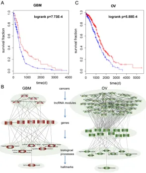
A representative prognostic module in GBM found to be associated with patient survival time (P = 7.73E-4, Figure 5A), included three synergistic pairs among four lncRNAs (Figure 5B). This module remained significantly associated with patient overall survival (Hazard Ratio = 0.65, p = 0.021) in a Cox multivariate analysis, after adjusting for patient age, gender and tumor subtype. The module includes the lncRNA RP11-419K12.1, which overlaps an experimentally validated GBM-associated lncRNA, CCDC26. A single nucleotide polymorphism (SNP) rs891835 in CCDC26 is reportedly associated with glioblastoma susceptibility. Analysis of the genes co-regulated by these four lncRNAs showed that they are mainly involved in signal transduction and included RIPK1, CFLAR, NOD1 and FLNA. This suggests identification and characterization of signal transduction pathways altered by lncRNA synergistic pairs may contribute to identification of therapeutic targets and enable more effective treatment in GBM. A module consisting of 91 synergistic pairs among 41 lncRNAs was associated with survival in OV (P = 5.88E-4, Figure 5C). Cox multivariate analysis indicated that this module was significantly associated with overall patient survival (Hazard Ratio = 0.7, p = 0.004) after adjusting for patient age, grade and tumor stage. Among these lncRNAs, MEG3 is affected in many cancer types, including cervical and prostate cancer. Another lncRNA in the module ENSG00000249548 shows more than 80% overlap with an experimentally validated PCa associated lncRNA, CTBP1-AS. Moreover, some lncRNAs in this module, such as MIR22HG [47] and MIR181A1HG [48], were found to be host genes for miRNAs known to play critical roles in various types of cancer.
Evidences also shows that activation of PAR1 (F2R) induces changes in cell morphology associated with increased cell motility and plays a critical role in cancer cell invasion and metastasis [49]. In addition, DUSP3 has been implicated in human cancer, though it has been alternatively described as having tumor suppressive and oncogenic properties [50]. Functional analysis of these genes revealed that this module mainly
regulates coagulation-related functions (Figure 5B). In the context of cancer biology, hemostatic components are interconnected in multiple ways. Consequently, while cancer cells are able to activate the coagulation system, hemostatic factors also play a role in tumor progression. This analysis potentially opens the way to development of bifunctional therapeutic approaches that capable of both attacking malignant processes and resolving coagulation impairment.
Collectively, our examples suggest that analysis of the structure of LFSNs is a simple and efficient method for detecting prognostic biomarkers in cancer lncRNA expression datasets using modules derived from the LFSNs.
DISCUSSION
In this study, we presented a multi-step method for constructing the LFSNs based on co-regulating functional modules, which enable an in-depth analysis of lncRNA regulation on a system-wide level. We applied this approach to three types of cancer and performed a systematic analysis of the properties of cancer-associated lncRNAs in the context of LFSNs. Although the LFSNs, cancer-associated lncRNAs and hub membership within the networks varied greatly from one cancer to another, our study revealed several distinct properties of cancer-associated lncRNAs, and the consistency of observed patterns across multiple cancers highlights their robustness. As general biological networks, LFSNs are scale free and modular. Hub lncRNAs are topologically central within LFSNs and have maximal informational connections with other lncRNAs. In addition to their high overall connectivity, we found cancer-associated lncRNAs to be enriched in the hub nodes of cancer-specific LFSNs. As hubs are associated with very high levels of activity involving both the receiving and sending of signals, disruption of their expression may contribute to the progression of cancers.
for OV from one recent study [52], we found that 92.68% of lncRNAs within the modules (Figure 5B) were detected and 63.16% were differentially expressed in OV (P < 0.05, Student’s t-test). These results suggest that the majority of lncRNAs are detectable in serum or ascites and may be applicable for clinical trial.
In our present study, we used a common simplification process to facilitate analysis of the topological structure of the LFSNs and to identify central lncRNAs and lncRNA modules within the LFSNs. This entailed setting the weights of all edges to 1. In addition, we also tried to consider the co-regulating strength for each lncRNA pair based on the number of shared modules. Analysis of the structure of the weighted LFSNs yielded results simiar to those obtained earlier and the degree distributions of the networks also followed power-law distributions (Supplementary Figure S1B). Similarly, the top 10% lncRNAs with the highest summary scores were defined as central lncRNAs within the weighted LFSNs, and the modules were further identified under different weight thresholds. Cfinder was also used to detect modules, and four different weight gradients were considered in GBM and OV.
Because most synergistic lncRNA pairs in PCa shared no more than two functional modules, we analyzed only two weight gradients for PCa. The weighted networks continued to show modular structures (Supplementary Figure S5), and cancer-related lncRNAs were enriched in the central lncRNA sets (Supplementary Figure S6 and S7). In summary, these results are indicative of the robustness of the structures of LFSNs and the topological features of cancer-associated lncRNAs.
[image:8.612.162.456.48.397.2]We not only identified the synergism among lncRNAs, we also revealed their underlying functional patterns. LncRNAs are a new class of ncRNAs functioning as regulators of gene expression, and their dysregulation of lncRNAs and the resultant dysregulated gene expression, has been shown to play critical roles in cancer. We have shown here that lncRNAs carry out their functions by acting in combination and proposed a hierarchical model to systematically understand the lncRNA regulatory networks in human cancers (Figure 5B). Four kinds of nodes were included in the model: lncRNA modules, genes, GO terms and cancer hallmarks. All of the illustrated examples suggest that lncRNA pairs function Figure 5: The prognostic lncRNA modules in cancer. A. Kaplan-Meier survival curves for GBM patients classified into high- and
synergistically through regulation of co-correlated genes to further impact cancer-related singaling pathways, thereby mediating tumor initiation and development.
Although our study provides a degree of insight into the emergent properties of cancer-related lncRNAs in the context of LFSNs, additional effort will be required to validate and extend our findings. A univariate linear regression model was used here to identify potential a lncRNA-correlated mRNA subset. The co-expression of lncRNAs and protein coding genes may reflect a variety of scenarios. For example, the genes may co-localize within the genome or may be co-regulated by the same transcription factors, or the lncRNA may be regulated by protein coding genes. To exclude these scenarios, we excluded co-localized and co-regulated genes from the lncRNA-correlated genes (details in Methods). Only 0.16% of lncRNA-gene pairs in GBM were co-localized or co-regulated by transcription factors, and only 0.36% in OV and 0.07% in PCa. After excluding these pairs from the lncRNA-gene pairs, we observed that all of the lncRNA pairs within the LFSNs also shared the common correlated genes. A large majority (99.90% in GBM, 98.34% in OV and 98.82% in PCa) of candidate functional modules co-regulated by lncRNA pairs in the LFSN were the same as in our earlier analyses. In addition, we believe that stringent functional enrichment analysis of GO biological processes and the filtering of candidate functional modules in the PPI network in later steps will compensate for this limitation, increasing our confidence in the functional synergistic lncRNA pairs we identified.
We are only beginning to understand the mechanism by which lncRNAs carry out their regulatory functions. A modular RNA regulatory code is an attractive hypothesis but remains to be tested. Understanding these principles will require identification of lncRNA-protein interactions. At present, however, the lncRNA-protein interaction dataset is limited [53]. Alternatively, we anticipate that by repurposing the publically available lncRNAs and protein-coding gene expression profiles, we will be able to accurately model the combinatorial functions of lncRNAs.
In conclusion, our research provides new insight into the properties of lncRNA regulation through construction of lncRNA functional synergistic networks at the system level. Although limitations exist with the current methods and datasets, the results presented here shed light on the key roles played by synergistic lncRNA regulation within complex diseases.
MATERIALS AND METHODS
Datasets
lncRNA and mRNA expression profiles across cancers The genome-wide paired lncRNA and mRNA expression profiles in cancers were obtained from a recent
study that repurposed publically available array-based data [54]. Briefly, the exon array data was downloaded from TCGA (https://tcga-data.nci.nih.gov/), after which probe sets for the Human Exon array were reannotated to the human genome (hg19). The expression level of a lncRNA or mRNA was calculated by summarizing the background-corrected intensity of all probes mapped to this gene. The expression of lncRNA and mRNA was quantile normalized across different biological samples, and Combat was used to remove potential batch effects [55]. This procedure yielded the expression of 10, 207 lncRNAs and 18, 292 mRNAs. All the expression profiles were log2 transformed. In our present study, three cancers were analyzed, including 450 samples of GBM tumor tissue, 584 samples of OV and 150 samples of PCa. Twenty-nine samples of healthy tissue were also analyzed. In addition, overall survival data for 419 GBM samples and 558 OV samples were downloaded from Synapse, the TCGA Pan-Cancer website [56] (https:// www.synapse.org/).
Functional annotation of human genes
The 825 Biological Process (BP) terms for Gene Ontology (GO) were downloaded from the Molecular Signatures Database (MsigDB) [57]. As in previous studies, process categories from GO are restricted to BP terms such that the number of genes annotated to a term is at least 5 and no more than 500. Ultimately, 792 GO BP terms that passed the filtering criteria were taken into account.
Human protein-protein interaction network
Protein-protein interaction data was assembled from the HPRD (Human Protein Reference Database) [58] and self-loop interactions were removed. The gene symbols for each interaction were mapped to corresponding Entrez gene identifiers. We extracted the maximum component of the whole protein interaction network, which contained 35,865 interactions among 9,028 genes.
Methods
Overview of the construction of lncRNA-lncRNA functional synergistic network
a LFSN, where nodes represented lncRNAs and edges represented their functional synergistic interactions. Identification of expression-correlated lncRNA genes
Although the function of most lncRNAs is unknown, a number of studies have shown that lncRNAs mainly carry out their functions via expression-correlated genes. As in earlier studies, we adopted a univariate linear regression model to identify the expression-correlated genes for specific lncRNAs. For each lncRNA, we assembled all mRNAs that were significantly associated with the expression of the lncRNA of interest at a significance level of 1.0E-10, which approximates the Bonferroni correction FDR = 0.05 (0.05/(10, 207*18,292).
Identification of lncRNA-lncRNA synergistic pairs For a given pair of lincRNAs, A and B, we identified the common LCGs with which they co-correlated, and a subset of common LCGs was required to have at least three genes. The biological processes enriched by the common LCGs were then identified by hypergeometric distribution. The probability for the common LCGs for a GO term i was calculated according to
where N is the number of all genes, Ki is the number of genes annotated in the GO term i, M is the size of the common LCGs, x is the number of common LCGs also annotated to term i and I is the total number of GO terms we considered. At a given significance level, we not only obtained the enriched GO terms but also captured the subset of common LCGs annotated to each term, GAB. Namely, GAB is the set of candidate function modules. We next further filtered these candidate function modules using two topological features of the protein interaction network: (i) the minimum distance from every gene to others in the subset is no larger than 2; (ii) the characteristic path length (CPL) is significantly shorter than random. The significant p-value was calculated using the edge-switching method and was defined as the fraction of the CPL for the same subset in random networks that was shorter than that in the real network. We generated 1,000 random networks using the software Mfinder [59]. After performing function enrichment and two topological restrictions in the network, lncRNAs A and B were considered to be synergistic if they co-regulated at least one functional module.
Construction of the lncRNA-lncRNA functional synergistic network
Finally, a LFSN was constructed for each cancer by assembling all significant lncRNA pairs identified above. A node represents a lncRNA, and two nodes are connected if they are functionally synergistic.
Topological measurements of the LFSN
In this study, we investigated several topological features of LFSN using the R package “igraph.” First, we evaluated whether the degree distribution of the synergistic network satisfied a power law model. The degree of connectivity of a node was defined as the total number of edges connecting the node, and the top 10% of the lncRNAs with the highest connectivity within the network were defined as hub nodes. Next, lncRNA functional synergistic modules, which were defined as cliques, were identified in each LFSN using the clique percolation clustering method. Cliques are all of the complete subgraphs, and all cliques in the LFSN were identified using igraph. Each module may overlap, allowing the same lncRNAs or the same interactions to occur in more than one module.
Collection of cancer-associated lncRNAs across cancers We obtained experimentally validated disease lncRNAs from a previous study, including disease lncRNAs in the LncRNADisease database [60], and manually searched from the published literature by our group [61]. All of these known cancer-associated lncRNAs were mapped to the lncRNAs in a microarray based to their genomic positions, and lncRNAs that had at least 80% reciprocal overlap with the known lncRNAs were regarded as experimentally validated cancer lncRNAs. The chromosome locations of disease lncRNAs and 10,207 lncRNAs used here were checked for overlap using BEDTools (-f set to 0.8 here), which yielded 67 experimentally validated disease lncRNAs. There were 32 validated disease lncRNAs in GBM, 21 in OV and 18 in PCa. As the experimentally validated cancer lncRNAs were relatively limited, cancer-associated lncRNA sets were extended in two ways: prognosis-related lncRNAs in GBM and OV or differentially expressed lncRNAs in PCa. The cancer-associated lncRNAs with raw Wald P -values < 0.05 generated from the univariate Cox regression model in GBM and OV were added. As clinical information about prostate cancer was not present on Synapse at the time of analysis, we identified the most differentially expressed ones as cancer-associated lncRNAs in PCa (FDR < 0.01, Student’s t-test with Bonferroni correction). With this approach, we identified 890 cancer-associated lncRNAs in GBM, 683 in OV and 991 in PCa.
Hub and module analysis of cancer associated lncRNAs A Wilcoxon test was used to evaluate differences in the connectivity of disease lncRNAs and other lncRNAs within the whole network. Here we considered cliques as modules. For each cancer type, we used a Fisher’s exact test to assess the enrichment of cancer-associated lncRNAs in hub nodes and modules.
Cancer-associated hallmarks
A list of GO terms defined to be related to the hallmarks of cancer was obtained from a previous study [62]. Within the LFSNs, we selected the lncRNA pairs whose common correlated genes were significantly enriched in
pi=1−F x
(
/N, Ki,M)
=1−t=0 x−1
∑
Ki t ⎛ ⎝⎜ ⎞ ⎠⎟N−Ki M−t ⎛ ⎝⎜ ⎞ ⎠⎟ N M ⎛ ⎝ ⎞⎠
cancer hallmark-related GO terms, then these lncRNA pairs were assembled into a cancer hallmark-related subnetwork. For each cancer hallmark, we calculated whether or not the cancer-associated hallmark was present in each LFSN. We considered it present when there was at least one enriched GO term also related to the cancer hallmark.
Identification of clinically related lncRNA modules We used the Markov Clustering Algorithm (MCL) method to identify modules in each hallmark subnetwork. Then to assess the clinical relevance of these modules in GBM and OV, tumor samples were classified into clusters based on the expression of lncRNAs in modules using K means agglomerative clustering (K = 2). The prognostic modules were then identified by testing the correlations of sample clusters with patient survival (log-rank tests, P < 0.05). Identification of genes co-localized and co-regulated with lncRNAs
The protein-coding genes within 5 kb of lncRNAs were regarded as being co-localized with the lncRNAs. In addition, we downloaded the TF-gene and TF-lncRNA regulations from the ChIPBase database [63] and then used linear regression to identify the active TF-gene and TF-lncRNA regulations in each cancer (FDR < 0.01, Supplementary Table S5). As in an earlier study [64], two overlap ratios were calculated for a protein coding gene A and a lncRNA B with different numbers of TFs: the proportion of TFs regulating A that were also regulating B (rAB), and the proportion of genes regulating B that were
also regulating A (rBA). We chose the formula r = (rAB*rBA)0.5
to describe the degree of coregulation. LncRNA-gene pairs with an r greater than 0.8 were regarded as co-regulated.
ACKNOWLEDGMENTS AND FUNDING
This work was supported by the National High Technology Research and Development Program of China [863 Program, Grant No. 2014AA021102], the National Natural Science Foundation of China [Grant Nos. 91439117, 61473106, and 61203264], the China Postdoctoral Science Foundation [Grant No. 2014T70364, 2015M571436 and LBH-Z14134], Natural Science Foundation of Heilongjiang Province [Grant Nos. QC2015020], WeihanYu Youth Science Fund Project of Harbin Medical University and Harbin Special Funds of Innovative Talents on Science and Technology Research Project [Grant No. RC2015QN003080].
CONFLICTS OF INTEREST
No potential conflicts of interest were disclosed.
REFERENCES
1. Derrien T, Johnson R, Bussotti G, Tanzer A, Djebali S, Tilgner H, Guernec G, Martin D, Merkel A, Knowles DG,
Lagarde J, Veeravalli L, Ruan X, Ruan Y, Lassmann T, Carninci P, et al. The GENCODE v7 catalog of human long noncoding RNAs: analysis of their gene structure, evolution,
and expression. Genome research. 2012; 22:1775–1789.
2. Lau E. Non-coding RNA: Zooming in on lncRNA
functions. Nature reviews genetics. 2014; 15:574–575.
3. Flintoft L. Non-coding RNA: Structure and function for lncRNAs. Nature reviews genetics. 2013; 14:598.
4. Xu J, Li CX, Li YS, Lv JY, Ma Y, Shao TT, Xu LD, Wang YY, Du L, Zhang YP, Jiang W, Li CQ, Xiao Y, Li X. MiRNA-miRNA synergistic network: construction via co-regulating functional modules and disease miRNA
topologi-cal features. Nucleic acids research. 2011; 39:825–836.
5. Xu J, Li Y, Li X, Li C, Shao T, Bai J, Chen H, Li X. Dissection of the potential characteristic of miRNA-miRNA functional synergistic regulations. Molecular bioSystems.
2013; 9:217–224.
6. Rinn JL, Chang HY. Genome regulation by long noncoding
RNAs. Annual review of biochemistry. 2012; 81:145–166.
7. Yang G, Lu X, Yuan L. LncRNA: a link between RNA and cancer. Biochimica et biophysica acta. 2014;
1839:1097–1109.
8. Pachnis V, Brannan CI, Tilghman SM. The structure and expression of a novel gene activated in early mouse
embryogenesis. The EMBO journal. 1988; 7:673–681.
9. Shi Y, Wang Y, Luan W, Wang P, Tao T, Zhang J, Qian J, Liu N, You Y. Long non-coding RNA H1 promotes gli-oma cell invasion by deriving miR-675. PLoS One. 2014; 9:e86295.
10. Rinn JL, Kertesz M, Wang JK, Squazzo SL, Xu X, Brugmann SA, Goodnough LH, Helms JA, Farnham PJ, Segal E, Chang HY. Functional demarcation of active and silent chromatin domains in human HOX loci by noncoding
RNAs. Cell. 2007; 129:1311–1323.
11. Gupta RA, Shah N, Wang KC, Kim J, Horlings HM, Wong DJ, Tsai MC, Hung T, Argani P, Rinn JL, Wang Y, Brzoska P, Kong B, Li R, West RB, van de Vijver MJ, et al. Long non-coding RNA HOTAIR reprograms chro-matin state to promote cancer metastasis. Nature. 2010;
464:1071–1076.
12. Wang P, Ren Z, Sun P. Overexpression of the long non-cod-ing RNA MEG3 impairs in vitro glioma cell proliferation.
Journal of cellular biochemistry. 2012; 113:1868–1874.
13. Tseng YY, Moriarity BS, Gong W, Akiyama R, Tiwari A, Kawakami H, Ronning P, Reuland B, Guenther K, Beadnell TC, Essig J, Otto GM, O’Sullivan MG, Largaespada DA, Schwertfeger KL, Marahrens Y, et al. PVT1 dependence in cancer with MYC copy-number increase. Nature. 2014;
512:82–86.
14. Shi X, Sun M, Liu H, Yao Y, Song Y. Long non-coding RNAs: a new frontier in the study of human diseases.
Cancer letters. 2013; 339:159–166.
M, Tsutsumi Y, Kobayashi T, Matsumoto Y, Horiike S, Kuroda J, Taniwaki M. Frequent PVT1 rearrangement and novel chimeric genes PVT1-NBEA and PVT1-WWOX occur in multiple myeloma with 8q24 abnormality. Cancer
research. 2012; 72:4954–4962.
16. Guan Y, Kuo WL, Stilwell JL, Takano H, Lapuk AV, Fridlyand J, Mao JH, Yu M, Miller MA, Santos JL, Kalloger SE, Carlson JW, Ginzinger DG, Celniker SE,
Mills GB, Huntsman DG, et al. Amplification of PVT1
contributes to the pathophysiology of ovarian and breast cancer.
Clinical cancer research : an official journal of the American Association for Cancer Research. 2007; 13:5745–5755.
17. Wilkosz J, Brys M, Rozanski W. Urine markers and pros-tate cancer. Central European journal of urology. 2011;
64:9–14.
18. Guttman M, Rinn JL. Modular regulatory principles of large
non-coding RNAs. Nature. 2012; 482:339–346.
19. Guttman M, Donaghey J, Carey BW, Garber M, Grenier JK, Munson G, Young G, Lucas AB, Ach R, Bruhn L, Yang X, Amit I, Meissner A, Regev A, Rinn JL, Root DE, et al. lincRNAs act in the circuitry controlling pluripotency and
differentiation. Nature. 2011; 477:295–300.
20. Tsai MC, Manor O, Wan Y, Mosammaparast N, Wang JK, Lan F, Shi Y, Segal E, Chang HY. Long noncoding RNA
as modular scaffold of histone modification complexes. Science. 2010; 329:689–693.
21. Ma W, Ay F, Lee C, Gulsoy G, Deng X, Cook S, Hesson J, Cavanaugh C, Ware CB, Krumm A, Shendure J, Blau CA, Disteche CM, Noble WS, Duan Z. Fine-scale chromatin interaction maps reveal the cis-regulatory landscape of
human lincRNA genes. Nature methods. 2015; 12:71–78.
22. Cabili MN, Trapnell C, Goff L, Koziol M, Tazon-Vega B,Regev A, Rinn JL. Integrative annotation of human large intergenic
noncoding RNAs reveals global properties and specific sub
-classes. Genes & development. 2011; 25:1915–1927.
23. Guo X, Gao L, Liao Q, Xiao H, Ma X, Yang X, Luo H, Zhao G, Bu D, Jiao F, Shao Q, Chen R, Zhao Y. Long non-coding RNAs function annotation: a global predic-tion method based on bi-colored networks. Nucleic acids research. 2013; 41:e35.
24. Liao Q, Liu C, Yuan X, Kang S, Miao R, Xiao H, Zhao G, Luo H, Bu D, Zhao H, Skogerbo G, Wu Z, Zhao Y. Large-scale prediction of long non-coding RNA functions in a coding-non-coding gene co-expression network. Nucleic
acids research. 2011; 39:3864–3878.
25. Li J, Chen Z, Tian L, Zhou C, He MY, Gao Y, Wang S, Zhou F, Shi S, Feng X, Sun N, Liu Z, Skogerboe G, Dong J,Yao R,
Zhao Y, et al. LncRNA profile study reveals a three-lncRNA
signature associated with the survival of patients with
oesopha-geal squamous cell carcinoma. Gut. 2014; 63:1700–1710.
26. Hu Y, Chen HY, Yu CY, Xu J, Wang JL, Qian J, Zhang X, Fang JY. A long non-coding RNA signature to improve prognosis prediction of colorectal cancer. Oncotarget. 2014;
5:2230–2242.
27. Pujana MA, Ruiz A, Badenas C, Puig-Butille JA, Nadal M, Stark M, Gomez L, Valls J, Sole X, Hernandez P, Cerrato C, Madrigal I, de Cid R, Aguilar H, Capella G, Cal S, et al. Molecular characterization of a t(9, 12)(p21;q13) balanced chromosome translocation in combination with
integra-tive genomics analysis identifies C9orf14 as a candidate
tumor-suppressor. Genes, chromosomes & cancer. 2007;
46:155–162.
28. Benetatos L, Vartholomatos G, Hatzimichael E. MEG3 imprinted gene contribution in tumorigenesis. International journal of cancer Journal international du cancer. 2011;
129:773–779.
29. Zhang X, Gejman R, Mahta A, Zhong Y, Rice KA, Zhou Y, Cheunsuchon P, Louis DN, Klibanski A. Maternally expressed gene 3, an imprinted noncoding RNA gene, is associated with meningioma pathogenesis and progression.
Cancer research. 2010; 70:2350–2358.
30. Sheng X, Li J, Yang L, Chen Z, Zhao Q, Tan L, Zhou Y,
Li J. Promoter hypermethylation influences the suppressive
role of maternally expressed 3, a long non-coding RNA, in the development of epithelial ovarian cancer. Oncology
reports. 2014; 32:277–285.
31. Tani H, Onuma Y, Ito Y, Torimura M. Long non-coding RNAs as surrogate indicators for chemical stress responses in human-induced pluripotent stem cells. PLoS One. 2014; 9:e106282.
32. Tani H, Torimura M. Identification of short-lived long
non-coding RNAs as surrogate indicators for chemical stress response. Biochemical and biophysical research
communi-cations. 2013; 439:547–551.
33. Hung T, Chang HY. Long noncoding RNA in genome reg-ulation: prospects and mechanisms. RNA biology. 2010;
7:582–585.
34. Berghoff EG, Clark MF, Chen S, Cajigas I, Leib DE, Kohtz JD. Evf2 (Dlx6as) lncRNA regulates ultraconserved enhancer methylation and the differential transcriptional control of
adjacent genes. Development. 2013; 140:4407–4416.
35. Zotenko E, Mestre J, O’Leary DP, Przytycka TM. Why do hubs in the yeast protein interaction network tend to be essential: reexamining the connection between the network topology and essentiality. PLoS computational biology. 2008; 4:e1000140.
36. Hanahan D, Weinberg RA. Hallmarks of cancer: the next
generation. Cell. 2011; 144:646–674.
37. Day BW, Stringer BW, Al-Ejeh F, Ting MJ, Wilson J, Ensbey KS, Jamieson PR, Bruce ZC, Lim YC, Offenhauser C, Charmsaz S, Cooper LT, Ellacott JK, Harding A, Leveque L, Inglis P, et al. EphA3 maintains tumorigenicity and is a therapeutic target in glioblastoma multiforme. Cancer cell.
2013; 23:238–248.
38. Li M, Mukasa A, Inda MM, Zhang J, Chin L, Cavenee W, Furnari F. Guanylate binding protein 1 is a novel effector of EGFR-driven invasion in glioblastoma. The Journal of
39. Nieman KM, Kenny HA, Penicka CV, Ladanyi A, Buell-Gutbrod R, Zillhardt MR, Romero IL, Carey MS, Mills GB, Hotamisligil GS, Yamada SD, Peter ME, Gwin K, Lengyel E. Adipocytes promote ovarian cancer metastasis and provide energy for rapid tumor growth. Nature medicine. 2011;
17:1498–1503.
40. Tan DS, Agarwal R, Kaye SB. Mechanisms of transcoe-lomic metastasis in ovarian cancer. The Lancet Oncology.
2006; 7:925–934.
41. Taverna G, Pedretti E, Di Caro G, Borroni EM, Marchesi F,
Grizzi F, et al. Inflammation and prostate cancer: friends or foe? Inflammation research : official journal of the European Histamine Research Society. 2015; 64:275–286.
42. Liu X, Goldstein AS. Inflammation promotes prostate
differentiation. Proceedings of the National Academy of Sciences of the United States of America. 2014;
111:1666–1667.
43. De Marzo AM, Platz EA, Sutcliffe S, Xu J, Gronberg H,
Drake CG, Nakai Y, Isaacs WB, Nelson WG. Inflammation
in prostate carcinogenesis. Nature reviews Cancer. 2007;
7:256–269.
44. Wu G, Stein L. A network module-based method for identi-fying cancer prognostic signatures. Genome biology. 2012; 13:R112.
45. Yang D, Sun Y, Hu L, Zheng H, Ji P, Pecot CV, Zhao Y, Reynolds S, Cheng H, Rupaimoole R, Cogdell D, Nykter M, Broaddus R, Rodriguez-Aguayo C, Lopez-Berestein G, Liu J, et al. Integrated analyses identify a master microRNA regulatory network for the mesenchymal subtype in serous
ovarian cancer. Cancer cell. 2013; 23:186–199.
46. Brennan CW, Verhaak RG, McKenna A, Campos B, Noushmehr H, Salama SR, Zheng S, Chakravarty D, Sanborn JZ, Berman SH, Beroukhim R, Bernard B, Wu CJ, Genovese G, Shmulevich I, Barnholtz-Sloan J, et al. The somatic genomic landscape of glioblastoma. Cell. 2013;
155:462–477.
47. Xu D, Takeshita F, Hino Y, Fukunaga S, Kudo Y, Tamaki A, Matsunaga J, Takahashi RU, Takata T, Shimamoto A, Ochiya T, Tahara H. miR-22 represses cancer progression by inducing cellular senescence. The Journal of cell
biol-ogy. 2011; 193:409–424.
48. He S, Zeng S, Zhou ZW, He ZX, Zhou SF. Hsa-microRNA-181a is a regulator of a number of cancer genes and a biomarker for endometrial carcinoma in patients: a bioinformatic and clinical study and the thera-peutic implication. Drug design, development and therapy.
2015; 9:1103–1175.
49. Kuhn SA, Martin M, Brodhun M, Kratzsch T, Hanisch UK, Haberl H. Overexpression of protease-activated receptor type 1 (PAR-1) in glioblastoma multiforme WHO IV cells and blood vessels revealed by NCAM-assisted glioblastoma border labeling. Neurological research. 2014;
36:709–721.
50. Amand M, Erpicum C, Bajou K, Cerignoli F, Blacher S, Martin M, Dequiedt F, Drion P, Singh P, Zurashvili T, Vandereyken M, Musumeci L, Mustelin T, Moutschen M, Gilles C, Noel A, et al. DUSP3/VHR is a pro-angiogenic
atypical dual-specificity phosphatase. Molecular cancer.
2014; 13:108.
51. Noerholm M, Balaj L, Limperg T, Salehi A, Zhu LD,
Hochberg FH, Breakefield XO, Carter BS, Skog J. RNA
expression patterns in serum microvesicles from patients with glioblastoma multiforme and controls. BMC cancer. 2012; 12:22.
52. Schumann T, Adhikary T, Wortmann A, Finkernagel F, Lieber S, Schnitzer E, Legrand N, Schober Y, Nockher WA, Toth PM, Diederich WE, Nist A, Stiewe T, Wagner U, Reinartz S, Muller-Brusselbach S, et al. Deregulation of PPARbeta/delta target genes in tumor-associated macro-phages by fatty acid ligands in the ovarian cancer microen-vironment. Oncotarget. 2015.
53. Li JH, Liu S, Zhou H, Qu LH, Yang JH. starBase v2.0: decoding miRNA-ceRNA, miRNA-ncRNA and protein-RNA interaction networks from large-scale CLIP-Seq data.
Nucleic acids research. 2014; 42:D92–97.
54. Du Z, Fei T, Verhaak RG, Su Z, Zhang Y, Brown M, Chen Y, Liu XS. Integrative genomic analyses reveal clinically relevant long noncoding RNAs in human cancer.
Nature structural & molecular biology. 2013; 20:908–913.
55. Johnson WE, Li C, Rabinovic A. Adjusting batch effects in microarray expression data using empirical Bayes methods.
Biostatistics. 2007; 8:118–127.
56. Omberg L, Ellrott K, Yuan Y, Kandoth C, Wong C, Kellen MR, Friend SH, Stuart J, Liang H, Margolin AA. Enabling transparent and collaborative computational anal-ysis of 12 tumor types within The Cancer Genome Atlas.
Nature genetics. 2013; 45:1121–1126.
57. Subramanian A, Tamayo P, Mootha VK, Mukherjee S, Ebert BL, Gillette MA, Paulovich A, Pomeroy SL, Golub TR, Lander ES, Mesirov JP. Gene set enrichment analysis: a knowl-edge-based approach for interpreting genome-wide expression
profiles. Proceedings of the National Academy of Sciences of the United States of America. 2005; 102:15545–15550.
58. Keshava Prasad TS, Goel R, Kandasamy K, Keerthikumar S, Kumar S, Mathivanan S, Telikicherla D, Raju R, Shafreen B, Venugopal A, Balakrishnan L, Marimuthu A, Banerjee S, Somanathan DS, Sebastian A, Rani S, et al. Human Protein Reference Database—2009 update. Nucleic acids
research. 2009; 37:D767–772.
59. Milo R, Shen-Orr S, Itzkovitz S, Kashtan N, Chklovskii D, Alon U. Network motifs: simple building blocks of complex
networks. Science. 2002; 298:824–827.
60. Chen G, Wang Z, Wang D, Qiu C, Liu M, Chen X, Zhang Q, Yan G, Cui Q. LncRNADisease: a database for long-non-coding RNA-associated diseases. Nucleic acids research.
61. Zhao T, Xu J, Liu L, Bai J, Xu C, Xiao Y, Li X, Zhang
L. Identification of cancer-related lncRNAs through inte -grating genome, regulome and transcriptome features.
Molecular bioSystems. 2015; 11:126–136.
62. Plaisier CL, Pan M, Baliga NS. A miRNA-regulatory net-work explains how dysregulated miRNAs perturb onco-genic processes across diverse cancers. Genome research.
2012; 22:2302–2314.
63. Yang JH, Li JH, Jiang S, Zhou H, Qu LH. ChIPBase: a database for decoding the transcriptional regulation of long
non-coding RNA and microRNA genes from ChIP-Seq
data. Nucleic acids research. 2013; 41:D177–187.