Sequencing and Tracking of Neuraminidase Evolution Using 454
Pyrosequencing
Guillaume Croville,a,bSébastien Mathieu Soubies,a,bJohanna Barbieri,a,bChristophe Klopp,cJérôme Mariette,cOlivier Bouchez,d,e Christelle Camus-Bouclainville,a,band Jean-Luc Guérina,b
Universite de Toulouse, INP, ENVT, Toulouse, Francea; INRA, UMR 1225, Toulouse, Franceb; Plateforme Bioinformatique Toulouse Midi-Pyrénées, UBIA, INRA, Auzeville
Castanet-Tolosan, Francec; INRA, UMR 444 Laboratoire de Génétique Cellulaire, INRA Auzeville, Castanet-Tolosan, Franced; and GeT-PlaGe, Genotoul, INRA Auzeville,
Castanet-Tolosan, Francee
Adaptation of avian influenza viruses (AIVs) from waterfowl to domestic poultry with a deletion in the neuraminidase (NA) stalk has already been reported. The way the virus undergoes this evolution, however, is thus far unclear. We address this ques-tion using pyrosequencing of duck and turkey low-pathogenicity AIVs. Ducks and turkeys were sampled at the very beginning of an H6N1 outbreak, and turkeys were swabbed again 8 days later. NA stalk deletions were evidenced in turkeys by Sanger se-quencing. To further investigate viral evolution, 454 pyrosequencing was performed: for each set of samples, up to 41,500 reads of ca. 400 bp were generated and aligned. Genetic polymorphisms between duck and turkey viruses were tracked on the whole genome. NA deletion was detected in less than 2% of reads in duck feces but in 100% of reads in turkey tracheal specimens col-lected at the same time. Further variations in length were observed in NA from turkeys 8 days later. Similarly, minority mutants emerged on the hemagglutinin (HA) gene, with substitutions mostly in the receptor binding site on the globular head. These critical changes suggest a strong evolutionary pressure in turkeys. The increasing performances of next-generation sequencing technologies should enable us to monitor the genomic diversity of avian influenza viruses and early emergence of potentially pathogenic variants within bird flocks. The present study, based on 454 pyrosequencing, suggests that NA deletion, an example of AIV adaptation from waterfowl to domestic poultry, occurs by selection rather thande novoemergence of viral mutants.
A
critical biological feature of influenza viruses, shared with allRNA viruses, is their high genetic variability generated by poor proofreading activity. As a consequence, influenza viruses appear as clouds of mutants, referred to as quasispecies, which can
hardly be assessed by a consensus sequence (15). Deep sequencing
is therefore an alternative approach to dissecting this genetic com-plexity by detecting minority mutants in complex viral popula-tions.
Low-pathogenicity avian influenza virus (LPAIV) infections in poultry are mostly nonclinical and therefore remain poorly inves-tigated despite the role of precursors of highly pathogenic (HP) strains.
The presence of a polybasic cleavage site in the hemagglutinin (HA) sequence is known as the main support of pathogenicity in avian hosts. However, determinants involved in the adaptation to gallinaceous birds have also been identified. Examples of such determinants are represented by deletions in the stalk of the neur-aminidase (NA), which occur during viral circulation in poultry. These deletions appear to contribute to viral adaptation to these
species (13,16) and to a change in viral tropism from the intestine
to the respiratory tract of birds (23). Reverse genetic studies
re-cently confirmed that NA stalk deletions are associated with an
increased virulence in poultry (17,20,24). These deletion events
have been recurrently observed in viral isolates infecting gallina-ceous birds and are phylogenetically close to strains circulating in
wild birds (2,4). Nevertheless, parental and truncated viruses have
not been tracked thus far in field conditions.
In this study, we investigate a case of H6N1 infection in Mus-covy ducks, which resulted in in-farm contamination of turkeys, and monitored genetic changes on the 8 viral segments. A
partic-ular interest was focused on NA: using deep sequencing, we were able to detect ultraminority deleted NA alleles in ducks, which were found in 100% of NA sequences in turkeys, and then evolved further with cocirculation of 2 forms of NA deletions in the same birds. Altogether, these results illustrate the invaluable contribu-tion of deep sequencing to the fine disseccontribu-tion of genetic species of influenza viruses.
MATERIALS AND METHODS
Clinical examination and sampling of birds.The poultry farm, localized in Pays de la Loire, France, included 2 sheds, which housed, at the time of the infectious episode in March 2009, 8,700 Muscovy ducks and 8,000 meat turkey poults, respectively. The 2 sheds were totally closed, bird-proofed, and independently cared for by the same farmer. On the first day of a clinical episode, a sampling of 10 diseased turkeys was performed, and at the same time, several samples of fecal material in the duck house were collected and pooled. A second necropsy was performed on 10 birds 8 days after the first one. Tracheal swabs and organ samples were taken from all
dead animals. Swabs were immediately stored at⫺80°C until further
pro-Received1 May 2012Returned for modification3 June 2012
Accepted10 June 2012
Published ahead of print20 June 2012
Address correspondence to Jean-Luc Guérin, jl.guerin@envt.fr.
G.C. and S.M.S. contributed equally to this article.
Supplemental material for this article may be found athttp://jcm.asm.org/.
Copyright © 2012, American Society for Microbiology. All Rights Reserved.
doi:10.1128/JCM.01142-12
on May 16, 2020 by guest
http://jcm.asm.org/
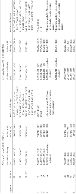
TABLE 1 Comparison of nucleotide and amino acid sequences of A/Muscovy duck/France/09010/09(H6N1) with A/turkey/France/09010 –1/09(H6N1) and A/turkey/France/09010 –2/09(H6N1) viruses Segment Protein A/Turkey/France/09010–1/09(H6N1) A/Turkey/France/09010–2/09(H6N1) Nucleotide identity (%) Amino acid identity (%) Amino acid changes Nucleotide identity (%) Amino acid identity (%) Amino acid changes 1 PB2 2,005/2,280 (87.9) 751/759 (98.9) R61K, T147I, V292I, V295I 2,005/2,280 (87.9) 751/759 (98.9) R61K, T147I, V292I, V295I V397A, V397A, I511V, T585P, D680G I511V, T585P, D680G 2 PB1 2,190/2,274 (96.3) 750/757 (99) K52N, R54K, A56T, M195I, L550I, R584H, N694S 2190/2274 (96.3) 750/757 (99) K52N, R54K, A56T, M195I, L550I, R584H, N694S PB1-F2 258/273 (94.5) 76/90 (84.4) E4G, R21 M, G22E, G23S, Q26R, L30P, T46 M, Q48R, A49V, K53R, Q54R, K65R, Q79R, S82L 258/273 (94.5) 76/90 (84.4) E4G, R21 M, G22E, G23S, Q26R, L30P, T46 M, Q48R, A49V, K53R, Q54R, K65R, Q79R, S82L 3 PA 2,059/2,151 (95.7) 714/716 (99.7) A70V, E538G 2,059/2,151 (95.7) 714/716 (99.7) A70V, E538G 4 HA 1,698/1,701 (99.8) 565/566 (99.8) K118R 1,698/1,701 (99.8) 565/566 (99.8) K118R 5 NP 1,497/1,497 (100) 498/498 (100) 1,497/1,497 (100) 498/498 (100) 6 NA 1,350/1,350 (excluding deletion) 449/449 (100) N.B.: amino acids 55–74 are absent in the turkey isolate (deleted region) 1,350/1,350 (excluding deletion) 449/449 (100) N.B.: amino acids 55–74 are absent in the turkey isolate (deleted region) 1,341/1,341 (excluding deletion) 446/446 (100) N.B.: amino acids 43–66 are absent in the turkey isolate (deleted region) 7 M1 759/759 (100) 252/252 (100) 759/759 (100) 252/252 (100) M2 294/294 (100) 97/97 (100) 294/294 (100) 97/97 (100) NS1 693/693 (100) 230/230 (100) 693/693 (100) 230/230 (100) 8 NEP 366/366 (100) 121/121 (100) 366/366 (100) 121/121 (100)
on May 16, 2020 by guest
http://jcm.asm.org/
[image:2.585.173.429.73.727.2]cessing. Histological examination of the trachea and lung of turkeys was performed using routine procedures.
RNA extraction and RT-PCR.All the swabs received at the National
Veterinary School of Toulouse were first vortexed in a 500-l PBS
solu-tion. Viral RNA was extracted from 150l of sample by use of the RNA
Virus kit (Macherey-Nagel, Düren, Germany) according to the
manufac-turer’s protocol. The RNA was eluted into 50l RNase-free water. Each of
the viral gene segments was amplified by 2-step reverse transcription-PCR
(RT-PCR). For reverse transcription, 20l of total RNA was mixed with
40 pmol of Uni12 primer (7) and incubated for 5 min at 65°C. Then, 8l
of 5⫻reaction buffer (Fermentas, Glen Burnie, MD) was added to this
mixture at the same time as 1l of RiboLock RNase inhibitor
(Fermen-tas), 4l of 10 mmol/liter deoxynucleoside triphosphate (dNTP) solution
(Finnzymes, Espoo, Finland), and 2l of RevertAid reverse transcriptase
(Fermentas). The reaction volume was completed to 40l with distilled
water. The RT reaction was performed at 42°C for 60 min and terminated by heating at 70°C for 10 min.
cDNAs were amplified by using Phusion High-Fidelity DNA
poly-merase (Finnzymes) and a set of gene-specific primers (7).
Eight separated PCR mixtures, corresponding to the 8 genes, were
prepared and contained 29.5l of distilled water, 10l of 5⫻buffer, 1l
of 10 mmol/liter dNTP solution (Finnzymes), 2.5l of 10mol/liter
primers, 4l of the cDNA, and 1 unit Phusion High-Fidelity DNA
poly-merase. The amplification program consisted of a first 30-s step at 98°C followed by 35 cycles with the following conditions: 98°C for 10 s, 61°C for 20 s, and 72°C for 1 min 30 s. The program ended with 1 step at 72°C for 10 min.
Sanger sequencing of the amplification products.The amplification products were separated by electrophoresis using 1% agarose gels. Ob-served bands were excised from the gel and purified using the Nucleospin Extract II kit (Macherey-Nagel) according to the manufacturer’s
proto-col. These PCR products were eluted into 50l of distilled water and
subsequently used in Sanger sequencing and pyrosequencing. By Sanger sequencing, we determined consensus whole-genome sequences for each of the 3 types of sampling.
Differential real-time PCR on neuraminidase alleles.Quantitative real-time PCR was also used to characterize the neuraminidase allele(s) present in duck feces or turkey tracheas. A set of primers and probes was designed for full-length neuraminidase or truncated neuraminidase de-tection, according to the Sanger sequencing results. The forward primer
was NA-F (5=-AGACAGGGAATCAGCACCAG-3=), and the reverse
primer was Delta-R (5=-TTGGGCAAAGAGATGCATTA-3=). The
Taq-Man probe sequences targeting the complete or the truncated NA were
5=-FAM-TGAAAACAACACTTGGGTAAATCAGACC-BHQ-3= and 5=
-JOE-AAAAGCATCATTCTTACTGAGCAAGCTGT-BHQ-3=,
respec-tively. Both were coupled with a Black Hole Quencher dye at the 3=end.
Viral RNA was retrotranscribed as described before, and cDNA was am-plified and detected in a 7000 Sequence Detection System (PE Applied Biosystems) by using the Platinum Quantitative PCR SuperMix-UDG
(Invitrogen SRL, Milan, Italy). The 25-l PCR mixture contained 5 pmol
of each primer and 50 pmol probe. After 2 thermal steps at 50°C for 2 min and 95°C for 2 min, 40 cycles of PCR at 95°C for 15 s and at 60°C for 30 s were performed.
454 pyrosequencing of the amplification products.The previously purified PCR products were quantified using a Quant-iT PicoGreen kit (Molecular Probes, Eugene, OR). Libraries were prepared after PCR prod-uct nebulization using the GS DNA Rapid Library Preparation kit accord-ing to the manufacturer’s recommendations (Roche, Basel, Switzerland). Three pools (corresponding to the 3 different sets of samplings), each containing 500 ng DNA, were used to generate the libraries; the 8 seg-ments were pooled in equimolar amounts. Each DNA pool was ligated to an adapter containing a multiplex identifier sequence (MID) for emulsion PCR and sequencing. Final libraries were quantified using the SlingShot kit, using the Fluidigm Digital Array for sample quantification, following the manufacturer’s instructions. Pyrosequencing using the 454/Roche GS
FLX Titanium chemistry was carried out with the GS Titanium SV emul-sion PCR kit and sequencing kit according to the manufacturer’s instruc-tions. The 3 samples were sequenced on 1 region of an 8-region picotiter plate.
Bioinformatic analysis.Sequences were first demultiplexed using Roche’s tool SFF file without allowing any error per MID. Reads were then
filtered using pyrocleaner (14), considering different criteria such as
length. Reads shorter or longer than the mean length of the reads⫾2
standard deviations were discarded. Reads were also filtered based on their complexity, which is computed using the compressed string length (library zip) on several subsequences generated using a sliding window approach. Moreover, the pyrocleaner script removes sequences for which all base pairs have a phred quality value under 20 or if the rate of unde-termined bases is higher than 4%, which has been proven to correlate with poor quality. Next, to carry out the single nucleotide polymorphism (SNP) calling, the remaining sequences were assembled on the reference to produce a consensus sequence for each sample. To do so, sequences
were aligned against the reference genome using bwa (11), and the
con-sensus was computed with the SAMtools (12) software package. Reads
were then realigned against this consensus sequence before SNP calling using other modules of the same package. For large deletion discovery, the
BLAT (10) aligner was used on the same reads for each sample. The
re-sulting files were then displayed within the IGV (22) browser.
Nucleotide sequence accession numbers.The complete sequences of A/Muscovy duck/France/09010/09, A/turkey/France/09010 –1/09, and A/turkey/France/09010 –2/09 (H6N1) viruses have been deposited in
GenBank under accession numbersJN860169toJN860193.
RESULTS
Case history and virological findings.Three days after the re-moval of ducks to the slaughterhouse, the turkey flock started to show prostration and respiratory signs, resulting in approximately 10% mortality. A sampling of 10 diseased turkeys was performed, and at the same time, several samples of fecal material in the duck house were collected and pooled. Necropsies were performed on the 10 birds, and all showed severe exudative tracheitis, airsaccu-litis, and pneumonia. A routine antibiotic treatment was per-formed to prevent secondary bacterial infections, but it did not stop mortality. A second necropsy was therefore performed on 10 other birds 8 days later. Tracheal swabs and organ samples were taken from all dead animals.
Real-time PCR assays were negative forMycoplasma
gallisepti-cumandMycoplasma synoviae. Real-time RT-PCR analyses of the
tracheal samples were negative for avian metapneumovirus (5)
but positive for avian influenza virus (AIV) (3).
FIG 1Typical agarose gel electrophoresis pattern of NA gene-specific RT-PCR products from duck and turkey clinical specimens, using internal primers
(NA-F: 5=-TGGTAATTGGAATAGTCAGTTTGA-3=; NA-R: 5=-ATCCCCTT
TGGAACCAATTC-3=). Lane 1: duck feces; lanes 2 to 5: representative early
turkey swabs, showing a 60-nucleotide deletion; lanes 6 to 9: representative late turkey swabs, showing various, mixed patterns of deletion. M: molecular mass
marker; *: full-length NA.⌬60 and⌬69: 60-nt and 69-nt deletion, respectively.
on May 16, 2020 by guest
http://jcm.asm.org/
[image:3.585.300.543.67.161.2]Immunostaining for influenza virus A revealed infected cells in the tracheal epithelium and in the pulmonary parenchyma at the time of the first sampling (data not shown).
Sequence analysis of duck and turkey isolates.RT-PCR
am-plifications encompassing all the viral genomic segments’ coding sequences were performed on both turkey tracheal swabs and duck feces using a set of gene-specific primers for influenza viruses
(7). This procedure, followed by Sanger sequencing, led to the
identification of the H6N1 subtype. Comparison of complete
se-quences of duck and turkey viruses (Table 1) revealed more than
99.8% nucleotide (nt) identity genome-wide, except for (i) one internal deletion of 60 nt in segment 6 resulting in a putative 20-aa deletion in the neuraminidase stalk in all of the first turkey isolates collected and (ii) a singular pattern of NA stalk deletions consist-ing of a mix of the 60-nt deleted allele and a new, second 69-nt deleted one in some turkeys swabbed after 8 days of infection, as confirmed by cloning and sequencing. Four of 10 birds sampled at this later stage of infection in the turkey barn showed this mixed
pattern (representative specimens shown inFig. 1).
Differential real-time PCR on Neuraminidase alleles.To
deter-TABLE 2Quantitative RT-PCR distinguishing between wild-type and
truncated NAa
Specimen type Probe sequence
Mean no. of copies⫾SD
Wild-type NA Truncated NA
Duck feces TGAAAACAACACTTGG
GTAAATCAGACC
787⫾311 ND
Turkey swabs AAAAGCATCATTCTTA
CTGAGCAAGCTGT
ND 5,109⫾1,585
a
ND, not detected. 5=primer sequence AGACAGGGAATCAGCACCAG; 3=primer
sequence, TTGGGCAAAGAGATGCATTA.
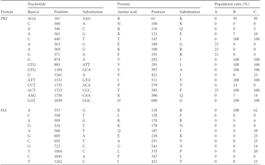
FIG 2Map of sequence depth of the whole influenza genome for duck (A), early turkey (B), and late turkey virus (C). For each segment, the coverage depth is plotted as a function of nucleotide position. PA: PA polymerase; HA: hemagglutinin; NP: nucleoprotein; NA: neuraminidase; M: matrix; NS: nonstructural.
on May 16, 2020 by guest
http://jcm.asm.org/
[image:4.585.41.286.88.168.2] [image:4.585.43.541.272.702.2]mine if the deleted forms were present, even at low copy number, in duck feces samplings, a real-time PCR assay was designed to discriminate between undeleted and deleted NA, using a unique couple of primers encompassing the deletion region of NA and 2
sequence-specific internal probes (Table 2). The copy number was
deduced from a plasmidic standard dilution (ranging from 10 to
106copies) containing the cognate sequences. This allele-specific
real-time PCR showed a very low threshold of less than 10 copies per reaction (data not shown) and was performed in triplicate on duck and turkey samples. Using this sensitive method, we readily detected the full-length NA but not the truncated segment in duck samples. The same analysis, when applied to early turkey tracheal
swabs, only detected the truncated form of the NA segment (Table 2).
Deep sequencing of complete viral genome of duck and tur-key viruses.Complete genomes were obtained for the 3 pools of samples, with a mean number of reads of 21,600 per pool and an average coverage depth of 680 for all segments. A comprehensive
coverage map is shown inFig. 2.
For each viral gene, SNPs were tracked on the basis of a 5%
threshold.Table 3summarizes the nonsynonymous SNPs and the
ratio of mutated viral sequences for PB2 and HA. Briefly, 8 SNPs on PB2 appeared within a few days in 100% of the turkey viral reads. Some minority mutants in the ducks disappeared as readily as in the turkeys. On HA, one SNP (K118R) was identified and remained preponderant in turkeys in the late samples. Eight other minority SNPs, concerning 6% to 32% of the reads, were
evi-denced in late samples on positions 170 to 241, corresponding to the receptor binding region of HA. Data concerning the 6 other segments are shown in Table SA1 in the supplemental material; SNPs were observed in turkeys on PB1 (7 positions), PB1-F2 (14 positions), and PA (3 positions).
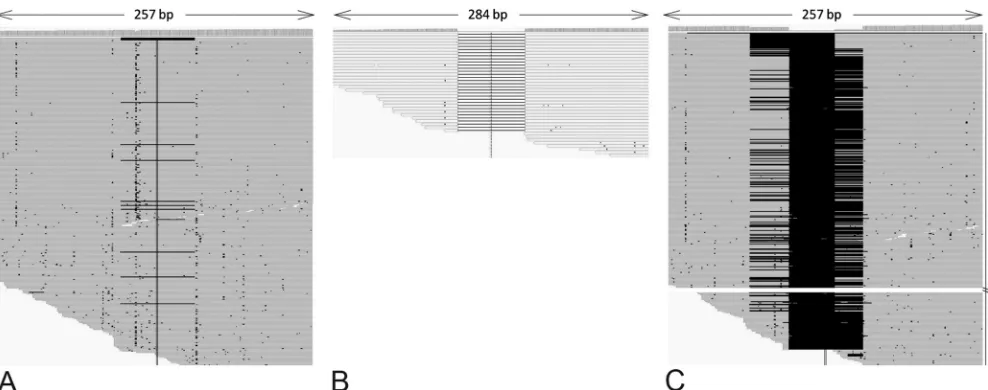
A particular analysis was performed on the NA gene; as expected, we identified a 100% 60-nt deleted population in early turkey samples and a mixed 60- and 69-nt deletion pattern in late turkey samples. Alignments of the NA genes could readily evidence a comprehensive
pattern of deletions (Fig. 3). More surprising is that the same analysis
on duck fecal material evidenced an ultraminority population of leted 60-nt NA alleles (13/670 reads, i.e., 1.94%), which was not
de-tected by other means (Fig. 3). This critical result demonstrates that
minute amounts of deleted NA alleles were present in the duck viral mutant cloud and “emerged” in the turkeys.
DISCUSSION
[image:5.585.39.554.77.399.2]In this study, we monitored the evolution of LPAIVs by pyrose-quencing performed directly on clinical samples. The whole ge-nomes of 3 pooled samples were subjected to deep sequencing and analysis of viral subpopulations. The global sequence coverage was sufficient to observe low population variation, which may possibly represent quasispecies, and to track the evolution of mi-nority mutants. For this purpose, a critical advantage of 454 py-rosequencing, when compared with other next-generation se-quencing platforms, relies on its ability to generate quite long
TABLE 3Nonsynonymous single nucleotide polymorphisms (SNPs) detected on PB2 and HAa
Protein
Nucleotide Protein Population ratio (%)
Base(s) Position Substitution Amino acid Position Substitution A B C
PB2 AGA 181 AAG R 61 K 0 95 95
C 300 A N 100 K 0 0 8
A 346 C K 116 Q 0 0 5
A 361 G K 121 E 0 7 10
C 440 T T 147 I 0 100 100
A 563 G E 188 G 25 0 0
A 569 G K 190 R 23 0 0
G 571 A E 191 K 21 0 0
G 874 A V 292 I 0 100 100
GTG 883 ATT V 295 I 0 100 100
GTG 1189 GCA V 397 A 0 100 100
G 1261 A V 421 I 0 0 23
ATT 1531 GTA I 511 V 0 100 100
CCT 1735 ACA P 579 T 0 14 0
ACT 1753 CCC T 585 P 25 100 100
AAG 1756 CAA K 586 Q 0 7 14
GAT 2038 GGC D 680 G 0 100 100
HA A 353 G K 118 R 0 100 62
C 358 T L 120 F 6 0 0
A 509 G K 170 R 0 0 6
G 534 T K 178 N 9 0 0
A 560 T Q 187 L 0 0 38
G 685 A E 229 K 0 0 25
C 692 T A 231 V 0 0 25
G 722 C G 241 A 0 0 14
T 1004 C L 335 P 0 0 20
C 1041 A F 347 L 0 0 6
T 1262 C I 421 T 0 0 23
a
For each library, corresponding to duck feces (A), early samples in turkeys (B), and late samples in turkeys (C), reads were aligned against the consensus sequence before SNP
calling. The percentage of reads sharing the SNP was determined with a threshold of 5%.
on May 16, 2020 by guest
http://jcm.asm.org/
reads, of roughly 400 nt (at the time of the experiment), facilitat-ing subsequent sequence reconstruction.
One of the most significant changes observed was the emer-gence of 2 avian influenza NA stalk deletion variants after virus transmission from the ducks to the turkeys. In this case study, the NA stalk deletions could be monitored in the field, at the scale of a single farm, with a direct link from the parental to the deletion variants.
Our data favor the idea that NA stalk truncation emerged early after turkey contamination, as a minute number of reads of de-leted NA could be detected in duck feces. Besides, we did not detect any full-length NA form in turkeys, even in early samples, suggesting a strong evolutionary advantage of the truncated form at the expense of the full-length one in this poultry species.
Very surprising is that after 8 more days of viral circulation in turkeys, some birds appeared to be coinfected with a second vari-ant harboring a larger deleted region, which did not totally overlap
with the 60-nt deletion previously observed. This later⌬69 allele
could not be detected in early turkey samples, maybe because the depth coverage of NA was quite low for this early sample. This finding suggests a higher plasticity of neuraminidase than previ-ously thought.
NA stalk truncation events, which reduce the protein enzy-matic activity, have been shown to be associated with changes in the glycosylation pattern of the HA globular head, resulting in a
modification of HA affinity for cellular receptors (5,17,19).
We tracked the evolution of HA and could evidence the emer-gence of minority mutants, with substitutions mostly in the recep-tor binding site on the globular head: these SNPs concerned up to 38% of viral species after 8 days of clinical evolution in the flock. None of these mutations altered glycosylation sites of HA. They may have contributed to the refinement of binding to cellular receptors of the turkeys’ upper respiratory tract and/or to
circum-vent the host’s immune response (18).
It is of interest to note that the observed mortality in turkeys was contemporary to NA deletion variant detection, whereas ducks did not show any clinical sign of infection. Even though NA
stalk deletion was recently associated with an increased virulence
in chickens infected by reverse-genetics-engineered viruses (8,
20), whether the deletions described in this field case are
respon-sible for the observed virulence in turkey remains to be demon-strated.
In this farm, the 2 species were bred separately in 2 different houses. The contamination of turkeys can likely be explained by a biosecurity failure, which probably occurred when ducks were transported to the slaughterhouse. This case suggests further that the turkey is a very peculiar host among poultry species and shows
very high receptivity and sensitivity to influenza A viruses (21).
Recent epidemiological modeling data suggest a very high
within-flock transmissibility of LPAIVs in turkeys (1). This case also
em-phasizes the risks of breeding together different avian species, i.e., terrestrial birds (chickens or turkeys) and waterfowl. Such a situ-ation, which seems relatively common in Asia, can result in the quick emergence of variants with altered phenotypes and in-creased pathogenicity. Moreover, it illustrates the genetic hetero-geneity of avian influenza viruses within a single flock. In a first attempt, our approach was intentionally based on the poultry population considered as a whole, irrespective of the individual level. The next step will be to investigate quasispecies variations between birds in a flock and then between different tissues (i.e., respiratory versus digestive tracts) in given birds.
In previous studies, next-generation sequencing approaches have already proven to be of interest for monitoring viral evolu-tion at precise viral genome posievolu-tions following evoluevolu-tionary
con-straints such as antiretroviral treatment during HIV infection (6)
or oseltamivir treatment during influenza infection (9). A broader
approach of viral genome evolution may allow us to monitor the
early apparition of mutants withouta prioriconsideration and to
follow complex chains of mutational events occurring on several positions of viral genomes. The 454 sequencing platform used here has the critical advantage of producing long reads—from 400 nt to more than 700 nt in recent updates—facilitating sequence reconstruction in a whole viral genome approach. Alternative technologies are in constant development and will likely provide
FIG 3Read alignments on the NA stalk region, focusing on deletion patterns; 454 reads were contiged and aligned. Deletion in the stalk region (positions 161 to 220) was found in only 13 of 670 reads in duck feces (A) but in 100% of reads in early samples from turkeys (B). A mix of 60- and 69-nt (positions 129 to 197) deletions was observed in turkeys 8 days later (C).
on May 16, 2020 by guest
http://jcm.asm.org/
[image:6.585.46.540.64.259.2]improved sequencing capacities in the near future. The develop-ment of next-generation sequencing approaches should enable us to monitor both viral genome diversity within bird flocks and the early apparition of potentially pathogenic variants.
Although our data do not explain how the NA stalk deletion appeared in the duck samples in the first place, this present study suggests that this deletion can exist as a minority mutant in water-fowl, awaiting a change of host to be selected.
ACKNOWLEDGMENTS
We thank Mariette Ducatez for helpful comments on the manuscript. We acknowledge the assistance of Jerome Lluch, Genomics platform of Tou-louse, France, with the 454 pyrosequencing.
This project was partly supported by French Region Midi-Pyrénées.
REFERENCES
1.Comin A, Klinkenberg D, Marangon S, Toffan A, Stegeman A.2011. Transmission dynamics of low pathogenicity avian influenza infections in
Turkey flocks. PLoS One6:e26935.
2.Di Trani L, et al.2004. Molecular characterization of low pathogenicity
H7N3 avian influenza viruses isolated in Italy. Avian Dis.48:376 –383.
3.Fouchier RA, et al.2000. Detection of influenza A viruses from different species by PCR amplification of conserved sequences in the matrix gene. J.
Clin. Microbiol.38:4096 – 4101.
4.Giannecchini S, et al.2006. Comparison of in vitro replication features of H7N3 influenza viruses from wild ducks and turkeys: potential
implica-tions for interspecies transmission. J. Gen. Virol.87:171–175.
5.Guionie O, et al.2007. Laboratory evaluation of a quantitative real-time reverse transcription PCR assay for the detection and identification of the
four subgroups of avian metapneumovirus. J. Virol. Methods139:150 –
158.
6.Hedskog C, et al.2010. Dynamics of HIV-1 quasispecies during antiviral treatment dissected using ultra-deep pyrosequencing. PLoS One
5:e11345.
7.Hoffmann E, Stech J, Guan Y, Webster RG, Perez DR.2001. Universal primer set for the full-length amplification of all influenza A viruses. Arch.
Virol.146:2275–2289.
8.Hoffmann TW, et al.2012. Length variations in the NA stalk of an H7N1 influenza virus have opposite effects on viral excretion in chickens and
ducks. J. Virol.86:584 –588.
9. Inoue M, et al.2010. Emergence of oseltamivir-resistant pandemic
(H1N1) 2009 virus within 48 hours. Emerg. Infect. Dis.16:1633–1636.
10. Kent WJ.2002. BLAT: the BLAST-like alignment tool. Genome Res.12: 656 – 664.
11. Li H, Durbin R.2009. Fast and accurate short read alignment with
Bur-rows-Wheeler transform. Bioinformatics25:1754 –1760.
12. Li H, et al.2009. The Sequence alignment/map format and SAMtools.
Bioinformatics25:2078 –2079.
13. Li J, Zu Dohna H, Cardona CJ, Miller J, Carpenter TE.2011. Emergence and genetic variation of neuraminidase stalk deletions in avian influenza
viruses. PLoS One6:e14722.
14. Mariette J, Noirot C, Klopp C.2011. Assessment of replicate bias in 454 pyrosequencing and a multi-purpose read-filtering tool. BMC Res. Notes
4:149.
15. Mathieu C, et al.2010. Pandemic (H1N1) 2009 in breeding turkeys,
Valparaiso, Chile. Emerg. Infect. Dis.16:709 –711.
16. Matrosovich M, Zhou N, Kawaoka Y, Webster R.1999. The surface glycoproteins of H5 influenza viruses isolated from humans, chickens, and
wild aquatic birds have distinguishable properties. J. Virol.73:1146 –1155.
17. Matsuoka Y, et al.2009. Neuraminidase stalk length and additional gly-cosylation of the hemagglutinin influence the virulence of influenza H5N1
viruses for mice. J. Virol.83:4704 – 4708.
18. McHardy AC, Ben Adams B.2009. The role of genomics in tracking the
evolution of influenza A virus. PLoS Pathog.5:e1000566.
19. Mitnaul LJ, et al.2000. Balanced hemagglutinin and neuraminidase ac-tivities are critical for efficient replication of influenza A virus. J. Virol.
74:6015– 6020.
20. Munier S, et al.2010. A genetically engineered waterfowl influenza virus with a deletion in the stalk of the neuraminidase has increased virulence
for chickens. J. Virol.84:940 –952.
21. Pillai SPS, Pantin-Jackwood M, Yassine HM, Saif YM, Lee CW.2010. The high susceptibility of turkeys to influenza viruses of different origins
implies their importance as potential intermediate hosts. Avian Dis.54:
522–526.
22. Robinson JT, et al.2011. Integrative genomics viewer. Nat. Biotechnol.
29:24 –26.
23. Sorrell EM, Song H, Pena L, Perez DR.2010. A 27-amino-acid deletion in the neuraminidase stalk supports replication of an avian H2N2
influ-enza A virus in the respiratory tract of chickens. J. Virol.84:11831–11840.
24. Zhou H, et al.2009. The special neuraminidase stalk-motif responsible for increased virulence and pathogenesis of H5N1 influenza A virus. PLoS
One4:e6277.