Heparin cofactor II inhibits arterial thrombosis
after endothelial injury
Li He, … , Daniel T. Eitzman, Douglas M. Tollefsen
J Clin Invest.
2002;
109(2)
:213-219.
https://doi.org/10.1172/JCI13432
.
Heparin cofactor II (HCII) is a plasma protein that inhibits thrombin rapidly in the presence of
dermatan sulfate, heparan sulfate, or heparin. HCII has been proposed to regulate
coagulation or to participate in processes such as inflammation, atherosclerosis, and wound
repair. To investigate the physiologic function of HCII, about 2 kb of the mouse
HCII
gene,
encoding the N-terminal half of the protein, was deleted by homologous recombination in
embryonic stem cells. Crosses of F
1HCII
+/–animals produced
HCII
–/–offspring at the
expected mendelian frequency. Biochemical assays confirmed the absence of dermatan
sulfate–dependent thrombin inhibition in the plasma of
HCII
–/–animals. Crosses of
HCII
–/–animals produced litters similar in size to those obtained from heterozygous matings. At 1
year of age, HCII-deficient animals were grossly indistinguishable from their wild-type
littermates in weight and survival, and they did not appear to have spontaneous thrombosis
or other morphologic abnormalities. In comparison with wild-type animals, however, they
demonstrated a significantly shorter time to thrombotic occlusion of the carotid artery after
photochemically induced endothelial cell injury. This abnormality was corrected by infusion
of purified HCII but not ovalbumin. These observations suggest that HCII might inhibit
thrombosis in the arterial circulation.
Article
Find the latest version:
http://jci.me/13432/pdf
Introduction
Heparin cofactor II (HCII) is a serine protease inhibitor (serpin) that inactivates thrombin rapidly in the presence of certain glycosaminoglycans (reviewed in ref. 1). HCII does not inhibit other proteases involved in coagulation or fibrinolysis. Thrombin stimulates platelet aggregation, promotes coagulation by cleavage of fibrinogen and acti-vation of factors V, VIII, XI, and XIII, and inhibits fibri-nolysis by activation of a plasma carboxypeptidase (2). Conversely, when thrombin binds to thrombomodulin on the surface of endothelial cells, it activates protein C, which inhibits further thrombin generation. Thrombin also engages in a variety of activities unrelated to hemo-stasis (3). For example, it causes proliferation of fibrob-lasts and other cells, induces monocyte chemotaxis, pro-motes adhesion of neutrophils to endothelial cells, and inhibits neurite outgrowth. HCII could potentially regu-late the activity of thrombin in one or more of these diverse biological processes.
The rate of inhibition of thrombin by HCII increases more than 1000-fold in the presence of heparin, heparan sulfate, or dermatan sulfate (4). HCII is unique among serpins in its ability to be stimulated by dermatan sulfate and binds to a minor subpopulation of dermatan sulfate oligosaccharides (5). By contrast, antithrombin binds to a specific pentasaccharide structure in heparin or
heparan sulfate and is thereby stimulated to inhibit thrombin and other coagulation proteases (especially factors Xa and IXa) (1). The distribution of specific gly-cosaminoglycans on the cell surface or in the ECM may serve to localize protease inhibition by HCII and antithrombin to different sites. Cultured fibroblasts and vascular smooth muscle cells synthesize proteoglycans that stimulate inhibition of thrombin by HCII, but endothelial cells do not, suggesting that HCII may inhib-it thrombin at sinhib-ites outside the vascular lumen (6, 7).
Although the physiologic function of HCII is unknown, the presence of thrombin-HCII complexes in human plasma indicates that HCII inhibits thrombin in vivo (8, 9). HCII is synthesized by the liver and circu-lates in human plasma at a concentration of about 1
µM (10). Turnover studies of labeled HCII in humans suggest that about 40% of the protein equilibrates with an extravascular compartment (11), but the tissue dis-tribution of HCII has not been thoroughly investigated. HCII has been detected in the intima of normal human arteries, and the ability of dermatan sulfate in the arte-rial wall to stimulate HCII is decreased in atheroscle-rotic lesions (12, 13). During pregnancy, both the mater-nal and the fetal blood contain trace amounts of a dermatan sulfate proteoglycan that stimulates throm-bin inhibition by HCII (14). The placenta is rich in
der-Heparin cofactor II inhibits arterial thrombosis
after endothelial injury
Li He,
1Cristina P. Vicente,
1Randal J. Westrick,
2Daniel T. Eitzman,
2and Douglas M. Tollefsen
11Division of Hematology, Department of Internal Medicine, and Department of Biochemistry and Molecular Biophysics,
Washington University, St. Louis, Missouri, USA
2Division of Cardiology, Department of Medicine, University of Michigan, Ann Arbor, Michigan, USA
Address correspondence to: Douglas M. Tollefsen, Division of Hematology, Box 8125,
Washington University School of Medicine, 660 South Euclid Avenue, St. Louis, Missouri 63110, USA. Phone: (314) 362-8830; Fax: (314) 362-8826; E-mail: tollefsen@im.wustl.edu.
Received for publication June 4, 2001, and accepted in revised form December 3, 2001.
Heparin cofactor II (HCII) is a plasma protein that inhibits thrombin rapidly in the presence of der-matan sulfate, heparan sulfate, or heparin. HCII has been proposed to regulate coagulation or to par-ticipate in processes such as inflammation, atherosclerosis, and wound repair. To investigate the phys-iologic function of HCII, about 2 kb of the mouse HCIIgene, encoding the N-terminal half of the protein, was deleted by homologous recombination in embryonic stem cells. Crosses of F1HCII+/– ani-mals produced HCII–/–offspring at the expected mendelian frequency. Biochemical assays confirmed the absence of dermatan sulfate–dependent thrombin inhibition in the plasma of HCII–/–animals. Crosses of HCII–/–animals produced litters similar in size to those obtained from heterozygous mat-ings. At 1 year of age, HCII-deficient animals were grossly indistinguishable from their wild-type lit-termates in weight and survival, and they did not appear to have spontaneous thrombosis or other morphologic abnormalities. In comparison with wild-type animals, however, they demonstrated a sig-nificantly shorter time to thrombotic occlusion of the carotid artery after photochemically induced endothelial cell injury. This abnormality was corrected by infusion of purified HCII but not ovalbu-min. These observations suggest that HCII might inhibit thrombosis in the arterial circulation.
matan sulfate and may be the source of the circulating proteoglycan, suggesting that HCII could be activated locally to inhibit coagulation in the placenta (15). Chemotactic peptides are released when HCII is par-tially degraded by neutrophil proteases, suggesting a possible direct role for HCII in inflammation (16). HCII could also participate in wound healing by regulating the mitogenic or chemotactic activities of thrombin.
Several patients with inherited HCII deficiency (∼50% of normal) and histories of venous thromboembolic dis-ease have been reported (17, 18). In one study, however, 4 of 379 apparently healthy individuals had HCII levels less than 60% of normal (19). Thus, heterozygous defi-ciency of HCII may be a coincidental finding in about 1% of patients with venous thrombosis and does not appear to be a strong risk factor for development of this disease (20). Two sisters who appeared to be homozy-gous for HCII deficiency (HCII activity 10–15% of nor-mal) have been reported (21); one of these individuals was also heterozygous for antithrombin deficiency and had recurrent venous thromboembolism. Human sub-jects who completely lack HCII have not been identified. With the aim of elucidating the physiologic function(s) of HCII, we have generated a mouse model of HCII defi-ciency by targeted gene disruption in embryonic stem cells. HCII-deficient mice are viable and fertile, and they do not appear to have spontaneous thrombosis or other morphologic abnormalities. In comparison with wild-type animals, however, they demonstrate a significantly shorter time to thrombotic occlusion of the carotid artery after photochemically induced endothelial cell injury. These observations suggest that a function of HCII might be to inhibit thrombosis in the arterial circulation.
Methods
Construction of the targeting vector. PCR primers (5′-CTTCTTTCTCTCATCACATTTATG and 5′- CTTG-GAAAAGCTGCAGGATGTTG) that amplify a 271-bp frag-ment in exon 1 of the mouse HCIIgene (22) were used to screen a bacterial artificial chromosome (BAC) library made from 129/SvJ mouse genomic DNA (Genome Systems Inc., St. Louis, Missouri, USA). Two clones were isolated, each containing more than 100 kb of genomic DNA. A 6-kb EcoRV fragment that includ-ed about 3 kb of 5′flanking sequence, exon 1, and about 2 kb of intron 1 was isolated from one of the BAC clones and subcloned into the EcoRV site of pBlue-script II SK(+) (Stratagene, La Jolla, California, USA). The vector was then cleaved with EcoRI to remove the exon 1 and intron 1 sequences and ligated to a 1.6-kb EcoRI fragment of pPGKneo-I(Embryonic Stem Cell Core, Washington University), which contained the phosphoglycerate kinase promoter and the neomycin
phosphotransferase gene (neo) flanked by loxP
sequences. Ligation of the neocassette occurred in the same transcription orientation as the HCIIgene. A 4-kb BamHI fragment extending from the middle of intron 1 through intron 3 of the HCIIgene was isolated from the other BAC clone and inserted into the BamHI
mul-tiple cloning site of pBluescript to yield the targeting vector shown in Figure 1a. The final construct con-tained a 5′arm of 2.9 kb and a 3′arm of 3.6 kb of HCII genomic sequence flanking the neocassette.
Generation of HCII-deficient mice. The targeting vector was linearized with NotI and introduced into 129/SvJ-derived RW-4 embryonic stem cells (Embryonic Stem Cell Core, Washington University) by electroporation. G418-resistant clones were screened for homologous
recombination at the HCII locus by Southern
hybridization of SacI digests, using a 329-bp probe (3′ -probe) to a DNA sequence in exon 4 and an approxi-mately 500-bp probe (5′-probe) to a DNA sequence in the 5′flanking region of the HCIIgene; both probes hybridized to sequences external to the targeting vec-tor (Figure 1a). Cells from a correctly targeted clone were injected into C57BL/6 blastocysts, which were then implanted into pseudopregnant females. The resulting chimeric males were crossed with wild-type C57BL/6 females to produce F1agouti offspring that
were genotyped by Southern hybridization of DNA from tail biopsies. F1heterozygotes were crossed to
pro-duce homozygous HCII-deficient mice. These mice were backcrossed for six generations with wild-type C57BL/6 animals to produce a nearly congenic strain of mice containing the HCII-null allele. N6 heterozy-gotes were crossed to produce HCII+/+and HCII–/–mice. mRNA analysis. Total RNA was purified from adult mouse livers by acid phenol–guanidinium thio-cyanate–chloroform extraction (23). Northern blots of the RNA were hybridized with an 891-bp cDNA probe encoding the 3′half of exon 2 and all of exons 3 and 4. The blots were hybridized separately with a 625-bp probe to the neocassette.
Western blots. Plasma (0.1 or 0.3 µl) was subjected to electrophoresis on a 7.5% SDS polyacrylamide gel under reducing conditions. The proteins were transferred to a nitrocellulose membrane, which was then blocked with 5% milk and incubated with affinity-purified goat anti-human HCII IgG (Affinity Biologicals Inc., Hamilton, Ontario, Canada) at a concentration of 3 µg/ml for 1 hour at room temperature. The membrane was subse-quently washed and incubated with horseradish perox-idase–conjugated mouse anti-goat IgG (A9452; Sigma Chemical Co., St. Louis, Missouri, USA) at a 1:5000 dilu-tion. Immobilized antibody was detected with Super-Signal West Pico Chemiluminescent Substrate (Pierce Chemical Co., Rockford, Illinois, USA) according to the manufacturer’s instructions.
Inhibitor activity assays. HCII activity was determined by measuring the amount of thrombin inhibited in mouse plasma supplemented with dermatan sulfate. Plasma (2 µl) was mixed with 50 µg/ml porcine skin dermatan sulfate (Sigma Chemical Co.) and 16 nM human α-thrombin (Haematologic Technologies Inc., Essex Junction, Vermont, USA) in a total volume of 100
µl of buffer containing 50 mM Tris-HCl, 150 mM
the reaction. After a 60-second incubation at room temperature, 500 µl of 100 µM tosyl-Gly-Pro-Arg-p -nitroanilide (Chromozym TH; Roche Molecular Bio-chemicals, Indianapolis, Indiana, USA) in TS/PEG was added, and the absorbance at 405 nm was determined continuously for 100 seconds. The rate of change of absorbance was proportional to the concentration of active thrombin that remained in the incubation. The molar concentration of HCII in plasma was calculated from the amount of thrombin inhibited, assuming 1:1 stoichiometry of inhibition.
Antithrombin activity was determined by measuring the amount of factor Xa inhibited in plasma supple-mented with heparin. Plasma (0.3 µl) was mixed with 1 U/ml porcine intestinal heparin (Elkins-Sinn Inc., Cherry Hill, New Jersey, USA) and 11 nM human factor Xa (Haematologic Technologies) in 100 µl of TS/PEG buffer. Factor Xa was added last to initiate the reaction. After a 60-second incubation at room temperature, 500
µl of 500 µM MeO-CO-D-cyclohexylglycyl-Gly-Arg-p -nitroanilide (Spectrozyme fXa; American Diagnostica Inc., Greenwich, Connecticut, USA) in TS/PEG was added, and the absorbance at 405 nm was determined continuously for 100 seconds. The rate of change of absorbance was proportional to the concentration of active factor Xa that remained in the incubation. The molar concentration of antithrombin was calculated from the amount of factor Xa inhibited, assuming 1:1 stoichiometry of inhibition.
Blood tests. Routine blood tests of four HCII–/–males,
four HCII–/–females, four HCII+/+ males, and three HCII+/+females, all approximately 6 months of age,
were performed using standard methods in the Divi-sion of Comparative Medicine, Washington University. The tests included complete blood counts with white blood cell differentials and assays for urea nitrogen, creatinine, total protein, alanine aminotransferase, and aspartate aminotransferase.
Necropsies. Necropsies of four HCII–/–mice
approxi-mately 6 months of age (two males and two females) were performed by a veterinary pathologist in the Divi-sion of Comparative Medicine, Washington University. Histologic examination included the lung, heart, stom-ach, intestinal tract, brain, liver, kidney, spleen, pan-creas, aorta, adrenal glands, ovary, uterus, testes, and accessory sex glands.
Arterial thrombosis model. Carotid artery thrombosis was induced as described previously (24). Briefly, adult male and female mice were anesthetized with intraperi-toneal sodium pentobarbital, secured in the supine position, and placed under a dissecting microscope. The right common carotid artery was isolated through a midline cervical incision, and an ultrasonic flow probe (model 0.5 VB; Transonic Systems Inc., Ithaca, New York, USA) was applied. A 1.5-mW, 540-nm laser beam (Melles Griot Inc., Carlsbad, California, USA) was applied to the artery from a distance of 6 cm. Rose ben-gal dye (50 mg/kg body weight; Fisher Scientific Co., Fair Lawn, New Jersey, USA) was then injected into the
tail vein, and flow in the vessel was monitored until complete occlusion occurred. Five minutes before the injection of rose bengal dye, some animals were inject-ed with purifiinject-ed human HCII (25) or ovalbumin (Sigma Chemical Co.) in PBS to achieve a plasma concentration of about 1 µM. Specifically, 6.7 µl of a 4.7 µM protein solution was injected per gram body weight; the plasma volume was assumed to be 31.5 µl/g. Carotid artery
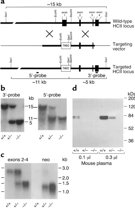
seg-Figure 1
Targeted disruption of the murine HCIIgene. (a) Restriction map of the HCIIlocus and design of a replacement vector. The boxes repre-sent exons 1–4 of the HCIIgene. The thick lines represent genomic DNA; the thin lines, vector DNA. The targeting vector was con-structed by insertion of the neomycin phosphotransferase gene (neo) between the EcoRI and BamHI sites of the HCIIgene. (b) Southern blots of genomic DNA isolated from the tails of 4- to 6-week-old mice and digested with SacI. The restriction fragments were detected with probes (5′-probe and 3′-probe) that hybridized with sequences external to the genomic DNA present in the targeting vector. (c) Northern blot of total liver RNA obtained from adult mice. The blot was first hybridized with a cDNA probe containing sequences pres-ent in exons 2–4 of the HCIIgene. It was then stripped and rehy-bridized with a probe containing sequences present in the neo cas-sette. (d) Western blot of mouse plasma probed with goat anti-human HCII IgG. The 68-kDa and 72-kDa bands represent two glycoforms of HCII that are present in normal mouse plasma (22).
[image:4.576.305.536.175.530.2]ments were fixed in 10% formalin, cross-sectioned, and stained with hematoxylin and eosin for examination by light microscopy.
Statistical analysis. Statistical significance was deter-mined with the Student two-tailed t test (http://facul-ty.vassar.edu/lowry/VassarStats.html). A Pvalue less than 0.05 was considered significant.
Results
A cDNA clone for murine HCII was isolated previously, and Southern hybridization of genomic DNA indicated the presence of a single HCIIgene (22). In the current study, genomic clones were isolated from a BAC library constructed with DNA from 129/SvJ mice. A partial restriction map of the HCIIlocus is shown in Figure 1a. A targeting vector was constructed by insertion of the neomycin phosphotransferase gene (neo) between the EcoRI and BamHI sites of the HCIIgene, which resulted in deletion of about 1 kb of 5′flanking sequence, exon 1, and part of intron 1. Since exon 1 encodes approximate-ly the N-terminal half of HCII, any gene product synthe-sized from the targeted locus would be expected to be inactive. RW-4 embryonic stem cells derived from 129/SvJ mice were transfected with the linearized targeting vector and selected for growth in the presence of G418. Homol-ogous recombination at the HCIIlocus was detected by Southern blotting with a probe that hybridized to a DNA sequence in exon 4, external to the genomic sequence included in the targeting vector. Digestion of DNA with SacI yielded a 15-kb fragment from the wild-type allele and a 5-kb fragment from the targeted allele.
Homologous recombination occurred in 5 of 684 G418-resistant embryonic stem cell clones. Embryonic stem cells from one clone with the targeted allele were injected into C57BL/6 blastocysts and implanted into pseudopregnant females. The resulting chimeric males were crossed with wild-type C57BL/6 females to pro-duce F1offspring that were genotyped by Southern
blot-ting of DNA from tail biopsies. Correct targeblot-ting of the HCIIlocus was verified with probes that hybridized to sequences both 5′and 3′to the DNA included in the tar-geting vector (Figure 1b). Crosses of heterozygous F1
animals yielded 250 offspring, with HCII–/–pups being
present at close to the expected mendelian frequency (24.8% HCII+/+, 53.6% HCII+/–, and 21.6% HCII–/–).
Sub-sequent crosses in which both parents were HCII–/–
pro-duced litters similar in size (6.8 ± 3.0 pups per litter, n= 11 litters) to those obtained from crosses of HCII+/–
mice (6.7 ± 3.1 pups per litter, n= 38 litters).
Northern blots of total liver RNA isolated from adult HCII+/+mice demonstrated a single HCII mRNA species
of about 2.5 kb, as expected from previous studies (22); this mRNA was absent in HCII–/–mice (Figure 1c). A
1.2-kb transcript containing neosequences was detected in HCII+/–and HCII–/–mice but not in wild-type animals. A
separate, 1.4-kb transcript containing sequences present in exons 2–4 of the HCIIgene was also present in HCII+/–
and HCII–/–mice. Since plasma from HCII-null mice did
not contain detectable HCII antigen or activity (see below), the 1.4-kb transcript was not investigated further. Western blots of plasma probed with a polyclonal anti-body indicated the presence of two previously described glycoforms of HCII (68 kDa and 72 kDa) in wild-type mice (22). Both glycoforms were absent in HCII–/–mice,
and intermediate levels were present in HCII+/–mice
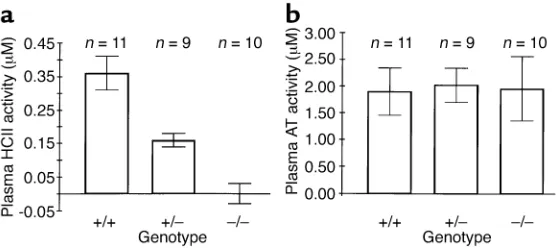
(Fig-ure 1d). No bands of lower molecular weight were observed in plasma of mice with the targeted HCIIallele, strongly suggesting that the abnormal 1.4-kb HCII tran-script did not give rise to HCII-like polypeptides in the circulation. Biochemical assays confirmed the absence of HCII activity (i.e., dermatan sulfate–dependent thrombin inhibition) in plasma obtained from HCII–/– mice
(0.00 ± 0.03 µM, mean ± SD) in contrast to HCII+/–
(0.16 ± 0.02 µM) and HCII+/+(0.36 ± 0.05 µM) mice
(Figure 2a). Antithrombin activity (i.e., heparin-depend-ent factor Xa inhibition) was similar in all three geno-types (Figure 2b).
At one year of age, homozygous HCII-deficient mice were indistinguishable from their wild-type and het-erozygous littermates in weight, appearance, and survival. Necropsies of four adult HCII–/–animals revealed no
gross or microscopic abnormalities. Blood tests of HCII–/–
mice (n= 8), including complete blood count with white blood cell differential, urea nitrogen, creatinine, total pro-tein, alanine aminotransferase, and aspartate amino-transferase, were within normal limits and did not differ significantly from those of wild-type mice (n= 7).
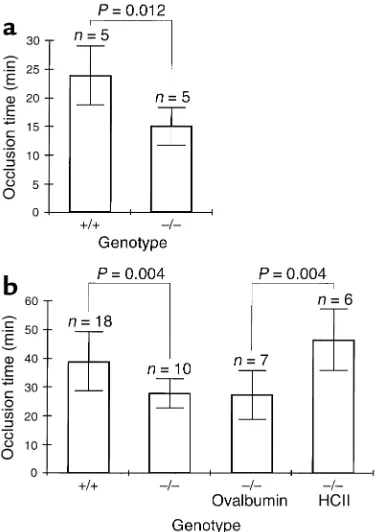
[image:5.576.63.341.55.179.2]The time to formation of an occlusive thrombus in the carotid artery following photochemically induced endothelial injury was shorter in HCII-deficient mice than in wild-type controls. Previous studies indicated that the base-line occlusion time in this model varies with
Figure 2
the mouse strain (26). For our initial experiments, HCII–/–
mice and their HCII+/+littermates were studied at 6–8
months of age. These animals were obtained from cross-es of heterozygous F1animals to insure that potential
modifier genes present in the C57BL/6 and 129/SvJ back-grounds would be distributed at equal frequencies in both sets of animals (Figure 3a). The time to arterial occlusion was 14.9 ± 3.2 minutes in HCII–/–mice
com-pared with 23.7 ± 5.1 minutes in HCII+/+mice (mean ±
SD, n= 5 in each group). This difference is statistically sig-nificant (P= 0.012). To determine whether the difference in occlusion time is reproducible in mice with a more homogeneous genetic background, HCIIheterozygotes were backcrossed for six generations with wild-type C57BL/6 animals to yield animals with about 97% C57BL/6 gene content. The N6 heterozygotes were crossed to produce HCII+/+and HCII–/–mice, which were
studied at 3–4 months of age. In the HCII–/–mice, the
time to arterial occlusion was 27.7 ± 5.0 minutes (n= 10)
compared with 38.6 ± 10.2 minutes in the HCII+/+mice
(n= 18) (Figure 3b). Although the occlusion times of both the wild-type and the HCII-null animals were longer than those of mice with a mixed genetic background, the absolute difference was similar (11 minutes vs. 9 minutes) and was statistically significant (P= 0.004). The abnor-mally short arterial occlusion time of the HCII–/–mice
was corrected by intravenous injection of purified HCII 5 minutes prior to induction of vascular injury (46.7 ± 10.8 minutes, n= 6) but not by injection of the noninhibitory serpin ovalbumin (27.4 ± 8.5 minutes, n= 7) (Figure 3b). Representative recordings of carotid blood flow are shown in Figure 4a. Cessation of blood flow was accompanied by the appearance of a pearly white occlu-sive filling defect in the lumen of the artery that was vis-ible through the dissecting microscope. Sections of the occluded arteries stained with hematoxylin and eosin revealed no gross histologic differences between throm-bi formed in HCII+/+and HCII–/–animals (Figure 4, b
and c). It was apparent from transient reductions in blood flow as well as direct observation through the dissecting microscope that a dynamic state of throm-bus formation and lysis occurred throughout the peri-od of vascular injury prior to complete occlusion.
Discussion
[image:6.576.88.277.56.322.2]Unlike antithrombin-deficient mice, which die in utero with extensive fibrin(ogen) deposition in the liver and myocardium (27), HCII-deficient mice apparently undergo normal fetal development and are born at the expected mendelian frequency. Their subsequent growth and survival is normal up to at least 1 year of age, and they do not demonstrate any obvious morphologic Figure 3
Effect of HCII deficiency on thrombotic occlusion of the carotid artery. Blood flow in the common carotid artery was monitored continuously with an ultrasonic flow probe. Local endothelial injury was induced by application of a 540-nm laser beam to the carotid artery followed by injection of rose bengal dye (50 mg/kg) into the lateral tail vein. (a) Mice with a mixed C57BL/6-129/SvJ genetic background. (b) Mice back-crossed for six generations into the C57BL/6 background. Five minutes before the injection of rose bengal dye, some of these mice were injected intravenously with purified human HCII or ovalbumin as indicated to achieve a plasma level of about 1 µM.
Figure 4
[image:6.576.228.522.581.734.2]abnormalities on gross or microscopic examination. Blood tests also indicate normal hematopoiesis as well as normal liver and kidney function. Thus, we find no evidence of thrombosis or bleeding secondary to dis-seminated intravascular coagulation, which are typical manifestations of deficiencies of several other anticoag-ulant proteins, including protein C (28), tissue factor pathway inhibitor (29), and antithrombin (27). Further experiments may show that HCII deficiency is similar to protein Z deficiency, in which a spontaneous prothrom-botic phenotype occurs only when combined with the homozygous factor VLeidengenotype (30).
Indirect evidence suggests that HCII activity may be increased in human pregnancy. During the third trimester, plasma HCII levels are about 150% of normal (31, 32), both maternal and fetal plasma contain trace amounts of a dermatan sulfate proteoglycan that stimu-lates thrombin inhibition by HCII (14), and thrombin-HCII complexes are elevated approximately fourfold (8). Conversely, HCII levels are decreased to about 50% of nor-mal in women with severe pre-eclampsia (32), suggesting that decreased HCII activity may be associated with pla-cental dysfunction. Nevertheless, HCII does not appear to be required for normal gestation in mice, since crosses in which both parents are HCII-deficient produce litters similar in size to those from heterozygous matings.
In contrast to the benign phenotype of unchallenged HCII-deficient mice, our experiments suggest that HCII plays an important role in maintaining blood flow after injury to the arterial endothelium. In these experi-ments, rose bengal dye in the lumen of the carotid artery was excited by transillumination with a green light laser to generate singlet oxygen. Previous studies have shown that this treatment causes membrane dam-age, resulting in detachment of the endothelial cells from a localized segment of the vessel wall followed by formation of a thrombus that is rich in platelets and fibrin (33). The mean time to thrombotic occlusion of the carotid artery was significantly shorter in HCII-defi-cient mice than in their wild-type littermates, both in a mixed C57BL/6-129/SvJ background and in a nearly congenic C57BL/6 strain. In a previous study, the carotid occlusion time was reduced to a similar degree in apoE-deficient mice fed a high-cholesterol diet (34). By contrast, the occlusion time was prolonged approx-imately twofold in mice deficient in plasminogen acti-vator inhibitor-1 (24, 35). Thus, the rose bengal model appears to be sensitive to structural alterations in the vessel wall associated with atherosclerosis as well as to systemic changes in the fibrinolytic system. The abnor-mally short occlusion time of HCII-deficient mice was corrected by intravenous injection of purified HCII (2.1
µg/g) to achieve a plasma concentration of about 1 µM prior to photochemical injury. As reported previously, intravenous injection of a much larger dose of purified HCII (84 µg/g) prior to photochemical injury of the femoral artery prolonged the occlusion time from 17 minutes to 31 minutes in wild-type rats (36). This experiment, together with our results, suggests that
HCII exerts an antithrombotic effect after photochem-ical injury to the arterial endothelium.
An important question raised by these studies is whether the protective effect of HCII depends on cataly-sis of the thrombin-HCII reaction by glycosaminoglycans in the vessel wall. Purified human HCII inhibits throm-bin with second-order rate constants of about 6 ×10⁴M–1
min–1in the absence of a glycosaminoglycan and about 2
×108M–1min–1in the presence of dermatan sulfate or
heparin (37). If one assumes that mouse HCII inhibits thrombin with similar kinetics and is present in plasma at a concentration of 0.35 µM (Figure 2), then the half-time for inhibition of thrombin by HCII in mouse plas-ma would be about 30 minutes in the absence of a gly-cosaminoglycan. However, it is likely that antithrombin dominates thrombin inhibition in the fluid phase, because it is present at a higher concentration in plasma (2 µM, Figure 2) and it inhibits thrombin more rapidly than HCII does in the absence of a glycosaminoglycan (1). Binding of HCII to subendothelial glycosaminoglycans could potentially concentrate the HCII and dramatically increase the rate of thrombin inhibition at the site of arte-rial injury. Consistent with this hypothesis, Hatton et al. (38) demonstrated uptake of radiolabeled HCII into the vessel wall and formation of high-Mrcomplexes
(pre-sumably thrombin-HCII) following damage to the rabbit aortic endothelium with a balloon catheter. Future exper-iments in which HCII-deficient mice are reconstituted with native recombinant HCII along with variants defec-tive in binding to dermatan sulfate or heparin (39, 40) may provide direct evidence for activation of HCII by gly-cosaminoglycans in the carotid thrombosis model.
The identification of an abnormal phenotype in the HCII knockout mouse does not eliminate the impor-tance of searching for additional abnormalities. Thus, it will be important to determine if HCII-deficient mice are more susceptible to venous thrombosis after endothelial injury or develop thrombi spontaneously within normal blood vessels when another throm-bophilic mutation (e.g., homozygosity for factor VLeiden)
is also present. Since HCII inhibits arterial thrombosis after endothelial injury, it might affect other arterial processes such as atherosclerosis, in which abnormali-ties in HCII-activating glycosaminoglycans have been demonstrated (13). The HCII-deficient mouse provides a valuable tool with which to explore the regulation of physiologic processes involving thrombin.
Acknowledgments
1. Tollefsen, D.M. 2001. Antithrombin deficiency. In The metabolic and molecular bases of inherited disease. 8th edition. C.R. Scriver, A.L. Beaudet, W.S. Sly, and D. Valle, editors. McGraw-Hill. New York, New York, USA. 4455–4471.
2. Jenny, N.S., and Mann, K.G. 2001. Thrombin. In Hemostasis and thrombo-sis: basic principles and clinical practice. 4th edition. R.W. Colman, J. Hirsh, V.J. Marder, A.W. Clowes, and J.H. George, editors. Lippincott Williams & Wilkins. Philadelphia, Pennsylvania, USA. 171–189.
3. Coughlin, S.R. 2000. Thrombin signalling and protease-activated recep-tors. Nature.407:258–264.
4. Tollefsen, D.M., Pestka, C.A., and Monafo, W.J. 1983. Activation of heparin cofactor II by dermatan sulfate. J. Biol. Chem. 258:6713–6716. 5. Maimone, M.M., and Tollefsen, D.M. 1990. Structure of a dermatan sul-fate hexasaccharide that binds to heparin cofactor II with high affinity. J. Biol. Chem. 265:18263–18271.
6. McGuire, E.A., and Tollefsen, D.M. 1987. Activation of heparin cofactor II by fibroblasts and vascular smooth muscle cells. J. Biol. Chem. 262:169–175. 7. Shirk, R.A., Church, F.C., and Wagner, W.D. 1996. Arterial smooth mus-cle cell heparan sulfate proteoglycans accelerate thrombin inhibition by heparin cofactor II. Arterioscler. Thromb. Vasc. Biol. 16:1138–1146. 8. Liu, L., et al. 1995. Inhibition of thrombin by antithrombin III and
heparin cofactor II in vivo. Thromb. Haemost. 73:405–412.
9. Andersson, T., et al. 1996. Thrombin-inhibitor complexes in the blood during and after delivery. Thromb. Res. 82:109–117.
10. Tollefsen, D.M., and Pestka, C.A. 1985. Heparin cofactor II activity in patients with disseminated intravascular coagulation and hepatic fail-ure. Blood. 66:769–774.
11. Sié, P., Dupouy, D., Pichon, J., and Boneu, B. 1985. Turnover study of heparin cofactor II in healthy man. Thromb. Haemost. 54:635–638. 12. Cooper, S.T., et al. 1996. Vascular localization of the heparin-binding
ser-pins antithrombin, heparin cofactor II, and protein C inhibitor. Clin. Appl. Thromb. Hemost. 2:185–191.
13. Shirk, R.A., Parthasarathy, N., San Antonio, J.D., Church, F.C., and Wag-ner, W.D. 2000. Altered dermatan sulfate structure and reduced heparin cofactor II-stimulating activity of biglycan and decorin from human ath-erosclerotic plaque. J. Biol. Chem. 275:18085–18092.
14. Andrew, M., et al. 1992. An anticoagulant dermatan sulfate proteoglycan circulates in the pregnant woman and her fetus. J. Clin. Invest. 89:321–326. 15. Brennan, M.J., Oldberg, A., Pierschbacher, M.D., and Ruoslahti, E. 1984. Chondroitin/dermatan sulfate proteoglycan in human fetal membranes: demonstration of an antigenically similar proteoglycan in fibroblasts. J. Biol. Chem. 259:13742–13750.
16. Church, F.C., Pratt, C.W., and Hoffman, M. 1991. Leukocyte chemoat-tractant peptides from the serpin heparin cofactor II. J. Biol. Chem.
266:704–709.
17. Sié, P., Dupouy, D., Pichon, J., and Boneu, B. 1985. Constitutional heparin co-factor II deficiency associated with recurrent thrombosis. Lancet. 2:414–416.
18. Tran, T.H., Marbet, G.A., and Duckert, F. 1985. Association of hereditary heparin co-factor II deficiency with thrombosis. Lancet. 2:413–414. 19. Andersson, T.R., Larsen, M.L., Handeland, G.F., and Abildgaard, U. 1986.
Heparin cofactor II activity in plasma: application of an automated assay method to the study of a normal adult population. Scand. J. Haematol.
36:96–102.
20. Bertina, R.M., van der Linden, I.K., Engesser, L., Muller, H.P., and Brom-mer, E.J.P. 1987. Hereditary heparin cofactor II deficiency and the risk of development of thrombosis. Thromb. Haemost. 57:196–200.
21. Villa, P., et al. 1999. Hereditary homozygous heparin cofactor II
defi-ciency and the risk of developing venous thrombosis. Thromb. Haemost.
82:1011–1014.
22. Zhang, G.S., Mehringer, J.H., Van Deerlin, V.M., Kozak, C.A., and Tollef-sen, D.M. 1994. Murine heparin cofactor II: purification, cDNA sequence, expression, and gene structure. Biochemistry. 33:3632–3642. 23. Sambrook, J., and Russell, D.W. 2001. Molecular cloning: a laboratory man-ual.Cold Spring Harbor Laboratory Press. Cold Spring Harbor, New York, USA. 7.4–7.8.
24. Eitzman, D.T., Westrick, R.J., Nabel, E.G., and Ginsburg, D. 2000. Plas-minogen activator inhibitor-1 and vitronectin promote vascular throm-bosis in mice. Blood. 95:577–580.
25. Tollefsen, D.M., Majerus, D.W., and Blank, M.K. 1982. Heparin cofactor II. Purification and properties of a heparin-dependent inhibitor of thrombin in human plasma. J. Biol. Chem. 257:2162–2169.
26. Eitzman, D.T., and Westrick, R.J. 2000. Vascular photochemical injury in the mouse. In Contemporary cardiology: vascular disease and injury: pre-clinical research. D.I. Simon and C. Rogers, editors. Humana Press. Totowa, New Jersey, USA. 95–101.
27. Ishiguro, K., et al. 2000. Complete antithrombin deficiency in mice results in embryonic lethality. J. Clin. Invest. 106:873–878.
28. Jalbert, L.R., et al. 1998. Inactivation of the gene for anticoagulant pro-tein C causes lethal perinatal consumptive coagulopathy in mice. J. Clin. Invest. 102:1481–1488.
29. Huang, Z.F., Higuchi, D., Lasky, N., and Broze, G.J., Jr. 1997. Tissue fac-tor pathway inhibifac-tor gene disruption produces intrauterine lethality in mice. Blood. 90:944–951.
30. Yin, Z.-F., et al. 2000. Prothrombotic phenotype of protein Z deficiency. Proc. Natl. Acad. Sci. USA. 97:6734–6738.
31. Massouh, M., Jatoi, A., Gordon, E.M., and Ratnoff, O.D. 1989. Heparin cofactor II activity in plasma during pregnancy and oral contraceptive use. J. Lab. Clin. Med. 114:697–699.
32. Bellart, J., et al. 1998. Heparin cofactor II: a new marker for pre-eclamp-sia. Blood Coagul. Fibrinolysis. 9:205–208.
33. Saniabadi, A.R., Umemura, K., Matsumoto, N., Sakuma, S., and Nakashima, M. 1995. Vessel wall injury and arterial thrombosis induced by a photochemical reaction. Thromb. Haemost. 73:868–872.
34. Eitzman, D.T., Westrick, R.J., Xu, Z., Tyson, J., and Ginsburg, D. 2000. Hyperlipidemia promotes thrombosis after injury to atherosclerotic ves-sels in apolipoprotein E-deficient mice. Arterioscler. Thromb. Vasc. Biol.
20:1831–1834.
35. Matsuno, H., et al. 1999. Differential role of components of the fibri-nolytic system in the formation and removal of thrombus induced by endothelial injury. Thromb. Haemost. 81:601–604.
36. Yamanaga, K., et al. 2000. Heparin cofactor II inhibits thrombus forma-tion in a rat thrombosis model. Thromb. Res. 98:95–101.
37. Derechin, V.M., Blinder, M.A., and Tollefsen, D.M. 1990. Substitution of arginine for Leu444 in the reactive site of heparin cofactor II enhances the rate of thrombin inhibition. J. Biol. Chem. 265:5623–5628. 38. Hatton, M.W.C., et al. 1999. Uptake of heparin cofactor II and
antithrombin into the aorta wall after a deendothelializing injury in vivo: comparison with the behaviors of prothrombin and fibrinogen. J. Lab. Clin. Med. 133:81–87.
39. Blinder, M.A., Andersson, T.R., Abildgaard, U., and Tollefsen, D.M. 1989. Heparin cofactor IIOslo. Mutation of Arg-189 to His decreases the affin-ity for dermatan sulfate. J. Biol. Chem. 264:5128–5133.