Copyright © 1998, American Society for Microbiology
Evaluation of PCR for Diagnosis of Bordetella pertussis and
Bordetella parapertussis Infections
LENA LIND-BRANDBERG,
1CHRISTINA WELINDER-OLSSON,
1TERESA LAGERGÅRD,
2JOHN TARANGER,
3BIRGER TROLLFORS,
3ANDGUNILLA ZACKRISSON
1*
Departments of Clinical Bacteriology,
1Medical Microbiology and Immunology,
2and Pediatrics,
3Go¨teborg University, Go¨teborg, Sweden
Received 5 May 1997/Returned for modification 10 October 1997/Accepted 17 December 1997
PCR, using primers PIp1 and PIp2, was evaluated for the detection of DNA from Bordetella pertussis in
bacterial strains and in nasopharyngeal samples from patients with a cough lasting at least 7 days. The assay
could detect DNA from 6 CFU of B. pertussis/10
m
l of sample. Results of the PCR assay were compared with
those of cultures, a determination of serum antibodies against pertussis toxin and filamentous hemagglutinin,
and a clinical evaluation of 2,442 coughing episodes. The overall sensitivity of PCR was 65% (623 of 956), which
was higher than the sensitivity of cultures (58%) (P < 0.001). Factors influencing the sensitivity of PCR were
the interval between the onset of symptoms and sampling and the vaccination status of the patient. The
specificity of PCR was 98% (1,451 of 1,486). The positive and negative predictive values were 95 and 81%,
respectively. Parapertussis PCR, using primers BPPA and BPPZ, was positive in 11 of 18 culture-positive cases
and was confirmed by serology in another 4 cases. In conclusion, PCR is a valuable complement to cultures and
can probably replace cultures for diagnosis of B. pertussis and Bordetella parapertussis infections.
Pertussis can be diagnosed by cultures and serological
meth-ods. Isolation of Bordetella pertussis from nasopharyngeal
se-cretions has a specificity close to 100%, but the sensitivity is
only about 60% if the culture is taken early during the disease
and even lower the longer the disease persists (11, 14).
Dem-onstrations of significant increases in serum antibodies against
pertussis toxin (PT) or filamentous hemagglutinin (FHA) are
also well-documented methods for diagnosing pertussis with
high specificity and sensitivity (3, 26, 27). Serology requires
paired sera taken about 1 month apart. Hence, results are not
available until the patient is recovering. Thus, there is a need
for a specific and sensitive diagnostic method for pertussis that
can be used in the early stage of the disease. Detection of B.
pertussis-specific DNA by PCR is a promising new technique
for rapid and reliable diagnosis of pertussis. Although several
studies have shown that PCR is more sensitive than cultures (1,
4, 7, 16, 24), questions about the specificity of PCR have been
raised (4, 19). The aim of the present study was to evaluate the
sensitivity and specificity of PCR for diagnosis of B. pertussis
and Bordetella parapertussis infections by comparison of PCR
with cultures, serology, and clinical diagnosis.
MATERIALS AND METHODS
Study design.All individuals in the present study were participants or family members of participants in a double-blind, placebo-controlled efficacy trial of a monocomponent pertussis toxoid vaccine performed in the Go¨teborg area of western Sweden (21, 22). In Sweden, there was no licensed pertussis vaccine between 1979 and 1996. Because a Swedish-made whole-cell vaccine was inef-fective, pertussis had already recurred during the early 1970s, with a yearly incidence rate in preschool children of about 10% (9, 10).
A total of 3,450 healthy infants were randomized to vaccination with diphthe-ria-tetanus toxoids with or without pertussis toxoid at 3, 5, and 12 months of age. There were 10,200 family members: 6,900 parents and 3,300 siblings. Families were enrolled in the study between September 1991 and June 1992. The study was completed in January 1995.
The vaccine trial was approved by the institutional review board of the
Na-tional Institute of Child Health and Human Development; the Food and Drug Administration; the Medical Products Agency, Uppsala, Sweden; and the Ethics Committee, Go¨teborg University. All parents gave their written consent after receiving oral and written information.
Follow-up of coughing episodes.Parents were instructed to contact the study nurse if anyone in the family coughed for$7 days. In addition a study nurse called each family once a month. Clinical and laboratory investigations were performed in the same way for study children and family members. A nasopha-ryngeal sample for culture and PCR and a serum sample for antibody determi-nation were obtained. A convalescent serum sample was taken$4 weeks later. Information about exposure to pertussis within and outside the family, duration of the cough, paroxysms, whooping, vomiting, fever, rhinitis, and use of antibi-otics was recorded.
Laboratory assays. (i) Nasopharyngeal secretions.Nasopharyngeal secretions were collected by swabbing with a rayon swab (PurFybr Inc., Munster, Ind.) and transported to the laboratory in a modified Stuart medium. The same samples were used for culture and PCR.
(ii) Culture.The swab was inoculated on Regan-Lowe medium (15) and then inserted into a tube containing enrichment medium. The enrichment medium had the same composition as the culture medium but with only half the concen-tration of charcoal agar (5, 6). The plates and the enrichment tubes were inoc-ulated at 35 to 37°C. A subculture from the enrichment medium was made after 72 h of incubation. The primary plates were inspected from the third through the seventh days after inoculation. The subcultures from the enrichment medium were incubated at 35 to 37°C for at least 4 days. Colonies of B. pertussis and B.
parapertussis were verified by Gram staining, agglutination with specific antisera
(Difco, Detroit, Mich.), and biochemical tests (6).
(iii) Pretreatment of the clinical samples for PCR.While the original swab was put into the enrichment tube for culture, a new sterile swab (OTE Sjukvård-sprodukter, Billdal, Sweden) was put into the transport tube to obtain material for PCR. The swab was tested to ensure that it did not inhibit the PCR (data not shown). The swab was kept in the transport medium for about 10 min before being inserted into an Eppendorf tube containing 100ml of sterile, double-distilled water. After 5 min of incubation at room temperature, the tube was shaken for 60 s and the swab was removed. The tubes were incubated in a heat block at 95 to 100°C for 10 min. The samples were stored at 4 to 8°C until they were used for PCR. If the samples had to be stored for more than 48 h they were kept frozen at220°C.
(iv) B. pertussis PCR.A modified PCR method was used as described by Houard et al. (8). Briefly, the B. pertussis PCR gave rise to a 121-bp fragment from the insertion sequence IS481, with forward primer PIp1 (59CCC ATA AGC ATG CCC GAT TGA C 39) and reverse primer PIp2 (59CGC ACA GTC GGC GCG GTG AC 39), corresponding to bp 110 to 131 and 211 to 230, respectively. The oligonucleotides were purchased from Scandinavian Gene Syn-thesis (SGS) AB, Ko¨ping, Sweden.
The PCR was performed in a total volume of 50 ml containing 20 mM (NH4)2SO4, 75 mM Tris-HCl (pH 9.0), 0.01% (wt/vol) Tween, 1.5 mM MgCl2,
200mM (each) deoxynucleoside triphosphate (dUTP was used instead of dTTP), 30 pmol of PIp1, 15 pmol of PIp2, 2 U of Taq DNA polymerase (Advanced
* Corresponding author. Mailing address: Department of Clinical
Bacteriology, Go¨teborg University, Guldhedsgatan 10, S-413 46
Go¨te-borg, Sweden. Phone: 46 31 60 46 91. Fax: 46 31 60 47 60. E-mail:
Gunilla.Zackrisson@sahlgrenska.se.
679
on May 15, 2020 by guest
http://jcm.asm.org/
Biotechnologies, Leatherhead, United Kingdom), and 10ml of the pretreated clinical sample. To be able to use the uracil-DNA-glycosylase enzyme (Roche Molecular Systems, Inc. Branchbury, N.J.), we used dUTP instead of dTTP to destroy any DNA already amplified (13).
The master mixture (containing all of the ingredients mentioned above except the sample) was prepared without MgCl2in a special area, with pipettes and tips
which were used only there, overlaid with 50ml of mineral oil, and frozen at 220°C until used for PCR, but no longer than 1 month. When used, the tubes were thawed and 6ml of 12.5 mM MgCl2was added to each tube under the
mineral oil.
The samples were amplified in a thermal reactor (Hybaid, Teddington, United Kingdom). DNA was denatured at 94°C for 30 s. Annealing and DNA chain extension were performed at 66°C for 2.5 min. After 40 cycles, a temperature delay step of 5 min at 66°C was done to complete the elongation. The thermal reactor was programmed to hold at 72°C until stopped, in case the UNG enzyme had to be used. Preparation of the master mixture, pretreatment of clinical samples, and amplification were performed in three different areas to avoid contamination.
Fifteen microliters of each amplified PCR product stained with ethidium bromide, was analyzed on a 2.0% agarose gel. The PCR products were visualized under UV light, and the gel was photographed.
(v) Specificity tests.To test the specificity of the PCR method for B. pertussis, suspensions of species related to B. pertussis and other potential pathogens and commensals of the respiratory tract were examined (Fig. 1 and 2). One colony of each species was put in 100ml of sterile double-distilled water, vortexed, and boiled for 10 min. Pertussis PCR was performed as described above.
(vi) Controls.As negative controls, sterile test tubes with sterile, double-distilled water and the master mixture were used. A negative control was pro-cessed after every fourth test tube. All specimens which were culture positive and PCR negative were processed once more. No inhibitory phenomenon was found in the examined specimens (data not shown).
(vii) Southern blot analysis of PCR products.Hybridization was performed by standard procedures (17). Briefly, 15ml of the PCR product was analyzed on a 2% agarose gel. DNA was transferred from the agarose gel to a nylon membrane (Hybond-N1; Amersham International pl., Little Chalfont, United Kingdom) by diffusion blotting in 0.4 M NaOH overnight. Membranes were preincubated for 20 min at 40°C in a hybridization solution containing 53SSPE (203SSPE contains 3 M NaCl, 0.2 M NaH2PO4zH2O, 20 mM EDTA [pH 7.4]), 0.5%
sodium dodecyl sulfate, 53Denhardt solution (503Denhardt solution contains 1% Ficoll, 1% polyvinylpyrrolidone, and 1% bovine serum albumin), and 10mg of sonicated salmon sperm DNA/ml. Hybridization was performed for 20 min at
40°C in 10 ml of hybridization solution with 5ml of streptavidin peroxidase dehydrogenase conjugate (500 U/ml; Boehringer Mannheim) and 1 pmol of 59 biotinylated PI probe (59GTG CCT GAA GCG GCC CGC GC 39; SGS AB)/ml (18). Subsequent washings were carried out three times in 20 ml of a solution containing 13SSPE and 0.1% sodium dodecyl sulfate for 10 min each time at 40°C. Detection procedures were performed according to the supplier’s instruc-tions with an ECL detection system (Amersham International pl.). Autoradiog-raphy was conducted for 3 to 5 min with Kodak X-OMAT S film and intensifying screens.
(viii) B. parapertussis PCR.A modified PCR method was used as described by van der Zee et al. (23). The specific DNA fragment (498 bp) is from IS1001. The oligonucleotide primers BPPA (59CGC CGC TTG ATG ACC TTG ATA 39), corresponding to bp 1211 to 1232, and BPPZ (59CAC CGC CTA CGA GTT GGA GAT 39), corresponding to bp 734 to 755, purchased from SGS AB, were used. The PCR was performed as described for B. pertussis, except that 12 pmol of primer BPPA and 12 pmol of primer BPPZ were used instead of PIp1 and PIp2. In 67 of 2,442 clinical samples, parapertussis PCR was not performed.
(ix) Serology.Antibodies against PT and FHA immunoglobulin G (IgG) were measured by enzyme-linked immunosorbent assay in duplicate in eight threefold dilutions starting from 1/10 (20, 22). PT lot 97/98 was obtained from North American Vaccine (Beltsville, Md.), and FHA lot 10,000 was obtained from the Institute Pasteur-Me´rieux (Marcy L’Etoile, France). The reference was Food and Drug Administration pertussis antiserum lot 3. Titers were defined as the reciprocal serum dilution corresponding to an absorbance of 0.2 above the background. Based on intra-assay and intraindividual variation, threefold rises were considered significant if the titer in the convalescent serum was$200. Titers of$6,000 for anti-PT IgG and anti-FHA IgG concurrently in the same convalescent serum were considered a sign of pertussis if an acute serum was not available (20, 22). Anti-PT IgM and IgA antibodies were determined by enzyme-linked immunosorbent assay in patients with significant FHA IgG increase and without significant PT IgG increase in order to differentiate between pertussis and parapertussis (20, 22).
A detailed description of all laboratory methods has been given in a technical report from the vaccine trial (20).
Case definitions.In the present study, all coughing episodes of the study participants and family members were classified into five categories based on results of cultures, serology, family exposure, and clinical symptoms but without the use of PCR (Table 1).
RESULTS
PCR assay.
Amplifications were carried out with PIp1 and
PIp2 as primers and purified DNA from B. pertussis, giving rise
FIG. 1. Electrophoretic analysis of the PCR products obtained byamplifica-tion of DNA belonging to B. pertussis and related species with primers PIp1 and PIp2. Lanes 1 and 11, 100-bp “S9ladder low” (Advanced Biotechnologies); lane 2, B. pertussis; lane 3, B. holmesii; lane 4, Bordetella bronchiseptica; lane 5, B.
[image:2.612.95.242.69.154.2]parapertussis; lane 6, Bordetella avium; lane 7, Bordetella trematum; lane 8, Bor-detella hinzii; lane 9, Alcaligenes xylosoxidans; lane 10, P. putida.
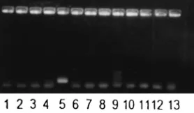
FIG. 2. Amplification of DNA from B. pertussis and other species appearing in the human respiratory tract with primers PIp1 and PIp2. Lane 1, Moraxella
catarrhalis; lane 2, Escherichia coli; lane 3, Haemophilus influenzae; lane 4, Hae-mophilus parainfluenzae; lane 5, B. pertussis; lane 6, negative control; lane 7, Klebsiella pneumoniae; lane 8, Legionella pneumophila; lane 9, Pseudomonas aeruginosa; lane 10, Staphylococcus aureus; lane 11, coagulase-negative
[image:2.612.100.239.587.668.2]staphy-lococci; lane 12, Streptococcus pneumoniae; lane 13, Streptococcus pyogenes.
TABLE 1. Classification of coughing episodes involving pertussis
and parapertussis based on cultures, serology, family exposure, and
clinical symptoms
Classification and criteriaPertussis
1. B. pertussis isolated from the nasopharynx
2. At least one of the following criteria fulfilled:
a. Significant increase in PT IgG
b. Significant increase in FHA IgG without other criteria for
parapertussis
c. Both PT IgG and FHA IgG
$6,000 in the same convalescent
serum
d. Family member with pertussis verified by culture or serology
3. Clinical pertussis with at least 3 weeks of paroxysmal cough and
known exposure to pertussis outside the family; serum
samples lacking or suboptimal timing in relation to onset of
symptoms
4. Known exposure to pertussis within or outside the household;
clinical symptoms not evaluable because of early
erythromycin treatment
Parapertussis
1. B. parapertussis isolated from the nasopharynx
2. Significant FHA IgG increase without an increase in PT IgG,
IgM, and IgA antibodies
on May 15, 2020 by guest
http://jcm.asm.org/
to the expected 121-bp fragment. On inspection of ethidium
bromide-stained gels, a single fragment was clearly visible
when 50 fg of B. pertussis template DNA was present,
equiva-lent to 10 genome copies, assuming a genome size of 3,800 kbp
(18). A band of the expected size was also visible when PCR
was performed on cultures of B. pertussis serially diluted in
double-distilled water and boiled. The same volume of sample
that was used for PCR was cultured before the boiling
proce-dure, revealing that 6 CFU per 10
m
l of sample, visible by
culture, was detected by PCR. This finding is consistent with
that of a previous study (8).
Visualization of amplified DNA from B. pertussis and other
human respiratory tract pathogens or species genetically
re-lated to B. pertussis is shown in Fig. 1 and 2. Amplification of
DNA from species closely related to B. pertussis (Fig. 1, lanes
2 to 9) gave rise to DNA fragments of the wrong size in most
cases. However, amplification of DNA belonging to Bordetella
holmesii (Fig. 1, lane 3) gave rise to a fragment
indistinguish-able from that of DNA belonging to B. pertussis. This band also
hybridized to the PI probe (data not shown). Amplified DNA
from Pseudomonas putida is shown in Fig. 1, lane 10. DNA
from this species also gave a band of about the same size as
that of B. pertussis when amplified with the PIp1 and PIp2
primers. This band, however, did not hybridize to the PI probe
(data not shown). Amplification of DNA from 11 other human
respiratory tract pathogens or commensals was negative, as
they did not give rise to any PCR product (Fig. 2).
Hybridization.
Sixty-seven samples randomly selected from
patients in the study were hybridized with a probe. All 17 of
those which were positive according to the expected 121-bp
fragment also gave a positive signal with hybridization. The 50
samples that were negative by PCR were also negative by
hybridization with the probe.
PCR of nasopharyngeal samples from patients with a cough.
A total of 2,442 nasopharyngeal specimens were available for
PCR and culture. The results of PCR in relation to cultures,
serology, clinical, and epidemiological data are given in Table
2. Assuming that not only the 942 patients with positive
cul-ture, serologic evidence, or known household exposure had
pertussis but also the 14 patients with only clinical and
epide-miological data, the overall sensitivity of PCR was 65% (623 of
956), the specificity was 98% (1,451 of 1,486), the positive
predictive value was 95% (623 of 658), and the negative
pre-dictive value was 81% (1,451 of 1,784) (Table 3).
The sensitivity of PCR was higher than the sensitivity of
cultures (65% [623 of 956] versus 58% [558 of 956]; P
,
0.001
by the binomial distribution test and McNemar’s test), but it
should be noted that PCR was negative in 36 of 558
culture-positive cases (6%).
Of the 35 patients who had a positive PCR but no other
laboratory or clinical indications of B. pertussis infection (false
positive), one case was the result of contamination with B.
pertussis DNA from whole-cell vaccine from equipment used
for taking the nasopharyngeal sample at a pediatric outpatient
clinic (19).
The capability to diagnose pertussis with PCR and culture
decreased with time from the onset of symptoms. The median
time from onset to sampling in 522 patients with positive
cul-ture and positive PCR was 9 days. In 297 patients with
serol-ogy-confirmed pertussis who had negative cultures and
nega-tive PCR, the corresponding time was 15 days (P
,
0.01;
unpaired t test).
PCR was less often positive in fully vaccinated than in
non-vaccinated children in the study with pertussis confirmed by
other methods. In the randomized study the assay was positive
for 70 of 148 (47%) participants who had received three doses
of pertussis toxoid and in 205 of 324 (63%) nonvaccinated
control children (P
,
0.001; Fisher’s exact test).
Parapertussis PCR was positive for 11 of 18 culture-positive
patients and for another 4 patients with serology-confirmed
parapertussis (i.e., increase in FHA IgG without increase in PT
IgG, IgM, or IgA). There was no case of positive parapertussis
by PCR without other laboratory confirmation, i.e., no
false-positive cases. PCR was negative for 7 culture-false-positive patients
and 2,353 culture-negative patients.
DISCUSSION
The present study showed that PCR, using the primers PIp1
and PIp2, was specific for B. pertussis with one exception: B.
holmesii gave rise to a band indistinguishable from that
in-duced by B. pertussis. This band also hybridized to the PI
probe. The risk of B. holmesii giving false-positive results by
pertussis PCR is minimal because the organism has not been
found in the respiratory tract and does not cause a disease
similar to pertussis. The organism has been isolated from the
blood of a few septic patients (12, 25). Bacterial suspensions of
the other 5 Bordetella species and of another 13 bacterial
spe-cies that can be found in the respiratory tract gave no
false-positive reactions.
[image:3.612.51.292.89.226.2]The study also showed that PCR for diagnosis of pertussis
had a high specificity (98%) and a higher sensitivity than
na-sopharyngeal culture in a large body of clinical material. A
sensitive method such as PCR, which can detect DNA from as
few as 6 CFU, is extremely susceptible to contamination that
TABLE 2. Results of cultures, serology, and clinical evaluation in
patients with positive and negative pertussis PCR
Results
Total patients with: Positive
PCR NegativePCR
Pertussis confirmed by culture
522
36
Pertussis confirmed by serology and/
or household exposure
87
297
Clinical pertussis; sera unavailable or
suboptimal timing
9
Exposure to pertussis; early
erythromycin treatment
5
Nonspecific cough; no laboratory data
indicating pertussis
35
a
1,429
Parapertussis
22
[image:3.612.308.548.89.187.2]aIncluding one case with known contamination during sampling (11).
TABLE 3. Results of PCR versus culture, serology, exposure, or
clinical pertussis
PCR
No. of patients with:
Total Pertussis diagnosed by
culture, serology, exposure, or clinical
symptoms
Nonspecific cough; no laboratory data indicating pertussis
Positive
623
35
658
cNegative
333
1,451
1,784
dTotal
956
a1,486
b2,442
aSensitivity, 65% (623 of 956). bSpecificity, 98% (1,451 of 1,486). cPositive predictive value, 95% (623 of 658). dNegative predictive value, 81% (1,451 of 1,784).
on May 15, 2020 by guest
http://jcm.asm.org/
may occur anywhere between the examination room and the
laboratory. One of the false-positive assays in this study was
due to contamination of the sampling material in a room that
was also used for vaccination with a whole-cell pertussis
vac-cine (19). Though difficult to prove, another source of
contam-ination in the examcontam-ination room could have been that a child
with pertussis was examined in the same room immediately
before a participant in this study. Contamination in the
labo-ratory is also a possibility, but this seems less likely in the
present study, because there was no clustering of false-positive
samples and because PCR and culture for pertussis were
per-formed by different technicians on separate floors in the same
building. The negative control used after every fourth sample
was negative throughout the study. Transient PCR positivity
can also be expected if an immune individual is exposed to
pertussis (4, 7).
The sensitivity of cultures for diagnosing pertussis was 58%
in this study, which is similar to or slightly higher than that
found in previous studies (2, 4, 5, 11). However, some
investi-gators have found a lower sensitivity in adults (4). PCR had a
significantly higher sensitivity (65%), even though the study
design did not favor PCR, as the original swab was used for
culture and a new sterile swab was then put into the transport
tube to get material for PCR. There is a possibility that this
swab missed DNA-positive material, which would explain why
6% of culture-positive cases were PCR negative.
Several methods for detecting different DNA sequences of
B. pertussis by PCR in nasopharyngeal secretions have been
described elsewhere. Consistent with other studies, we noted
that the sensitivity of PCR is higher than that of cultures (1, 4,
7, 16, 24), but culture-confirmed cases with negative PCR have
been reported (16, 24). Previous studies have reported varying
sensitivity of pertussis PCR. The overall sensitivity of PCR for
diagnosing pertussis was 65% in the present study, where
se-rology was used to confirm the great majority of cases. In two
large studies that also used culture and serology for verifying
the diagnosis, the sensitivities were 94 (16) and 21% (24),
respectively. In these two studies, the target sequences were
the PT operon and IS1001, respectively. Such comparisons,
however, do not necessarily imply that one method is superior
to another, because differences in the clinical material may also
influence the results. In our study and in one previous study
(24), the intervals between the onset of symptoms and
sam-pling were found to influence the results. The present study
also demonstrated that the vaccination status of the patient is
of importance, since a higher proportion of samples from
non-vaccinated children with pertussis than from non-vaccinated
chil-dren with the disease were PCR positive.
The specificity of PCR can be estimated only if a highly
sensitive reference method is available. In the present study
and some previous studies it was possible to compare PCR and
serology. In two of the largest studies, with 2,421 and 833
nasopharyngeal samples, the specificities were considered to
be 99 and 98%, respectively (16, 24), i.e., specificities similar to
that obtained in the present study.
The present study confirmed that PCR for detection of
DNA from B. parapertussis can be a useful addition to cultures,
as has been shown elsewhere (24). Four culture-negative
pa-tients with serologic evidence of parapertussis had positive
parapertussis PCR, and there were no false-positive tests.
When comparing numbers of patients with positive cultures
and positive PCR for parapertussis, it should be emphasized
again that the study design, with priority for cultures, may have
treated PCR unfavorably.
In conclusion, PCR detecting a 121-bp fragment from the
gene IS481 of B. pertussis had both high sensitivity and high
specificity and can be used as a complement to or replacement
for cultures. A positive PCR, however, must always be
evalu-ated in relation to the clinical picture because transient
colo-nization or contamination may lead to false-positive results at
a low frequency. The decisive advantage of PCR over culture
is its rapidity: PCR can be performed in 1 day, while results of
culture are available in 4 to 7 days.
ACKNOWLEDGMENTS
We thank Elisabeth Pettersson for excellent technical assistance and
Valter Sundh for data management.
The vaccine trial was supported by a contract (NO1-HD-9-2905)
from the National Institute of Child Health and Human Development.
PCR analyses were financed by Go¨teborg University and the Go¨teborg
Medical Society (91/178).
REFERENCES
1. Ba¨ckman, A., B. Johansson, and P. Olce´n. 1994. Nested PCR optimized for detection of Bordetella pertussis in clinical nasopharyngeal samples. J. Clin. Microbiol. 32:2544–2548.
2. Granstro¨m, G., B. Wretlind, and M. Granstro¨m. 1991. Diagnostic value of clinical and bacteriological findings in pertussis. J. Infect. 22:17–26. 3. Granstro¨m, M., G. Granstro¨m, A. Lindfors, and P. Askelo¨f. 1982. Serologic
diagnosis of whooping cough by an enzyme-linked immunosorbent assay using fimbrial hemagglutinin as an antigen. J. Infect. Dis. 146:741–745. 4. Grimprel, E., P. Be´gue´, I. Anjak, F. Betsou, and N. Guiso. 1993. Comparison
of polymerase chain reaction, culture, and Western immunoblot serology for diagnosis of Bordetella pertussis infection. J. Clin. Microbiol. 31:2745–2750. 5. Hallander, H. O., J. Storsaeter, and R. Mo¨llby. 1991. Evaluation of serology and nasopharyngeal cultures for diagnosis of pertussis in a vaccine efficacy trial. J. Infect. Dis. 163:1046–1054.
6. Hallander, H. O., E. Reizenstein, B. Renemar, G. Rasmuson, L. Mardin, and
P. Olin.1993. Comparison of nasopharyngeal aspirates with swabs for cul-ture of Bordetella pertussis. J. Clin. Microbiol. 31:50–52.
7. He, Q., J. Mertsola, H. Soini, M. Skurnik, O. Ruuskanen, and M. K. Viljanen. 1993. Comparison of polymerase chain reaction with culture and enzyme im-munoassay for diagnosis of pertussis. J. Clin. Microbiol. 31:642–645. 8. Houard, S., C. Hackel, A. Herzog, and A. Bollen. 1989. Specific identification
of Bordetella pertussis by the polymerase chain reaction. Res. Microbiol.
140:477–487.
9. Isacson, J., B. Trollfors, J. Taranger, G. Zackrisson, and T. Lagergård. 1993. How common is whooping cough in a nonvaccinating country? Pediatr. Infect. Dis. J. 12:284–288.
10. Krantz, I., J. Taranger, and B. Trollfors. 1989. Estimating incidence of whooping cough over time: a cross-sectional recall study of four Swedish birth cohorts. Int. J. Epidemiol. 18:959–963.
11. Kwantes, W., D. H. M. Joynson, and W. O. Williams. 1983. Bordetella pertussis isolation in general practice: 1977–1979 whooping cough epidemic in West Glamorgan. J. Hyg. Lond. 90:149–158.
12. Lindquist, S. W., D. J. Weber, M. E. Mangum, D. G. Hollis, and J. Jordan. 1995. Bordetella holmesii sepsis in an asplenic adolescent. Pediatr. Infect. Dis. J. 14:813–815.
13. Longo, M. C., M. S. Berninger, and J. L. Hartley. 1990. Use of uracil DNA glycosylase to control carry-over contamination in polymerase chain reac-tions. Gene 93:125–128.
14. Onorato, I. M., and G. F. Wassilak. 1987. Laboratory diagnosis of pertussis: the state of the art. Pediatr. Infect. Dis. J. 6:145–151.
15. Regan, J., and F. Lowe. 1977. Enrichment medium for the isolation of
Bordetella. J. Clin. Microbiol. 6:303–309.
16. Reizenstein, E., L. Lindberg, R. Mo¨llby, and H. O. Hallander. 1996. Vali-dation of nested Bordetella PCR in pertussis vaccine trial. J. Clin. Microbiol.
34:810–815.
17. Sambrook, J., E. F. Fritsch, and T. Maniatis. 1989. Molecular cloning: a laboratory manual, 2nd ed. Cold Spring Harbor Laboratory Press, Cold Spring Harbor, N.Y.
18. Stibitz, S., and T. L. Garletts. 1992. Derivation of a physical map of the chromosome of Bordetella pertussis Tohama I. J. Bacteriol. 174:7770–7777. 19. Taranger, J., B. Trollfors, L. Lind, G. Zackrisson, and K. Beling-Holmquist.
1994. Environmental contamination leading to false-positive polymerase chain reaction for pertussis. Pediatr. Infect. Dis. J. 13:936–937.
20. Taranger, J., B. Trollfors, and T. Lagergård. 1995. Clinical trials of a mono-component pertussis toxoid vaccine (NICHD Ptxd): a technical report. The Go¨teborg Pertussis Vaccine Study, Go¨teborg University. Graphic Systems, Go¨teborg, Sweden.
21. Taranger, J., B. Trollfors, T. Lagergård, L. Lind, V. Sundh, G. Zackrisson,
D. A. Bryla, and J. B. Robbins.1997. Unchanged efficacy of a pertussis toxoid vaccine throughout the 2 years after the third vaccination of infants. Pediatr. Infect. Dis. J. 16:180–184.
on May 15, 2020 by guest
http://jcm.asm.org/
22. Trollfors, B., J. Taranger, T. Lagergård, L. Lind, V. Sundh, G. Zackrisson,
C. U. Lowe, W. Blackwelder, and J. B. Robbins.1995. A placebo-controlled trial of a pertussis-toxoid vaccine. N. Engl. J. Med. 333:1045–1050. 23. Van der Zee, A., C. Agterberg, M. Peeters, J. Schellekens, and F. R. Mooi.
1993. Polymerase chain reaction assay for pertussis: simultaneous detection and discrimination of Bordetella pertussis and Bordetella parapertussis. J. Clin. Microbiol. 31:2134–2140.
24. Van der Zee, A., C. Agterberg, M. Peeters, F. Mooi, and J. Schellekens. 1996. A clinical validation of Bordetella pertussis and Bordetella parapertussis poly-merase chain reaction: comparison with culture and serology using samples from patients with suspected whooping cough from a highly immunized population. J. Infect. Dis. 174:89–96.
25. Weyant, R. S., D. G. Hollis, R. E. Weaver, M. F. M. Amin, A. G. Steigerwalt,
S. P. O’Connor, A. M. Whitney, M. I. Daneshvar, C. W. Moss, and D. J. Brenner.1995. Bordetella holmesii sp. nov., a new gram-negative species associated with septicemia. J. Clin. Microbiol. 33:1–7.
26. Zackrisson, G., I. Krantz, T. Lagergård, P. Larsson, R. Sekura, N. Sigurs, J.
Taranger, and B. Trollfors.1988. Antibody response to pertussis toxin in patients with clinical pertussis measured by enzyme-linked immunosorbent assay. Eur. J. Clin. Microbiol. Infect. Dis. 7:149–154.
27. Zackrisson, G., F. Arminjon, I. Krantz, T. Lagergård, N. Sigurs, J. Taranger,
and B. Trollfors.1988. Serum antibody response to filamentous hemagglu-tinin in patients with clinical pertussis measured by an enzyme-linked im-munosorbent assay. Eur. J. Clin. Microbiol. Infect. Dis. 7:764–770.