Genes that modify the hemochromatosis
phenotype in mice
Joanne E. Levy, … , Lynne K. Montross, Nancy C. Andrews
J Clin Invest.
2000;
105(9)
:1209-1216.
https://doi.org/10.1172/JCI9635
.
Hereditary hemochromatosis (HH) is a prevalent human disease caused by a mutation in
HFE
, which encodes an atypical HLA class I protein involved in regulation of intestinal iron
absorption. To gain insight into the pathogenesis of hemochromatosis, we have bred
Hfe
knockout mice to strains carrying other mutations that impair normal iron metabolism.
Compound mutant mice lacking both Hfe and its interacting protein, beta-2 microglobulin
(B2m), deposit more tissue iron than mice lacking Hfe only, suggesting that another
B2m-interacting protein may be involved in iron regulation. Hfe knockout mice carrying mutations
in the iron transporter DMT1 fail to load iron, indicating that hemochromatosis involves iron
flux through DMT1. Similarly, compound mutants deficient in both Hfe and hephaestin
(Heph) show less iron loading than do
Hfe
knockout mice, indicating that iron absorption in
hemochromatosis involves the function of Heph as well. Finally, compound mutants lacking
Hfe and the transferrin receptor accumulate more tissue iron than do mice lacking Hfe
alone, consistent with the idea that interaction between these two proteins contributes to the
control of normal iron absorption. In addition to providing insight into the pathogenesis of
HH, our results suggest that each of these genes might be a candidate modifier of the
human hemochromatosis phenotype.
Article
Introduction
Hereditary hemochromatosis (HH) is a prevalent genet-ic disorder, affecting up to one in 250 individuals of European descent (1). It is characterized by iron deposi-tion in hepatocytes, cardiac myocytes, and other cells, as a consequence of a small, chronic increase in intestinal iron absorption. Over several decades, patients with HH accumulate up to tenfold more body iron than do nor-mal individuals. Iron deposition leads to tissue damage and fibrosis and ultimately to organ failure if the dis-ease is not recognized and treated. HH is inherited as a monoallelic, recessive disorder, in which clinical signs and symptoms are manifest in homozygotes. However, there is clear evidence that both genetic and environ-mental factors modify disease severity (2).
Insight into the molecular basis of HH first came almost 25 years ago, when the phenotype was shown to be linked to the HLA locus on human chromosome 6 (3). This observation led to the identification of the causative gene, HFE, in 1996 (4). HFEencodes an atyp-ical MHC class I–like molecule that is widely expressed and forms a heterodimer with beta-2 microglobulin (B2m). Its role in regulation of intestinal iron absorp-tion is not well understood. It appears to act through a high-affinity protein-protein interaction with the transferrin receptor (Trfr) (5–9). Most patients with HH are homozygous for a missense mutation in the HFEgene that results in a cysteine→tyrosine
substi-tution at amino acid 282 of the HFE protein (C282Y) (4). However, there is marked variability in the pheno-type of C282Y homozygotes and heterozygotes that is only partially explained by environmental factors. Not all individuals homozygous for the C282Y mutation develop iron overload. At the other extreme, some indi-viduals with marked iron overload are heterozygous for the C282Y mutation, with one apparently normal HFEallele. The biologic basis for this phenotypic vari-ability is unknown.
Although many tissues are affected in hemochro-matosis, the liver is the primary storage depot for iron in excess of immediate body needs. Liver non-heme iron increases in linear relationship to increasing body iron stores until late in the disease, when liver iron stor-age sites become saturated (10). For this reason, liver iron content is a useful measure of total body iron stores during the early, iron-loading phase of hemochromatosis (reviewed in ref. 11).
We previously generated two murine models of HH (12). The Hfegene was disrupted by targeted removal of essential coding sequence and transmitted through the germline to produce mice homozygous for a null Hfe allele. In parallel, the disease-causing C282Y mutation was introduced into the murine Hfe allele. Both mutations result in loss of protein function and in an iron overload similar to that seen in the human disease. The null mutation produces a more severe
Genes that modify the hemochromatosis phenotype in mice
Joanne E. Levy,
1,2Lynne K. Montross,
1,3and Nancy C. Andrews
1,3,41Division of Hematology-Oncology, Children’s Hospital, 2Division of Hematology, Brigham and Women’s Hospital, 3Howard Hughes Medical Institute, and
4Department of Pediatrics, Harvard Medical School, Boston, Massachusetts, USA
Address correspondence to: Nancy C. Andrews, Children’s Hospital, Enders 720, 300 Longwood Avenue, Boston, Massachusetts 02115, USA. Phone: (617) 355-7265; Fax: (617) 734-6791; E-mail: nandrews@rascal.med.harvard.edu.
Received for publication February 16, 2000, and accepted in revised form March 16, 2000.
Hereditary hemochromatosis (HH) is a prevalent human disease caused by a mutation in HFE, which encodes an atypical HLA class I protein involved in regulation of intestinal iron absorption. To gain insight into the pathogenesis of hemochromatosis, we have bred Hfeknockout mice to strains carrying other mutations that impair normal iron metabolism. Compound mutant mice lacking both Hfe and its interacting protein, beta-2 microglobulin (B2m), deposit more tissue iron than mice lacking Hfe only, suggesting that another B2m-interacting protein may be involved in iron regulation. Hfe knockout mice carrying mutations in the iron transporter DMT1 fail to load iron, indicating that hemochromatosis involves iron flux through DMT1. Similarly, compound mutants deficient in both Hfe and hephaestin (Heph) show less iron loading than do Hfeknockout mice, indicating that iron absorption in hemochromatosis involves the function of Heph as well. Finally, compound mutants lacking Hfe and the transferrin receptor accumulate more tissue iron than do mice lacking Hfe alone, consistent with the idea that interaction between these two proteins contributes to the control of normal iron absorption. In addition to providing insight into the pathogenesis of HH, our results suggest that each of these genes might be a candidate modifier of the human hemochromatosis phenotype.
phenotype than does the missense mutation (12). Other investigators have independently shown that Hfe knockout mice develop iron overload (13, 14). We wanted to use our mutant mice to study the effects of mutations in other genes on the expression of the hemochromatosis phenotype.
To evaluate potential genetic modifiers of mutations at the Hfelocus, we crossed Hfemutant mice with mice carrying other spontaneous and induced mutations affecting defined steps in iron metabolism. We have used two spontaneous mutant strains, mk and sla. Mice homozygous for the microcytosis (mk)trait have a severe loss-of-function mutation in the gene encod-ing DMT1 (formerly Nramp2/DCT1), the major trans-membrane iron import molecule (15–17). This results in a defect in absorption of dietary iron from the intes-tinal lumen and in defective iron utilization by ery-throid precursors (reviewed in ref. 18). Mice homozy-gous or hemizyhomozy-gous for the sex-linked anemia (sla) mutation carry a deletion in the Hephaestin gene (Heph), found on the X chromosome. These animals have a defect in export of iron from epithelial cells due to deficiency of the protein Hephaestin (Heph). Heph is homologous to ceruloplasmin and is postulated to act as a membrane-bound, copper-dependent ferroxi-dase (19). In sla mice, dietary iron appears to enter absorptive duodenal enterocytes normally but is not efficiently transferred across the basolateral mem-brane to reach the circulation.
We have also tested two strains of knockout mice car-rying null mutations in the genes encoding B2m (20) and Trfr (21). B2m is a dimerization partner for typical and atypical major histocompatibility class I proteins, including Hfe. Disruption of the B2mgene results in immunological abnormalities, as well as iron loading (22). Knockout mice homozygous for a null mutation in the Trfrgene die from severe anemia during embry-onic development (21). The formation of most tissues does not appear to be impaired, with the exception of the nervous system and the erythron. Surprisingly, het-erozygous mice carrying only one functional Trfr allele develop mild tissue iron depletion (21). Decreased lev-els of tissue iron may result from a lower “set point” for iron absorption in the heterozygous animals.
Here, we report the iron status of animals carrying mutations in the Hfelocus in combination with muta-tions in other genes important in iron metabolism. The results of our studies help to define the relationship between genotype and phenotype in this murine model of hereditary hemochromatosis and give insight into the pathogenesis of iron overload. In addition, these observations identify candidate modifier genes that may influence the course of HH in human patients.
Methods
Animal care and genotyping. Mice were housed in the barri-er facility at Children’s Hospital and maintained on stan-dard mouse diet. All mouse production and experimen-tation were in compliance with the guidelines of the
Institutional Animal Care and Use Committee. Hfe–/–and
Trfr+/–mice used in this study have been described
previ-ously (12, 21). C57BL/6J-B2m–/–and MK/ReJ-mk/+ mice
were derived from breeding stocks purchased from The Jackson Laboratory (Bar Harbor, Maine, USA). The C57BL/6J-sla mutant stock was obtained from a colony maintained by L. Peters at The Jackson Laboratory.
Animals were housed for different lengths of time prior to analysis. Some investigations were done at 6 weeks of age, and others were done at 10 to 11 weeks of age. Because iron accumulation is progressive, the size of the iron stores is dependent upon age. For this rea-son, experimental and control animals of the same age were used for each comparison.
DNA was prepared from mouse tail fragments using a DNeasy Tissue kit (QIAGEN Inc., Valencia, California, USA). Trfrmutant mice were genotyped as described previously (21). Hfe mutant mice were genotyped using a PCR assay. Radiolabeled primers were used to ampli-fy a 202-bp product from the wild-type allele
(5′-ATCAAATGGCATCTCTGGCA-3′and 5′ -GTGGCGAGT-CACTTTCACCA-3′), and a 190-bp product from the tar-geted allele (5′-CTAGCTTCGGCCGTGACG-3′ and
5′-AGTTGGGAGTGGTGTCCGA-3′). PCR products were electrophoresed through 6% acrylamide gels and detect-ed by exposure of the gels to x-ray film. Themkmutant mice were genotyped using radiolabeled primers (5′ -GAAATCGTCCCTGGGTTTTT-3′ and 5′ -CCCCTGGCCT-TACCATATTT-3′)for PCR amplification of DMT1 DNA flanking the G185R missense mutation (15). The result-ing 129-bp PCR products from the wild-type and mutant DMT1 alleles were distinguished by differential migration through 5% nondenaturing polyacrylamide gels (23). B2m mutant mice were genotyped using primers that amplify a 262-bp product from the wild-type B2mallele (5′-CTGAGCTCTGTTTTCGTCTG-3′and
5′-TATCAGTCTCAGTGGGGGTG-3′)and a 768-bp product from the targeted allele (5′-GCTATTCGGCTATGACTGGG 3′and 5′-TATCAGTCTCAGTGGGGGTG-3′). In this case, the PCR products were fractionated on 1% agarose gels. The mutant slamice were maintained in our laboratory as homozygous sla/sla females and hemizygous sla/Y males. The breeding scheme used in crosses with Hfe mutant mice involved breeding homozygous sla/sla females to Hfe–/–males and then backcrossing the obli-gate sla/Y, Hfe+/–male progeny to their sla/slamothers. The progeny from this cross were bred to obtain ani-mals with various Hfegenotypes. Because all of these animals were homozygous or hemizygous for sla, molec-ular genotyping was not necessary. Animals of similar genetic background were generated as Hfe–/–controls
with wild-type slaalleles.
value of P < 0.05 was regarded as significant. Statview (SAS Institute, Inc., Cary, North Carolina, USA) and InStat (Graph Pad Software, San Diego, California, USA) software were used for statistical evaluation.
Results
Absence of B2m increases iron loading in Hfe knockout mice. B2m heterodimerizes with MHC class I proteins, including Hfe. It has been presumed that Hfe requires B2m for its regulatory effect on iron metabolism, par-ticularly because mice lacking B2m (B2m–/–) develop
iron overload (24). We wanted to determine how iron loading in mice lacking both Hfe and B2m compared with iron loading in mice lacking Hfe alone. To study the effect of B2mmutations on the Hfe–/–phenotype, we
interbred B2m–/–and Hfe–/–mice. We studied compound
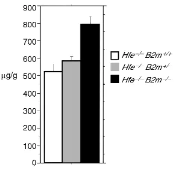
mutant animals whose genetic background was mixed, averaging half C57BL/6J and half 129/SvEvTacfBR. Mice were analyzed at 10–13 weeks of age. Surprising-ly, liver iron deposition was greater in mice lacking both Hfe and B2m than in mice lacking Hfe alone (Fig-ure 1). The difference was highly significant.
Iron loading of Hfe knockout mice requires the apical iron transporter DMT1. The severe phenotype of homozy-gous mk mice, carrying a deleterious missense muta-tion in the gene encoding DMT1, indicates that DMT1 is an important component of the major path-way for intestinal iron absorption in normal animals. We wanted to determine whether the increased iron absorption seen in HH resulted from increased iron flux through DMT1 or from the activation of an
alter-native, accessory, iron absorption pathway. To answer this question, we interbred mice carrying the mk muta-tion in DMT1 with Hfe–/–mice. The compound
mutant offspring had a mixed genetic background that included contributions from MK/ReJ (25), C57BL/6J, and 129/SvEvTacfBR strains. Liver iron deposition was analyzed at 4.5–6.5 weeks of age (Fig-ure 2). There is a marked difference in iron accumula-tion between Hfe–/–mice that carry at least one
wild-type allele of the DMT1 gene and Hfe–/–mice that are
homozygous for the mkallele of DMT1. In these ani-mals, mutation of the Hfe gene did not improve the extremely low iron stores of homozygous mk mutant mice. This indicates that the iron accumulation seen in this model of hemochromatosis is prevented by mutations in DMT1, suggesting that iron accumula-tion occurs predominantly through an absorpaccumula-tion pathway mediated by DMT1.
[image:4.612.95.268.53.221.2]Interestingly, although liver non-heme iron levels were indistinguishable from those of age-matched mk/mk mice with normal Hfe protein, the animals that were homozygous for both the mkmutation in DMT1 and the Hfeknockout allele appeared healthier. Typically, fewer than 10% of mk/mk mutant mice survive to wean-ing (ref. 26, and M.D. Flemwean-ing, unpublished study). In contrast, on the Hfe knockout background, most mk/mk mice survived (data not shown). We have previ-ously shown that DMT1 protein containing the mk amino acid substitution retains a low level of iron trans-port function (17). We speculate that, in the absence of Hfeprotein, there is increased intestinal iron absorption in mk/mkmice. However, the increased absorption is
Figure 1
Liver iron in mice carrying Hfeand B2mmutations. In Figures 1, 2, 3, and 5, histograms show levels of liver iron expressed as micrograms of non-heme iron per gram wet weight of liver. Genotypes are specified by color coding. Absolute quantities are given in parentheses as mean + SE followed by the number of samples analyzed. Pvalues were cal-culated by the unpaired Student’s ttest with the Welch correction, with Pvalues < 0.05 considered significant. This figure shows liver iron lev-els in mice carrying mutant alleles of Hfeand B2min various combina-tions: Hfe–/–B2m+/+(526.18A+ 40.39; n= 16), Hfe–/–B2m+/–(581.71B
+ 26.08; n= 24), Hfe–/–B2m–/–(794.12C+ 45.40; n= 15). Pvalues are
[image:4.612.310.520.450.593.2]as follows: A vs. C, P = 0.0001; B vs. C, P = 0.0005; A vs. B, P = NS.
Figure 2
Liver iron in mice carrying Hfeand DMT1 mutations. This figure shows liver iron levels in mice carrying mutant alleles of Hfeand the gene encoding DMT1 in various combinations. The notation +/+ is used for animals that are wild type at both DMT1 alleles; mk/mk is used for animals that carry the mkmutation (G185R) in both of their DMT1 alleles. Values are given here as described for Figure 1. Hfe+/+
+/+ (84.02A+ 7.72; n= 6), Hfe+/–+/+ (142.35B+ 14.31; n= 7),
Hfe+/–mk/mk (19.24C+ 1.39; n= 9), Hfe–/–+/+ (365.96D+ 51.87; n=
5), Hfe–/–mk/mk(22.32E+ 2.46; n= 8). P values: C vs. E, P = NS; A
marginal and not detectable as an increase in liver iron stores because the iron is diverted to the erythron, where it is used for erythropoiesis.
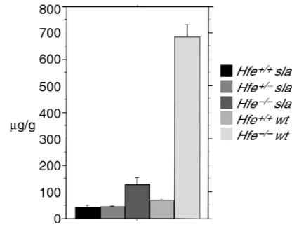
Compound mutant mice lacking Heph and Hfe. Heph is thought to be a component of the basolateral iron transport apparatus, probably acting as a ferroxidase to facilitate transmembrane iron transport. Mice that are homozygous or hemizygous for the X-linked slaphenotype have a partial disruption of intestinal iron absorption. Although the sladeletion in Hephis presumed to be a null mutation (19), slamice are less iron deficient than are mkmice lacking functional DMT1. If the sla mutation does result in total loss of protein function, then Heph must aid in intestinal iron absorption, but not be strictly required. To determine whether increased iron absorption in hemochromatosis involves the iron uptake pathway that is enhanced by Heph function, we bred Hfe–/–
[image:5.612.77.285.54.216.2]mice to slamice and studied 10- to 12-week-old mice. In this case, the genetic background was a mix of C57BL/6J and 129/SvEvTacfBR. Mice homozygous for mutations in both Hephand Hfehad less hepatic iron than did Hfeknockout mice at the same age (Figure 3). Mutations in Hfemodified the sla pheno-type; loss of the Hfe gene ameliorated the iron defi-ciency of slamice; iron stores in mice lacking both Heph and Hfe were higher than in mice lacking Heph alone and were higher than in wild-type mice (Figure 3). It appears likely that iron loading occurs through a pathway involving Heph, but, as previ-ously inferred, Heph is not absolutely required for intestinal iron absorption.
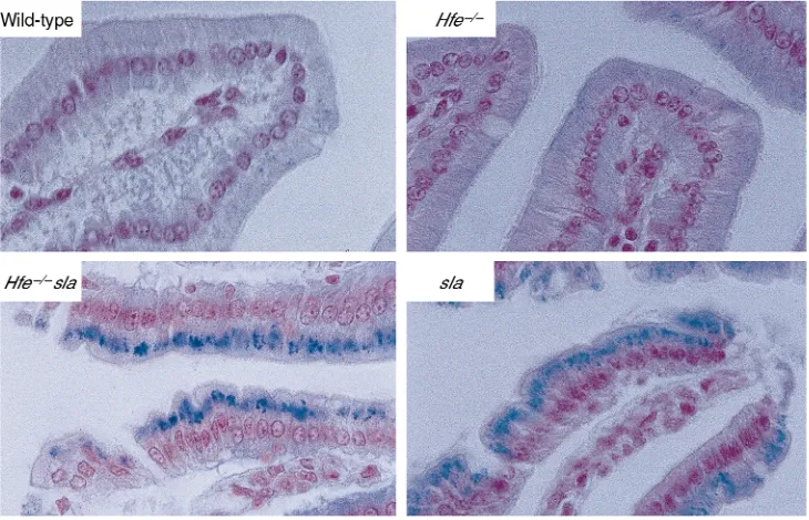
Homozygous and hemizygous sla mice accumulate iron in the intestinal mucosa, as a result of diminished basolateral transfer (27). Iron is reported to be decreased in the villus enterocytes of patients with HH (28). We examined the distribution of iron in duodenal epithe-lial cells from mice with the sla mutation in Heph, the Hfeknockout mutation, and both mutations combined. As shown in Figure 4, all mice carrying the slamutation accumulate mucosal iron, regardless of whether they do or do not have Hfe. This has implications for models of Hfe function, as discussed later here.
Transferrin receptor interacts functionally with Hfe to regu-late iron homeostasis. In a previous study, we showed that animals lacking one Trfr allele (Trfr+/– mice) had
decreased hepatic iron stores, indicating altered iron homeostasis (21). Because Trfr has no role in apical intestinal iron absorption, this result cannot simply be explained by a direct effect on uptake of dietary iron from the intestinal lumen. One explanation is that decreased gene dosage for Trfrmight change the stoi-chiometric ratio of Trfr/Hfe and that this somehow downregulates intestinal iron absorption. This possi-bility is difficult to investigate using biochemical assays because the in vivo function of the Hfe-Trfr complex is not fully understood. Alternatively, it might be that there is less Trfr expression on hepatocytes, and, there-fore, less liver iron uptake. This seems unlikely, because hepatocytes do not normally have a large complement of cell surface Trfr, and posttranscriptional regulation of Trfr by iron regulatory proteins (IRPs) is thought to adjust the amount of Trfr mRNA in response to iron needs (reviewed in ref. 29).
To learn more about the role of Trfr in iron home-ostasis and the functional consequences of the Hfe-Trfr interaction, we bred mice heterozygous for a null Trfr allele (Trfr+/–mice) to mice carrying mutations in Hfe.
In this case, all strains had a pure 129/SvEvTacfBR background. Mice were analyzed at 4–6 weeks of age. As shown in Figure 5, we found that absence of one Trfr allele did not inhibit iron loading in Hfe–/–mice.
Sur-prisingly, Hfe–/– mice lacking one Trfr allele actually have significantly greater hepatic iron deposition than do Hfe–/–mice with a normal complement of Trfr. This
result is paralleled by the result of the cross between Trfr mutant mice and HfeC282Y/C282Y mice. The fact that
heterozygosity for Trfr has opposite effects on mice that have or lack Hfe has implications for the function of the Hfe/Trfr protein complex, as discussed later here.
Discussion
Genetic variability in human patients with HFE-asso-ciated HH has led to a complex spectrum of phenotyp-ic expression. Although there is no doubt that the C282Y mutation in HFE is a disease-causing mutation, there is a small subgroup of C282Y homozygotes who do not develop clinically significant iron overload (2). On the other hand, some C282Y homozygotes develop severe iron overload early in life, and some C282Y het-erozygotes develop hemochromatosis that is
indistin-Figure 3
Liver iron in mice carrying Hfeand Heph mutations. This figure shows liver iron levels in mice carrying mutant alleles of Hfeand Heph in var-ious combinations. slamice indicates a genotype of either Hephsla/sla
or Hephsla/Y. Wild-type (wt) indicates a Hephgenotype that is either
Heph+/+or Heph+/Y.Values are given here as described for Figure 1.
Hfe+/+sla (41.23A+ 9.54; n= 4), Hfe+/–sla(45.93B+ 3.90; n= 21),
Hfe–/–sla(128.47C+ 27.02; n= 22), Hfe+/+wt(70.64D+ 4.10; n= 9),
Hfe–/–wt(685.97E+ 47.88; n= 8). Pvalues: A vs. C, P = 0.0057; B vs.
guishable from that of the homozygotes. The variabil-ity cannot be explained by environmental factors alone; there must be other modifier genes that influence the HH disease phenotype.
Mice carrying targeted mutations in the Hfegene pro-vide an excellent model system to study the pathogen-esis of HH. They have the advantage that they are inbred, and Hfe mutations have been engineered in the context of a defined genetic background. Furthermore, there are a variety of mouse strains with spontaneous and induced mutations in other genes affecting iron metabolism, and interbreeding offers an opportunity to test selectively for modifying effects. In this study, we have taken advantage of mouse genetics to investigate the pathogenesis of HH and to examine the influence of possible modifying genes.
Hfe belongs to a large family of MHC class I–like pro-teins. At present, it remains unclear how a molecule from this group acts to regulate intestinal iron absorption. The tantalizing finding that it interacts with Trfr offers a clue to its activity, but has not yet elucidated its mode of action. The first question that we addressed was whether Hfe is unique among MHC class I–like proteins, or whether there are other, related proteins affecting iron homeostasis. We previously observed that Hfe knockout mice still have substantial regulation of intestinal iron absorption. As they age, their iron overload proceeds, until the amount of tissue iron reaches a plateau (data not shown). Although similar to human patients (30), this is in striking contrast to another mouse model of iron overload (31). Homozygous hpxmice carry a muta-tion in the transferrin (Trf) gene that severely abrogates
Trf expression. Age-matched hpx mice accumulate 15–20 times as much hepatic iron as do Hfeknockout mice (32). They have accelerated intestinal iron absorption that con-tinues indefinitely as the animals age; tissue iron levels do not appear to plateau. These findings suggest that there are additional regulatory pathways governing intestinal iron absorption. Our finding that mice lacking both B2m and Hfe develop more iron overload than do mice lack-ing Hfe alone suggests that there may be another, still unidentified, molecule that interacts with B2m and reg-ulates intestinal iron absorption. It is possible that it is an atypical MHC class I–like molecule, similar to Hfe. Alter-natively, it is possible that the compromised immune sys-tem of B2m knockout mice exacerbates the iron loading phenotype of Hfe–/–mice by some indirect mechanism that is not yet understood.
[image:6.612.117.482.453.688.2]Studies of human patients provide genetic evidence for an additional locus on chromosome 6p, distinct from HFE, that modifies the hemochromatosis phe-notype (33). Furthermore, a second polymorphism in HFE, resulting in a histidine→aspartic acid substitu-tion (H63D), is highly prevalent but only rarely associ-ated with clinical hemochromatosis in the homozy-gous state (reviewed in ref. 34). It is possible that the H63D polymorphism does not, itself, significantly per-turb HFE function. Rather, a unique H63D haplotype might be genetically linked to a mutation in a nearby gene, within the HLA complex, that encodes a molecule that functions in a manner similar to HFE. In this case, H63D would not be a disease-causing mutation, but rather a marker in linkage disequilibrium with a dis-ease-causing mutation in another gene. On the basis of
Figure 4
the increased iron loading observed in Hfe–/–B2m–/–
compound mutant mice, we speculate that there may be another nearby MHC class I–like gene that similar-ly regulates iron absorption. Alternativesimilar-ly, another iron regulatory, B2m-interacting protein may be encoded by a gene located on a different chromosome. If so, it might be a candidate for the causative gene in non-HFE hemochromatosis (35, 36) or in juvenile hemochro-matosis (37). Each of these diseases resembles HH clin-ically, but can be distinguished genetically.
Several different iron uptake pathways have been pro-posed to explain intestinal iron absorption (38). Studies of severely iron-deficient mkmice (39) and brats (40), carrying a severe loss-of-function mutation in DMT1 (15, 41), indicate that DMT1 is the primary apical iron transporter involved in the uptake of dietary iron in nor-mal mamnor-mals. We wanted to determine whether increased iron absorption associated with hemochro-matosis involved DMT1 or induction of an alternative transport mechanism. To answer this question, we bred Hfe–/–mice to mkmice. Our results unequivocally show
that DMT1 mediates iron absorption in HH. It is not yet clear whether increased iron uptake in hemochromato-sis is associated with increased levels of DMT1 protein. Although there are reports that levels of DMT1 mRNA are increased in patients with HH (42) and Hfeknockout mice (43), we have not found significant induction of
DMT1 levels in our Hfe mutant mice (M.D. Fleming, J.E. Levy, and N.C. Andrews, unpublished study). We favor a model supported by several clinical studies, in which basolateral iron transfer is increased in HH (28, 44). However, regardless of the mechanism, the fact that iron overload in HH depends on DMT1 has an important clinical implication. If a compound were to selectively block DMT1 activity at the brush border, it might be developed into an oral pharmaceutical agent to prevent iron absorption in patients with hemochromatosis.
To investigate basolateral iron transfer in Hfe knock-out mice, we bred Hfe–/–animals to slaanimals, which
have impaired basolateral iron transport as a result of a mutation in Heph. The decreased liver iron loading seen in compound mutant mice carrying loss-of-func-tion mutaloss-of-func-tions in both Hfe and Heph indicate that Heph is also a component of the enterocyte iron uptake apparatus functional in HH. However, loss of Heph function does not block iron transfer complete-ly, either in slamice, in which the mutation was initial-ly described, or in Hfe–/–/Heph sla/slacompound mutants.
The absorptive enterocyte is reported to be relatively iron depleted in HH (28). This has fueled speculation that cellular iron deficiency might lead to activation of IRPs, which might in turn induce the expression of DMT1 (43). In contrast, the sla mutation in Heph results in iron accumulation within duodenal entero-cytes. We examined these cells in animals with muta-tions in both Hfeand Hephand found that the com-pound mutants have abundant, stainable, non-heme iron, similar to that of slaanimals. Although we have not measured the activity of IRPs in the duodenal mucosa, and it remains possible that the abundant intracellular iron is somehow sequestered from the IRP regulatory system, the fact that enterocyte non-heme iron is increased should be taken into account in mak-ing models for Hfe function. We observe that com-pound mutants deficient in both Hfe and Heph have larger liver iron stores than do wild-type animals, indi-cating that they absorb more iron in spite of their increased mucosal iron content.
The most intriguing results come from analysis of mice with the genotype Hfe–/–/Trfr+/–. These animals
lack Hfe and have only one functional Trfr allele. Sur-prisingly, they accumulate more hepatic iron than do mice lacking Hfe alone. This result was unexpected because we previously found that mice with intact Hfe alleles but missing one Trfrallele have lower body iron stores than do normal mice (21).
[image:7.612.58.293.52.197.2]This apparent paradox can be resolved in the follow-ing way. We hypothesize that an interaction between Hfe and Trfr establishes a dominant set point for iron homeostasis. In the absence of one functional Trfrallele, the set point would be decreased, as a result of the changed stoichiometry (Hfe relative to Trfr). In the absence of Hfe, this set-point mechanism is disrupted and iron absorption increases, unregulated by this level of control. When Hfe is absent and only one Trfrallele is functional, the Hfe/Trfr-related set point is already
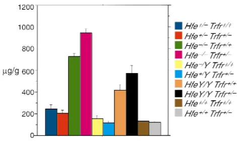
Figure 5
Liver iron in mice carrying Hfeand Trfrmutations. This figure shows liver iron levels in mice carrying mutant alleles of Hfeand Trfrin var-ious combinations. Hfemutant mice carrying the C282Y mutation are listed as HfeY/Yor Hfe+/Y. Hfemutant mice carrying the null
muta-tion are listed as Hfe–/–orHfe+/–. Values are given here as described
for Figure 1. Navy blue: Hfe+/–Trfr+/+(246.71 + 34.73; n= 6). Red:
Hfe+/–Trfr+/–(208.17 + 26.08; n= 6). Green: Hfe–/–Trfr+/+(725.36A+
32.04; n= 25). Pink: Hfe–/–Trfr+/–(947.20B+ 36.64; n= 29). Yellow:
Hfe+/YTrfr+/+(155.76 + 25.03; n= 3). Light blue: Hfe+/YTrfr+/–(113.07
+ 17.92; n= 5). Orange: HfeY/YTrfr+/+(416.93 + 50.38; n= 5). Black:
HfeY/YTrfr+/–(574.73 + 67.19; n= 4). Brown: Hfe+/+Trfr+/+(127.75 +
6.99; n= 8). Gray: Hfe+/+Trfr+/–(115.02 + 12.63; n= 3). Pvalue: A
vs. B, P < 0.0001. The difference in iron loading between Hfe+/+Trfr+/+
and Hfe+/+Trfr+/–animals was not statistically significant in this
experiment, because only three Hfe+/+Trfr+/–animals were analyzed.
perturbed and cannot be further affected because there is no Hfe to form the complex. In this setting, an alter-native regulatory mechanism is revealed, and iron stores increase beyond the level seen in Hfe knockout mice. We cannot yet identify the putative, alternative regulatory mechanism, but we speculate that it may be related to the “erythroid regulator,” which modulates intestinal iron absorption in response to the needs of the erythron (45, 46). We have previously shown that mice heterozy-gous for the null Trfr allele have small erythrocytes that contain less hemoglobin than normal red cells because erythroid precursors have reduced levels of Trfr and therefore cannot take up enough iron for normal hemo-globinization (21). Hfe–/–/Trfr+/– mice also have iron
defi-cient erythropoiesis, in spite of tissue iron overload (data not shown). It is well established that erythroid iron deficiency stimulates intestinal iron absorption through an unknown humoral signal. We suggest that this signal, in response to decreased iron uptake by ery-throid precursors, acts independently of Hfe to further increase iron absorption in Hfe–/–/Trfr+/–mice.
In summary, we have used mice with defined genetic backgrounds to explore the relationship between Hfe and other genes important for the regulation of iron metabolism. We have shown that B2m, DMT1, Heph, and Trfr can act as modifiers of the HH phenotype in mice. Our results give insight into the pathogenesis of hemochromatosis, by suggesting the existence of another B2m-associated protein involved in iron home-ostasis. Furthermore, we have established that increased iron absorption in HH occurs through an iron transport pathway involving DMT1 and Heph. We have begun to characterize the molecular aspects of complex regulatory mechanisms controlling intestinal iron absorption. It seems likely that variability in the human orthologues of the genes we have studied could similarly modify HH in human patients. It will be important to search for polymorphisms and mutations in these genes in patients with exceptionally severe and exceptionally mild cases of HH. It is possible that loss-of-function mutations in B2M exacerbate the pheno-type of C282Y homozygotes. In addition, mild loss-of-function mutations in the genes encoding DMT1 and HEPH may ameliorate HH. Furthermore, mutations in TRFR and other proteins important for erythroid iron utilization may have complex effects on patients who are heterozygous or homozygous for the C282Y muta-tion in HFE.
Acknowledgments
We thank members of the Andrews laboratory for stim-ulating discussions and help with mouse care. M.D. Fleming maintained some of the mutant mouse strains used for these experiments. The mutant slamice were originally provided by L. Peters. This work was partially supported by grants K08 HLO30505 to J.E. Levy and R01 HL51057 to N.C. Andrews from the National Institutes of Health. N.C. Andrews is an Associate Investigator of the Howard Hughes Medical Institute.
1. Edwards, C.Q., et al. 1988. Prevalence of hemochromatosis among 11,065 presumably healthy blood donors. N. Engl. J. Med. 318:1355–1362. 2. Olynyk, J.K., et al. 1999. A population-based study of the clinical
expres-sion of the hemochromatosis gene. N. Engl. J. Med.341:718–724. 3. Simon, M., Bourel, M., Fauchet, R., and Genetet, B. 1976. Association of
HLA-A3 and HLA-B14 antigens with idiopathic haemochromatosis. Gut. 17:332–334.
4. Feder, J.N., et al. 1996. A novel MHC class I-like gene is mutated in patients with hereditary haemochromatosis. Nat. Genet. 13:399–408. 5. Parkkila, S., et al. 1997. Association of the transferrin receptor in human
placenta with HFE, the protein defective in hereditary hemochromato-sis. Proc. Natl. Acad. Sci. USA.94:13198–13202.
6. Lebron, J.A., et al. 1998. Crystal structure of the hemochromatosis pro-tein HFE and characterization of its interaction with transferrin recep-tor. Cell. 93:111–123.
7. Feder, J.N., et al. 1998. The hemochromatosis gene product complexes with the transferrin receptor and lowers its affinity for ligand binding.
Proc. Natl. Acad. Sci. USA.95:1472–1477.
8. Bennett, M.J., Lebron, J.A., and Bjorkman, P.J. 2000. Crystal structure of the hereditary haemochromatosis protein HFE complexed with trans-ferrin receptor. Nature.403:46–53.
9. Lebron, J.A., West, A.P., Jr., and Bjorkman, P.J. 1999. The hemochro-matosis protein HFE competes with transferrin for binding to the trans-ferrin receptor. J. Mol. Biol.294:239–245.
10. Mandelli, C., et al. 1992. Saturability of hepatic iron deposits in genetic hemochromatosis. Hepatology. 16:956–959.
11. Kaltwasser, J.P., and Werner, E. 1989. Diagnosis and clinical evaluation of iron overload. Baillieres Clin. Haematol.2:363–389.
12. Levy, J.E., Montross, L.K., Cohen, D.E., Fleming, M.D., and Andrews, N.C. 1999. The C282Y mutation causing hereditary hemochromatosis does not produce a null allele. Blood.94:9–11.
13. Zhou, X.Y., et al. 1998. HFE gene knockout produces mouse model of hereditary hemochromatosis. Proc. Natl. Acad. Sci. USA.95:2492–2497. 14. Bahram, S., et al. 1999. Experimental hemochromatosis due to MHC class I HFE deficiency: immune status and iron metabolism. Proc. Natl. Acad. Sci. USA.96:13312–13317.
15. Fleming, M.D., et al. 1997. Microcytic anemia mice have a mutation in Nramp2, a candidate iron transporter gene. Nat. Genet.16:383–386. 16. Gunshin, H., et al. 1997. Cloning and characterization of a mammalian
proton-coupled metal-ion transporter. Nature.388:482–488. 17. Su, M.A., Trenor, C.C., Fleming, J.C., Fleming, M.D., and Andrews, N.C.
1998. The G185R mutation disrupts function of iron transporter Nramp2. Blood.92:2157–2163.
18. Andrews, N.C., Fleming, M.D., and Levy, J.E. 1999. Molecular insights into mechanisms of iron transport. Curr. Opin. Hematol.6:61–64. 19. Vulpe, C.D., et al. 1999. Hephaestin, a ceruloplasmin homologue
impli-cated in intestinal iron transport, is defective in the sla mouse. Nat. Genet. 21:195–199.
20. Koller, B.H., Marrack, P., Kappler, J.W., and Smithies, O. 1990. Normal development of mice deficient in beta 2M, MHC class I proteins, and CD8+ T cells. Science.248:1227–1230.
21. Levy, J.E., Jin, O., Fujiwara, Y., Kuo, F., and Andrews, N.C. 1999. Trans-ferrin receptor is necessary for development of erythrocytes and the nerv-ous system. Nat. Genet.21:396–399.
22. de Sousa, M., et al. 1994. Iron overload in beta2-microglobulin deficient mice. Immunol. Lett.39:105–111.
23. Warren, W., Hovig, E., Smith-Sorensen, B., and Borresen, A.-L. 1997. In
Current protocols in human genetics. N.C. Dracopoli, editor. John Wiley & Sons. New York, New York, USA. 7.4.1–7.4.19.
24. de Sousa, M., et al. 1994. Iron overload in beta 2-microglobulin-deficient mice. Immunol. Lett.39:105–111.
25. Russell, E.S., et al. 1970. Characterization and genetic studies of micro-cytic anemia in house mouse. Blood.35:838–850.
26. Russell, E.S., McFarland, E.C., and Kent, E.L. 1970. Low viability, skin lesions, and reduced fertility associated with microcytic anemia in the mouse. Transplant. Proc.2:144–151.
27. Bannerman, R.M. 1976. Genetic defects of iron transport. Fed. Proc. 35:2281–2285.
28. McLaren, G.D., Nathanson, M.H., Jacobs, A., Trevett, D., and Thomson, W. 1991. Regulation of intestinal iron absorption and mucosal iron kinetics in hereditary hemochromatosis. J. Lab. Clin. Med.117:390–401. 29. Muckenthaler, M., and Hentze, M.W. 1997. Mechanisms for posttran-scriptional regulation by iron-responsive elements and iron regulatory proteins. Prog. Mol. Subcell. Biol.18:93–115.
30. Smith, P.M., Godfrey, B.E., and Williams, R. 1969. Iron absorption in idiopathic haemochromatosis and its measurement using a whole-body counter. Clin. Sci.37:519–531.
31. Craven, C.M., et al. 1987. Tissue distribution and clearance kinetics of non-transferrin-bound iron in the hypotransferrinemic mouse: a rodent model for hemochromatosis. Proc. Natl. Acad. Sci. USA.84:3457–3461. 32. Trenor, C.C., III, et al. 2000. The molecular defect in
33. Pratiwi, R., et al. 1999. Linkage disequilibrium analysis in Australian haemochromatosis patients indicates bipartite association with clinical expression. J. Hepatol.31:39–46.
34. Cullen, L.M., Anderson, G.J., Ramm, G.A., Jazwinska, E.C., and Powell, L.W. 1999. Genetics of hemochromatosis. Annu. Rev. Med.50:87–98. 35. Pietrangelo, A., et al. 1999. Hereditary hemochromatosis in adults
with-out pathogenic mutations in the hemochromatosis gene. N. Engl. J. Med. 341:725–732.
36. Camaschella, C., et al. 1999. Inherited HFE-unrelated hemochromato-sis in Italian families. Hepatology.29:1563–1564.
37. Roetto, A., et al. 1999. Juvenile hemochromatosis locus maps to chro-mosome 1q. Am. J. Hum. Genet.64:1388–1393.
38. Conrad, M.E., Umbreit, J.N., and Moore, E.G. 1999. Iron absorption and transport. Am. J. Med. Sci.318:213–229.
39. Edwards, J.A., and Hoke, J.E. 1972. Defect of intestinal mucosal iron uptake in mice with hereditary microcytic anemia. Proc. Soc. Exp. Biol. Med.141:81–84.
40. Oates, P.S., and Morgan, E.H. 1996. Defective iron uptake by the
duo-denum of Belgrade rats fed diets of different iron contents. Am. J. Physi-ol.270:G826–G832.
41. Fleming, M.D., et al. 1998. Nramp2 is mutated in the anemic Belgrade (b) rat: evidence of a role for Nramp2 in endosomal iron transport. Proc. Natl. Acad. Sci. USA.95:1148–1153.
42. Zoller, H., Pietrangelo, A., Vogel, W., and Weiss, G. 1999. Duodenal metal-transporter (DMT-1, NRAMP-2) expression in patients with hereditary haemochromatosis. Lancet.353:2120–2123.
43. Fleming, R.E., et al. 1999. Mechanism of increased iron absorption in murine model of hereditary hemochromatosis: increased duodenal expres-sion of the iron transporter DMT1. Proc. Natl. Acad. Sci. USA.96:3143–3148. 44. Powell, L.W., Campbell, C.B., and Wilson, E. 1970. Intestinal mucosal uptake of iron and iron retention in idiopathic haemochromatosis as evidence for a mucosal abnormality. Gut.11:727–731.
45. Finch, C. 1994. Regulators of iron balance in humans. Blood. 84:1697–1702.