Coexpression of ATP-binding cassette proteins
ABCG5 and ABCG8 permits their transport to
the apical surface
Gregory A. Graf, … , Jonathan C. Cohen, Helen H. Hobbs
J Clin Invest.
2002;
110(5)
:659-669.
https://doi.org/10.1172/JCI16000
.
Mutations in either ATP-binding cassette (ABC) G5 or ABCG8 cause sitosterolemia, an
autosomal recessive disorder of sterol trafficking. To determine the site of action of ABCG5
and ABCG8, we expressed recombinant, epitope-tagged mouse ABCG5 and ABCG8 in
cultured cells. Both ABCG5 and ABCG8 underwent N-linked glycosylation. When either
protein was expressed individually in cells, the N-linked sugars remained sensitive to
Endoglycosidase H (Endo H). When ABCG5 and ABCG8 were coexpressed, the attached
sugars were Endo H–resistant and neuraminidase-sensitive, indicating that the proteins
were transported to the
trans
-Golgi complex. The mature, glycosylated forms of ABCG5 and
ABCG8 coimmunoprecipitated, consistent with heterodimerization of these two proteins.
The Endo H–sensitive forms of ABCG5 and ABCG8 were confined to the endoplasmic
reticulum (ER), whereas the mature forms were present in non-ER fractions in cultured
hepatocytes. Immunoelectron microscopy revealed ABCG5 and ABCG8 on the plasma
membrane of these cells. In polarized WIF-B cells, recombinant ABCG5 localized to the
apical (canalicular) membrane when coexpressed with ABCG8, but not when expressed
alone. To our knowledge this is the first direct demonstration that trafficking of an ABC
half-transporter to the cell surface requires the presence of its dimerization partner.
Article
Cardiology
Find the latest version:
Introduction
Mutations in the ATP-binding cassette (ABC) half-transporters ABCG5and ABCG8cause sitosterolemia (1, 2), a rare autosomal recessive disorder characterized by elevated plasma levels of both plant and animal sterols (3, 4). Normal individuals absorb less than 5% of dietary sitosterol, the major plant sterol, and effi-ciently excrete plant sterols into bile (5, 6). Plasma sitos-terol concentrations rarely exceed 1 mg/dl, even in indi-viduals with very high intakes of dietary sitosterol. In contrast, sitosterolemic subjects absorb about 15–20% of dietary sitosterol and have a profoundly reduced capacity to excrete sitosterol into bile. Consequently, sitosterol accumulates in the blood and body tissues of patients with this genetic disease (5, 7–11). A greater fraction of both animal- and plant-derived sterols are
absorbed from the diet in affected individuals. The increased fractional absorption and reduced biliary excretion of cholesterol in sitosterolemia results in hypercholesterolemia and premature coronary artery disease (4, 11). The phenotype of sitosterolemia sug-gests that ABCG5 and ABCG8 limit intestinal absorp-tion and promote biliary excreabsorp-tion of neutral sterols. ABC transporters share a common molecular archi-tecture that includes two nucleotide-binding folds and two transmembrane domains containing 6–11 mem-brane-spanning α-helices (12, 13). ABC transporters are organized as full transporters containing two trans-membrane domains and two nucleotide-binding folds, or as half-transporters that form homo- or het-erodimers (14). ABCG5 and ABCG8 are members of the G subfamily of ABC transporters, which are pre-dicted to contain a single magnesium-dependent ATP catalytic domain N-terminal to six transmembrane segments (1). Mutations in either ABCG5or ABCG8 cause an identical phenotype, which is consistent with these two gene products functioning as a heterodimer (1, 15), though this has yet to be demonstrated.
Most ABC half-transporters reside in intracellular membranes (14). The best-characterized mammalian half transporters are TAP1 and TAP2 (ABCB2 and ABCB3), which are endoplasmic reticulum (ER) resi-dent proteins that transport peptides from the cytosol into the lumen of this compartment (16). All four ABC
Coexpression of ATP-binding cassette
proteins ABCG5 and ABCG8 permits
their transport to the apical surface
Gregory A. Graf,
1Wei-Ping Li,
2Robert D. Gerard,
1Ingrid Gelissen,
1Ann White,
1Jonathan C. Cohen,
1,3and Helen H. Hobbs
11McDermott Center for Human Growth and Development, Department of Molecular Genetics, 2Department of Cell Biology, and
3Center for Human Nutrition, University of Texas Southwestern Medical Center at Dallas, Dallas, Texas, USA
Mutations in either ATP-binding cassette (ABC) G5 or ABCG8 cause sitosterolemia, an autosomal recessive disorder of sterol trafficking. To determine the site of action of ABCG5 and ABCG8, we expressed recombinant, epitope-tagged mouse ABCG5 and ABCG8 in cultured cells. Both ABCG5 and ABCG8 underwent N-linked glycosylation. When either protein was expressed individually in cells, the N-linked sugars remained sensitive to Endoglycosidase H (Endo H). When ABCG5 and ABCG8 were coexpressed, the attached sugars were Endo H–resistant and neuraminidase-sensitive, indicating that the proteins were transported to the trans-Golgi complex. The mature, glycosylated forms of ABCG5 and ABCG8 coimmunoprecipitated, consistent with heterodimerization of these two proteins. The Endo H–sensitive forms of ABCG5 and ABCG8 were confined to the endoplasmic reticulum (ER), whereas the mature forms were present in non-ER fractions in cultured hepatocytes. Immunoelectron microscopy revealed ABCG5 and ABCG8 on the plasma membrane of these cells. In polarized WIF-B cells, recombinant ABCG5 localized to the apical (canalicular) membrane when coexpressed with ABCG8, but not when expressed alone. To our knowledge this is the first direct demonstration that trafficking of an ABC half-transporter to the cell surface requires the presence of its dimerization partner.
J. Clin. Invest.110:659–669 (2002). doi:10.1172/JCI200216000.
Received for publication May 24, 2002, and accepted in revised form July 9, 2002.
Address correspondence to: Helen H. Hobbs, Department of Molecular Genetics, University of Texas Southwestern Medical Center at Dallas, 5323 Harry Hines Boulevard, Dallas, Texas 75390-9046, USA. Phone: (214) 648-6724; Fax: (214) 648-7539; E-mail: Helen.Hobbs@UTSouthwestern.edu.
Conflict of interest: No conflict of interest has been declared. Nonstandard abbreviations used: ATP-binding cassette (ABC); endoplasmic reticulum (ER); aminopeptidase N (APN); hemagglutinin (HA); polyclonal antibody (pAb); peptide N-glycosidase F (PNGase F); Endoglycosidase H (Endo H); LDL receptor–related protein (LRP); β-galactosidase (β-gal).
half-transporters in the D subfamily are associated with the peroxisomal membrane (17). The Drosophila ABCG transporters white, brown and scarlet are locat-ed within membranes of intracellular pigment granules (18). The only mammalian ABC half-transporter that has been localized to the plasma membrane is the mul-tidrug resistance protein ABCG2 (BCRP), which is associated with the canalicular membrane in human liver (19). ABCG5and ABCG8are most highly expressed in the enterocytes of the intestine and hepatocytes of the liver (20), but the subcellular location of the two proteins is not known.
In this paper, we have examined the cellular itinerary and fate of recombinant, epitope-tagged mouse ABCG5 and ABCG8 in three cell types: CHO-K1 cells, cultured rat hepatocytes (CRL-1601 cells), and polar-ized WIF-B cells (21, 22). We provide evidence that ABCG5 and ABCG8 are physically associated in cells and that they chaperone each other out of the ER to the plasma membrane. In polarized WIF-B cells, the trans-porters colocalized with a resident apical membrane protein, aminopeptidase N (APN), on the canalicular membrane. Our data are consistent with ABCG5 and ABCG8 forming a heterodimer in the ER prior to being transported to apical membranes.
Methods
Development of epitope-tagged recombinant ABCG5 and ABCG8. A 105-bp fragment encoding three copies of a myc epitope (EQKLISEEDLN) (23) and a 93-bp frag-ment containing three copies of a hemagglutinin (HA) epitope (YPYDVPDYAG) (24) were added to the 3′ends of the murine ABCG5 and ABCG8 cDNAs by overlap PCR. The ABCG5-myc, ABCG5-HA, ABCG8-myc, and ABCG8-HA cDNAs were individually cloned into pcDNA3.1(+) and pcDNA3.1/Zeo (Invitrogen Corp., Carlsbad, California, USA). The fidelity of each con-struct was confirmed by DNA sequencing. The initial ABCG8-myc construct used in these studies had a pro-line at amino acid residue 220. Subsequently, Yu et al. (25) reported a sequence containing arginine at this position. Therefore an expression construct that encoded an arginine at residue 220 was also made.
Cell culture. CHO-K1 cells (American Type Culture Collection, Manassas, Virginia, USA) were cultured in Ham’s F12/DMEM 50% (vol/vol) medium containing 5% (vol/vol) FBS, 100 U/ml penicillin, and 100 µg/ml streptomycin (Life Technologies Inc., Grand Island, New York, USA) in a humidified incubator (8.8% CO2). CRL-1601 cells (American Type Culture Collection) were cultured in DMEM (glucose, 1 g/l) containing 10% (vol/vol) FBS, 100 U/ml penicillin, and 100 µg/ml strep-tomycin in a humidified incubator (5% CO2). WIF-B cells were a generous gift from Ann Hubbard (Johns Hopkins University, Baltimore, Maryland, USA) and were cultured as previously described (22).
Immunoprecipitation of epitope-tagged ABCG5 and ABCG8. Epitope-tagged ABCG5 and ABCG8 expres-sion constructs were transiently transfected either
alone or together into CHO-K1 cells using FuGene (Roche Molecular Biochemicals, Mannheim, Ger-many) according to the manufacturer’s protocol. Immunoprecipitations were performed as previously described (26) with the following modification: Incu-bations of primary antibodies with cell lysates were limited to 6 hours. Mouse anti-HA antibody (12CA5) and anti-myc antibody (9E10; Roche Molecular Bio-chemicals) were used to immunoprecipitate ABCG8 and ABCG5, respectively.
SDS-PAGE and immunoblot analysis of ABCG5 and ABCG8. The protein concentrations of cell lysates and fractions were determined by Bio-Rad Dc assay according to the manufacturer’s protocol (Bio-Rad Laboratories Inc., Hercules, California, USA). Pro-teins were size-fractionated on either 8% or 4–20% gradient (wt/vol) SDS-polyacrylamide gels and trans-ferred to nitrocellulose membranes. Membranes were immunoblotted as described (27) using polyclonal antibodies (pAb’s) directed against the myc (A14) and HA (Y11) epitopes (Santa Cruz Biotechnology Inc., Santa Cruz, California, USA).
Pulse-chase analysis of ABCG5 and ABCG8 synthesis in cul-tured cells. CHO-K1 cells were transfected with ABCG5-myc and ABCG8-HA expression plasmids either alone or together. After 48 hours, the cells were washed twice with PBS (37°C) and incubated in methionine- and cysteine-free DMEM for 2 hours. Newly translated proteins were metabolically labeled for 15 minutes with methionine-and cysteine-free DMEM containing 100 µCi/ml [35 S]-Met/Cys (Perkin-Elmer Life Sciences Inc., Boston, Mass-achusetts, USA). Cells were washed twice with PBS (37°C) and incubated with DMEM for up to 48 hours. Cell lysates were prepared and immunoprecipitations were conducted as described above; primary antibodies were incubated with lysates for 18 hours at 22°C. The immunoprecipitates were subjected to SDS-PAGE and the gels fixed for 30 minutes (40% methanol, 10% acetic acid). Signals from the labeled proteins were amplified by fluorography (30 minutes, 1 M salicylate), and the gels were dried and exposed to autoradiography film (–80°C, 8–48 hours). The relative signal intensities were determined by densitometry (Eagle Eye II; Stratagene, La Jolla, California, USA). The values obtained for each time point were corrected for background signal, log-trans-formed, and used to calculate the t1/2of each protein by linear regression using the least-squares method.
Analysis of glycosylation of ABCG5 and ABCG8. The glyco-sylation state of ABCG5-myc and ABCG8-HA was ana-lyzed in transiently transfected CHO-K1 cells. Cells were cultured in 10-cm dishes and transfected, and the pro-teins were immunoprecipitated as described above. Immunoprecipitated proteins were recovered from pro-tein A-agarose beads by heating in 0.1% SDS, 1 mM
Corp., San Diego, California, USA), 5 U of Endoglycosi-dase H (Endo H; Roche Molecular Biochemicals), 50 mU neuraminidase (Roche Molecular Biochemicals), or 2.5 mU O-glycosidase (Roche Molecular Biochemicals) for 2 hours at 37°C following digestion with neuraminidase according to the manufacturers’ protocols. Samples were subjected to SDS-PAGE and immunoblot analysis.
Generation of cell lines stably expressing ABCG5-myc and ABCG5-myc plus ABCG8-HA. CRL-1601 cells were trans-fected at 60% confluence as described for CHO-K1 cells. Cells were selected in medium containing 700 µg/ml G418 sulfate and 700 µg/ml Zeocin (Invitrogen Corp.). Cells were grown under continuous selection, and sin-gle colonies were selected, subcultured, and screened for expression of ABCG5-myc and ABCG8-HA by immunoblot analysis as described above.
Cell fractionation. Cell lysates were fractionated by den-sity gradient centrifugation in Nycodenz (Sigma, St. Louis, Missouri, USA) to separate the ER from other organelles (28). Fractions (n = 36) were collected after tube puncture. Protein sample buffer was added to a final concentration of 1×, and 100 µl of each odd-num-bered fraction was subjected to SDS-PAGE and immunoblot analysis. The separation of the ER from other intracellular compartments was confirmed by immunoblot analysis of proteins associated with the ER (calnexin; StressGen Biotechnologies Corp., Victoria, British Columbia, Canada) with the Golgi complex (mannosidase II; Kelly Mormen, University of Georgia, Athens, Georgia, USA), with the endosomes (Rab11; Santa Cruz Biotechnology Inc.), and with the plasma membrane and endosomes (LDL receptor–related pro-tein [LRP] 85-kDa form) (29).
Immunofluorescence and immunoelectron microscopy. The subcellular localization of ABCG5-myc and ABCG8-HA in cultured cells was determined by immunocytochemical analysis as described previous-ly (30). Cells were imaged by confocal microscopy using a Leica TCS SP microscope (Leica Microsystems Inc., Heidelberg, Germany).
For immunoelectron microscopy, cells were cultured in 100-mm dishes to 95% confluence and fixed with 3% (wt/vol) paraformaldehyde and 0.2% glutaraldehyde in PBS (100 mM sodium phosphate [pH 7.4] 0.15 M NaCl, 4 mM KCl, 2 mM MgCl2) for 5 minutes at 37°C, followed by fixation in the same fixative for 25 minutes (22°C). After brief rinses with PBS, the samples were immersed with PBS containing 50 mM NH4Cl for 30 minutes and enclosed in 2% agarose. The cell pellets were immersed in 2 M sucrose containing 15% polyvinylpyrrolidone (10 kDa) and processed for frozen ultrathin sectioning according to the method of Tokuyasu (31). Cryosections were conditioned on droplets of conditioning buffer (PBS with 1% BSA, 0.01% Triton X-100, and 0.01% Tween-20) for 20 min-utes and then incubated for 2 hours in the presence of either anti-myc pAb or anti-HA pAb (10 µg/ml in con-ditioning buffer). The IgG was localized by incubating sections for 1 hour with 10 nm gold-labeled goat
anti-rabbit IgG diluted 1:40 in conditioning buffer. Electron micrographs were taken with a JEOL 1200 electron microscope (JEOL USA Inc., Peabody, Massachusetts, USA) operating at 80 kV.
Generation of recombinant adenoviruses expressing ABCG5-myc and ABCG8. Recombinant adenoviral vectors con-taining ABCG5, ABCG5-myc, ABCG8, and ABCG8-HA were generated by in vitro cre-lox recombination (32). The adenovirus particles were used to express ABCG5-myc and untagged ABCG8 in WIF-B cells that were cul-tured as described above. Ten days following plating, cells were infected with 1 ×10⁹particles of virus express-ing β-galactosidase (β-gal) ABCG5-myc, ABCG8, or both ABCG-myc and ABCG8 cDNAs.
Results
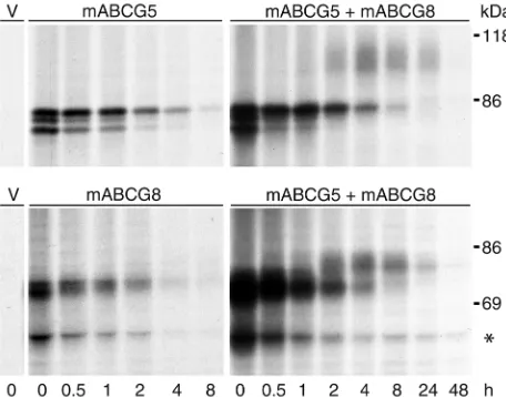
Coexpression of ABCG5 and ABCG8 induces posttransla-tional modification of the proteins. To examine the size, posttranslational processing, and half-lives of ABCG5 and ABCG8, the cDNAs encoding the two proteins were modified to contain epitope tags at the C-termi-ni and then expressed independently in CHO-K1 cells (Figure 1, left). Transfected cells were labeled with [35S]-Met/Cys for 15 minutes and then chased with excess Met/Cys for the indicated time periods. ABCG5-myc and ABCG8-HA were immunoprecipitated from cell lysates using mAb’s to the epitope tags. At the 0 time point, three proteins of 65 kDa, 70 kDa, and 73 kDa were observed for ABCG5, and two proteins of 66 and 68 kDa for ABCG8-HA. The t1/2of both ABCG5-myc and ABCG8-HA was approximately 3 hours. When ABCG5-myc and ABCG8-HA were coexpressed (Figure 1, right), a series of higher–molecular weight proteins (90–100 kDa for ABCG5-myc and about 78 kDa for ABCG8-HA) appeared at 2 hours and remained detectable at 24 hours, when the lower– molecular weight proteins were barely visible. No high-er–molecular weight proteins were detected with longer exposures in cells expressing either ABCG5 or ABCG8 alone (data not shown). These results suggest that both ABCG5 and ABCG8 undergo an initial pro-cessing event that is not dependent on the presence of the other protein. After 1 to 2 hours, the proteins undergo further modifications, which are contingent on the expression of both proteins. The sequential appearance of higher–molecular weight proteins is consistent with a precursor-product relationship.
The identity of a 48-kDa protein that immunopre-cipitated with the anti-HA antibody and decreased in intensity over the course of the experiment is not known. A protein of this size was detected in CHO-K1 cells expressing ABCG8-HA, but not when this protein was expressed in CRL-1601 cells.
endoglycosidases (Figure 2). In cells expressing ABCG5-myc or ABCG8-HA alone, the higher–molecu-lar weight forms of the proteins were sensitive to PNGase F, which removes all N-linked sugars, and to Endo H, which removes high-mannose sugars that have not yet undergone full maturation. Mouse ABCG8 has one canonical site for the addition of N-linked sugars at residue 619, and ABCG5 has three potential sites for addition of N-linked sugars (residues 440, 585, and 592), but one of these sites is predicted to be within a few residues of the first mem-brane-spanning domain and is not conserved in the human protein (Figure 2, bottom). The stepwise increases in molecular masses of these proteins in transfected CHO-K1 cells are consistent with the cova-lent addition of one N-linked sugar to ABCG8-HA and two N-linked sugars to ABCG5-myc (Figure 1). Treat-ment with neuraminidase or with neuraminidase plus O-glycosidase did not change the migration of either ABCG5-myc or ABCG8-HA when the proteins were expressed individually. The LDL receptor was exam-ined in the same experiment, and the expected approx-imately 40-kDa decrease in apparent molecular mass
following treatment with O-glycosidase was detected (data not shown) (33). Thus, when ABCG5-myc or ABCG8-HA was expressed alone, N-linked sugars were added in the ER but the proteins did not appear to traffic to the Golgi complex.
Coexpression of ABCG5-myc and ABCG8-HA result-ed in the appearance of additional higher–molecular weight forms, as described above (Figure 1). Im-munoblotting revealed that in the steady state the higher–molecular weight forms of the proteins pre-dominated, especially for ABCG8-HA. The more slow-ly migrating proteins collapsed into a single protein upon treatment with PNGase F but were resistant to treatment with Endo H, indicating that the more slow-ly migrating forms of the proteins had reached the medial Golgi complex, whereas the lower–molecular weight forms were retained in the ER. Neuraminidase, which removes sialic acid residues added in the trans -Golgi complex (34), caused a slight but consistent reduction in the apparent molecular mass of the larg-er forms of ABCG5-myc and ABCG8-HA. No furthlarg-er changes in protein mass were apparent with O-gly-cosidase treatment. Taken together, these results indi-cated that ABCG5-myc and ABCG8-HA were retained in the ER unless expressed together, and they suggest-ed that ABCG5 and ABCG8 form a heterodimer in order to exit the ER.
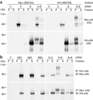
Recombinant ABCG5 and ABCG8 coimmunoprecipitate in CHO-K1 cells. To determine whether ABCG5 and ABCG8 are physically associated in cells, we expressed myc-tagged ABCG5 and HA-myc-tagged ABCG8 alone or togeth-er in CHO-K1 cells (Figure 3a). The anti-myc antibody precipitated a protein of the size expected for ABCG5-myc in cells expressing both ABCG5-ABCG5-myc and ABCG8-HA, but not in cells expressing ABCG8-HA alone; simi-larly, the anti-HA antibody immunoprecipitated a protein of the size expected for ABCG8-HA in cells expressing both transporters but not in cells expressing only ABCG5-myc. More than 95% of each protein was immunoprecipitated by mAb’s to their respective epi-tope tags. ABCG8-HA coimmunoprecipitated with ABCG5-myc and ABCG5-myc coimmunoprecipitated with ABCG8-HA in cells expressing both proteins.
To determine whether the association between ABCG5-myc and ABCG8-HA occurred before or after cell lysis, cells were first transfected independently with ABCG5-myc, ABCG5-HA, ABCG8-myc, or ABCG8-HA (Figure 3b). Lysates from the independently transfect-ed cells were mixtransfect-ed (5&5, 5-myc and 5-HA; 5&8, 5-myc and 8-HA; 8&8, 8-myc and 8-HA) and then subjected to immunoprecipitation and immunoblot analysis. The anti-myc mAb failed to immunoprecipitate the HA-tagged transporters, and the myc-HA-tagged transporters did not coimmunoprecipitate with the anti-HA mAb. Thus, the association of ABCG5-myc and ABCG8-HA did not occur after cell lysis.
[image:5.576.60.288.57.236.2]To test for the formation of homo-oligomeric com-plexes, we cotransfected cells with ABCG5-myc and ABCG5-HA (5+5), with ABCG5-myc and ABCG8-HA
Figure 1
(5+8), or with ABCG8-myc and ABCG8-HA (8+8, Figure 3b). A small fraction of ABCG5 and a significant frac-tion of ABCG8 coimmunoprecipitated in cells express-ing both the myc- and the HA-tagged forms of the same proteins. In both cases, only the lower–molecular weight forms of the proteins were detected, indicating that if homodimers form, they are retained in the ER. In con-trast, both the lower– and the higher–molecular weight forms were observed in cells expressing both ABCG5-myc and ABCG8-HA and coimmunoprecipitated with antibodies to either epitope. These results are consistent with the half-transporters forming heterodimers and then trafficking to the Golgi complex.
To ensure that the substitution of proline for argi-nine at codon 220 of ABCG8 did not affect the associ-ation or trafficking of ABCG8, the pulse-chase, endo-glycosidase digestion, and coimmunoprecipitation experiments were repeated using an expression con-struct encoding an arginine at residue 220, and identi-cal results were obtained (data not shown).
ABCG8 expression affects the trafficking of ABCG5. To trace the intracellular trafficking of ABCG5 and ABCG8 in a physiologically relevant cell type, rat hepa-tocyte–derived cell lines (CRL-1601) expressing either recombinant ABCG5-myc alone or ABCG5-myc and ABCG8-HA together were selected and subcloned. No endogenous ABCG5 or ABCG8 mRNA was detected by Northern blotting in these cells (data not shown). Steady-state levels of ABCG5-myc and ABCG8-HA were analyzed by immunoblotting in both cell lines (Figure 4a). A single protein of 68 kDa was detected in cells expressing ABCG5-myc alone, whereas addition-al higher–molecular weight forms of the protein (89–100 kDa) were present in cells expressing both ABCG5-myc and ABCG8-HA.
To determine the subcellular location of the half-trans-porters in the steady state, lysates from cells expressing ABCG5-myc alone or ABCG5-myc plus ABCG8-HA were fractionated on Nycodenz gradients to separate the ER from other cellular compartments (Figure 4b). Im-munoblot analysis of the gradient fractions indicated that the lower–molecular weight, Endo H–sensitive forms of ABCG5-myc and ABCG8-HA comigrated with the ER resident protein calnexin. Only the higher–molecular weight, Endo H–resistant forms were present in non-ER fractions. The higher–molecular weight forms of both ABCG5-myc and ABCG8-HA were also present in the ER fractions. The ER fractions of the gradients did not con-tain proteins present in the plasma membrane (LRP, 85 kDa), endosomes (LRP, Rab11), or Golgi complex (p58K). ABCG5 and ABCG8 localize to the cell membrane of CRL-1601 cells. To determine the subcellular localization of ABCG5 and ABCG8, CRL-1601 cells expressing ABCG5-myc and ABCG8-HA were grown on glass cov-erslips and processed for immunofluorescence and immunoelectron microscopy. Immunoreactive ABCG5 colocalized with ER resident proteins (GRP-78, cal-nexin) in cells expressing ABCG5 alone, but with the cell surface protein APN in cells expressing both ABCG5 and ABCG8 (data not shown). To determine whether ABCG5 and ABCG8 were on the cell surface or in closely apposed subplasmalemmal vesicles, we performed immunoelectron microscopy on cells expressing both half-transporters. Gold particles were associated with the plasma membrane and ER for ABCG5-myc (Figure 5b) and for ABCG8-HA (Figure 5c). No signal was detected in cells containing the expression vector alone (Figure 5a).
[image:6.576.63.315.475.738.2]ABCG5 and ABCG8 traffic to the apical surface in polar-ized WIF-B cells. Since CRL-1601 cells are not polarized,
Figure 2
these cells cannot be used to determine whether ABCG5 and ABCG8 are specifically targeted to the bile canalicular (apical) surface. Therefore, we obtained a polarized cell line (WIF-B) derived from a fusion between a rat hepatoma and human fibroblast cells (22). These cells form canaliculi and sort proteins in a manner similar to that observed in vivo (35) and have been used to define the subcellular localization of bil-iary ABC transporters (30). Northern blot analysis using probes specific for rat ABCG5 and ABCG8
indi-cated that both endogenous genes were expressed in these cells (data not shown). Human chromosome 2 was not retained in this cell line (21), so these cells cannot synthesize human ABCG5 or ABCG8. To determine whether ABCG5-myc traffics beyond the ER when expressed in WIF-B cells, a pulse-chase experiment was performed in cells infected with ade-noviruses encoding ABCG5-myc alone, or ABCG5-myc and ABCG8. Mature, fully glycosylated forms of ABCG5-myc were only observed when ABCG8 was coexpressed (data not shown), indicating that if het-erodimers between rat and mouse ABCG5 and ABCG8 form, they are incapable of escaping the ER.
[image:7.576.58.356.51.372.2]To determine the subcellular local-ization of ABCG5-myc in the absence or presence of ABCG8, cells were infected with adenoviruses encoding cDNAs for β-gal, ABCG5-myc, or ABCG5-myc and ABCG8 and processed for immunofluorescence microscopy (Figure 6). The anti-myc antibody detected ABCG5-myc in cells expressing ABCG5-myc alone or ABCG5-myc plus ABCG8, but not in cells expressing β-gal (Figure 6, a–c). In the absence of ABCG8, ABCG5-myc colocalized with BiP, but not with basolateral (HA321) (36) or api-cal (APN) plasma membrane mark-ers (Figure 6, d–l), which indicated that in WIF-B cells, as in CRL-1601 cells, ABCG5 was confined to the ER when expressed alone. In the pres-ence of ABCG8, ABCG5-myc colocal-ized with the apically sorted protein APN (Figure 6, s–u). However, no colocalization was seen with HA321 or mannosidase II. A faint signal was seen on the basolateral surface of some cells (Figure 6p). Whether this represents proteins that first go to the basolateral membranes before trafficking to the apical surface or is an artifact of overexpression is not known. These results indicated that ABCG5 is sorted to the apical surface when coexpressed with ABCG8, and they sug-gested that ABCG5/ABCG8 heterodimers traffic to the canalicular surface of hepatocytes. While immunofluorescence microscopy does not provide sufficient resolution to exclude a subplasmalemmal localization, the results of the electron microscopy studies in the CRL-1601 cells suggest that the two proteins are located in the plasma membrane.
Figure 3
Discussion
In this study we have examined the cellular itinerary and fate of recombinant, epitope-tagged ABCG5 and ABCG8 in three cell types: CHO-K1 cells, nonpolar-ized cultured rat hepatocytes (CRL-1601 cells), and polarized hepatocytes (WIF-B cells). We provide evi-dence that ABCG5 and ABCG8 are physically associ-ated in these cells and that they chaperone each other out of the ER en route to the plasma membrane. The transporters colocalized with each other in the ER and plasma membranes of stably expressing cultured hepatocytes and were present on the apical (canalicu-lar) membranes of WIF-B cells. Collectively, these data indicate that an ABCG5/ABCG8 complex is assem-bled in the ER, moves through the Golgi complex, and is targeted to the apical surface of cultured cells, sug-gesting that ABCG5 and ABCG8 may function as a heterodimer on the apical surfaces of cells to export neutral sterols. To our knowledge this is the first direct demonstration that trafficking of an ABC half-transporter to the cell surface requires the presence of its dimerization partner.
All experiments in this study were performed using epitope-tagged versions of ABCG5 and ABCG8, since antibodies that recognize the native proteins are not available. Although we cannot exclude the possibility that epitope tagging of the proteins alters their behav-ior in cells, experiments using tagged and untagged
versions of ABCG8 indicate that this possibility is unlikely. The exit of ABCG5 from the ER requires coexpression of ABCG8. Both tagged and nontagged forms of ABCG8 allow transport of ABCG5 to the plasma membrane. These results indicate that the for-mation of ABCG5/ABCG8 complex and its trafficking to the cell surface are not influenced by the presence of the epitope tag. Similarly, the substitution of a pro-line residue for arginine at residue 220 of ABCG8 had no effect on the half-life, processing, or formation of heterodimers in CHO-K1 cells.
[image:8.576.71.531.401.638.2]Both ABCG5 and ABCG8 are N-glycosylated. When each transporter is expressed individually in cells, the added sugars do not undergo maturation and the pro-teins are more rapidly degraded. When the two propro-teins are coexpressed, higher–molecular weight, Endo H–resistant, neuraminidase-sensitive forms of both proteins appear. The higher–molecular weight forms of the proteins have significantly longer half-lives, sug-gesting that formation of the complex between the two half-transporters in the ER promotes their transit from the ER to the trans-Golgi complex and thus diverts them from a degradative pathway. Cell fractionation and immunolocalization studies in cultured hepato-cytes confirm that ABCG5 is retained in the ER unless ABCG8 is expressed in the same cells. These data indi-cate that the cellular localizations of the proteins are contingent on their oligomeric structure.
Figure 4
The observation that mutations in either ABCG5or ABCG8cause an identical phenotype (1, 15) is also con-sistent with ABCG5and ABCG8forming a complex. Since ABC half-transporters contain only a single nucleotide-binding fold and six putative transmem-brane segments, they must form homodimers or het-erodimers to be active (14). Some half-transporters, such as TAP1 and TAP2, function as heterodimers; mutations in either gene result in the loss of peptide transport activity (16, 37). The four peroxisomal half-transporters (ABCD1–ABCD4) form homodimers and heterodimers in vitro (17), but the composition of the functional complexes of these proteins is not known. The ABCG subfamily members scarlet, brown, and white also function as heterodimers (38), whereas ABCG2 transports substrates when expressed alone in insect cells, suggesting that it functions as a homod-imer (39). The finding that ABCG5 and ABCG8 coim-munoprecipitate indicates that the two proteins are associated in cells and suggests that they het-erodimerize. The interaction between these two pro-teins is specific and is not a post–cell lysis artifact, since the half-transporters do not coimmunoprecipi-tate if lysates from cells expressing each half-trans-porter alone are mixed. Coimmunoprecipitation of the high–molecular weight, Endo H–resistant forms of ABCG5-myc and ABCG8-HA is almost quantitative, further indicating that ABCG5/ABCG8 complex for-mation is required for ER exit.
A small fraction of expressed ABCG5 and ABCG8 also immunoprecipitates as homo-oligomeric com-plexes. For both proteins, only the lower–molecular weight forms are present in these complexes, indicat-ing that if ABCG5 or ABCG8 form homodimers or other homo-oligomers, these complexes fail to exit the ER. Consistent with this interpretation, ABCG5-myc is not transported to the apical membrane in WIF-B cells if expressed alone. In contrast, ABCG2 appears to func-tion as a homodimer and has been observed on the bile canalicular membrane (19). Further studies will be required to determine whether the ER-associated forms
of ABCG5 and ABCG8 function as homodimers, rep-resent nonspecific protein aggregates, or are mis-paired half-transporters that fail to achieve the appropriate conformation to escape the ER.
The observation that ABCG5-myc and ABCG8-HA reach the trans–Golgi complex only when the two pro-teins are coexpressed indicates that monomeric and multimeric homodimers of these proteins are specifi-cally retained in the ER. The mechanism for ER reten-tion of ABCG5 and ABCG8 is not known. Other oligomeric membrane proteins contain ER retention motifs that are masked when the proteins complex. For example, heterodimerization is required for the T cell antigen receptor (TCR), the H2a subunit of the asialoglycoprotein receptor (ASGPR), and the GABAB receptor to exit the ER (40–42). Each subunit of these proteins contains a different ER retention motif (GLRILLLKV, EGHRG, and RXR for TCR, ASGPR, and GABAB, respectively) that is masked by het-erodimerization, allowing the protein complex to exit the ER. Only the RXR motif is present in mouse ABCG5 and ABCG8. One copy of this motif is con-served between the mouse and human proteins (residues 242–244 in ABCG5 and residues 196–198 in ABCG8), but the role of these residues in ER retention remains to be examined. Other mechanisms for the retention of improperly complexed oligomers include binding of proteins to ER chaperones that contain KDEL motifs, and thiol retention whereby the mask-ing of cysteine residues by disulfide bondmask-ing allows proteins to exit the ER (43, 44). ABCG5 and ABCG8 contain numerous cysteine residues, but whether intramolecular or intermolecular disulfide bonds exist in the ABCG5/ABCG8 complex has not been exam-ined. Additional studies will be required to determine the mechanisms by which ABCG5 and ABCG8 monomers, and perhaps oligomers, are recognized by the ER quality control system.
[image:9.576.62.537.57.189.2]A potentially interesting observation is the finding of higher–molecular weight forms of ABCG5 and ABCG8 in the ER, as well as in the non-ER, fractions
Figure 5
of cultured hepatocytes expressing both half-transporters. The presence of the mature forms in the ER fractions could rep-resent contamination of these fractions with non-ER components of the cell, although none of the non-ER marker pro-teins are present in the ER fractions (Figure 4b). The fully mature, glycosylated forms of the proteins may have recycled from the Golgi complex compartment back to the ER, as has been described for SREBP cleav-age–activating protein (SCAP) (45). More experiments will be required to determine the source of the mature forms of the half-transporters in the ER.
[image:10.576.262.541.54.739.2]ABCG5 and ABCG8 colocalize at the cell surface in cultured hepatocytes, and immu-noelectron microscopy confirms that the proteins are associated with the plasma membrane in these cells. The proteins are located predominantly on the apical plasma membrane, suggesting that ABCG5 and ABCG8 may participate directly in the trans-port of neutral sterols across the apical membrane into bile. It is not known whether neutral sterols are the primary transport substrate of ABCG5 and ABCG8 or whether another molecule is actively transported and sterol efflux is secondary. Phospholipids are the primary transport substrate for a num-ber of other ABC lipid transporters, and phospholipid transport and cholesterol transport into bile are tightly interrelated. For example, biliary cholesterol excretion is markedly reduced in mice lacking the phos-phatidylcholine transporter ABCB4 (MDR2) (46). Sitosterolemic patients have a marked reduction in fractional excretion of biliary sterols but have normal biliary excretion of phospholipids (7). Conversely, overexpres-sion of ABCG5 and ABCG8 in mice causes a selective increase in cholesterol excretion
Figure 6
into the bile (47). These findings suggest that neutral sterols or a neutral sterol carrier protein, rather than phospholipids, are the primary transport substrate of ABCG5 and ABCG8.
The observation that ABCG5 and ABCG8 form a het-erodimer does not exclude the possibility that either protein may form functional complexes with other ABC half-transporters. In Drosophila, white forms func-tional complexes with both scarlet and brown to trans-port different substrates (38). The mammalian ABCG transporter subfamily includes six known members. Although the tissue expression patterns of ABCG5 and ABCG8 differ from those of the other ABCG subfami-ly members, both ABCG1 and ABCG2 are expressed in the liver and intestine (19, 48, 49). ABCG3 and ABCG4 are expressed at only low levels in the liver and intestine of mice (data not shown); the tissue distribution of expression of these proteins in humans is not known. Thus, it remains possible that ABCG5 and ABCG8 are not monogamous and that the half-transporters asso-ciate with other subfamily members to transport dif-ferent substrates. The formation of dimer pairs between various members of this subfamily of proteins may permit transport of a more diverse array of sub-strates, or provide tissue- and/or substrate-specific con-trol of solute transport.
Acknowledgments
We thank Ann Hubbard and Lita Braiterman for the generous gift of the WIF-B cells and helpful discus-sions. We thank Liangcai Nie, Jin Wang, and Lisa Beat-ty for excellent technical assistance; Kathleen Neill for assistance with the manuscript preparation; and David W. Russell and Richard Anderson for helpful discus-sions. This work was supported by the National Heart, Lung, and Blood Institute (POI HL2098), the W.M. Keck Foundation, the Perot Family Fund, and the Don-ald W. Reynolds Cardiovascular Clinical Research Cen-ter (Dallas, Texas, USA).
1. Berge, K.E., et al. 2000. Accumulation of dietary cholesterol in sitos-terolemia caused by mutations in adjacent ABC transporters. Science.
290:1771–1775.
2. Lee, M.H., et al. 2001. Identification of a gene, ABCG5, important in the regulation of dietary cholesterol absorption. Nat. Genet.27:79–83. 3. Bhattacharyya, A.K., and Connor, W.E. 1974. Beta-sitosterolemia and xanthomatosis. A newly described lipid storage disease in two sisters.
J. Clin. Invest.53:1033–1043.
4. Bjorkhem, I., Boberg, K., and Leitersdorf, E. 2001. Inborn errors in bile acid biosynthesis and storage of sterols other than cholesterol. In The metabolic and molecular bases of inherited disease. Volume II. C. Scriver, A. Beaudet, W. Sly, and D. Valle, editors. McGraw-Hill. New York, New York, USA. 2961–2988.
5. Salen, G., Ahrens, E.H., Jr., and Grundy, S.M. 1970. Metabolism of
beta-sitosterol in man. J. Clin. Invest.49:952–967.
6. Gould, R.G., Jones, R.J., LeRoy, G.V., Wissler, R.W., and Taylor, C.B. 1969. Absorbability of beta-sitosterol in humans. Metabolism.
18:652–662.
7. Miettinen, T.A. 1980. Phytosterolaemia, xanthomatosis and premature atherosclerotic arterial disease: a case with high plant sterol absorp-tion, impaired sterol elimination and low cholesterol synthesis. Eur. J. Clin. Invest.10:27–35.
8. Salen, G., et al. 1989. Increased sitosterol absorption, decreased removal, and expanded body pools compensate for reduced cholesterol synthesis in sitosterolemia with xanthomatosis. J. Lipid Res.
30:1319–1330.
9. Lutjohann, D., Bjorkhem, I., Beil, U.F., and von Bergmann, K. 1995. Sterol absorption and sterol balance in phytosterolemia evaluated by deuterium-labeled sterols: effect of sitostanol treatment. J. Lipid Res.
36:1763–1773.
10. Nguyen, L.B., et al. 1990. Decreased cholesterol biosynthesis in sitos-terolemia with xanthomatosis: diminished mononuclear leukocyte 3-hydroxy-3-methylglutaryl coenzyme A reductase activity and enzyme protein associated with increased low-density lipoprotein receptor func-tion. Metabolism.39:436–443.
11. Salen, G., et al. 1992. Sitosterolemia. J. Lipid Res.33:945–955. 12. Higgins, C. 1992. ABC transporters: from microorganisms to man.
Annu. Rev. Cell Biol. 8:67–113.
13. Dean, M., and Allikmets, R. 1995. Evolution of ATP-binding cassette transporter genes. Curr. Opin. Genet. Dev.5:779–785.
14. Dean, M., Hamon, Y., and Chimini, G. 2001. The human ATP-binding cassette (ABC) transporter superfamily. J. Lipid Res.42:1007–1017. 15. Lu, K., et al. 2001. Two genes that map to the STSLlocus cause
sitos-terolemia: genomic structure and spectrum of mutations involving sterolin-1 and sterolin-2, encoded by ABCG5and ABCG8, respectively.
Am. J. Hum. Genet.69:278–290.
16. Kleijmeer, M.J., et al. 1992. Location of MHC-encoded transporters in the endoplasmic reticulum and cis-Golgi. Nature.357:342–344. 17. Liu, L.X., et al. 1999. Homo- and heterodimerization of peroxisomal
ATP-binding cassette half-transporters. J. Biol. Chem.274:32738–32743. 18. Mackenzie, S.M., Howells, A.J., Cox, G.B., and Ewart, G.D. 2000. Sub-cellular localisation of the white/scarlet ABC transporter to pigment granule membranes within the compound eye of Drosophila melanogaster.
Genetica.108:239–252.
19. Maliepaard, M., et al. 2001. Subcellular localization and distribution of the breast cancer resistance protein transporter in normal human tis-sues. Cancer Res.61:3458–3464.
20. Repa, J.J., et al. 2002. Regulation of ATP-binding cassette sterol trans-porters, ABCG5 and ABCG8, by the oxysterol receptors, LXRalpha and beta. J. Biol. Chem.277:18793–18800.
21. Cassio, D., Hamon-Benais, C., Guerin, M., and Lecoq, O. 1991. Hybrid cell lines constitute a potential reservoir of polarized cells: isolation and study of highly differentiated hepatoma-derived hybrid cells able to form functional bile canaliculi in vitro. J. Cell Biol.115:1397–1408. 22. Ihrke, G., et al. 1993. WIF-B cells: an in vitromodel for studies of
hepa-tocyte polarity. J. Cell Biol.123:1761–1775.
23. Sakai, J., et al. 1998. Molecular identification of the sterol-regulated luminal protease that cleaves SREBPs and controls lipid composition of animal cells. Mol. Cell.2:505–514.
24. Tokiwa, G., Tyers, M., Volpe, T., and Futcher, B. 1994. Inhibition of G1 cyclin activity by the Ras/cAMP pathway in yeast. Nature.371:342–345. 25. Lu, K., et al. 2002. Molecular cloning, genomic organization, genetic variations, and characterization of murine sterolin genes Abcg5 and Abcg8. J. Lipid Res.43:565–578.
26. Sakai, J., et al. 1997. Identification of complexes between the COOH-terminal domains of sterol regulatory element-binding proteins (SREBPs) and SREBP cleavage-activating protein. J. Biol. Chem.
272:20213–20221.
27. Graf, G.A., Connell, P.M., van der Westhuyzen, D.R., and Smart, E.J. 1999. The class B, type I scavenger receptor promotes the selective uptake of high density lipoprotein cholesterol ethers into caveolae.
J. Biol. Chem.274:12043–12048.
28. Hammond, C., and Helenius, A. 1994. Quality control in the secretory pathway: retention of a misfolded viral membrane glycoprotein involves cycling between the ER, intermediate compartment, and Golgi appara-tus. J. Cell Biol.126:41–52.
29. Willnow, T.E., Armstrong, S.A., Hammer, R.E., and Herz, J. 1995. Func-tional expression of low density lipoprotein receptor-related protein is controlled by receptor-associated protein in vivo. Proc. Natl. Acad. Sci. USA.92:4537–4541.
30. Sai, Y., Nies, A.T., and Arias, I.M. 1999. Bile acid secretion and direct tar-geting of mdr1-green fluorescent protein from Golgi to the canalicular membrane in polarized WIF-B cells. J. Cell Sci.112:4535–4545. 31. Tokuyasu, K.T. 1980. Immunochemistry on ultrathin frozen sections.
Histochem. J.12:381–403.
32. Aoki, K., Barker, C., Danthinne, X., Imperiale, M.J., and Nabel, G.J. 1999. Efficient generation of recombinant adenoviral vectors by Cre-lox recombination in vitro. Mol. Med.5:224–231.
33. Cummings, R.D., et al. 1983. Biosynthesis of N- and O-linked oligosac-charides of the low density lipoprotein receptor. J. Biol. Chem.
258:15261–15273.
34. Kornfeld, R., and Kornfeld, S. 1985. Assembly of asparagine-linked oligosaccharides. Annu. Rev. Biochem.54:631–664.
35. Song, W., Apodaca, G., and Mostov, K. 1994. Transcytosis of the poly-meric immunoglobulin receptor is regulated in multiple intracellular compartments. J. Biol. Chem.269:29474–29480.
selective loss into the bile. J. Biol. Chem.267:6099–6106.
37. Kelly, A., et al. 1992. Assembly and function of the two ABC transporter proteins encoded in the human major histocompatibility complex.
Nature.355:641–644.
38. Sullivan, D.T., and Sullivan, M.C. 1975. Transport defects as the phys-iological basis for eye color mutants of Drosophila melanogaster. Biochem. Genet.13:603–613.
39. Ozvegy, C., et al. 2001. Functional characterization of the human mul-tidrug transporter, ABCG2, expressed in insect cells. Biochem. Biophys. Res. Commun.285:111–117.
40. Shenkman, M., Ehrlich, M., and Lederkremer, G.Z. 2000. Masking of an endoplasmic reticulum retention signal by its presence in the two sub-units of the asialoglycoprotein receptor. J. Biol. Chem.275:2845–2851. 41. Margeta-Mitrovic, M., Jan, Y.N., and Jan, L.Y. 2000. A trafficking check-point controls GABABreceptor heterodimerization. Neuron.27:97–106. 42. Bonifacino, J.S., Cosson, P., and Klausner, R.D. 1990. Colocalized trans-membrane determinants for ER degradation and subunit assembly explain the intracellular fate of TCR chains. Cell.63:503–513. 43. Munro, S., and Pelham, H.R. 1987. A C-terminal signal prevents
secre-tion of luminal ER proteins. Cell.48:899–907.
44. Reddy, P.S., and Corley, R.B. 1998. Assembly, sorting, and exit of oligomeric proteins from the endoplasmic reticulum. Bioessays.
20:546–554.
45. Nohturfft, A., DeBose-Boyd, R.A., Scheek, S., Goldstein, J.L., and Brown, M.S. 1999. Sterols regulate cycling of SREBP cleavage-activating pro-tein (SCAP) between endoplasmic reticulum and Golgi. Proc. Natl. Acad. Sci. USA.96:11235–11240.
46. Smit, J.J., et al. 1993. Homozygous disruption of the murine Mdr2 P-glycoprotein gene leads to a complete absence of phospholipid from bile and to liver disease. Cell.75:451–462.
47. Yu, L, et al. 2002. Overexpression of ABCG5 and ABCG8 promotes bil-iary cholesterol secretion and reduces fractional absorption of dietary cholesterol. J. Clin. Invest. 110:671–680. doi:10.1172/JCI200216001. 48. Klucken, J., et al. 2001. ABCG1(ABC8), the human homolog of the
Drosophial white gene, is a regulator of macrophage cholesterol and phospholipid transport. Proc. Natl. Acad. Sci. USA.97:817–822. 49. Kennedy, M.A., et al. 2001. Characterization of the human ABCG1 gene:
liver X receptor activates an internal promoter that produces a novel transcript encoding an alternative form of the protein. J. Biol. Chem.