Na+/myo-inositol transport is regulated by
basolateral tonicity in Madin-Darby canine
kidney cells.
A Yamauchi, … , E Imai, T Kamada
J Clin Invest.
1996;
97(1)
:263-267.
https://doi.org/10.1172/JCI118401
.
We investigated the effects of change in basolateral osmolality on Na(+)-dependent
myo-inositol uptake in Madin-Darby canine kidney cells to test our hypothesis that the
Na+/myo-inositol transporter (SMIT), an osmolyte transporter, is mainly regulated by osmolality on the
basolateral surface. A significant osmotic gradient between both sides of the epithelium
persisted at least 10 h after basolateral osmolality was increased. [3H]myo-inositol uptake
increased in a basolateral osmolality-dependent manner. The magnitude of the increase is
comparable to that for making both sides hypertonic. Apical hypertonicity also increased the
uptake on the basal side, but the magnitude of the increase was significantly smaller than
the basolateral or both sides hypertonicity. Betaine-gamma-amino-n-butyric acid transporter
activity, measured by [3H]gamma-amino-n-butyric uptake, showed a pattern similar to SMIT
activity in response to basolateral hypertonicity. The most plausible explanation for the
polarized effect of hypertonicity is that the basal membrane is much more water permeable
than the apical membrane. These results seem to be consistent with the localization and
regulation of the SMIT in vivo.
Research Article
Find the latest version:
J. Clin. Invest.
© The American Society for Clinical Investigation, Inc. 0021-9738/96/01/0263/05 $2.00
Volume 97, Number 1, January 1996, 263–267
Rapid Publication
Na
1/
Myo
-inositol Transport Is Regulated by Basolateral Tonicity
in Madin-Darby Canine Kidney Cells
Atsushi Yamauchi,* Toshihiro Sugiura,* Takahito Ito,* Akiko Miyai,* Masaru Horio,‡ Enyu Imai,* and Takenobu Kamada*
*First Department of Medicine and ‡Department of Clinical Laboratory Science, Osaka University School of Medicine, Osaka, 565 Japan
Abstract
We investigated the effects of change in basolateral osmolal-ity on Na1-dependent myo-inositol uptake in Madin-Darby canine kidney cells to test our hypothesis that the Na1/myo -inositol transporter (SMIT), an osmolyte transporter, is mainly regulated by osmolality on the basolateral surface. A significant osmotic gradient between both sides of the epi-thelium persisted at least 10 h after basolateral osmolality was increased. [3H]myo-inositol uptake increased in a baso-lateral osmolality–dependent manner. The magnitude of the increase is comparable to that for making both sides hy-pertonic. Apical hypertonicity also increased the uptake on the basal side, but the magnitude of the increase was signifi-cantly smaller than the basolateral or both sides hypertonic-ity. Betaine-g-amino-n-butyric acid transporter activity, measured by [3H]g-amino-n-butyric uptake, showed a
pat-tern similar to SMIT activity in response to basolateral hy-pertonicity. The most plausible explanation for the polar-ized effect of hypertonicity is that the basal membrane is much more water permeable than the apical membrane. These results seem to be consistent with the localization and regulation of the SMIT in vivo. (J. Clin. Invest. 1996. 97: 263–267.) Key words: osmolytes • betaine • g-amino-n
-bu-tyric acid • polarity • water permeability
Introduction
Many kinds of cells respond to extracellular hypertonicity by the accumulation of high concentrations of small organic sol-utes, which are referred to as “compatible osmolytes,” that protect cells from the perturbing effects of high intracellular concentrations of electrolytes (1, 2). It has been shown that
myo-inositol and betaine function as organic osmolytes in vari-ous tissues and types of cells (3–5). Madin-Darby canine
kid-ney (MDCK)1 cells accumulate these osmolytes through
spe-cific Na1-coupled transporters when cultured in a hypertonic
medium (6, 7). Cloning of the cDNAs encoding osmolyte transporters such as Na1/myo-inositol transporter (SMIT) (8) and Na1/Cl2/betaine-g-amino-n-butyric acid (GABA) trans-porter (BGT-1) (9) offers insight into understanding of molec-ular mechanism for accumulation of these osmolytes. The abundance of the mRNAs for these transporters and the tran-scription rate of the genes increased when the cells were cul-tured in a hypertonic medium (10, 11), suggesting that tran-scription is the primary step in regulation of these transporters by hypertonicity. The canine BGT-1 gene has been recently cloned (12), and a hypertonic stress–responsive element has
been identified from the 59-flanking region of the BGT-1
gene (13).
Myo-inositol is the only osmolyte found in substantial
amounts in the cortex and outer medulla in addition to the in-ner medulla (14). Our previous report (15) showed that SMIT mRNA abundance was highest in the outer medulla rather than in the inner medulla, and a small but significant amount of SMIT mRNA was present in the cortex. SMIT mRNA in the cortex as well as that in the medulla was significantly in-duced by dehydration. We have recently shown using in situ hybridization (16) that SMIT mRNA is predominantly present in the thick ascending limb of Henle’s loop (TALH) and mac-ula densa cells, and that NaCl loading rapidly increases the sig-nals. Basal expression and magnitude of the induction by NaCl administration seemed to be uniform throughout the TALH as well as macula densa cells. The fact that furosemide markedly reduced the signals indicates that SMIT is not regulated by lu-minal osmolality. Our question is what is the signal for the ex-pression of SMIT.
Although we cannot deny the idea that SMIT is regulated by unknown factor(s) other than osmolality, there has been no report regarding such a factor in spite of extensive studies of this transporter. We speculate that SMIT expression may change with osmolality on the basolateral surface, which is supposed to be dependent on NaCl reabsorption of the tubular cells. To investigate this issue, we examined the effects of the change in basolateral osmolality on Na1-dependent myo -inosi-tol uptake in MDCK cells cultured on a porous support. We used MDCK cells because a large amount of data regarding osmolyte transporters and their regulations has been accumu-lated and established for MDCK cells. There are no other cell
Address correspondence to Atsushi Yamauchi, First Department of Medicine, Osaka University School of Medicine, 2-2, Yamadaoka, Suita, Osaka, 565 Japan. Phone: 81-6-879-3632; FAX: 81-6-879-3639.
Received for publication 30 June 1995 and accepted in revised form 19 October 1995.
1. Abbreviations used in this paper: ADH, antidiuretic hormone; BGT-1, betaine GABA transporter; GABA, g-amino-n-butyric acid; MDCK, Madin-Darby canine kidney; SMIT, Na1/myo-inositol
lines available which clearly express SMIT and BGT-1. The re-sults obtained demonstrate that SMIT and BGT-1 activities are predominantly regulated by basolateral osmolality in MDCK cells. This seems to be consistent with the localization and regulation of SMIT in vivo.
Methods
Cell culture. MDCK cells were a generous gift from the Japanese Cancer Research Resources Bank (National Institute of Health, To-kyo, Japan) and grown in DME supplemented with 10% FCS, 50 U/ ml of penicillin, and 50 mg/ml of streptomycin equilibrated with 5% CO2–95% air at 378C. Cells were carried on tissue culture plastic
dishes and seeded at confluent density on tissue culture–treated poly-carbonate filters (Transwell™, Costar Corp., Cambridge, MA). Each
5-cm2 Transwell™ filter (Nuclepore™, 3-mm pore) cup was placed in a
35-mm well of a Cluster Six tissue culture dish (Costar Corp.). 7 d af-ter seeding, some were switched to the medium made hypertonic (350–450 mosmol/kg) by addition of raffinose on either side of the ep-ithelium. Hypotonic medium was made by simple dilution of growth medium with water. Medium osmolality was measured with a freez-ing point osmometer (OM-6010; Kyoto-Daiichi-Kagaku, Kyoto, Ja-pan). Others were maintained in isotonic (300 mosmol/kg) medium.
Myo-inositol uptake was determined 6 h after basolateral osmolality increased. All experiments were performed using 25–30 passages of MDCK cells.
MDCK cells are tolerant of acute increase in osmolality up to 500 mosmol/kg when osmolalities of both sides are the same. In case of basolateral hypertonicity, however, the cells sometimes began to de-tach within 6 h over 500 mosmol/kg, so we used media up to 450 mos-mol/kg. There were no significant differences in protein content be-tween isotonic and hypertonic cells for at least 6 h after the switch.
Measurement of SMIT and BGT-1 activities. Because myo -ino-sitol is taken up mostly on the basal side of MDCK cells (17), we measured basolateral uptake of myo-inositol in MDCK cells on a po-rous support. Na1-dependent myo-inositol uptake was measured
es-sentially as described previously with slight modifications (17). To measure uptake into MDCK cells grown on filters, the cells were rinsed twice on both surfaces with PBS and then incubated for 30 min at 378C in preincubation solution (150 mM NaCl or 150 mM LiCl, 5 mM KCl, 2 mM CaCl2, 1.2 mM MgCl2, 10 mM Hepes/Tris, pH 7.4). This
solution was replaced by the same solution containing 10 mM myo -inositol (uptake medium). [3H]myo-inositol (20 Ci/mmol; New
En-gland Nuclear, Boston, MA) was added (0.5 mCi/ml) to the solution on the basal side of the filter. To end the uptake period, the filter on its support was dipped into three beakers containing 100 ml ice-cold stop solution (PBS + 1 mM phlorizin) to stop the uptake and remove extracellular tracer. The filter with its attached epithelium was then cut out of the filter support with a surgical blade and put into a scintil-lation vial where the cells were solubilized overnight in 500 ml of 1 N NaOH. Then 500 ml of 1 N HCl was added to the solution. Duplicate aliquots of 25 ml were used for protein determination (Protein Assay kit; Bio-Rad Laboratories, Richmond, CA) using BSA as a standard. 10 ml of ACSII (Amersham Corp., Arlington Heights, IL) was added to the remainder of the sample for liquid scintillation counting. The uptake by MDCK cells was found to be linear with time for at least 60 min. Accordingly, uptake measured after 30 min of incubation was taken as the initial uptake rate.
We also examined the effects of basolateral hypertonicity on BGT-1 activity. BGT-1 activity was determined by measuring the ini-tial rate of [3H]GABA uptake into MDCK cells with the same
proce-dures as myo-inositol uptake. The uptake buffer contained 10 mM of GABA and 0.5 mCi/ml of [3H]GABA (40 Ci/mmol, New England
Nuclear).
In every experiment, just before the last uptake period ended, ali-quots were taken from the solution on each side of the filter to check for transepithelial leakage. Transepithelial leakage or transport of
ra-diolabeled myo-inositol or GABA was negligible; tracer added to one surface was scarcely (, 0.1%) detected in the solution on the other surface of the epithelium, indicating that there was little trans-epithelial transport or leakage of myo-inositol or GABA. In experi-ments in which the cells were in hypertonic medium, PBS, preincubation medium, uptake medium, and stop solution were made hypertonic by adding mannitol. To study transport in the absence of sodium, lithium was used to replace all sodium.
Data analysis. The results shown are means6SD. When no error bar is shown in a figure, the SD is smaller than the symbol. The data were statistically analyzed by the one-way ANOVA. Statistical signif-icance was set at P , 0.05.
Results
Previous reports have shown that monolayers of MDCK cells grown on a permeable support transport water at a low rate (18, 19). To examine how long a significant osmotic gradient between both sides of the epithelium persists, medium osmo-lality of the samples from each side of the epithelium was mea-sured after basolateral osmolality was increased (Fig. 1). Os-molality of basal medium gradually decreased and that of apical medium gradually increased. A significant osmotic gra-dient persisted for at least 10 h. 16 h after the switch,
[image:3.612.313.559.58.202.2]osmolali-Figure 1. Time course of change in osmolality after basolateral me-dium osmolality in-creased to 450 mosmol/ kg. On time 0, confluent cells cultured in isotonic medium (300 mosmol/ kg) were switched to the same medium made hypertonic (450 mos-mol/kg) by addition of raffinose on basal side. Medium osmolality of the sample from each side of the epithelium was measured successively. Results are means6SD of three independent experiments. *P , 0.01 vs apical medium.
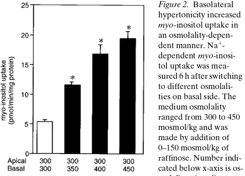
Figure 2. Basolateral hypertonicity increased
myo-inositol uptake in an osmolality-depen-dent manner. Na1
[image:3.612.316.556.529.701.2]ties of both sides became very close. From this result, we de-cided that uptake experiments would be performed 6 h after the switch.
To examine whether the myo-inositol transport increases
as basolateral osmolality increases, we measured Na1
-depen-dent myo-inositol uptake from basal side 6 h after basolateral medium was switched to media with different osmolalities (Fig. 2). The medium osmolality ranged from 300 to 450 mos-mol/kg which were made by addition of 0–150 mosmos-mol/kg of raffinose. The uptake rate increased as basolateral osmolality increased until 450 mosmol/kg.
To see if there is a difference in the increase in SMIT activ-ity between apical and basal hypertonicactiv-ity, we investigated the
effect of hypertonicity on either side or both sides of the epi-thelium (Fig. 3 A). When basolateral osmolality increased, the magnitude of the increase was almost the same as that for making both sides hypertonic. Apical hypertonicity also in-creased the uptake, but the magnitude of the increase was sig-nificantly smaller than basolateral or both sides hypertonicity. The polarized effect of hypertonicity may be restricted to induction of SMIT activity or may be common to induction of other osmoregulatory transporters. To clarify this issue, we measured BGT-1 activity, another osmolyte transporter in
MDCK cells. BGT-1 activity, measured by [3H]GABA uptake,
showed a pattern similar to SMIT in response to basolateral hypertonicity. It increased in a basolateral osmolality–depen-dent manner (data not shown). Basolateral hypertonicity in-creased BGT-1 activity significantly more than apical hyperto-nicity (Fig. 3 B). Thus, the predominant effect of basolateral hypertonicity is common to osmolyte transporters.
We measured myo-inositol uptake from the apical side
un-der different conditions. There was some myo-inositol uptake
from the apical side that was induced by hypertonicity as well (17). Either apical or basal hypertonicity increased both apical and basal transports. Although basal hypertonicity was more effective than apical hypertonicity, the ratio of basal transport to apical transport was almost constant under different condi-tions (z 20–30 times). These results confirmed that apical
up-take was always negligible compared to basal upup-take.
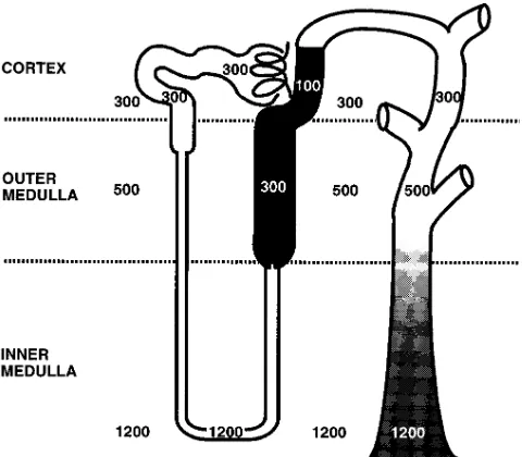
[image:4.612.63.273.61.474.2]In situ hybridization study indicated that SMIT predomi-nantly expressed TALH (16), where a large transepithelial os-motic gradient exists (Fig. 4) (20). So we examined the possi-bility that the transepithelial osmotic gradient might induce SMIT in MDCK cells. Hypotonic media were made simply by dilution of the growth medium with water. When the isotonic cells were switched to hypotonic medium (100 mosmol/kg) on
Figure 3. Basolateral hypertonicity increased myo-inositol (A) and GABA (B) uptake more than apical hypertonicity. Na1-dependent
myo-inositol (A) or GABA (B) uptake was measured 6 h after switching to hypertonic medium (400 mosmol/kg) on either side or both sides of the epithelium. Number indicated below x-axis is osmo-lality of medium on each side (mosmol/kg). Each bar is mean of three independent experiments; error lines are SD. *P , 0.01 vs isotonic (300 mosmol/kg) cells; ‡P , 0.01 vs apical hypertonicity; §P , 0.05 vs
apical hypertonicity.
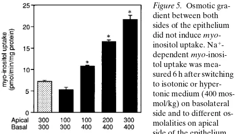
[image:4.612.316.556.437.647.2]apical side, no increase was observed in myo-inositol transport (Fig. 5). We also examined the effect of apical hypotonicities in case of basal hypertonicity. The isotonic cells were switched to hypertonic medium (400 mosmol/kg) on basolateral mem-brane and to the media with different osmolalities, ranging from 100 to 300 mosmol/kg on the apical side. As shown in Fig. 5, osmotic gradient per se did not accelerate SMIT activity. It is apparent, however, that basolateral hypertonicity predomi-nantly affects SMIT activity rather than apical hypotonicity be-cause SMIT activity significantly increased even when apical osmolality was only 100 mosmol/kg. When the osmolality of each side was reversed in this experiment, apical hypertonicity did not significantly increase the transport activity in case of basolateral hypotonicity (data not shown). This result further suggests the predominance of basolateral hypertonicity for in-ducing osmolyte transporters.
Discussion
In the present study, SMIT activity as well as BGT-1 activity is predominantly regulated by basolateral osmolality in MDCK cells. These results could be explained by the difference in wa-ter permeability between apical and basolawa-teral plasma mem-branes. The fact that the transepithelial gradient holds for sev-eral hours (Fig. 1) indicates that one or both of the membranes is water impermeable. This result is consistent with a previous report showing that water permeability of MDCK cell mono-layers was in the lower range of the values reported for biolog-ical membranes (19). Generally, the basolateral plasma mem-brane of epithelia is water permeable, and the apical plasma membrane water permeability varies among epithelia. In case of the antidiuretic hormone (ADH)-responsive epithelia, api-cal membranes have a low osmotic water permeability, whereas basolateral membranes are relatively permeable to water (21). Since addition of vasopressin or dibutyryl cAMP to MDCK cell monolayers induced an increase in water perme-ability (19), they apparently belong to ADH-responsive epi-thelia. These results suggest that the rate-limiting site for water and solute transport in MDCK cells is at the apical (luminal) epithelial surface, at least in the absence of ADH. Further-more, it has been shown that plasma membrane lipid order is asymmetrical in MDCK cells (22), and ADH modulates lipid order of apical plasma membrane (23). Taken together, it is
very likely that water permeability of the apical membrane is lower than that of basal membrane in MDCK cells.
When the basolateral membranes are water permeable and the apical membranes are water impermeable, the high baso-lateral osmolality is supposed to be rapidly balanced with the intracellular osmolality (or ionic strength), which is considered to be a trigger for stimulation of osmoregulatory genes (24). In contrast, apical (luminal) tonicity does not affect the intracel-lular osmolality so much. This hypothesis seems to be consis-tent with the results of the present study for SMIT and BGT-1 transports in MDCK cells.
In case of the TALH cells in vivo, where SMIT expresses most predominantly (Fig. 4) (16), apical membranes have an extremely low osmotic water permeability, whereas basolat-eral membranes are relatively permeable to water (25). Thus, the same notion could be applicable to TALH cells in vivo. SMIT expression did not correspond to luminal osmolality, since SMIT highly expressed throughout the TALH and was universally induced by acute NaCl administration. When NaCl loading to TALH cells increases, the primary event to stimu-late NaCl reabsorption is considered to be the activation of the
Na1+K1-pump located in the basolateral membrane. In this
case, it is possible that the osmolality close to basolateral plasma membrane of the tubular cells, that is peritubular os-molality (20), may be very high throughout the TALH. There is direct evidence of peritubular hypertonicity in case of mac-ula densa cells. The chloride concentration or osmolality of juxtaglomerular interstitium, that is, the area located on basal side of macula densa cells, has been shown to be very high and to change with the tubular flow rate in Amphiuma (26). NaCl transport into the interstitium at the glomerular vascular pole proceeds through the water impermeable tubular epithelium, which results in basolateral hypertonicity during elevated flow rates. Because the mechanism of NaCl absorption across mac-ula densa cells is at least qualitatively similar to that of TALH cells, we assume that the osmolality close to the TALH cells is also hypertonic.
As shown in Fig. 4, there is an osmotic gradient between apical and basal sides of this nephron segment (20). We first assume that the transepithelial osmotic gradient may be a sig-nal for the induction of SMIT. Osmotic gradient, however, did not accelerate the increase in SMIT activity in MDCK cells (Fig. 5). In this experiment, it is possible that the dilution of some component on the apical fluid may be important in
regu-lating myo-inositol uptake because we made hypotonic media
by simply diluting the growth media. To clarify this issue, we tried to do the experiments in Fig. 5 using the isotonic media (300 mosmol/kg) made by addition of 100 mosmol/kg of raffi-nose to hypotonic (200 mosmol/kg) media. Comparing apical hypotonicity to both sides isotonic, there is no significant dif-ference in the transport rate (data not shown). Thus, transepi-thelial osmotic gradient per se seemed not to induce the trans-port activity in MDCK cells.
[image:5.612.56.299.568.705.2]In summary, we investigated the effects on SMIT activity of the change in osmolality on either side of MDCK cells. SMIT activity increased in a basolateral osmolality–dependent man-ner, and basolateral hypertonicity increased the activity signifi-cantly more than apical hypertonicity. BGT-1 activity showed a similar pattern as SMIT. These results were consistent with our hypothesis that SMIT is mainly regulated by osmolality on the basolateral surface. The polarized effect of hypertonicity might be due to the difference in water permeability between
Figure 5. Osmotic gra-dient between both sides of the epithelium did not induce myo -inositol uptake. Na1
basal and apical membranes. Further studies will be required to clarify whether SMIT is regulated in vivo by the same mech-anism in TALH cells.
Acknowledgments
We thank Dr. Joseph S. Handler (The Johns Hopkins University, Baltimore, MD) for helpful advice.
This research was supported in part by a Grant-in-Aid for Scien-tific Research from the Ministry of Education, Science, and Culture of Japan and by a Grant from the Osaka Kidney Foundation (OKF 95-0003).
References
1. Yancey, P. H., M. E. Clark, S. C. Hand, R. D. Bowlus, and G. N. Somero. 1982. Living with water stress: evolution of osmolyte systems. Science (Wash. DC). 217:1214–1222.
2. Bagnasco, S., R. Balaban, H. M. Fales, Y. Yang, and M. Burg. 1986. Pre-dominant osmotically active organic solutes in rat and rabbit renal medullas. J. Biol. Chem. 261:5872–5877.
3. Garcia-Perez, A., and M. B. Burg. 1991. Renal medullary organic os-molytes. Physiol. Rev. 71:1081–1115.
4. Miyai, A., A. Yamauchi, T. Nakanishi, M. Sugita, Y. Takamitsu, K. Yokoyama, T. Itoh, A. Andou, T. Kamada, N. Ueda, and Y. Fujiwara. 1995. Na1/myo-inositol cotransport is regulated by tonicity in cultured rat mesangial cells. Kidney Int. 47:473–480.
5. Gullans, S. R., and J. G. Verbalis. 1993. Control of brain volume during hyperosmolar and hypoosmolar conditions. Annu. Rev. Med. 44:289–301.
6. Nakanishi, T., R. J. Turner, and M. B. Burg. 1989. Osmoregulatory changes in myo-inositol transport by renal cells. Proc. Natl. Acad. Sci. USA. 86: 6002–6006.
7. Nakanishi, T., R. J. Turner, and M. B. Burg. 1990. Osmoregulation of be-taine transport in mammalian renal medullary cells. Am. J. Physiol. 258:F1061– F1067.
8. Kwon, H. M., A. Yamauchi, S. Uchida, A. S. Preston, A. Garcia-Perez, M. B. Burg, and J. S. Handler. 1992. Cloning of the cDNA for a Na1/myo- inosi-tol cotransporter, a hypertonicity stress protein. J. Biol. Chem. 267:6297–6301.
9. Yamauchi, A., S. Uchida, H. M. Kwon, A. S. Preston, R. B. Robey, P. A. Garcia, M. B. Burg, and J. S. Handler. 1992. Cloning of a Na1- and Cl2 -depen-dent betaine transporter that is regulated by hypertonicity. J. Biol. Chem. 267: 649–652.
10. Yamauchi, A., S. Uchida, A. S. Preston, H. M. Kwon, and J. S. Handler. 1993. Hypertonicity stimulates transcription of gene for Na1-myo-inositol cotransporter in MDCK cells. Am. J. Physiol. 264:F20–F23.
11. Uchida, S., A. Yamauchi, A. S. Preston, H. M. Kwon, and J. S. Handler. 1993. Medium tonicity regulates expression of the Na1-and Cl2-dependent be-taine transporter in Madin-Darby canine kidney cells by increasing
transcrip-tion of the transporter gene. J. Clin. Invest. 91:1604–1607.
12. Takenaka, M., S. M. Bagnasco, A. S. Preston, S. Uchida, A. Yamauchi, H. M. Kwon, and J. S. Handler. 1995. The canine betaine g-amino-n-butyric acid transporter gene: diverse mRNA isoforms are regulated by hypertonicity and are expressed in a tissue-specific manner. Proc. Natl. Acad. Sci. USA. 92: 1072–1076.
13. Takenaka, M., A. S. Preston, H. M. Kwon, and J. S. Handler. 1994. The tonicity-sensitive element that mediates increased transcription of the betaine transporter gene in response to hypertonic stress. J. Biol. Chem. 269:29379– 29381.
14. Wirthensohn, G., S. Lefrank, M. Schmolke, and W. G. Guder. 1989. Regulation of organic osmolyte concentrations in tubules from rat renal inner medulla. Am. J. Physiol. 256:F128–F135.
15. Yamauchi, A., T. Nakanishi, Y. Takamitsu, M. Sugita, E. Imai, T. Nogu-chi, Y. Fujiwara, T. Kamada, and N. Ueda. 1994. In vivo osmoregulation of Na/
myo-inositol cotransporter mRNA in rat kidney medulla. J. Am. Soc. Nephrol.
5:62–67.
16. Yamauchi, A., A. Miyai, S. Shimada, Y. Minami, M. Tohyama, E. Imai, T. Kamada, and N. Ueda. 1995. Localization and rapid regulation of Na1/myo -inositol cotransporter in rat kidney. J. Clin. Invest. 96:1195–1201.
17. Yamauchi, A., H. M. Kwon, S. Uchida, A. S. Preston, and J. S. Handler. 1991. Myo-inositol and betaine transporters regulated by tonicity are basolat-eral in MDCK cells. Am. J. Physiol. 261:F197–F202.
18. Misfeldt, D. S., S. T. Hamamoto, and D. R. Pitelka. 1976. Transepithe-lial transport in cell culture. Proc. Natl. Acad. Sci. USA. 73:1212–1216.
19. Giocondi, M., and C. L. Grimellec. 1991. Water permeation in Madin-Darby canine kidney cells is modulated by membrane fluidity. Biochim. Bio-phys. Acta. 1064:315–320.
20. Roy, D. R., H. E. Layton, and R. E. Jamison. 1992. Countercurrent mechanism and its regulation. In The Kidney. W. D. Seldin and G. Giebischs, editors. Raven Press, New York, 1649–1692.
21. Hebert, S. C., and T. Andreoli. 1982. Water permeability of biological membranes: lessons from antidiuretic hormone-responsive epithelia. Biochim. Biophys. Acta. 650:267–280.
22. Grimellec, C. L., G. Friedlander, and M. C. Giocondi. 1988. Asymmetry of plasma membrane lipid order in Madin-Darby kidney cells. Am. J. Physiol.
255:F22–F32.
23. Giocondi, M. C., G. Friedlander, and C. L. Grimellec. 1990. ADH mod-ulates plasma membrane lipid order of living MDCK cells via cAMP-depen-dent process. Am. J. Physiol. 259:F95–F103.
24. Uchida, S., A. Garcia-Perez, H. Murphy, and M. Burg. 1989. Signal for induction of aldose reductase in renal medullary cells by high external NaCl.
Am. J. Physiol. 256:C614–C620.
25. Hebert, S. C. 1986. Hypertonic cell volume regulation in mouse thick limbs I. ADH dependency and nephron heterogeneity. Am. J. Physiol. 250: C907–C919.
26. Persson, B., T. Sakai, and J. D. Marsh. 1988. Juxtaglomerular interstitial hypertonicity in Amphiuma: tubular origin-TGF signal. Am. J. Physiol. 254: F445–F449.