0095-1137/95/$04.0010
Copyrightq1995, American Society for Microbiology
Comparison of In Vitro Culture, Immunohistochemical Staining,
and PCR for Detection of Borrelia burgdorferi in Tissue
from Experimentally Infected Animals
ANNE-METTE LEBECH,
1* OLE CLEMMENSEN,
2AND
KLAUS HANSEN
1Borrelia Laboratory, Department of Infection-Immunology, Statens Seruminstitut, Copenhagen,
1and Department of Dermatology, Odense Sygehus, Odense,
2Denmark
Received 9 January 1995/Returned for modification 15 March 1995/Accepted 28 April 1995
An avidin-biotin-amplified immunophosphatase staining method with a purified polyclonal rabbit
anti-Borrelia burgdorferi
hyperimmune serum was developed for identification of
B. burgdorferi
in tissue specimens.
The diagnostic efficacy was compared with those of in vitro culture and PCR with fresh and fixed,
paraffin-embedded tissues. A nested PCR assay was developed for identification of a 276-bp fragment of the
B.
burgdorferi
flagellin gene. The diagnostic sensitivities of the different techniques were evaluated with spleen,
renal, and urinary bladder tissues from eight experimentally infected gerbils. A systemic infection was
verified by positivity of 23 of 24 (96%) organ cultures.
B. burgdorferi
was visualized immunohistochemically
in 9 of 23 (39%) of the specimens. Among these nine specimens, an average of 33% of the 15 sections
examined were positive. The spirochetes accumulated in discrete clusters and were associated with focal
lymphocytic infiltration. The diagnostic sensitivity obtained by PCR with fixed, paraffin-embedded tissue was
21%, considerably lower than that with fresh tissue (71%). Thus, the reliable demonstration of
B. burgdorferi
by immunohistochemical staining is possible but extremely laborious, and considering the fact that the density
of
B. burgdorferi
in human tissue is even lower than that in experimentally infected animals, the method is not
useful in a clinical setting. It may, however, still be valuable in pathogenetic research. Detection of
B.
burgdorferi
DNA by PCR should be performed with fresh tissue specimens and not with fixed,
paraffin-embedded specimens.
Ever since the discovery of Borrelia burgdorferi, a main issue
has been the development of specific assays for direct detection
of the spirochete or components of it in specimens from
pa-tients with Lyme borreliosis. In this respect, the fundamental
problem seems to be the extremely small number of
spiro-chetes in pathological lesions and body fluids, which explains
why so far, even 12 years after the discovery of the spirochete,
no reliable routine test for direct detection of the organism
exists.
In vitro cultivation is a reliable method for the direct
dem-onstration of B. burgdorferi in clinical samples and is still
re-garded as the ‘‘gold standard.’’ However, cultivation of B.
burg-dorferi from clinical specimens is a difficult, time-consuming,
and (except with skin specimens) low-yield procedure. A
pos-itive culture from skin biopsies in 40 to 70% of patients with
erythema migrans (EM) or acrodermatitis chronica
atrophi-cans (ACA) is reported (3, 7), but cultures are rarely positive
until 2 to 4 weeks of incubation. In Lyme neuroborreliosis the
success rate with cerebrospinal fluid may reach 10% within the
first week of disease (20), and successful isolation from blood
has only rarely been reported (5, 33).
Histochemical staining with silver has been used to visualize
B. burgdorferi in different tissues from patients with Lyme
bor-reliosis (6, 10, 12, 19) or in tissue sections from experimentally
infected animals (4, 11, 29). However, silver staining methods
are not specific. B. burgdorferi-specific immunohistochemical
staining of skin biopsies from patients with dermatoborreliosis
by using either polyclonal or monoclonal antibodies has been
reported (1, 27), but for a limited number of patients. The
diagnostic sensitivities of silver impregnation and specific
im-munohistochemical staining techniques for identification of B.
burgdorferi in tissue sections have been neither systematically
evaluated nor compared with in vitro culture or PCR.
Regarding the diagnostic performance of PCR with clinical
specimens, the data so far available are variable, and with the
exception of use with skin biopsies, the value of PCR as a
reliable diagnostic test is not yet clear. In tissue from
experi-mentally infected animals (16, 22, 25) as well as in skin biopsies
from patients with EM and ACA (23, 30), PCR has a
diagnos-tic sensitivity comparable to that of culture. PCR with
cere-brospinal fluid from patients with defined Lyme
neuroborre-liosis is positive only in 20 to 25% of cases (2, 21), whereas in
a recent but single paper, PCR was reported successfully to
detect B. burgdorferi in synovial fluid from 75 of 88 patients
with Lyme arthritis (24).
So far, all PCR studies with clinical specimens have been
performed with fresh, nonfixed tissue. An adaptation of a B.
burgdorferi-specific PCR for formalin-fixed, paraffin-embedded
tissue would be very advantageous, since both
histopathologi-cal analysis and diagnostic PCR could be performed with the
same biopsy. If possible, this would also allow the study of
stored tissue blocks.
The aim of the present study was, by the use of in vitro
culture-positive tissue specimens from experimentally infected
animals, (i) to investigate the diagnostic sensitivity of a B.
burgdorferi-specific avidin-biotin-amplified immunphosphatase
staining method and (ii) to evaluate the diagnostic
perfor-mance of PCR with both fresh and fixed, paraffin-embedded
tissues. The results are expected to predict the efficacies of the
different methods as diagnostic tools for human Lyme
borre-liosis.
* Corresponding author. Mailing address: Borrelia Laboratory, De-partment of Infection-Immunology, Division of Biotechnology, Statens Seruminstitut, Artillerivej 5, DK-2300 Copenhagen S, Denmark. Phone: 45-32683285. Fax: 45-32683871.
2328
on May 15, 2020 by guest
http://jcm.asm.org/
MATERIALS AND METHODS
Animal model.Eight male gerbils of the strain Merioners unguiculatus (31) (weight, 50 to 70 g; Shamrock, United Kingdom) were infected intraperitoneally with 108
B. burgdorferi DK1 (subculture no. 9) organisms in order to obtain a
systemic infection. Three noninfected animals served as controls. On day 24 after infection, the animals were sacrificed and the kidney, spleen, and urinary bladder were removed. Specimens from every organ were (i) inoculated in BSK medium (3a) for culture of B. burgdorferi, (ii) immediately frozen at2808C in 0.9% NaCl until tissue preparation for PCR, or (iii) fixed in phosphate-buffered formalin (10%) and embedded in paraffin for immunohistochemical staining and PCR.
Three cultures of each organ (kidney, spleen, and urinary bladder) were initiated in BSK medium by inoculation of a 1- to 2-mm-thick tissue slice without further preparation. The organ cultures were incubated at 328C and examined by dark-field microscopy for growth of spirochetes once a week for 6 weeks. For immunohistological staining and PCR, the samples were prepared as described below.
Immunohistochemical staining.Gerbil tissue was fixed in phosphate-buffered formalin (10%) (pH 7.0) at room temperature for 16 to 20 h, embedded in paraffin, and sectioned. The paraffin was removed from series of 5-mm sections with xylene, and the sections were rehydrated through a graded ethanol series. The sections were immersed in a 0.1% solution of trypsin (type II, no. T-8128; Sigma) (pH 7.8) at 378C for 20 min and neutralized by washing with tap water and Tris-buffered saline (pH 7.6). Slides were blocked by immersion in 10% goat serum (code x-907; DAKO, Copenhagen, Denmark) for 10 min to suppress nonspecific immunoglobulin staining and then incubated with the purified rabbit polyspecific antibody to B. burgdorferi (BN98) diluted 1:16,000 in Tris-buffered saline at 48C overnight. (The production of the antibody BN98 has been de-scribed previously [15]). The slides were then treated consecutively with (i) biotinylated goat anti-rabbit immunoglobulin (code E432; DAKO) diluted 1:300, (ii) alkaline phosphatase-conjugated streptavidin (code D396; DAKO) diluted 1:100, and (iii) biotinylated alkaline phosphatase (code K391; DAKO) diluted 1:1,000. All reagents were diluted in Tris-buffered saline and incubated for 30 min. Washes were performed after each incubation step. Slides were incubated with the New Fuchsin substrate system (code K698; DAKO), and the color reaction was allowed to develop for 12 to 20 min. Following the staining reaction, the slides were washed thoroughly in tap water, counterstained with Mayer’s hematoxylin (code 254; Bie & Berntsen A/S, Rødovre, Denmark), dehydrated, and mounted with coverslips. Controls included were (i) positive controls (hu-man postmortem skin injected with a live B. burgdorferi DK1 culture), (ii) neg-ative controls (tissue sections from noninfected control animals), and (iii) tissue sections from B. burgdorferi-infected animals for which incubation with the B.
burgdorferi-specific indicator antibody was omitted.
From each organ a series of at least 15 sections (range, 15 to 42) was examined for the presence of B. burgdorferi. An organ specimen was considered positive if at least two consecutive slides contained B. burgdorferi antigen. The sections from infected and noninfected animals were coded and the microscopic examination was performed blindly by one of the authors (A.-M.L.).
In addition, three sections of each organ specimen were stained with hema-toxylin and eosin for histopathological examination.
Sample preparation for PCR. (i) Formalin-fixed and paraffin-embedded ger-bil tissue.Tissue was fixed in phosphate-buffered formalin (10%) (pH 7.0) at room temperature for 16 to 20 hours, dehydrated in a graded ethanol series, cleared in xylene, and embedded in paraffin. The paraffin-embedded tissue blocks were prepared according to a previously described protocol (34) with minor modifications. Briefly, 10 sections of 5mm were cut from the blocks, excess paraffin was removed, and the tissue sections were transferred to a microcentri-fuge tube. The paraffin was removed from the sections with xylene, and the sections were washed with 70% ethanol to remove the organic solvent. The desiccated tissue was then resuspended in 200ml of digestion buffer (50 mM Tris [pH 8.5], 1 mM EDTA, 0.5% Tween 20) with proteinase K (200mg/ml) and then incubated at 378C overnight or at 608C for 3 h. One hundred fifty microliters of a 5% Chelex-100 resin solution (catalog no. 142-2832; Bio-Rad, Richmond, Calif.) was added to the preparation, which then was heated to 1008C for 10 min, centrifuged at 3,0003g for 1 min, and subsequently chilled on ice. The DNA
concentration in the supernatant was estimated by agarose gel electrophoresis, and approximately 50 to 100 ng of the preparation was used as the source of template DNA.
(ii) Fresh gerbil tissue.Total DNA from gerbil tissue was prepared according to a previously described protocol (9). The DNA concentration was estimated by agarose gel electrophoresis, and 100 ng of the preparation was used as template DNA.
PCR.A nested PCR assay was developed. Oligonucleotide primers were based on the nucleotide sequence of the highly conserved flagellin-encoding gene of B.
burgdorferi (14). To obtain a B. burgdorferi-specific amplification, the primers
were placed in areas nonhomologous to the nucleotide sequences of the Borrelia
hermsii (28) and Treponema pallidum (8, 26) flagellin genes.
The outer primer set F1-F3 amplified a 791-bp fragment of the flagellin gene, and the inner primer set F6-F8 amplified a 275-bp fragment. The sequences and positions of the primers and the verification probe within the flagellin gene are shown in Table 1. Oligonucleotide primers were synthesized on an Applied Bio-Systems PCR-Mate DNA synthesizer. They were used after ethanol
precip-itation and resuspension in Tris-EDTA buffer. PCR was performed in a reaction volume of 50ml containing 10 mM Tris hydrochloride (pH 8.3), 50 mM KCl, 0.01% gelatin, 200mM (each) deoxynucleoside triphosphates (dATP, dCTP, dTTP, and dGTP), and 1 U of Taq DNA polymerase (Amplitaq; Perkin-Elmer Cetus, Norwalk, Conn.). The outer primer set F1-F3 required 3.5 mM MgCl2,
and the inner primer set F6-F8 required only 1.5 mM MgCl2. An amount of 10
pmol of each of the primers F1 and F3 was utilized. After an initial denaturation at 948C for 2 min, the PCR conditions were 35 cycles of denaturation for 948C for 1 min, annealing at 418C for 2 min, and extension at 668C for 3 min. Two microliters of the PCR mixture was then used as template DNA in the second amplification. The inner primer set F6-F8 (50 pmol) was used for another 35 cycles. After an initial denaturation at 948C for 2 min, the PCR conditions were denaturation for 948C for 1 min, annealing at 508C for 2 min, and extension at 728C for 3 min.
The PCR mixture was overlaid with 45ml of mineral oil (M-3516; Sigma). All reactions were performed in a thermal cycler (Techne PH-C-1; Techne Ltd., Cambridge, United Kingdom). The safety conditions to avoid false-positive PCRs due to contamination were as previously specified (21). Each PCR exper-iment included negative controls in which water replaced template DNA and a positive control which contained 1 ng of purified DNA from B. burgdorferi DK1. Reloading of template DNA for amplification with the inner primer set was performed in a third laboratory.
The specificity of the PCR products was confirmed by DNA-DNA hybridiza-tion. Southern blotting and slot blotting were performed as described previously (21). The oligodeoxynucleotide F7 was end labeled with [a-32P]dCTP by using a
terminus labeling kit (ENZO Diagnostics, Inc., New York, N.Y.). Membranes were hybridized and washed at 458C and were subsequently autoradiographed. The analytical sensitivity for PCR detection of purified DNA was established with serially diluted, purified B. burgdorferi DK1 DNA. The detection limit obtained was approximately 0.01 pg when the amplified fragments were detected with the radioactively labeled probe F7. The sensitivity of the PCR assay in terms of the minimum number of in vitro-cultivated B. burgdorferi cells without prior DNA extraction was determined with spirochetal solutions containing 106
to 10 spirochetes per 10ml. A reproducible amplification was achieved when 10 spi-rochetes were added to the PCR mixture. These samples were prepared as previously described (22).
The present PCR assay equally amplified DNAs from a panel of 20 different
B. burgdorferi isolates representing all three genospecies of B. burgdorferi sensu
lato (21). The inner primer set was responsible for the B. burgdorferi-specific amplification (28), since the outer primer set was previously shown to also amplify B. hermsii (22).
Statistical analysis.The diagnostic sensitivities of immunohistochemical stain-ing and PCR with either fixed, paraffin-embedded tissue or fresh tissue were compared by McNemar’s test. The calculations were performed pairwise by using the total number of organs for each method.
RESULTS
In vitro culture.
Renal, spleen, and urinary bladder tissues
from eight experimentally infected gerbils (total, 24 organ
specimens) and three noninfected gerbils (total, 9 organ
spec-imens) were examined for the presence of B. burgdorferi by in
vitro culture. Of the 24 organ specimens, 23 (96%) were
cul-ture positive (Table 2). All nine specimens from noninfected
animals were culture negative.
Immunohistochemical
staining.
Spirochetal
structures
stained intensely red in contrast to the hematoxylin-stained
background tissue. The connective tissue and muscle fibers
stained diffuse light red. Figure 1 shows the
immunohisto-TABLE 1. Sequences and positions of oligonucleotide primers for PCR
Primer Sequence Coding
strand Location
(nt)a
F1 ATT AAC GCT GCT AAT CTT AGT 1 52–72
F3 GTA CTA TTC TTT ATA GAT TC 2 823–842
F6 TTC AGG GTC TCA AGC GTC TTG GAC T 1 492–516
F8 GCA TTT TCA ATT TTA GCA AGT GAT G 2 743–767
F7b
CTC TGG TGA GGG AGC TCA AAC 1 594–614
a
Nucleotide (nt) positions are numbered according to the published sequence of the gene for the 41-kDa B. burgdorferi flagellin (14).
b 32
P-labeled oligonucleotide probe.
on May 15, 2020 by guest
http://jcm.asm.org/
[image:2.612.318.555.92.185.2]chemical staining of a section from the positive control (human
postmortem skin injected with B. burgdorferi).
The specificity of the immunostaining assay was controlled
by (i) omission of the anti-B. burgdorferi indicator antibody
(BN98) and (ii) investigation of tissue from noninfected
ger-bils. In none of these cases was there a staining reaction.
Tissues from the eight experimentally infected gerbils (24
organ specimens) and three noninfected gerbils (9 organ
spec-imens) were immunostained and examined for the presence of
B. burgdorferi. Of 23 organ specimen (one block was
acciden-tally destroyed during preparation), 9 (39%) stained positive
(Table 2). On the basis of the examination of at least 15
sections, spirochetes were found in 11 to 70% (average, 33%)
of the sections from a positive organ.
As summarized in Table 2, the presence of spirochetes
var-ied among tissues. No spirochetes were found in renal
speci-mens. In spleen and urinary bladder specimens, the spirochetes
occurred in small focal clusters, as usually three or four
organ-isms were observed in one area whereas other areas were
completely devoid of organisms.
The presence of spirochetes was associated with an
inflam-matory cell infiltrate dominated by lymphocytes but with an
admixture of eosinophils and plasma cells (Fig. 2). In the
spleen, five animals showed a moderate to marked infiltration
of lymphocytes surrounding the lymphoid follicles
accompa-nied by giant cells. In the urinary bladder, submucosal
infil-trates of lymphocytes was observed in six animals, whereas only
in one animal was the muscle layer involved. No histological
abnormalities were found in the kidneys.
Detection of
B. burgdorferi
DNA.
B. burgdorferi DNA was
detectable by PCR in 10 of 14 (71%) nonfixed renal and spleen
specimens from infected animals. Bladder tissue from all eight
animals and spleen tissue from two animals were not
exam-ined, because no tissue was left after in vitro culture,
immu-nohistochemical staining, and PCR with fixed,
paraffin-embed-ded tissue were performed. Regarding the paraffin-embedparaffin-embed-ded
organ specimens from eight infected animals, B. burgdorferi
DNA was detectable in only 5 of 24 specimens (21%). The
amplified 275-bp fragment was visible by both agarose gel
electrophoresis and Southern blotting (Fig. 3). All results
ob-tained by in vitro culture, immunohistochemical staining, and
PCR with both fresh and fixed, paraffin-embedded tissues are
summarized in Table 2. PCR with fresh tissue was significantly
more sensitive than specific immunohistochemical staining and
PCR with fixed, paraffin-embedded tissue.
DISCUSSION
[image:3.612.316.556.72.298.2]In 1983, Berger et al. (6) convincingly demonstrated
spiro-chetes in 4 of 14 skin biopsies from patients with EM by using
TABLE 2. Demonstration of B. burgdorferi in different organs ofeight experimentally infected gerbils by culture, immunohistochemical staining, and PCR
Organ
Sensitivity (no. positive/no. tested) of:
Culture
Immunohisto-chemical
staining
PCR with:
Paraffin-embedded
tissue
Fresh tissue
Kidney 8/8 0/7a 1/8 5/8
Spleen 7/8 5/8 1/8 5/6b
Urinary bladder
8/8 4/8 3/8 NDc
Total 23/24 (96%) 9/23 (39%) 5/24 (21%) 10/14 (71%)d
aOne block was accidentally destroyed during preparation.
bFor two animals no tissue was left over after the three other analyses were
performed.
cND, Not done because no tissue was left over after the three other analyses
were performed.
dP,0.05 versus immunohistochemical staining; P,0.01 versus PCR with
[image:3.612.59.299.448.697.2]paraffin-embedded tissue.
FIG. 1. Biopsy of human skin injected postmortem with B. burgdorferi DK1 (positive control). Immunohistochemical staining was performed with a polyspe-cific anti-B. burgdorferi antibody. (Magnification,3250.)
FIG. 2. Spleen specimen from an experimentally infected gerbil. Immuno-histochemical staining was performed with a polyspecific anti-B. burgdorferi an-tibody (BN98). Several single spirochetes associated with a lymphohistiocytic infiltrate are seen. (Magnification,3250.)
on May 15, 2020 by guest
http://jcm.asm.org/
a silver staining method. Some authors still use silver staining
techniques (10, 12, 19). However, in general, specific
immu-nostaining methods should be preferred because of a higher
specificity and no risk of artifacts due to interpretation of
argentophilic structures as spirochetes.
Despite this preference, only a few reports on B.
burgdorferi-specific immunostaining have been published (1, 27). Park et
al. (27) demonstrated B. burgdorferi in a single skin biopsy from
a patient with EM by using a murine monoclonal antibody
(H9724) directed against a genus-specific epitope of the
Bor-relia flagellum. On the basis of this single biopsy, they found
silver staining to be superior to immunostaining. Another study
used a human polyclonal anti-B. burgdorferi serum from a
pa-tient with ACA as the indicator antibody and reported the
successful identification of B. burgdorferi in approximately 30%
of skin biopsies from patients with EM and ACA (1). However,
B. burgdorferi-like structures were also identified in patients
with other skin diseases usually not attributed to Lyme
borre-liosis.
Studies that systematically evaluate the diagnostic sensitivity
of immunohistochemical staining have so far not been
re-ported. Our present study thus intended, by the use of organs
from experimentally infected animals, systematically to
inves-tigate two issues: (i) the utility of an avidin-biotin-amplified
immunophosphatase staining method with a purified
poly-clonal rabbit anti-B. burgdorferi hyperimmune serum and (ii)
the utility of a B. burgdorferi-specific PCR with fixed,
paraffin-embedded tissue for diagnostic identification of B. burgdorferi.
Although we were aware that a monoclonal antibody would
ensure a higher specificity of the immunostaining method, we
used a polyclonal, polyspecific anti-B. burgdorferi serum
be-cause previous experience with a monospecific anti-Borrelia
flagellum antibody revealed nonhomogeneous staining of the
spirochetes, thus lowering the visual discrimination (27). By
use of the immunostaining method, B. burgdorferi could be
demonstrated in 39% of the specimens, compared with 96% by
culture. The visual appearance of the spirochetes was distinct
and clear, but their sparsity required a meticulous search. Like
in a previous study (4), the spirochetes were located
extracel-lularly, showed a predilection for connective tissue, and were
recognized in clusters. Such foci typically contained three or
four spirochetes, whereas adjacent areas were totally devoid of
spirochetes. In the present study, the density of spirochetes
varied significantly between organs. Whereas B. burgdorferi was
easily visualized in approximately 50% of spleen and urinary
bladder specimens, no spirochetes were detected in the
kid-neys. This was unexpected, since renal infection was
docu-mented by culture and PCR. In accordance with the lack of
spirochetes, we found no histological abnormalities in the
kid-neys. Previous studies using silver staining techniques with
tissue from experimentally infected hamsters and mice (4, 11)
showed spirochetes in both spleen and renal specimens; there
was no mention of a difference in density. Those authors
re-ported spirochetes to be located in the renal tubules (11) or
proximal to the adventitia of the renal arteries (4). We were
unable to reproduce these findings. In a previous study the
number of organisms in different organs was found to depend
on the duration of the experimental infection, with the highest
number of spirochetes seen by 2 weeks (4). This may explain
the finding by Preac Mursic et al. (29), who 6 weeks after
experimentally infecting gerbils found visible spirochetes only
in abdominal lymphnodes and stomach tissue, whereas culture
proved a systemic infection, since positive cultures were
ob-tained from skin, heart, and joint tissues but not from renal or
spleen tissue. Urinary bladder tissue was not examined by
culture. Since the organs used in our study were collected on
day 24 after infection, this may explain the lower content of
spirochetes. In agreement with other reports (4, 29), despite a
systemic infection we found only a slight to moderate
inflam-matory host reaction, which nevertheless frequently was
help-ful in identifying the location of spirochetes.
The number of spirochetes in skin biopsies from patients
with EM seems to be considerably lower than that in tissue
from experimentally infected gerbils (8a). Thus, we assume
that the diagnostic sensitivity of the immunohistochemical
staining with clinical specimens will be too low to be of any
diagnostic value.
Confirming a previous report (22), this study found that
PCR with fresh tissue from experimentally infected animals
had a diagnostic sensitivity comparable to that of culture. A
similar conclusion was drawn by Hofmeister et al. (16) and
Pachner et al. (25), although Pachner et al. did not report the
precise value obtained for the sensitivity of either PCR or
culture. Because the amount of tissue used for culture exceeds
that used for PCR and immunohistochemical staining, a direct
comparison of the diagnostic efficacies of the methods is not
fully justifiable; however, because of the inherent necessity of
tissue for cultivating B. burgdorferi, this comparison is
inevita-ble. Previous comparisons of in vitro culture and PCR efficacy
also could not take this inequality into account. In any case, we
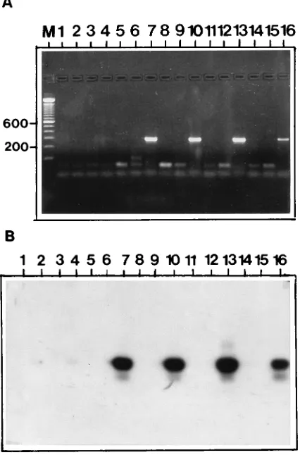
FIG. 3. (A) Agarose gel showing representative PCR results withparaffin-embedded tissue specimens from experimentally infected animals. The template DNAs were DNAs from urinary bladder tissues from noninfected animals (lanes 1 to 6) and from experimentally infected animals (lanes 7 to 14). Lane 15, negative control; lane 16; 1 ng of purified DNA from B. burgdorferi DK1 as positive control; lane M, DNA size markers in base pairs. (B) Southern blot hybridization of the gel shown in panel A. The amplification products were identified with the32P-end-labeled oligonucleotide F7.
on May 15, 2020 by guest
http://jcm.asm.org/
[image:4.612.72.285.76.399.2]decided to include in vitro culture, since this method is still the
most accepted documentation for B. burgdorferi infection. In
our present study, the amount of bladder tissue unfortunately
was not sufficient to perform all four procedures. Thus, we
decided to omit PCR with fresh bladder tissue because PCR
with this tissue source previously proved to be highly sensitive
(22).
Adaptation of PCR for the detection of B. burgdorferi in
formalin-fixed, paraffin-embedded tissue would be very
advan-tageous. The same skin biopsy could then be used for both
histological examination and diagnostic PCR. Furthermore,
this would allow the study of stored material retrospectively. So
far, the detection of B. burgdorferi DNA by PCR in extracts
from fixed, paraffin-embedded skin or tissue specimens has not
been reported. In the present study, we showed that the
diag-nostic sensitivity obtained for PCR with fixed,
paraffin-embed-ded specimens was only 21%, compared with 71% when DNA
was extracted from fresh tissue. PCR has successfully been
used to detect genes from eukaryotic and viral genomic DNAs
in formalin-fixed, paraffin-embedded tissue (17, 32). However,
fixatives cause DNA degradation, and PCR with DNA
ex-tracted from fixed sections is generally less efficient than PCR
with DNA prepared from fresh tissue (18). The degradation of
DNA depends strongly on the fixative and the fixation time
(18). Thus, we chose neutral buffered formalin fixation for less
than 24 h because this protocol was reported to be the least
destructive (13, 18). Furthermore, and in agreement with
re-sults of other studies (13, 34), we observed Taq polymerase
inhibition by DNA extracted from fixed tissue. This effect was
partly overcome by using a nested PCR approach. From a
practical point of view, however, the nested PCR is not
attrac-tive because of an increased risk of contamination. These
fac-tors probably contribute to the very reduced sensitivity
ob-tained by PCR with fixed, paraffin-embedded tissue. Another
reason is that the number of spirochetes present in lesions
from patients with Lyme borreliosis is very low, which is in
contrast to the case for most viral infections, in which the
number of viral particles is higher. It might be argued that the
observed differences in the sensitivity of PCR with fixed and
fresh tissue could be partly attributed to the larger amount of
fresh tissue used for DNA extraction. However, since the same
amount of extracted DNA was used as template DNA in the
two situations, this seems unlikely. Thus, from these results we
assume that PCR with paraffin-embedded tissue from human
skin biopsies is unlikely to be a useful diagnostic approach.
We conclude that B. burgdorferi can be detected in tissue
sections from experimentally infected animals by an
avidin-biotin-amplified immunophosphatase staining method but that
the diagnostic sensitivity is low. The immunohistochemical
staining technique is extremely laborious, and considering the
fact that the density of B. burgdorferi in human tissue is even
lower than that in experimentally infected animals, the method
is not useful in a clinical setting. It might, however, still be a
valuable tool in pathogenetic research. The diagnostic
sensi-tivity obtained by PCR with fixed, paraffin-embedded tissue
from experimentally infected animals was low. In contrast to
PCR with unfixed skin biopsies, diagnostic PCR with
paraffin-embedded tissue from human skin biopsies is unlikely to
be-come a useful diagnostic approach.
ACKNOWLEDGMENTS
We thank Marianne Tønder for perfect technical assistance; David Hougaard, Department of Molecular Cell Biology, Statens Seruminsti-tut, Copenhagen, Denmark, for the synthetic oligonucleotide primers; and Severin Olesen Larsen, Department of Biostatistics, Statens Se-ruminstitut, for statistical calculations.
Financial support from Fonden til Lægevidenskabes Fremme and Research Center for Medical Biotechnology under the Danish Bio-technological Research Program is gratefully acknowledged.
REFERENCES
1. Aberer, E., and R. Neumann. 1989. Lyme disease spirochetes in the skin: an immunohistochemical study, p. 141–145. In G. Stanek, W. Kristoferitsch, M. Pletschette, A. G. Barbour, and H. Flamm (ed.), Lyme borreliosis II. Gustav Fischer, Stuttgart, Germany.
2. Amouriaux, P., M. Assous, D. Margarita, G. Baranton, and I. Saint Girons. 1993. Polymerase chain reaction with the 30-kb circular plasmid of Borrelia burgdorferi B31 as a target for detection of the Lyme borreliosis agents in cerebrospinal fluid. Res. Microbiol. 144:211–219.
3. Asbrink, E., and A. Hovmark. 1985. Successful cultivation of spirochetes from skin lesions of patients with erythema chronicum migrans Afzelius and acrodermatitis chronica atrophicans. Acta Pathol. Microbiol. Immunol. Scand. Sect. B 93:161–163.
3a.Barbour, A. G. 1984. Isolation and cultivation of Lyme disease spirochetes. Yale J. Biol. Med. 57:521–525.
4. Barthold, S. W., D. H. Persing, A. L. Armstrong, and R. A. Peeples. 1991. Kinetics of Borrelia burgdorferi dissemination and evolution of disease after intradermal inoculation of mice. Am. J. Pathol. 139:263–273.
5. Benach, J. L., E. M. Bosler, J. P. Hanrahan, J. L. Coleman, G. S. Habicht, T. F. Bast, D. J. Cameron, J. L. Ziegler, A. G. Barbour, W. Burgdorfer, R. Edelman, and R. A. Kaslow.1983. Spirochetes isolated from the blood of two patients with Lyme disease. N. Engl. J. Med. 308:740–742.
6. Berger, B. W., O. J. Clemmensen, and A. B. Ackermann. 1983. Lyme disease is a spirochetosis. A review of the disease and evidence of its cause. Am. J. Dermatopathol. 5:111–124.
7. Berger, B. W., R. C. Johnson, C. Kodner, and L. Coleman. 1992. Cultivation of Borrelia burgdorferi from erythema migrans lesions and perilesional skin. J. Clin. Microbiol. 30:359–361.
8. Champion, C. I., J. N. Miller, M. A. Lovett, and D. R. Blanco. 1990. Cloning, sequencing, and expression of two class B endoflagellar genes of Treponema
pallidum subsp. pallidum encoding the 34.5- and 31.0-kilodalton proteins.
Infect. Immun. 58:1697–1704. 8a.Clemmensen, O. Unpublished data.
9. Coen, D. M. V. 1990. Quantitation of rare DNAs by the polymerase chain reaction, p. 15.3.1–15.3.4. In F. A. Ausubel, R. Brent, R. E. Kingston, D. D. Moore, J. G. Seidman, J. A. Smith, and K. Struhl (ed.), Current protocols in molecular biology. Greene Publishing, New York.
10. de Koning, J., R. B. Bosma, and J. A. Hoogkamp Korstanje. 1987. Demon-stration of spirochaetes in patients with Lyme disease with a modified silver stain. J. Med. Microbiol. 23:261–267.
11. Duray, P. H., and R. C. Johnson. 1986. The histopathology of experimentally infected hamsters with the Lyme disease spirochete, Borrelia burgdorferi. Proc. Soc. Exp. Biol. Med. 181:263–269.
12. Duray, P. H., and A. C. Steere. 1986. The spectrum of organ and systems pathology in human Lyme disease. Zentralbl. Bakteriol. Mikrobiol. Hyg. A 263:169–178.
13. Fiallo, P., D. L. Williams, G. P. Chan, and T. P. Gillis. 1992. Effects of fixation on polymerase chain reaction detection of Mycobacterium leprae. J. Clin. Microbiol. 30:3095–3098.
14. Gassmann, G. S., M. D. Kramer, U. B. Go¨bel, and R. Wallich.1989. Nucle-otide sequence of a gene encoding the Borrelia burgdorferi flagellin. Nucleic Acids Res. 17:3590.
15. Hansen, K., J. M. Bangsborg, H. Fjordvang, N. S. Pedersen, and P. Hin-dersson.1988. Immunochemical characterization of and isolation of the gene for a Borrelia burgdorferi immunodominant 60-kilodalton antigen com-mon to a wide range of bacteria. Infect. Immun. 56:2047–2053.
16. Hofmeister, E., R. B. Markham, J. E. Childs, and R. R. Arthur. 1992. Comparison of polymerase chain reaction and culture for detection of
Bor-relia burgdorferi in naturally infected Peromyscus leucopus and experimentally
infected C.B-17 scid/scid mice. J. Clin. Microbiol. 30:2625–2631. 17. Impraim, C. C., R. K. Saiki, H. A. Erlich, and R. L. Teplitz. 1987. Analysis
of DNA extracted from formalin-fixed, paraffin-embedded tissues by enzy-matic amplification and hybridization with sequence-specific oligonucleo-tides. Biochem. Biophys. Res. Commun. 142:710–716.
18. Jackson, D. P., F. A. Lewis, G. R. Tayllor, A. W. Boylston, and P. Quirke. 1990. Tissue extraction of DNA and RNA and analysis by the polymerase chain reaction. J. Clin. Pathol. 43:499–504.
19. Johnston, Y. E., P. H. Duray, A. C. Steere, M. Kashgarian, J. Buza, and P. W. Askenase.1985. Lyme arthritis. Spirochetes found in synovial microangio-pathic lesions. Am. J. Pathol. 118:26–34.
20. Karlsson, M., K. Hovind Hougen, B. Svenungsson, and G. Stiernstedt. 1990. Cultivation and characterization of spirochetes from cerebrospinal fluid of patients with Lyme borreliosis. J. Clin. Microbiol. 28:473–479.
21. Lebech, A. M., and K. Hansen. 1992. Detection of Borrelia burgdorferi DNA in urine samples and cerebrospinal fluid samples from patients with early and late Lyme neuroborreliosis by polymerase chain reaction. J. Clin. Mi-crobiol. 30:1646–1653.
22. Lebech, A. M., P. Hindersson, J. Vuust, and K. Hansen. 1991. Comparison
on May 15, 2020 by guest
http://jcm.asm.org/
of in vitro culture and polymerase chain reaction for detection of Borrelia
burgdorferi in tissue from experimentally infected animals. J. Clin. Microbiol.
29:731–737.
23. Melchers, W., J. Meis, P. Rosa, E. Claas, L. Nohlmans, R. Koopman, A. Horrevorts, and J. Galama.1991. Amplification of Borrelia burgdorferi DNA in skin biopsies from patients with Lyme disease. J. Clin. Microbiol. 29:2401– 2406.
24. Nocton, J. J., F. Dressler, B. J. Rutledge, P. N. Rys, D. H. Persing, and A. C. Steere.1994. Detection of Borrelia burgdorferi DNA by polymerase chain reaction in synovial fluid from patients with Lyme arthritis. N. Engl. J. Med. 330:229–234.
25. Pachner, A. R., N. Ricalton, and E. Delaney. 1993. Comparison of poly-merase chain reaction with culture and serology for diagnosis of murine experimental Lyme borreliosis. J. Clin. Microbiol. 31:208–214.
26. Pallesen, L., and P. Hindersson. 1989. Cloning and sequencing of a
Trepo-nema pallidum gene encoding a 31.3-kilodalton endoflagellar subunit
(FlaB2). Infect. Immun. 57:2166–2172.
27. Park, H. K., B. E. Jones, and A. G. Barbour. 1986. Erythema chronicum migrans of Lyme disease: diagnosis by monoclonal antibodies. J. Am. Acad. Dermatol. 15:406–410.
28. Picken, R. N. 1992. Polymerase chain reaction primers and probes derived from flagellin gene sequences for specific detection of the agents of Lyme
disease and North American relapsing fever. J. Clin. Microbiol. 30:99–114. 29. Preac Mursic, V., E. Patsouris, B. Wilske, S. Reinhardt, B. Gross, and P. Mehraein.1990. Persistence of Borrelia burgdorferi and histopathological alterations in experimentally infected animals. A comparison with his-topathological findings in human Lyme disease. Infection 18:332–341. 30. Schwartz, I., G. P. Wormser, J. J. Schwartz, D. Cooper, P. Weissensee, A.
Gazumyan, E. Zimmermann, N. S. Goldberg, S. Bittker, G. L. Campbell, and C. S. Pavia.1992. Diagnosis of early Lyme disease by polymerase chain reaction amplification and culture of skin biopsies from erythema migrans lesions. J. Clin. Microbiol. 30:3082–3088.
31. Schwentker, V. 1963. The gerbil—a new laboratory animal. III. Vet. 6:5–9. 32. Shibata, D., N. Arnheim, and W. J. Martin. 1988. Detection of human papilloma virus in paraffin-embedded tissue using the polymerase chain reaction. J. Exp. Med. 167:225–230.
33. Steere, A. C., R. L. Grodzicki, A. N. Kornblatt, J. E. Craft, A. G. Barbour, W. Burgdorfer, G. P. Schmid, E. Johnson, and S. E. Malawista.1983. The spirochetal etiology of Lyme disease. N. Engl. J. Med. 308:733–740. 34. Wright, D. K., and M. M. Manos. 1990. Sample preparation from
paraffin-embedded tissues, p. 153–158. In M. A. Innis, D. H. Gelfand, J. J. Sninsky, and T. J. White (ed.), PCR protocols: a guide to methods and applications. Academic Press, Inc., New York.