and influences encephalitogenic Th1 responses
in EAE
Gregory D. Gregory, … , Susan Winandy, Melissa A. Brown
J Clin Invest. 2006;
116(5)
:1327-1336.
https://doi.org/10.1172/JCI27227
.
When exposed to a pathogen, a naive CD4
+T cell is forced to make a cell fate decision that
leads to a polarized population of Th1 IFN-
g
– or Th2 IL-4– producing cells. Although IL-4
has traditionally been considered a factor that promotes Th2 cell differentiation, recent
evidence has demonstrated that the site and timing of IL-4 expression in an immune
response determines its ultimate effects on CD4
+T cell fate. Using a mast cell (MC)
reconstitution model, we demonstrate that MC-derived IL-4 promoted Th1 responses in vivo.
Furthermore, MCs from genetically disparate mouse strains varied in their potential for IL-4
expression. Independent of the activation mode, MCs from Th1-prone C57BL/6 mice
exhibited a more robust Il4 response than did the Th2-prone strain Balb/c. The hierarchy of
IL-4 expression potential was directly associated with the degree of basal chromatin
accessibility at cis-regulatory elements conserved noncoding sequence–1 and V
Aenhancer
within the Th2 locus. GATA1/2 and Ikaros, factors with opposing roles in chromatin
remodeling, acted at these sites. We propose that GATA and Ikaros proteins coordinately
fine-tune accessibility at the Il4 locus during development to variably regulate IL-4
expression. These events likely contribute to the genetically determined heterogeneity in
Th1 responses that underlie susceptibility to many diseases.
Research Article
Immunology
Find the latest version:
Research article
Mast cell IL-4 expression is regulated
by Ikaros and influences encephalitogenic
Th1 responses in EAE
Gregory D. Gregory,1,2 Shveta S. Raju,3 Susan Winandy,1 and Melissa A. Brown1
1Department of Microbiology-Immunology, Northwestern University Feinberg School of Medicine, Chicago, Illinois, USA. 2Graduate Program in Immunology
and Molecular Pathogenesis and 3Department of Pathology and Laboratory Medicine, Emory University School of Medicine, Atlanta, Georgia, USA.
When exposed to a pathogen, a naive CD4
+T cell is forced to make a cell fate decision that leads to a polarized
population of Th1 IFN-
g
– or Th2 IL-4– producing cells. Although IL-4 has traditionally been considered a
fac-tor that promotes Th2 cell differentiation, recent evidence has demonstrated that the site and timing of IL-4
expression in an immune response determines its ultimate effects on CD4
+T cell fate. Using a mast cell (MC)
reconstitution model, we demonstrate that MC-derived IL-4 promoted Th1 responses in vivo. Furthermore,
MCs from genetically disparate mouse strains varied in their potential for IL-4 expression. Independent of the
activation mode, MCs from Th1-prone C57BL/6 mice exhibited a more robust
Il4
response than did the
Th2-prone strain Balb/c. The hierarchy of IL-4 expression potential was directly associated with the degree of basal
chromatin accessibility at
cis
-regulatory elements conserved noncoding sequence–1 and V
Aenhancer within
the Th2 locus. GATA1/2 and Ikaros, factors with opposing roles in chromatin remodeling, acted at these
sites. We propose that GATA and Ikaros proteins coordinately fine-tune accessibility at the
Il4
locus during
development to variably regulate IL-4 expression. These events likely contribute to the genetically determined
heterogeneity in Th1 responses that underlie susceptibility to many diseases.
Introduction
IL-4 is a multifunctional cytokine that regulates both innate and adaptive immunity (1). Cells of several lineages produce IL-4, including CD4 and CD8 T cells, NKT cells, eosinophils, mast cells (MCs), and basophils. It is likely that expression by each cell type contributes to distinct as well as overlapping physiologic respons- es. IL-4 is perhaps best known for its role in promoting Th2 cell dif-ferentiation and limiting Th1 responses; however, recent evidence shows that IL-4 can also direct Th1 responses (2). IL-4 can act on a wide variety of other target cells including B cells, macrophages, DCs, and MCs as well as nonimmune cells such as epithelial cells and goblet cells (3–6). This broad range of effects necessitates strict controls on the expression of this cytokine. Inappropriate expres-sion is associated with allergic disease, autoimmunity, and failure to clear certain infections (7).
Most information regarding mechanisms of IL-4 gene (Il4
) regu-lation comes from studies in Th cells and MCs. In addition to acute signals that elicit active Il4 transcription in both cell types, a cru-cial determinant of IL-4 expression is locus accessibility acquired
during development (8). Il4
is closely linked to the genes encod-ing IL-5 (Il5) and IL-13 (Il13). These genes are clustered within an approximately 200-kb region, termed the Th2 locus, on chromo-some 11 in mice and chromosome 5 in humans. During Th cell differentiation, the Th2 locus undergoes a series of chromatin modifications that confer the relative potential for transcription
from Il4 (8).
DNAse I hypersensitive site (HS) analyses and cross-species sequence comparisons have allowed the identification of
a number of cis-regulatory elements implicated in controlling Il4
expression. Included among these are regions between Il13 and
Il4 (conserved noncoding sequence–1 [CNS-1]), within the Il4
promoter (HS I) and second intron (intronic enhancer [IE]), and distal to Il4 (HS IV, V, and VA) (9–13). In addition, a locus control region is located upstream of Il13 at the 3′ end of Rad50 (14). The histones associated with these sites become hyperacetylated in developing Th2 cells, correlating with the potential to express IL-4, whereas the Th2 locus is hypoacetylated and relegated to pericen- tromeric heterochromatin in Th1 cells (15–17). Resting bone mar-row–derived MCs (BMMCs) express most of the same HS sites as
Th2 cells within the Il4 locus. Although most currently defined
sites have enhancing activity, Rao and colleagues have defined HS IV as a conserved silencer of Il4 in both T cells and MCs (9).
The Il4 regulation paradigm is based primarily on results of
experiments utilizing T cells and MCs from only 2 common strains of mice: C57BL/6 (B6) and Balb/c. These strains have been useful in studying genetically based disease susceptibility due to
their disparate immune responses to some antigens, events per-haps most clearly manifested in Leishmania major infection (18).
B6 mice clear the infection due to a Th1-dominated response, whereas Balb/c mice fail to clear the parasite despite a strong but nonprotective Th2-skewed response. Most studies have focused on possible intrinsic differences in the response of B6- versus Balb/c-derived T cells, such as instability in expression of the
IL-12 receptor b2 subunit in Balb/c T cells (19). We considered
the possibility that strain-specific differences in bystander cell cytokine expression exist that modulate the Th1/Th2 balance. A genetically determined difference in the kinetics or level of IL-4 expression by MCs, for example, could directly alter the character of the T cell response or act indirectly through effects on DC
Nonstandard abbreviations used: B6, C57BL/6; BMMC, bone marrow–derived MC; CD40L, CD40 ligand; ChIP, chromatin immunoprecipitation; CNS-1, conserved non-coding sequence–1; FceRI, high affinity IgE receptor; HDAC, histone deacetylase; HS, hypersensitive site; IE, intronic enhancer; MC, mast cell; MOG, myelin oligodendro-cyte glycoprotein peptide; NuRD, nucleosome remodeler and deacetylase; VAE,
VA enhancer; W/Wv, WBB6-F1/J-KitW/KitWv.
Conflict of interest: The authors have declared that no conflict of interest exists.
maturation. In this report, we demonstrate that MC IL-4 was critical to the generation of myelin-specific Th1 cells and con-tributed to severe EAE, a murine model of MS. Furthermore, we
show that there were strain-specific differences in murine MC Il4
expression potential that may influence the heterogeneity of Th1
versus Th2 responses. Variability in the expression of Il4 occurred
irrespective of the mode of activation and correlated with distinct patterns of basal H3 and H4 histone acetylation at several sites
within the Th2 locus, including CNS-1 and VA enhancer (VAE)
elements. GATA1, GATA2, and Ikaros, DNA-binding proteins with established roles in chromatin remodeling, associated with
CNS-1 and VAE in vivo. We provide direct evidence that Ikaros
has a suppressive role in MC Il4 transcription, as Ikarosnull MCs
had elevated IL-4 mRNA expression and chromatin accessibil-ity across the Th2 locus. Our data is consistent with a model whereby the interplay of GATA and Ikaros proteins determines
the variable potential for MC Il4 expression, which subsequently
exerts a direct effect on Th1/Th2 responses.
Results
MC-derived IL-4 influences EAE severity by enhancing encephalitogenicity of Th1 cells. We hypothesized that MCs, by virtue of their ability to express cytokines that modulate T cell responses, have a role in adaptive immune responses (20). Consistent with this idea are
results of previous studies showing that MC-derived TNF-a
regu-lates in vitro T cell reactivity (21) and that MCs are essential for optimal generation of encephalitogenic Th1 cells during EAE (22). Although IL-4 is best known as a Th2-polarizing cytokine, IL-4 can also have distinct effects on Th1 responses (2, 23, 24). Thus it
was of interest to investigate whether MC IL-4 expression could modulate the quality of a Th1-mediated response.
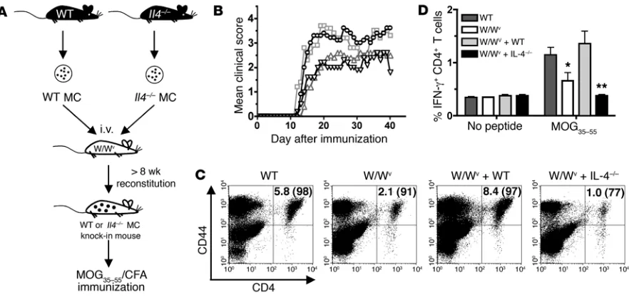
Using a MC knock-in model in which MC-deficient WBB6-F1/ J-KitW-KitWv (W/Wv) mice were selectively reconstituted with either
WT or IL-4–/– BMMCs (referred to as W/Wv + WT and W/Wv +
IL-4–/–, respectively; Figure 1A), we assessed the specific effects of MC IL-4 on myelin oligodendrocyte glycoprotein peptide–induced (MOG35–55-induced) EAE. As previously reported, W/Wv mice exhibited a significantly reduced overall mean clinical score com-pared with WT littermate controls (P
< 0.01; Figure 1B). Reconsti-tution of W/Wv mice with MCs deficient in CD40 ligand (CD40L),
a cell surface costimulatory molecule expressed on several cell types, restores disease to WT levels and served as a positive control in this experiment (25). In contrast, mice reconstituted with IL-4–/– MCs failed to display severe disease.
Development of EAE and MS is dependent upon trafficking of activated, myelin-specific T cells to the CNS. Consistent with reduced disease severity, reduced CNS infiltration of CD4 T cells
was observed in W/Wv and W/Wv + IL-4–/– mice compared with
WT and W/Wv + WT mice (Figure 1C). Furthermore, appreciably
fewer T cells entering the CNS of W/Wv and W/Wv + IL-4–/– mice
were activated, as measured by high CD44 surface expression. Together, these results suggest that MC IL-4 promotes encepha-litogenic Th1 responses.
MOG35–55-specific T cell responses were also evaluated in WT
and reconstituted mice. As shown in Figure 1D, W/Wv T cell
responses were reduced relative to WT as previously reported (22).
While WT BMMCs restored the frequency of MOG35–55-specific
[image:3.585.72.513.82.294.2]IFN-g–producing CD4 T cells, IL-4–/– BMMCs did not (P < 0.01).
Figure 1
research article
Similar results were obtained in experiments using immunization
with OVA323–339, a nonencephalitogenic peptide (data not shown).
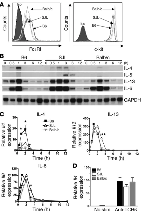
MCs, but not Th2 cells, exhibit strain-specific differences in Il4 expression. The demonstration that MC IL-4 enhanced Th1 responses raised the possibility that the propensity for Th heterogeneity in various strains of mice might be due to intrinsic differences in the magni-tude of MC IL-4 expression. For example, MCs from Th1-prone mice may have relatively higher IL-4 expression compared with a Th2-dominated strain. MCs from the B6 and Balb/c strains were
chosen to examine potential strain-specific differences in MC Il4
expression due to their dichotomous responses to L. major
infec-tion (18). The SJL strain was also examined as it has documented defects in T cell IL-4 expression and IgE production in vivo (26). MCs were differentiated from bone marrow by culturing in the presence of SCF and IL-3. BMMCs from all 3 strains expressed comparable levels of c-kit and the high affinity IgE receptor
(FceRI) after 6–8 weeks of culture (Figure 2A). Cells were activated
by cross-linking of FceRI, and IL-4 mRNA levels were measured
at various times after stimulation. As shown in Figure 2, B and C,
B6 BMMCs exhibited quick and relatively robust expression of Il4
compared with BMMCs from Balb/c or SJL mice (P < 0.05 at 1 and
2 hours). SJL BMMCs showed intermediate and more prolonged
expression of Il4, whereas Balb/c MCs displayed a relatively weak
expression profile, demonstrating the strain-specific variations in the duration and magnitude of the IL-4 response. The expression of other genes such as Il5, Il13, and Il6
also displayed notable varia-tions between strains. For example, appreciable Il5 expression was
observed only in SJL MCs.
To examine whether this difference was also evident in T cells,
naive CD4+
T cells from the same strains were isolated, differen-tiated under Th2 skewing conditions, and analyzed for cytokine expression. Unlike BMMCs, Th2 cells cultured in vitro showed no significant strain-specific variation in expression of IL-4 mRNA after restimulation through the T cell receptor (Figure 2D).
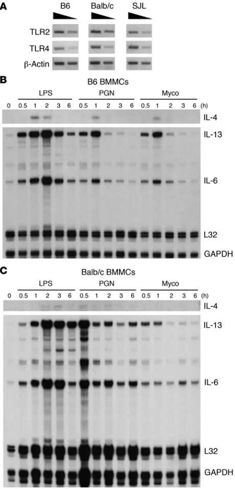
B6-derived BMMCs express higher levels of IL-4 mRNA irrespective of activating agonist. The unique profiles of MC Il4 expression could
be intrinsic to FceRI signaling; alternatively, they may reflect
more global differences in basal chromatin accessibility at the Il4
locus, leading to distinct expression potentials regardless of the mode of stimulation. To distinguish between these possibilities, the expression of IL-4 mRNA was compared after TLR-induced signaling. TLR2 and TLR4 mRNA expression was confirmed in cells derived from all 3 strains (Figure 3A). Cells were then stimu-lated with peptidoglycan (a TLR2 agonist), LPS (a TLR4 agonist),
or heat-killed Mycobacterium tuberculosis, which can signal through
bothTLR2 and TLR4. B6-derived MCs expressed higher levels of
IL-4 mRNA in response to all agonists compared with Balb/c MCs (Figure 3, B and C). This result was consistent with the idea that
B6 MCs (IL-4hi
) have a greater potential for IL-4 expression irre-spective of the activating stimulus.
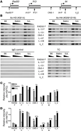
Il4 expression potential correlates with Th2 locus accessibility.Guo et al. demonstrated that the probability of a Th2 cell producing relatively high or low levels of IL-4 directly correlates with the degree of histone acetylation, a marker of chromatin accessibility, at the Th2 cytokine locus (27). To examine whether strain-based differences in the ability of MCs to express IL-4 are reflected in
distinct chromatin states that affect accessibility within the Il4
[image:4.585.46.284.81.437.2]locus, histone acetylation was evaluated in resting MCs by chro-matin immunoprecipitation (ChIP) using antibodies specific for acetylated histones H3 and H4. IP templates were analyzed by PCR using primers specific for previously defined regulatory elements within the Th2 locus (Figure 4A). Several differences were noted in histone H3 and H4 acetylation between strains (Figure 4, B–D). Of particular interest is the consistent and striking difference in acet-ylation observed at CNS-1 and VAE in the IL-4hi B6 and IL-4int SJL strains relative to Balb/c. Both of these elements are necessary for
Figure 2
a strong transcriptional response in T cells, and VAE is essential for optimal MC Il4 expression (11, 13, 28). In addition, accessibility at both these sites is a key determinant in regulating the potential for high versus low IL-4 expression in T cells (27). Thus the potential
for MC Il4
expression correlated with a unique pattern of chroma-tin accessibility at several sites within the Th2 locus.
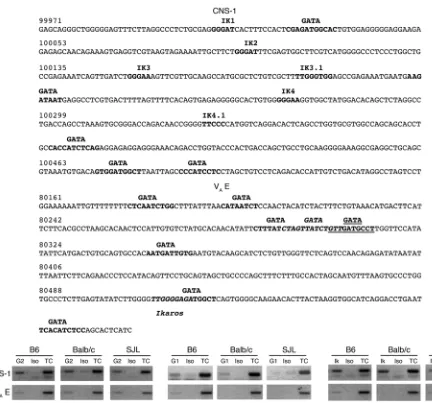
GATA and Ikaros family members associate with CNS-1 and the VAE in
vivo. The mechanism regulating Il4 locus accessibility in MCs as
well as T cells is poorly understood. Our finding that the acetyla-tion state of cis-regulatory elements CNS-1 and VA
E is commen-surate with Il4
expression potential warranted a further investiga-tion into the factors regulating these sites in MCs. No base pair polymorphisms were detected within these elements, indicating
that strain-specific differences in Il4 transcription are not due to
alterations in transactivating factor binding sites within CNS-1
or VAE. A number of putative GATA and Ikaros binding sites are
present within both CNS-1 and VAE (Figure 5A). As shown by ChIP
analysis in Figure 5B, GATA1, GATA2, and Ikaros were found to
associate with the CNS-1 and VAE elements. Binding to VAE was
generally more variable compared with CNS-1. The demonstration that GATA and Ikaros associated with these sites in nonstimulated MCs suggests that they are involved in regulating the Th2 locus.
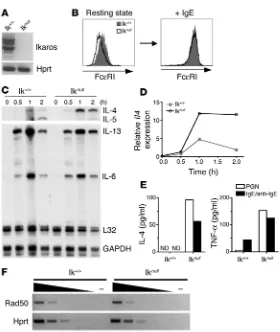
Ikaros regulates IL-4 expression in MCs. We have previously shown
that GATA1 and GATA2, like GATA3 in Th2 cells, are positive reg-ulators of Il4 expression in MCs. Association of these factors with
the IE is critical for intron acetylation and subsequent transcrip- tion (29–31). In contrast to the sole positive actions of GATA fam-ily members, Ikaros can act as either an activator or a suppressor
of transcription, activating expression of CD8a
in T cells and sup-pressing λ5 expression in B cells (32, 33). To investigate the effect
of Ikaros on Il4 expression, BMMCs were derived from (B6 × 129)
F1 mice containing a targeted deletion of Ikaros (Ikarosnull) and
their wild-type littermates (Ikaros+/+
) using standard differentia-tion culture conditions. RT-PCR assays revealed that Ikaros+/+ MCs
expressed a variety of Ikaros isoforms and confirmed the absence of Ikaros expression in Ikarosnull MCs (Figure 6A). Like Ikaros+/+, Ikarosnull MCs were c-kithi and FceRI+ and contained the charac-teristic metachromatic granules (Figure 6B and data not shown), indicating that there were no defects in the ability to generate MCs
from bone marrow. Although basal expression of Fce
RI was con-sistently lower in Ikarosnull cells, treatment with monomeric IgE
resulted in identical increases in receptor expression, confirming
that downstream FceRI-signaling pathways were intact (Figure 6B)
(34). Ikarosnull BMMCs demonstrated elevated and more prolonged
expression of Il4 in response to FceRI cross-linking compared with
Ikaros+/+ MCs (Figure 6, C and D). To corroborate these findings at
the protein level, activation-induced IL-4 secretion was measured in both groups (Figure 6E). IL-4 was detected in supernatants
from Ikarosnull BMMCs 22 hours after FceRI cross-linking and
TLR2-mediated stimulation, but not in Ikaros+/+ BMMCs. TNF-a
secretion was measured as a positive control for activation in
both groups. While Ikarosnull BMMC expressed lower levels of IL-5
mRNA, expression of Il13, as well as Il6 (a cytokine gene encoded
outside of the Th2 locus), was not appreciably affected. Transcrip-tion of Rad50, a gene located within the Th2 locus but expressed
constitutively in most cell types and under separate control than
Il4, Il5, and Il13, was not affected in resting Ikarosnull
BMMCs (Fig-ure 6F). Taken together with the evidence of in vivo Ikaros binding
to Il4 regulatory elements, these data demonstrate that Ikaros is
specifically involved in regulation of the Il4 locus in MCs.
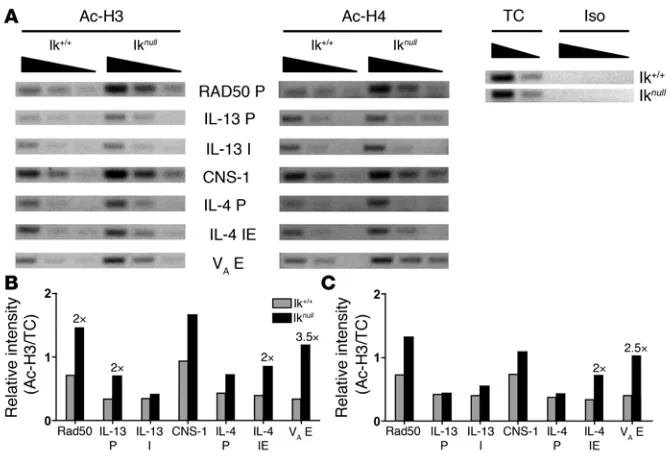
Ikaros controls Il4 expression at the chromatin level . To our knowl-edge, a direct mechanism by which Ikaros exerts its influence on
the CD8a and λ5 loci has not been described previously (32, 33).
The increased Il4 expression observed in Ikarosnull BMMCs could
be indicative of an overall suppressive effect on chromatin acces-Figure 3
[image:5.585.51.281.82.560.2]research article
sibility at the Th2 locus. Ikaros can associate with the higher order nucleosome remodeler and deacetylase (NuRD) complex contain-ing histone deacetylase 1 (HDAC1) and HDAC2. Thus if Ikaros is
acting to recruit HDAC activity to the Il4 locus, the lack of Ikaros
expression was predicted to result in higher histone acetylation levels within the Th2 locus. As expected, acetylation of histones H3 and H4 was increased at most of the Th2 locus regulatory elements
examined in Ikarosnull BMMCs compared with Ikaros+/+ BMMCs
(Figure 7). Relative levels of histone acetylation at the Gapdh
pro-moter were equivalent in both cell populations (data not shown).
Thus Ikaros regulated the potential for Il4 expression in MCs by
reducing chromatin accessibility at the Th2 locus.
Discussion
It is well accepted that cells of the innate immune system can pro-foundly influence the character of the adaptive immune response. There has been much speculation that MCs in particular can affect
the type and magnitude of a T cell response through the elaboration of immunoregulatory cytokines or expres-sion of co-stimulatory molecules (35, 36). Most data supporting such a role have come from in vitro studies using long-term T cell and MC lines (35). An underlying hypothesis of the present investigation was that strain-specific variations in MC IL-4 expression contribute to the genetically determined heterogeneity in Th respons-es. Several results of our study support this hypothesis. First, we showed that there were intrinsic differences in the ability of BMMCs, but not Th2 cells, derived from the B6, Balb/c, and SJL backgrounds to express IL-4. These differences were independent of the cell activa-tion mode and correlated with distinct patterns of basal chromatin accessibility at the Th2 locus.
How could a variable pattern of accessibility within
the Th2 locus be achieved that would give rise to dis-tinct potentials for Il4 expression? A clue is provided
by the observation that GATA and Ikaros, factors with
apparent opposing activity at the Il4 locus, bound in
vivo to regulatory elements of the Th2 locus. Notably, members of both of these families of transcription fac-tors impact the development of specific cell lineages through their ability to modulate chromatin accessibil-ity (37–39). In Th2 cells, GATA3 is essential for Th2 differentiation
and the events associated with Il4 locus opening (40). This factor
is thought to create and maintain an open chromatin state in rest-ing T cells (41), “poising” the locus for the interaction of inducible factors such as nuclear factor of activated T cells (NFAT) with their cognate sites upon cell activation (42–44). GATA3 enhances gene expression by increasing histone acetylation, presumably through the recruitment of CBP/p300-like proteins containing histone acetyltransferase activity. GATA3 can also antagonize the recruit-ment of DNA methyltransferase–1 and methyl CpG–binding domain protein–2 to CNS-1 in Th2 cells, 2 factors that would oth-erwise mediate repression of locus accessibility (45, 46). In MCs, GATA1 and GATA2 appear to play an analogous role to GATA3. However, while GATA2 is absolutely required for MC differentia-tion, a more predominant role in regulating gene expression in mature MCs has been assigned to GATA1 (39, 47). Both factors
associate with the Il4
IE in resting MCs and are necessary for main-Figure 4
[image:6.585.42.320.83.532.2]taining a demethylated DNA and acetylated histone chromatin state required for transcription (29–31). Based on these previous studies, GATA family members are considered positive regulators of chromatin accessibility in both cell types.
In contrast, our data indicated that Ikaros exerts a negative influence on expression from the Th2 locus. Ikaros was first char- acterized as an integral component of chromatin remodeling com-plexes in cells of the lymphoid lineage. Mice containing a targeted
deletion in Ikaros
have profound defects in both B and T cell devel-opment (37). The defined molecular targets of Ikaros have been
limited to only 3 genes expressed during lymphoid cell develop-ment: CD8a, TdT, and λ5 (32, 33, 48). Our study provides what
we believe to be the first direct evidence that Ikaros is also involved in Il4 regulation. Not only did Ikaros associate with cis-regulatory elements of Il4, but Ikarosnull MCs derived from (B6 × 129) F1 mice
had dysregulated Il4 expression. The increased IL-4 mRNA and
protein expression as well as elevated histone acetylation at CNS-1,
VAE, and other sites observed in IkarosnullMCs confirms that this
factor exerts a suppressive influence on Il4.
Ikaros was initially implicated in gene repression by virtue of its association with epigenetically silenced genes in pericentromeric heterochromatin (49). In Th1 cells, CNS-1 was essential for the
localization of Il4 to this nuclear compartment, an event that is
associated with transcriptional blockade (16). It is hypothesized that
Ikaros mediates this process through direct effects on the CNS-1 ele-ment. As IL-4 mRNA expression was still observed in Ikaros+/+ MCs,
it is likely that Ikaros acts to modulate rather than completely sup-press overall potential for transcription. The negative role of Ikaros in chromatin remodeling is also assumed to be due, in part, to its ability to form complexes with NuRD (50). Ikaros’s repressive action
on Il4
[image:7.585.60.492.78.481.2]transcription in MCs may be the result of targeting of NuRD-associated HDAC activity to relevant cis-regulatory elements.
Figure 5
research article
Our data correlating chromatin accessibility with IL-4 expres-sion potential in MCs reveals a possible parallel to mechanisms governing the regulation of this gene in Th cells. Several studies
have documented that transcription from the Il4 locus in Th2 cells
is a stochastic process whereby the probability of expression from either allele is associated with the degree of chromatin accessibil-ity (27, 51, 52). It is hypothesized that this process allows for a more flexible means of regulation and adds an additional level of
control to cytokine gene expression. In Th cells, CNS-1 and VAE are
the currently defined targets of probabilistic control, making them
key determinants in regulating Il4 expression potential (27). These
same sites appeared to influence differential potential for MC Il4
expression. It will be of interest to determine whether there is vari-ability at the single-cell level within MCs of a particular genetic background, as in Th2 cells (51).
Based on our data, we propose a model whereby the integration of GATA and Ikaros signals, which can exert opposing effects on chromatin conformation, are responsible for distinct levels of accessibility at the Th2 locus (Figure 8). There are several possible
scenarios. For example, Ikaros may be associated with relevant Il4
cis-regulatory elements in early myeloid-lymphoid progenitors and
silence expression in these cells. During MC development, lineage- specific transcription factors such as GATA family members func- tion as “pioneering” proteins to decompact the condensed chro-matin and promote a cell lineage–specific gene expression profile (53). The extent of GATA pioneering activity may be dependent on relative expression levels or activation states. Thus GATA2 may act as a pioneering factor for initial opening of the Il4 locus in early MC
progenitors. Further accessibility leading to a strong Il4 transcriptional response may be mediated by GATA1.
The observed variability in chromatin accessibility between strains may be inherent to very early MC pre- cursors and/or result from variations in signals down-stream of IL-3 and SCF during in vivo and/or in vitro differentiation. Whereas signals delivered by IL-4 and antigens instruct locus opening in Th2 cells, MC growth factors IL-3 and SCF likely work in parallel fashion to affect remodeling during differentiation. Of note, both GATA1 and GATA2 have been implicated as essential transacti-vators downstream of IL-3 signaling (54). Continued association of Ikaros with relevant elements may allow for a more adaptable locus that can readily alter chromatin accessibility depending on the cells’ local microenvironment. This model does not purport that the binding of Ikaros or GATA at individual sites alone can control relative accessibility. Rather, it is the sum of interactions at multiple Th2 locus elements that is likely to dictate the high versus low expression potential. Other possible mechanisms con-tributing to genetically determined variability in accessibility at this locus could include differences in specific binding patterns of these and other factors, altered ratios of Ikaros to GATA binding, or expression of unique Ikaros isoforms that have distinct func-tional capabilities. It is also predicted that the interplay between the actions of GATA3 and Ikaros may influence chromatin acces-sibility in naive and differentiating CD4 T cells, leading to the extremes in expression potential in Th1/2 cells.
Using a MC knock-in model, we also demonstrated that MCs
were required for an optimal antigen-specific IFN-g response by
[image:8.585.42.322.76.408.2]CD4 T cells. More importantly, MC-derived IL-4 was a critical cytokine for this response, supporting the idea that the variable MC IL-4 response in B6 versus Balb/c mice is physiologically relevant. We propose that in certain immune settings, the relatively high IL-4 response of B6 MCs leads to the Th1-dominated responses charac- teristic of this strain. Conversely, Balb/c MCs do not express suf-ficient IL-4 to drive a strong Th1 response and default to the Th2 pathway. While this interpretation appears somewhat paradoxical with respect to the conventional Th1/Th2 cell paradigm, these
Figure 6
Ikaros suppresses Il4 transcription in MCs. (A) Resting BMMC express a number of Ikaros isoforms. Total RNA was analyzed by RT-PCR for the expression of Ikaros using prim-ers specific for exons 1 and 7. Expression of the housekeep-ing gene Hprt was included as a positive control. Absence of Ikaros mRNA expression in Ikarosnull BMMCs was also
con-firmed. (B) Surface expression of FceRI on BMMCs derived from Ikaros+/+ and Ikarosnull mice. Cells were left untreated
or were cultured with monomeric IgE (5 mg/ml) overnight. FceRI expression was analyzed by flow cytometry. (C and
D) Ikaros+/+ and Ikarosnull BMMCs were stimulated by FceRI
cross-linking, and cytokine mRNA expression was analyzed by ribonuclease protection assay. IL-4 mRNA expression was quantified as in Figure 2C. Data are representative of 4 independently derived BMMC cultures. (E) Cytokine secre-tion by activated Ikaros+/+ and Ikarosnull BMMCs measured
22 hours after activation with peptidoglycan or FceRI cross-linking (IgE/anti-IgE). Mean of 2 independent experiments is shown. ND, not detected. (F) Rad50 and Hprt mRNA expression was analyzed from resting Ikaros+/+ and Ikarosnull
data highlight the importance of the spatial and temporal expres-sion of IL-4 in the event of a response in vivo. They also support a model proposed by Biedermann et al., who demonstrated that
the duration of IL-4 treatment during L. major infection of Balb/c
mice directs the character of the Th response (2). When present only during DC maturation, IL-4 induces an IL-12–producing DC1 phenotype promoting Th1 differentiation. If IL-4 is administered for longer periods and is present during T cell priming in the sec- ondary lymphoid organs, a typical Th2-dominated response is gen-erated. The ability of MCs to express IL-4 in response to a variety of stimuli and their close proximity to DCs in many tissues including the skin make them likely candidates to modulate DC function in inflammatory settings (55).
There is some indirect evidence to support the idea that genes regulating IL-4 expression potential and chromatin accessibility influence disease heterogeneity. Genes controlling susceptibility
to and IL-4 expression during L. major infection map to a number
of loci, including L. major response 6 (Lmr6). Located at the near
end of chromosome 11, Ikaros is found within Lmr6
and is a candi-date susceptibility gene (56, 57). Our identification of Ikaros as a gene controlling Th2 cytokine production strengthens these find-ings. Teuscher and colleagues have also implicated this region in EAE disease severity (58). Further investigations using mice with cell-specific targeted deletions of Ikaros may yield a better under-standing of the role of Ikaros in these disease settings.
[image:9.585.41.377.80.310.2]In summary, although the role of Ikaros is well defined in hematopoietic cell development, little is known about its function in the regulation of genes expressed during immune responses. Our finding that Ikaros was intricately involved in regulation of the Th2 locus in MCs reveals what we believe to be a novel and important role for Ikaros in regulating genes of mature effector cells. Our data also expand our current under- standing of the molecular basis for genetically influenced het-erogeneity in immune responses and reinforce the idea that MCs are critical regulatory/amplifying components of type 1 immune responses. Such information is critical for refining strategies for changing the class of an immune response, particularly in the setting of vaccine development.
Figure 8
GATA and Ikaros collaborate at the Th2 locus to govern Il4 expres-sion potential. (A) Model proposing that Ikaros and GATA proteins — which can associate with Th2 locus cis-regulatory elements con-currently — competitively remodel the local histone state via the recruitment of HDAC or histone acetyltransferase (HAT) activity, respectively. (B) The combination of Ikaros and GATA binding to multiple sites within the Th2 locus, in addition to each factors’ inher-ent ability to recruit suppressive (e.g., HDAC) versus activating (e.g., HAT) complexes, sets the potential for high, intermediate, or low Il4 expression in MCs derived from genetically disparate strains. In Th2 cells, relatively high GATA3 expression leads to efficient opening of the Th2 locus, resulting in a permissive chromatin state.
Figure 7
Ikaros regulates chromatin accessibility of the Th2 locus. (A) Th2 locus accessibility in Ikarosnull BMMC was assessed by H3
[image:9.585.292.543.558.741.2]research article
Methods
Mice. Balb/cJ, SJL/J, B6, and B6-Il4tm1Nnt/J female mice (5–6 weeks old) were
purchased from The Jackson Laboratory. Female MC-deficient W/Wv
mice and their congenic littermate controls (WBB6-F1/J-Kit+-Kit+) were
obtained at 3 to 5 weeks of age. Ikarosnull mice (B6 ×
Sv129) were origi-nally generated by K. Georgopoulos (Massachusetts General Hospital, Charlestown, Massachusetts, USA) (37). Mice were housed in the animal care facilities at Emory University and Northwestern University. All ani-mal experiments were approved by the Institutional Animal Care and Use Committee at Emory University and the Animal Care and Use Committee at Northwestern University.
Generation of BMMCs and Th2 cells. Bone marrow was harvested from 5- to
6-week-old mice and differentiated with recombinant IL-3 (rIL-3; 5 ng/ml) and SCF (12.5 ng/ml) in complete RPMI (15% heat-inactivated FBS, 2 mM
glutamine, 1% penicillin-streptomycin, 1 mM sodium pyruvate, and 50 mM
2-b-ME). At 6–8 weeks of culture, these populations were >95% MCs as
assessed by c-kit and FceRI expression and toluidine blue staining. Th2
cells were differentiated in vitro with plate-bound anti-CD28 and anti-TCRb as previously described (44).
For optimal cytokine expression, MCs were cultured at 5 × 10⁵ cells/ml
in 5 ng/ml rIL-3 (termed day 0) and stimulated on day 3. Fce
RI cross-link-ing was accomplished using a previously described mode of IgE/anti-IgE activation (59). RNA was obtained using RNA-STAT60 (Tel-Test Inc.). For
activation via TLR agonists, cells were suspended at 5 × 10⁵ cells/ml in
complete rIL-3–containing RPMI on day 3 and stimulated with 50 ng/ml
LPS or 100 mg/ml sonicated peptidoglycan (Sigma-Aldrich) or heat-killed
M. tuberculosis strain H37RA (BD Diagnostics). Th2 cells were activated for
24 hours by plate-bound anti-TCRb (0.5 m
g/ml). For cytokine protein anal-ysis, activated BMMCs were cultured at 2 × 10⁶ cells/well in 200 m
l com-plete DMEM supplemented with IL-3. Culture supernatants were collected at 22 hours, and cytokine levels were assayed using LiquiChip Workstation (Qiagen) as previously described (22).
Ribonuclease protection assays
. Assays were performed per the manufactur-er’s instructions (RiboQuant Multi-Probe RNase Protection Assay System; BD Biosciences). Gels were visualized by autoradiography and/or quanti-fied via phosphorimaging and ImageQuant software (GE HealthCare).
ChIP.Analysis of histone H3 and H4 acetylation and in vivo binding of
GATA1/2 and Ikaros was performed using ChIP per the manufacturer’s instructions (Upstate USA Inc.) and as previously described (29) using resting BMMCs. A polyclonal Ab to Ikaros (H-100) was obtained from Santa Cruz Biotechnologies Inc. Immunoprecipitates were resuspended in 50 ml of H2O, and serial 3-fold dilutions of 1 ml template were used for each
reaction. Sequences of primers used in PCR were as follows: Rad50 pro-moter forward, 5′-GTGCGAGCTTACGGTTGCTGGCTCGC-3′; Rad50
promoter reverse, 5′-AACCTCAAGGCGGAAGCTGGGCAAG-3′; IL-13
promoter forward, 5′-TCCAAACCATGCATTGCTTTGGTG-3′
; IL-13 pro-moter reverse, 5′-TTTGTTGGGCATTATCTGAAAACC-3′; IL-13 intron
forward, 5′-GTGAGTAGCACACACAGCCCCTCC-3′; IL-13 intron reverse,
5′-TGATAAACAGTGGTCGCCACTCC-3′; CNS-1 forward, 5′
-TGATTTCTC-GGCAGCCAGGGAGGGCC-3′; CNS-1 reverse, 5′
-GGTGCCTGCGT-CACCTCTGACCACAC-3′; IL-4 promoter forward, 5′
-ACTCATTTTCCCTTG-GTTTCAGC-3′; IL-4 promoter reverse, 5′-GATTTTTGTCGCATCCGTGG-3′;
IL-4 intron forward, 5′-TCTGCTTGGACATCTCTCTTCCC-3′; IL-4 intron
reverse, 5′-ACCACCCCACAGGTCTTTGTTC-3′; VAE forward, 5′
-CAG-GTCCTGATGCCACCTTAGTAAG-3′; VAE reverse, 5′
-TTTAAATCCA-GATTCTGCATAGAATGG-3′. PCR conditions were 95°C denaturation,
60°C annealing, and 72°C extension for 30–36 cycles. PCR products were visualized with ethidium bromide on a 1.5% agarose gel.
RT-PCR. RNA was obtained from resting MCs as described above. RNA
was reverse transcribed using oligo-dT and SuperScript II. The following primers, complementary to exon 1 and exon 7 sequences, were used to
amplify Ikaros isoforms: Ik forward, 5′
-GATAGATCTATGGATGTCGAT-GAGGGTCAAGAC-3′; Ik reverse, 5′
-GATGAATTCTTAGCTCAGGTGGTA-ACGATGCTC-3′ (60); Hprt forward, 5′
-GTTGGATACAGGCCAGACTTT-GTTG-3′; Hprt reverse, 5′-GAGGGTAGGCTGGCCTATAGGCT-3′. TLR2
and TLR4, amplified for 35 cycles, were used as previously described (61).
Reconstitution and disease induction. Four-week-old W/Wv
mice were recon-stituted with 4 × 10⁶ BMMCs by i.v. transfer. Selective reconstitution of
the MC compartment was confirmed by hematocrit, histology, and/or flow cytometry at >8 weeks after transfer (Supplemental Figure 1; avail-able online with this article; doi:10.1172/JCI27227DS1; and data not
shown). EAE was induced by s.c. injection of 300 mg MOG35–55 emulsified
in 5 mg/ml CFA and 250 ng pertussis toxin i.p. Mice were scored daily for paralysis as previously described (22).
Intracellular cytokine staining and flow cytometry
. Preparation of CNS-infiltrating lymphocytes and in vitro restimulation of splenocytes with MOG35–55 peptide was performed as described previously (22). Intracellular
cytokine expression for IFN-gexpression was assayed using the Cytofix/
Cytoperm Kit (BD Biosciences) and analyzed by flow cytometry.
Statistics
. Statistical analysis was performed using GraphPad Prism soft-ware (version 4.0a). Repeated-measures ANOVA followed by Dunnett’s multiple-comparison post-test was used for comparison of mean clinical scores in EAE experiments. Intracellular cytokine expression was ana-lyzed by unpaired Student’s t test. P values less than 0.05 were considered to be statistically significant. Acknowledgments This work was supported by NIH grant R01A047992 and grant RG3104 from the National Multiple Sclerosis Society (to M.A. Brown). Received for publication October 26, 2005, and accepted in revised form February 28, 2006. Address correspondence to: Melissa A. Brown, Department of Microbiology-Immunology, Northwestern University Feinberg School of Medicine, Tarry Medical Research Building 6-758, mail code S213, 320 East Superior Street, Chicago, Illinois 60611-3010, USA. Phone: (312) 503-0108; Fax: (312) 503-4839; E-mail: m-brown12@northwestern.edu. 1. Weiss, D.L., and Brown, M.A. 2001. Regulation of IL-4 production in mast cells: a paradigm for cell-type-specific gene expression. Immunol. Rev.
179:35–47.
2. Biedermann, T., et al. 2001. IL-4 instructs TH1 responses and resistance to Leishmania major in sus-ceptible BALB/c mice. Nat. Immunol. 2:1054–1060. 3. Min, B., et al. 2004. Basophils produce IL-4 and
accumulate in tissues after infection with a Th2-inducing parasite. J. Exp. Med. 200:507–517. 4. Launois, P., et al. 2002. Rapid IL-4 production by
Leishmania homolog of mammalian RACK1-reac-tive CD4(+) T cells in resistant mice treated once with anti-IL-12 or -IFN-gamma antibodies at the onset of infection with Leishmania major instructs Th2 cell development, resulting in nonhealing lesions. J. Immunol. 168:4628–4635.
5. Brown, M.A., et al. 1987. B cell stimulatory factor-1/ interleukin-4 mRNA is expressed by normal and transformed mast cells. Cell. 50:809–818. 6. Noben-Trauth, N., Hu-Li, J., and Paul, W.E. 2002.
IL-4 secreted from individual naive CD4+ T cells acts in an autocrine manner to induce Th2 differ-entiation. Eur. J. Immunol. 32:1428–1433.
7. Brown, M.A., and Hural, J. 1997. Functions of IL-4 and control of its expression. Crit. Rev. Immunol.
17:1–32.
8. Ansel, K.M., Lee, D.U., and Rao, A. 2003. An epi-genetic view of helper T cell differentiation. Nat. Immunol. 4:616–623.
9. Ansel, K.M., et al. 2004. Deletion of a conserved Il4 silencer impairs T helper type 1-mediated immu-nity. Nat. Immunol. 5:1251–1259.
Immunity. 16:649–660.
11. Solymar, D.C., Agarwal, S., Bassing, C.H., Alt, F.W., and Rao, A. 2002. A 3′ enhancer in the IL-4 gene regulates cytokine production by Th2 cells and mast cells. Immunity. 17:41–50.
12. Lee, G.R., Spilianakis, C.G., and Flavell, R.A. 2005. Hypersensitive site 7 of the TH2 locus control region is essential for expressing TH2 cytokine genes and for long-range intrachromosomal inter-actions. Nat. Immunol. 6:42–48.
13. Mohrs, M., et al. 2001. Deletion of a coordinate regulator of type 2 cytokine expression in mice.
Nat. Immunol. 2:842–847.
14. Fields, P.E., Lee, G.R., Kim, S.T., Bartsevich, V.V., and Flavell, R.A. 2004. Th2-specific chromatin remodel-ing and enhancer activity in the th2 cytokine locus control region. Immunity. 21:865–876.
15. Grogan, J.L., et al. 2001. Early transcription and silencing of cytokine genes underlie polarization of T helper cell subsets. Immunity. 14:205–215.
16. Grogan, J.L., et al. 2003. Basal chromatin modifica-tion at the IL-4 gene in helper T cells. J. Immunol.
171:6672–6679.
17. Avni, O., et al. 2002. T(H) cell differentiation is accompanied by dynamic changes in histone acety-lation of cytokine genes. Nat. Immunol. 3:643–651.
18. Sacks, D., and Noben-Trauth, N. 2002. The immu- nology of susceptibility and resistance to Leishma-nia major in mice. Nat. Rev. Immunol. 2:845–858. 19. Hondowicz, B.D., Park, A.Y., Elloso, M.M., and Scott,
P. 2000. Maintenance of IL-12-responsive CD4+ T cells during a Th2 response in Leishmania major-infected mice. Eur. J. Immunol. 30:2007–2014. 20. Robbie-Ryan, M., and Brown, M. 2002. The role of
mast cells in allergy and autoimmunity. Curr. Opin. Immunol. 14:728–733.
21. Nakae, S., et al. 2005. Mast cells enhance T cell acti-vation: importance of mast cell-derived TNF. Proc. Natl. Acad. Sci. U. S. A. 102:6467–6472.
22. Gregory, G.D., Robbie-Ryan, M., Secor, V.H., Saba-tino, J.J., Jr., and Brown, M.A. 2005. Mast cells are required for optimal autoreactive T cell responses in a murine model of multiple sclerosis. Eur. J. Immunol. 35:3478–3486.
23. Hochrein, H., et al. 2000. Interleukin (IL)-4 is a major regulatory cytokine governing bioactive IL-12 production by mouse and human dendritic cells. J. Exp. Med. 192:823–833.
24. Yao, Y., Li, W., Kaplan, M.H., and Chang, C.H. 2005. Interleukin (IL)-4 inhibits IL-10 to promote IL-12 production by dendritic cells. J. Exp. Med.
201:1899–1903.
25. Grewal, I.S., et al. 1996. Requirement for CD40 ligand in costimulation induction, T cell activa-tion, and experimental allergic encephalomyelitis.
Science. 273:1864–1867.
26. Yoshimoto, T., Bendelac, A., Hu-Li, J., and Paul, W.E. 1995. Defective IgE production by SJL mice is linked to the absence of CD4+, NK1.1+ T cells that promptly produce interleukin 4. Proc. Natl. Acad. Sci. U. S. A. 92:11931–11934.
27. Guo, L., et al. 2002. In TH2 cells the Il4 gene has a series of accessibility states associated with dis-tinctive probabilities of IL-4 production. Proc. Natl. Acad. Sci. U. S. A. 99:10623–10628.
28. Loots, G.G., et al. 2000. Identification of a coordinate regulator of interleukins 4, 13, and 5 by cross-species sequence comparisons. Science. 288:136–140. 29. Kwan, M., Powell, D.R., Nachman, T.Y., and Brown,
M.A. 2005. An intron GATA-binding site regulates chromatin accessibility and is essential for IL-4 gene expression in mast cells. Eur. J. Immunol.
35:1267–1274.
30. Henkel, G., et al. 1992. A DNase I-hypersensitive site in the second intron of the murine IL-4 gene defines a mast cell-specific enhancer. J. Immunol.
149:3239–3246.
31. Hural, J.A., Kwan, M., Henkel, G., Hock, M.B., and Brown, M.A. 2000. An intron transcriptional enhancer element regulates IL-4 gene locus acces-sibility in mast cells. J. Immunol. 165:3239–3249. 32. Sabbattini, P., et al. 2001. Binding of Ikaros to the
lambda5 promoter silences transcription through a mechanism that does not require heterochroma-tin formation. EMBO J. 20:2812–2822.
33. Harker, N., et al. 2002. The CD8alpha gene locus is regulated by the Ikaros family of proteins. Mol. Cell.
10:1403–1415.
34. Asai, K., et al. 2001. Regulation of mast cell survival by IgE. Immunity. 14:791–800.
35. Galli, S.J., Nakae, S., and Tsai, M. 2005. Mast cells in the development of adaptive immune responses.
Nat. Immunol. 6:135–142.
36. Gregory, G.D., and Brown, M.A. 2005. Mast cells in allergy and autoimmunity: implications for adap-tive immunity. In Mast cells: methods and protocols. G. Krishnaswamy and D.S. Chi, editors. Humana Press Inc. Totowa, New Jersey, USA. 35–50. 37. Georgopoulos, K., et al. 1994. The Ikaros gene is
required for the development of all lymphoid lin-eages. Cell. 79:143–156.
38. Migliaccio, A.R., et al. 2003. GATA-1 as a regulator of mast cell differentiation revealed by the pheno-type of the GATA-1low mouse mutant. J. Exp. Med.
197:281–296.
39. Tsai, F.Y., and Orkin, S.H. 1997. Transcription fac-tor GATA-2 is required for proliferation/survival of early hematopoietic cells and mast cell formation, but not for erythroid and myeloid terminal differ-entiation. Blood. 89:3636–3643.
40. Lee, H.J., et al. 2000. GATA-3 induces T helper cell type 2 (Th2) cytokine expression and chromatin remodeling in committed Th1 cells. J. Exp. Med.
192:105–115.
41. Yamashita, M., et al. 2004. Essential role of GATA3 for the maintenance of type 2 helper T (Th2) cytokine production and chromatin remodel-ing at the Th2 cytokine gene loci. J. Biol. Chem.
279:26983–26990.
42. Lee, G.R., Fields, P.E., and Flavell, R.A. 2001. Regu- lation of IL-4 gene expression by distal regula-tory elements and GATA-3 at the chromatin level.
Immunity. 14:447–459.
43. Monticelli, S., and Rao, A. 2002. NFAT1 and NFAT2 are positive regulators of IL-4 gene tran-scription. Eur. J. Immunol. 32:2971–2978.
44. Agarwal, S., Avni, O., and Rao, A. 2000. Cell-type-restricted binding of the transcription factor NFAT to a distal IL-4 enhancer in vivo. Immunity.
12:643–652.
45. Hutchins, A.S., et al. 2002. Gene silencing quanti-tatively controls the function of a developmental trans-activator. Mol. Cell. 10:81–91.
46. Makar, K.W., et al. 2003. Active recruitment of DNA methyltransferases regulates interleukin 4 in thy-mocytes and T cells. Nat. Immunol. 4:1183–1190. 47. Harigae, H., et al. 1998. Differential roles of GATA-1
and GATA-2 in growth and differentiation of mast cells. Genes Cells. 3:39–50.
48. Trinh, L.A., et al. 2001. Down-regulation of TDT transcription in CD4(+)CD8(+) thymocytes by Ikaros proteins in direct competition with an Ets activator. Genes Dev. 15:1817–1832.
49. Cobb, B.S., et al. 2000. Targeting of Ikaros to peri- centromeric heterochromatin by direct DNA bind-ing. Genes Dev. 14:2146–2160.
50. Kim, J., et al. 1999. Ikaros DNA-binding proteins direct formation of chromatin remodeling com-plexes in lymphocytes. Immunity. 10:345–355.
51. Guo, L., Hu-Li, J., and Paul, W.E. 2005. Probabilis-tic regulation in TH2 cells accounts for monoallelic expression of IL-4 and IL-13. Immunity. 23:89–99. 52. Guo, L., Hu-Li, J., and Paul, W.E. 2004. Probabilistic
regulation of IL-4 production in Th2 cells: acces-sibility at the Il4 locus. Immunity. 20:193–203.
53. Cirillo, L.A., et al. 2002. Opening of compacted chro-matin by early developmental transcription factors HNF3 (FoxA) and GATA-4. Mol. Cell. 9:279–289.
54. Yu, Y.L., Chiang, Y.J., and Yen, J.J. 2002. GATA fac-tors are essential for transcription of the survival gene E4bp4 and the viability response of interleu-kin-3 in Ba/F3 hematopoietic cells. J. Biol. Chem.
277:27144–27153.
55. Jawdat, D.M., Albert, E.J., Rowden, G., Haidl, I.D., and Marshall, J.S. 2004. IgE-mediated mast cell activation induces Langerhans cell migration in vivo. J. Immunol. 173:5275–5282.
56. Lipoldova, M., et al. 2000. Susceptibility to Leish-mania major infection in mice: multiple loci and heterogeneity of immunopathological phenotypes.
Genes Immun. 1:200–206.
57. Beebe, A.M., Mauze, S., Schork, N.J., and Coffman, R.L. 1997. Serial backcross mapping of multiple loci associated with resistance to Leishmania major in mice. Immunity. 6:551–557.
58. Butterfield, R.J., et al. 1998. New genetic loci that control susceptibility and symptoms of experi-mental allergic encephalomyelitis in inbred mice.
J. Immunol. 161:1860–1867.
59. Hock, M.B., and Brown, M.A. 2003. Nuclear factor of activated T cells 2 transactivation in mast cells: a novel isoform-specific transactivation domain confers unique FcepsilonRI responsiveness. J. Biol. Chem. 278:26695–26703.
60. Hahm, K., et al. 1994. The lymphoid transcription factor LyF-1 is encoded by specific, alternatively spliced mRNAs derived from the Ikaros gene. Mol. Cell. Biol. 14:7111–7123.
61. Supajatura, V., et al. 2002. Differential responses of mast cell Toll-like receptors 2 and 4 in allergy and innate immunity. J. Clin. Invest. 109:1351–1359. doi:10.1172/JCI200214704.