Somatostatin is required for masculinization of
growth hormone–regulated hepatic gene
expression but not of somatic growth
Malcolm J. Low, … , Yogesh C. Patel, Marcelo Rubinstein
J Clin Invest.
2001;
107(12)
:1571-1580.
https://doi.org/10.1172/JCI11941
.
Pulsatile growth hormone (GH) secretion differs between males and females and regulates
the sex-specific expression of cytochrome P450s in liver. Sex steroids influence the
secretory dynamics of GH, but the neuroendocrine mechanisms have not been conclusively
established. Because periventricular hypothalamic somatostatin (SST) expression is
greater in males than in females, we generated knockout (
Smst
–/–) mice to investigate
whether SST peptides are necessary for sexually differentiated GH secretion and action.
Despite marked increases in nadir and median plasma GH levels in both sexes of
Smst
–/–compared with
Smst
+/+mice, the mutant mice had growth curves identical to their sibling
controls and retained a normal sexual dimorphism in weight and length. In contrast, the liver
of male
Smst
–/–mice was feminized, resulting in an identical profile of GH-regulated hepatic
mRNAs between male and female mutants. Male
Smst
-/-mice show higher expression of
two SST receptors in the hypothalamus and pituitary than do females. These data indicate
that SST is required to masculinize the ultradian GH rhythm by suppressing interpulse GH
levels. In the absence of SST, male and female mice exhibit similarly altered plasma GH
profiles that eliminate sexually dimorphic liver function but do not affect dimorphic growth.
Article
Find the latest version:
Introduction
The episodic release of growth hormone (GH) from pituitary somatotrophs results from a complex inter-play primarily between two hypothalamic peptides, the stimulatory GH-releasing hormone (GHRH) and the inhibitory somatostatin (SST) (1). Activation of the GH secretagogue receptor (GHS-R) in hypothal-amus and pituitary further modulates the actions of the two classically defined regulators of GH secretion (2). Direct measurement of GHRH and SST in hypophyseal-portal blood of male rats indicates a rhythmic pattern of release of these two neuropep-tides at regular 3- to 4-hour intervals about 180° out of phase (3). In contrast, female rats appear to have a different release pattern of the two peptides charac-terized by continuous secretion of SST and episodic, rather than rhythmic bursts of GHRH. As a result, GH secretory profiles in females are distinct from males and may account for the striking sex difference in somatic growth (4). Male rats exhibit narrow GH
pulses with a frequency of about one pulse every 3–4 hours and prolonged nadir values below 1–2 ng/ml. Female rats exhibit relatively broader pulses with an irregular frequency and nadir values of 5–20 ng/ml (4). In humans, a similar sexual diergism (functional dimorphism) of GH secretion exists between men and women, indicating phylogenetic conservation of neuroendocrine hormone rhythms (5).
The underlying mechanisms for such a sharp sex-spe-cific difference in GH secretion remain unclear. Periven-tricular hypothalamic SST peptide and mRNA levels are higher in male compared with female rats and mice (6, 7), leading to the suggestion that sex differences in hypothalamic SST signaling to pituitary somatotrophs play a key role in the sexually diergic GH responses (1). Ontogenic studies have demonstrated that these dif-ferences are first evident at postnatal day 5 and contin-ue to develop in an orderly cascade through puberty into adulthood (8, 9). There are five known SST recep-tors (SSTRs) with distinct cellular distributions and
Somatostatin is required for masculinization of growth
hormone–regulated hepatic gene expression but not
of somatic growth
Malcolm J. Low,
1Veronica Otero-Corchon,
1,2Albert F. Parlow,
3Jose L. Ramirez,
4Ujendra Kumar,
4Yogesh C. Patel,
4and Marcelo Rubinstein
21Vollum Institute, Oregon Health & Science University, Portland, Oregon, USA
2Instituto de Investigaciones en Ingeneria Genetica y Biologia Molecular, CONICET, and Departamento de Ciencias
Biológicas, FCEN, University of Buenos Aires, Buenos Aires, Argentina
3National Hormone and Peptide Program, Harbor-UCLA Medical Center Research and Education Institute,
Torrance, California, USA
4Fraser Laboratories, Department of Medicine and Department of Neurology and Neurosurgery, McGill University and
Royal Victoria Hospital, Montreal, Quebec, Canada
Address correspondence to: Malcolm J. Low, Vollum Institute, L474, Oregon Health & Science University, 3181 SW Sam Jackson Park Road, Portland, Oregon 97201, USA.
Phone: (503) 494-4672; Fax: (503) 494-4976; E-mail: low@ohsu.edu.
Malcolm J. Low and Veronica Otero-Corchon contributed equally to this work.
Received for publication December 5, 2000, and accepted in revised form May 7, 2001.
Pulsatile growth hormone (GH) secretion differs between males and females and regulates the sex-specific expression of cytochrome P450s in liver. Sex steroids influence the secretory dynamics of GH, but the neuroendocrine mechanisms have not been conclusively established. Because periventricu-lar hypothalamic somatostatin (SST) expression is greater in males than in females, we generated knockout (Smst–/–) mice to investigate whether SST peptides are necessary for sexually differentiated
GH secretion and action. Despite marked increases in nadir and median plasma GH levels in both sexes of Smst–/–compared with Smst+/+mice, the mutant mice had growth curves identical to their
sib-ling controls and retained a normal sexual dimorphism in weight and length. In contrast, the liver of male Smst–/–mice was feminized, resulting in an identical profile of GH-regulated hepatic mRNAs
between male and female mutants. Male Smst-/-mice show higher expression of two SST receptors in
the hypothalamus and pituitary than do females. These data indicate that SST is required to mas-culinize the ultradian GH rhythm by suppressing interpulse GH levels. In the absence of SST, male and female mice exhibit similarly altered plasma GH profiles that eliminate sexually dimorphic liver function but do not affect dimorphic growth.
subtle differences in intracellular signaling that medi-ate the actions of SST peptides (10). Furthermore, sex-ual diergism has been noted for expression of SSTR1, 2, 3, and 5 in the anterior pituitary and for SSTR1 but not SSTR2 in the rat hypothalamus (11–13). Although estrogen and testosterone both influence hypothalam-ic Smst gene expression and play a major developmental role in organizing the sexual diergism of GH secretion, the effect of gonadal steroids is not limited to the criti-cal perinatal period of imprinting. Treatment of adults with either 17-β-estradiol or testosterone feminizes or masculinizes the GH secretory profile of male and female rats, respectively (14, 15).
Sex differences in GH pulsatility are ultimately respon-sible for sexual differentiation of the liver, a major target organ of GH (4). In particular, many members of the cytochrome P450 (CYP) and the major urinary protein (MUP) gene families are differentially regulated by the sex-specific pattern of GH secretion (16, 17). A key deter-minant of hepatic gene regulation in males is the long interpulse nadir of GH levels, which permits the resensi-tization of GH receptor signaling by Janus kinase/signal transducer and activation of transcription (JAK-STAT) cascades (18) specifically involving STAT 5a and 5b pro-teins (19–23). The hepatic microsomal enzymes regulat-ed by GH have important physiological roles in the hydroxylation and metabolism of both endogenous steroid hormones and xenotoxins, thereby providing a possible link between sexual differentiation of the brain and sex differences in susceptibility to chemical toxicity and carcinogenicity (24).
The goal of our present studies was to test directly the hypothesis that SST is a physiological determi-nant of masculinized GH secretory dynamics and sex-ually dimorphic patterns of somatic growth. Immunological, antisense, and pharmacological approaches to the abrogation of SST function all have important limitations in their selectivity, specificity, and extent of action. Although both the SSTR2 and SSTR1 genes have been knocked out in mice (25, 26), there have been no reports on the consequences of these mutations for sexually dimorphic GH release in vivo. Therefore, we chose to address this problem by the generation of gene-knockout mice lacking all forms of proSST-derived peptide ligands. We then analyzed growth, expression of SSTR1-5 in hypothal-amus and pituitary gland, and GH secretion by a combination of direct and indirect assays in both sexes of somatostatin-deficient mice.
Methods
Gene targeting. Mouse genomic DNA fragments used for construction of the Smsttargeting vector were isolated from a 129S6 cosmid library, generated by standard tech-niques in our laboratory, by hybridization to a rat SST cDNA probe (27). E14TG2A embryonic stem (ES) cells derived from 129P2/OlaHsd mice were electroporated and selected in G418 as described previously (28). Homol-ogous recombination of both long and short arms was
confirmed by Southern blot analysis of genomic DNA, and correctly targeted ES cells were microinjected into C57BL/6J (The Jackson Laboratory, Bar Harbor, Maine, USA) blastocysts to generate the founder chimeric mice. Chimeric males were mated with C57BL/6J females, and the resulting F1offspring were crossed to produce F2
hybrid mice (129P2/OlaHsd, C57BL/6J). F1
heterozy-gotes (Smst+/–) were also backcrossed for five successive generations to produce N5incipient congenic C57BL/6J
mice. Genotyping was performed by touchdown PCR using three primers: R460 (SST sense) 5′- AGTTTCTGCA-GAAGTCTCTGGC-3′; S229 (PGK-neosense) 5′-
CTAAAGCG-CATGCTCCAGAC-3′; and R465 (SST antisense)
5′-CAGGATGTGAATGTCTTCCAG-3′.
DNA fragments of 950 and 450 bp were amplified from the wild-type (+) and mutant (–) alleles,
respec-tively. Phenotypic analyses were first performed using sibling offspring from Smst+/–F
1 mating pairs and were
then confirmed and extended using sibling offspring from Smst+/–N
5 mating pairs. All procedures involving
mice were approved by the Institutional Animal Care and Use Committee and followed the Public Health Service’s guidelines.
Histology and immunocytochemistry. Histological analy-ses were performed on tissue fixed in 4% paraformalde-hyde, and 50-µm sections of brain were collected with a vibratome. Sections were incubated with 5% normal goat serum followed by incubation with one of the fol-lowing primary antisera at the indicated dilutions: anti–SST-14 (1:2,000) (29); anti-SST prohormone cryp-tic peptide (1:1,000) (29); anti-mouse GHRH (1:5,000) (30); or one of a panel of anti-peptide SSTR1-5 antisera 1:400 (31) at 4°C overnight. With intervening washes in Tris-buffered saline, the sections were incubated with biotinylated goat anti-rabbit secondary antibody at room temperature for 30 minutes, followed by expo-sure to avidin-biotin horseradish peroxidase complex (Vectastain Elite ABC Kit; Vector Laboratories, Burlingame, California, USA) in PBS at room temper-ature for 30 minutes. The reaction was revealed by chromagen diaminobenzidine containing 0.1% hydro-gen peroxide and yielded a brown reaction product. Controls used to validate the specificity of the SSTR immunoreactivity included preimmune serum in place of primary antibody and primary antibody adsorbed with excess antigen.
has a minimum sensitivity of 0.5 ng/ml using 10 µl plasma. Inter- and intra-assay variations were 9% and 5%, respectively. Estradiol, testosterone, and thyrox-ine were quantified by core RIA laboratories using standard methodologies.
Semiquantitative RT-PCR analysis. mRNAs for SSTR1-5, SST, and cortistatin (CST) were analyzed by semiquantitative RT-PCR using modifications of meth-ods described elsewhere (32). Total RNA was isolated from pools of hypothalamic blocks or whole pituitary glands obtained from 16 each of male and female
Smst+/+and Smst–/– mice using the guanidine-isothio-cyanate phenolchloroform extraction method. The RNA was reverse transcribed, and the resulting cDNA samples were amplified by PCR using rat SSTR1-5 and mouse SST and CST primers. The sequences for SSTR1-5 primers have been published previously (32). The primers used for SSTR2 only amplify the mouse SSTR2A isoform. The following primers were used for PCR amplification of SST and CST: SST (sense)
5′-GCTCCCTCGGACCCCAGACTG-3′ (nucleotides [nt]
73–93); SST (antisense) 5′- ACAGGATGTGAAT-GTCTTCCAG-3′(nt 992–1013); CST (sense) 5′-
TGGCC-GAGGCACAGGAGGCAAG-3′(nt 9–30); and CST
(anti-sense) 5′-TCAGGTCTCGTTGGCATCTCACA-3′ (nt 1413–1435). The SSTRs and SST and CST cDNAs were coamplified with β-actin. PCR products were separated by electrophoresis, and Southern blots were probed with [32P]-labeled cDNA probes followed by
autoradi-ography and densitometric quantitation. Values of SSTR1-5 mRNA expression were normalized to those of β-actin mRNA on the same gels. All experiments were performed four times, and each mRNA quantification represents the average of at least three measurements.
Northern blots of liver mRNA. Total RNA was prepared from 100 mg of liver homogenized in 3 ml of 4 M guanidinium and electrophoresed on 1.5% agarose formaldehyde gels. After RNA transfer, the Duralon nylon membranes (Stratagene, La Jolla, California, USA) were sequentially hybridized with [32
P]-radiola-beled probes, imaged on a Molecular Dynamics phos-phorimager (Molecular Dynamics, Sunnyvale, Califor-nia, USA), stripped, and rehybridized. Antisense oligodeoxynucleotide sequences used for end labeling with T4 DNA kinase were (MUPs I/II) 5′ -GAG-CACTCTTCATCTCTTACAGT-3′;(MUPs III) 5′ -TCGCAGT-CATTTCGGTGC-3′; (albumin) 5′
-TCTACAGCAGGGCT-GCCTCG-3′ (18S RNA) 5′
-ACGGTATCTGATCGTCTT-CGAACC-3′. Mouse cDNA sequences used for random-primer labeling were a 500-bp NcoI-BamHI prolactin receptor (33) and a 550-bp BamHI-EcoRI Cyp450-15α2A4(34) cloned fragments.
Statistics. Data were analyzed by multifactor ANOVA using StatView Power PC version for Macintosh (ver-sion 5.1; SAS Institute Inc., Cary, North Carolina, USA) except where otherwise stated. Post hoc com-parisons between groups were performed by Fisher’s protected least significant difference (PSLD), and P
values < 0.05 were considered significant. GH data
sets with different means were compared by χ2
[image:4.576.308.531.147.463.2]analy-ses after calculating Z scores [Z = (x – µ)/σ] for each data point. The Z transformation permits an approx-imation of the probabilities for each x using the probabilities derived from a standard normal distri-bution (µ= 0; σ= 1).
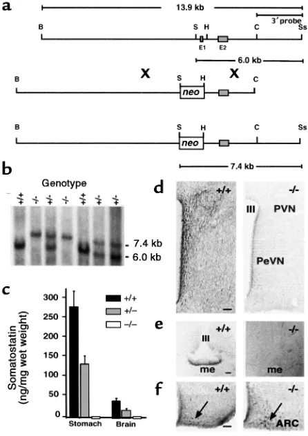
Figure 1
Generation of SST-deficient mice by homologous recombination. (a) Restriction maps of the mouse Smst gene locus (top), targeting vec-tor (middle), and predicted Smst null allele after homologous recom-bination (bottom) indicate the replacement of exon 1 (E1) and the
Smstpromoter by a PGK-neomycin selection cassette (neo). B, BamHI; S, SalI; H, HindIII; C, ClaI; Ss, SspI. (b) A Southern blot of genomic DNA digested with SalI and SspI was hybridized with the 3′probe indi-cated in a. The 6.0-kb band represents the wild-type allele and the 7.4-kb band represents the Smstnull allele in the offspring of an
Smst+/–mating pair. (c) RIA of SST in extracts of stomach and total brain (n = 3) demonstrates a 50% reduction in Smst+/–mice and lev-els below the assay sensitivity in Smst–/–mice. (d) Hemicoronal hypo-thalamic sections at the level of the periventricular nucleus (PeVN) and anterior paraventricular nucleus (PVN) of an Smst+/+and an
Results
Generation of an Smst null allele by homologous recombina-tion. The gene targeting strategy replaced critical Smst
promoter elements and exon 1, which contains the translational start site for preproSST, with a neo selec-tion cassette (Figure 1a). One correctly targeted ES cell clone produced germline-transmitting chimeras that were the founders for a colony of F2 hybrid mice
(129P2/OlaHsd, C57BL/6J) and, subsequently, N5
incipient congenic C57BL/6J mice. The Smstnull allele was transmitted from mating pairs of Smst+/–mice in a Mendelian ratio of 1:2:1 for Smst+/+:Smst+/–:Smst–/– geno-types (Figure 1b). Smst–/–mice of both genetic back-grounds were healthy and fertile and raised normal lit-ters. To verify that the gene mutation resulted in the loss of SST peptides, we analyzed tissue extracts by a RIA that detects both the 14 and 28 amino acid forms of SST. SST-like immunoreactivity was reduced by 50% in stomach and brain from Smst+/–mice and was below the RIA sensitivity in Smst–/–mice (Figure 1c).
Because SST-containing neurons within the hypo-thalamus are most directly involved in neuroen-docrine regulation, we examined this brain area for evidence of compensatory changes secondary to the loss of SST. Immunohistochemistry utilizing an SST-14–directed antiserum revealed a dense plexus of SST immunoreactive fibers and cell bodies within the periventricular (PeVN), paraventricular (PVN), arcu-ate (Arc), and ventromedial (VMH) nuclei of Smst+/+ mice. No specific reaction product was observed in the hypothalami of Smst–/–mice (Figure 1d), which appeared structurally normal based on Nissl and acetylcholinesterase stains (data not shown). A second antiserum raised against an epitope in the prohor-mone region of proSST (29) strongly labeled fibers in the external zone of the median eminence in Smst+/+ but not in Smst–/–mice (Figure 1e). Using a mouse GHRH antiserum, no apparent differences in either the number of GHRH-positive neuronal perikarya or density of fibers in the Arc (Figure 1f) were observed between Smst+/+or Smst–/–mice (30).
Effects of sex and genotype on SST and SSTR expression. Semiquantitative RT-PCR was used to determine whether SST and SSTR1-5 exhibited baseline sexual diergism in the mouse as reported previously for SSTR1 and 2 in rat (11), and whether the complete absence of SST would alter expression of the structurally related peptide CST or SSTRs in a sex-dependent pattern. There was a clear sexual diergism in SST gene expres-sion in wild-type hypothalamus and pituitary gland; male mice had SST mRNA levels more than twofold those of female mice in both tissues, and no expression was detected in the Smst–/–mice (Table 1). In contrast, CST expression was similar between males and females in hypothalamus, undetectable in pituitary, and unchanged in the absence of SST (data not shown).
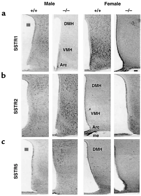
[image:5.576.70.533.578.686.2]A complex pattern of altered SSTR expression was detected in both tissues of Smst–/–mice (Table 1). SSTR1 expression decreased by more than 50% in the hypothalamus of both male and female Smst–/–mice but was unchanged in the pituitaries of these same mice. In contrast to the absence of sexual diergism in SSTR1 expression, there was a significant interaction between sex and genotype on the expression profiles of SSTR2 and 5. SSTR2 mRNA was increased more than twofold in the hypothalamus of Smst–/–males compared with Smst+/+males but was decreased by about 70% in the pituitary of both sexes of Smst–/– mice. SSTR5 mRNA levels increased twofold in the hypothalamus of both sexes of Smst–/–mice in con-trast to the twofold increase in pituitary SSTR5 expression that occurred only in Smst–/–males com-pared with Smst+/+males. There were no differences between sexes or genotypes in SSTR3 and 4 mRNA quantitation (data not shown). An immunocyto-chemical analysis of SSTR expression showed no qual-itative differences in receptor localization between genotypes (Figure 2, a–c). The intensity of immunos-taining closely paralleled the relative mRNA levels, indicating that the semiquantitative RT-PCR accu-rately predicted the changes in SSTR abundance in the hypothalamus.
Table 1
Differential expression of SSTR1, 2, and 5, and SST mRNAs in the hypothalamus and pituitary gland of Smst+/+and Smst–/–mice analyzed by semiquantitative RT-PCR
Gene Tissue Female Smst+/+ Female Smst–/– Male Smst+/+ Male Smst–/–
SSTR1 Hypothalamus 0.941 ± 0.247 0.437 ± 0.072A 0.992 ± 0.104 0.368 ± 0.040A
Pituitary 0.623 ± 0.143 0.440 ± 0.044 0.518 ± 0.087 0.528 ± 0.017
SSTR2 Hypothalamus 0.326 ± 0.070 0.337 ± 0.048B 0.267 ± 0.009 0.571 ± 0.021A
Pituitary 0.132 ± 0.018 0.040 ± 0.005A 0.170 ± 0.035 0.056 ± 0.019A
SSTR5 Hypothalamus 0.066 ± 0.008 0.134 ± 0.019A 0.054 ± 0.009 0.116 ± 0.011A
Pituitary 0.414 ± 0.023B 0.486 ± 0.013A,B 0.492 ± 0.006 0.906 ± 0.017A
SST Hypothalamus 1.917 ± 0.342B ND 4.710 ± 1.11 ND
Pituitary 0.225 ± 0.012B ND 0.533 ± 0.049 ND
SSTR mRNA was quantified from Southern hybridization signal of RT-PCR products by densitometry using the software Vista Scan (version 2.4; UMAX Tech-nologies Inc., Fremont, California, USA) and the total intensity (average optical density times area in pixels) was quantified with the Igor Pro analysis software package (version 3.13; WaveMetrics Inc., Lake Oswego, Oregon, USA). The arbitrary units were corrected for background and expressed as a ratio of β-actin mRNA in the same sample. Data are expressed as the mean ± SEM of four independent experiments. SSTR, somatostatin receptor; SST, somatostatin; AP <
Normal growth despite altered GH secretion in SST-deficient mice. It was apparent from observation of the first litters of F2mice that the absence of SST did not result in
either gigantism or a loss of sexual dimorphism in body size. Growth curves obtained from both F2(data not
shown) and N5mice (Figure 3a) confirmed this
impres-sion, although a gradual increase in body weight due to mild obesity did emerge for male Smst–/–mice after age 18 weeks [repeated measures ANOVA; F(1,14) = 6.1,
P < 0.05 main effect of genotype; F(10,140) = 3.5,
P < 0.001 genotype ×time interaction]. Smst–/– mice exhibited the normal sexual dimorphism in body length that becomes evident during puberty, and there was no significant increase in body length for either sex of
Smst–/–mice compared with their sibling controls (Fig-ure 3b). Organ weights of liver and kidney were also nor-mal in Smst–/–mice (data not shown).
Direct measurement of pulsatile GH in the mouse, unlike the rat, has been hampered by technical difficul-ties of chronic blood sampling in unrestrained animals. In a single reported study, male and female mice showed equivalent pulse amplitudes, but approximately 1.5-fold greater pulse frequency in females (35). Therefore to determine whether SST deficiency was associated with altered GH secretion, we obtained randomly timed blood samples by rapid tail bleeds between the hours of 0500 and 2300 from adult male and female mice of both genotypes over the course of 6 days. A total of 408 plasma samples from 107 mice (26–28 per group) were assayed for GH (Figure 3c). The box plots demonstrate that male and female Smst+/+mice had identical median GH levels of 3 ng/ml. Furthermore, the overall distri-bution of values was similar, with the exception that no female value was greater than 36 ng/ml whereas five of 107 male values ranged from 50 to 122 ng/ml. In marked contrast, the distribution of GH values was shifted higher for both male and female Smst–/–mice, which had medians of 21 and 17 ng/ml, respectively (P < 0.0001, Mann-Whitney Utest for each paired com-parison between Smst+/+and Smst–/–groups). Less than 10% of the random GH values from Smst–/–mice were below 6 ng/ml compared with 75% for both sexes of
Smst+/+mice (χ2= 166.1, P < 0.0001).
Pituitary size was unchanged between the Smst+/+and Smst–/–male and female mice. However, pituitary GH content was reduced by 50% in both sexes of Smst–/–mice compared with sex-matched controls (F(3,14) = 9.12,
P ≤0.01 for each comparison) (Figure 3d), possibly reflecting an altered balance between GH synthesis and release. Despite the changes in plasma and pituitary GH, there was no difference for IGF-1 betweenSmst+/+ and Smst–/–mice [F(1,21) = 0.25, P = 0.6] (Figure 3e).
To facilitate a comparison between the present data and that of MacLeod et al. (35), who directly measured pulsatile GH secretion in cannulated mice at 15-minute intervals over 8-hour periods, we performed Z transformations of our GH data sets and the values from MacLeod’s representative pulsatile GH profiles (three male and three female outbred CD-1 [ICR] mice;
[image:6.576.303.540.54.384.2]n = 99 data points for each sex; see original data in ref. 35). The Z transformation approximates a normal dis-tribution for a data set and then calculates a Z score for each data point that is equivalent to the number of nor-malized SDs relative to the group mean. Each of the six Z-score histograms is skewed to the left (median < mean) and has a tail extending to the right by as much as 6 SDs (Figure 4). By visual inspection, the four Z-score histograms of male and female Smst+/+mice and male and female ICR mice are most similar to each other, whereas the male and female Smst–/–mice appear to constitute a distinct set because of a greater propor-tion of 0 < Z < 1. The small percentages of extreme GH values constituting the positive tails (Z ≥1) represent the peak amplitudes of individual GH pulses in the
Figure 2
MacLeod data sets. Even though the female ICR mice were directly shown to have a greater GH pulse fre-quency than male ICR mice (35), resulting in nominal-ly larger fractions of Z scores ≥1 of 14/99 and 9/99, respectively (Figure 4, lower panels), a χ2test of this
comparison failed to indicate a significant difference (Fisher’s Exact P = 0.38) for the group sizes of n = 99.
Hepatic feminization in male SST-deficient mice. To ana-lyze further the alterations in GH secretory dynamics of the Smst–/–mice, we took advantage of the firmly established role of pulsatile GH in regulating sexual differentiation of the liver (17, 18). Northern blots were used to quantify mRNA levels of several GH-regulated genes, and the data were normalized to 18S RNA
[image:7.576.123.470.55.274.2]con-tent (Figure 5). The male-predominant gene transcripts for both MUPs group I/II and III exhibited levels that were approximately fivefold higher in Smst+/+ male compared with Smst+/+female mice [F(3,20) = 48.40, P < 0.001]. However, there was a dramatic reduction in MUPs expression in Smst–/–male mice to levels indis-tinguishable from females. Identical results were found in a separate cohort of F2hybrid mice (data not shown).
Figure 3
SST-deficient mice retain a normal sexual dimorphism in body size but exhibit altered pituitary GH secretion. (a) Serial body weights of sibling
Smst+/+and Smst–/–mice (n = 16) generated from N
5B6 congenic Smst+/–parents demonstrate normal growth patterns. The small increase in weight
of male Smst–/–mice after 18 weeks of age is due to increased white adipose tissue without organomegaly (n = 8) (data not shown). (b) Nose-to-anus body lengths of the mice in aat age 24 weeks are significantly different between sexes but not between genotypes. (c) Box plots of random-ly obtained serum GH levels from N5B6 congenic mice of each sex and genotype (n = 97–107) demonstrate significant differences among the
median values of the four groups (P < 0.0001, Kruskal-Wallis test). Individual values in the upper and lower 10% of the population distributions are plotted as open circles. Data are expressed as nanograms per milliliter in terms of the mouse GH AFP10783B reference preparation. (d) Pitu-itary GH content at age 20 weeks (n = 4–7) of sibling Smst+/+and Smst–/–mice generated from F
2hybrid Smst+/–parents is decreased by 50% in both
sexes of Smst–/–mice. (e) Serum IGF-1 levels of the mice in dshow no difference between genotypes when the data are collapsed across sex.
Figure 4
Z-score histograms demonstrate similar distributions of GH values in data sets obtained from randomly timed blood sampling (F+/+,
M+/+, F–/–, and M–/–) or from serial blood sampling in cannulated
[image:7.576.324.513.509.731.2]A similar pattern of gene expression across the four groups was found for albumin, although the increase in Smst+/+male compared to Smst+/+female mice was less than twofold [F(3,20) = 6.04, P < 0.01]. In contrast, pro-lactin receptor mRNA levels were more than threefold higher in Smst+/+females compared with Smst+/+males (P < 0.02, one-tailed t test), consistent with previously published results in mice for this typical female-pre-dominant gene (36). The absence of SST resulted in an increase of prolactin receptor expression in Smst–/–male compared with Smst+/+male mice [F(3,20) = 4.91, P < 0.05] but no significant change in Smst–/–female mice. Finally, we analyzed expression of the female predomi-nant Cyp450-15α2A4mRNA in liver (34). Both Smst–/– male and female mice exhibited a significant increase in hybridization signal compared with Smst+/+male mice [F(3,20) = 6.13, P < 0.05 and < 0.01, respectively]. When all five of the sexually diergic hepatic mRNAs are considered together, there is a compelling pattern of feminization in the liver of Smst–/–male mice compared to wild types, without reciprocal changes in the Smst–/– female mice. These differences are not due to alterations in circulating sex steroids in the male mutants because we found no significant differences between genotypes for either serum testosterone [Smst+/+males, 4.6 ± 1.7
ng/ml vs. Smst–/–males, 7.4 ± 2.0 ng/ml; F(1,30)=1.19,
P = 0.28] or estradiol [Smst+/+ males, 2.0 ± 0.6 pg/ml vs.
Smst–/–males, 9.1 ± 4.5 pg/ml; F(1,30)=2.48, P = 0.13].
SST receptors are also localized on pituitary thy-rotrophs and SST can inhibit thyroid-stimulating hor-mone secretion, however serum thyroxine levels did not
differ between sexes or genotypes (Smst+/+ mice, 3.4 ± 0.4
µg/dl vs. Smst–/– mice, 3.7 ± 0.4 µg/dl).
Discussion
SST is synthesized in neural, endocrine and inflammato-ry cells in the brain and peripheinflammato-ry and acts on multiple targets through a family of five receptors, SSTR1-5, to produce a broad spectrum of biologic effects (reviewed in ref. 10). However, many questions still remain concern-ing the physiological function of SST especially in the brain. Our present studies were undertaken to create a mouse model of complete SST deficiency from which we could infer the functions of the peptide by an analysis of the phenotype resulting from its absence. A primary aim was to determine the effects of chronic SST deficiency on GH secretion and test the hypothesis that male SST-defi-cient mice would exhibit a more altered phenotype than females because of the normally greater SST expression in male hypothalamus. Smst–/–mice were investigated on both an F2 hybrid and incipient congenic C57BL/6J
genetic background, and equivalent results were obtained in both strains, indicating that our results are unlikely to have been biased by selective epistatic interactions of the
Smstgene mutation with other gene alleles from either of the genetic backgrounds.
The direct measurement of GH pulsatility in mice represents a formidable challenge and has been report-ed by only one laboratory (35) to our knowlreport-edge. Our approach to this problem relied on obtaining suffi-ciently large data sets (n≅100 per group) of randomly timed plasma GH levels to approximate the true
distri-Figure 5
GH-dependent hepatic gene expression is feminized in male SST-deficient mice. The phosphorimager scans of Northern blots below the graphs show three representative samples from each group. Volumetric quan-titation of each hybridization signal is expressed as a ratio to the corresponding 18S RNA signal in arbitrary units (N5B6 congenic
mice, n = 6). MUPs I, II, and III and albumin mRNA levels are depressed in male Smst–/– mice to levels equivalent with all females. In contrast, levels of the female predominant transcripts for PrlR and CYP2A4 are signifi-cantly increased in male Smst–/–mice to levels equal or greater than female Smst+/+mice. Although the cDNA probe used to hybridize with Cyp450-15α2A4mRNA also detects a related, but not sexually diergic, Cyp450-15α2A5transcript of identical size (34), the increased hybridization signal is presumably due only to a rise in the GH regulated 2A4
[image:8.576.63.341.50.357.2]bution of values that might be obtained by serial meas-urement of GH from cannulated animals in a formal pulse-analysis study. The approach was validated by a comparison of our data from Smst+/+mice with those of the representative GH secretory profiles in wild-type mice published by MacLeod et al. (35) following Z-transformations to normalize each data set (Figure 4). It is apparent that the bona fide pulsatile GH data rep-resented in the bottom two panels have positively skewed (median < mean), non-Gaussian probability distributions. The peak values from GH pulses in both the male and female ICR mice are represented by Z scores ≥1 in the histogram tails. The highly similar dis-tributions of Z scores between the C57BL/6 Smst+/+and MacLeod’s ICR mice suggest that the C57BL/6 Smst+/+ mice secrete GH in a pulsatile rhythm that is similar to the ICR mice and characterized by large amplitude pulses and low interpulse nadir levels in both sexes.
In contrast to the Smst+/+ mice, male and female Smst–/–mice had very few random GH levels < 3 ng/ml and clearly elevated median GH levels, but the maxi-mal amplitude of GH was not changed. The Z-score distributions were similar to the Smst+/+and wild-type ICR mice based on positive skew and the presence of tails ≥1, but differed in their larger fraction of scores between 0 and 1. These data suggest that Smst–/–mice continue to secrete GH in a pulsatile rhythm, although SST is apparently required to inhibit GH secretion between GHRH pulses and thereby maintain low nadir interpulse GH levels. The decreased pituitary content of GH is also consistent with increased secretion of GH into the circulation. However, the Z-transformed data sets do not have sufficient statistical power to conclude whether the frequency of GH pulses is altered in the Smst–/–mice.
Experiments in rats and mice have demonstrated that there are at least two groups of hepatic genes whose sex-ually diergic expression is GH dependent, but which dif-fer in the mechanism of GH regulation. Male-predom-inant genes, including MUPs and the cytochrome P450
Cyp2d9, are directly activated by pulses of GH secretion separated by relatively long intervals during which GH levels are below 1–2 ng/ml (18, 19). This activation of male-predominant genes is prevented by either the absence of circulating GH, or conversely, continuous infusion of GH and subsequent desensitization of the GH receptor-JAK2/STAT5 signaling complex. Interest-ingly, even a subtle increase in GH pulse frequency from six to seven per day, delivered by cannula to hypophy-sectomized rats, does not support the activation of male-predominant genes presumably because of the shortened interpulse interval and suboptimal GH receptor resensitization (18). In male mice, the inter-mittent GH pulses also directly repress expression of female-specific genes including the cytochrome P450
Cyp2a4 by an unknown mechanism (19). Transcrip-tional regulation of these same genes differs in female liver in which there is a constitutive activation of female-specific genes (Cyp2a4) and repression of male-specific
genes (Cyp2d9)that is independent of pituitary GH (37). An important factor contributing to the female pattern is normal expression of estrogen receptor-α, presumably because of its importance in establishing the sexually dimorphic patterns of GH secretion (34).
Male SST-deficient mice exhibit a striking reduction in expression of the male-specific genes encoding MUPs to levels equivalent with normal females. This is clearly not due to the absence of GH, testosterone, or thyroxine and, therefore, is strong confirmatory evi-dence that the normal male pattern of pulsatile GH secretion is altered in the male Smst–/–mice. The trans-formation could be due solely to elevated interpulse levels of GH, consistent with the measured random GH levels. However, it is also possible that GH pulse fre-quency is increased according to the model of Tannen-baum that predicts an intrinsic once-per-hour pul-satility of GHRH in the absence of a SST-mediated short-loop feedback inhibition from GH (1). Further studies using cannulated mice will be necessary to definitively demonstrate altered GH pulse frequency in the SST-deficient mice. That IGF-1 levels are not ele-vated in SST-deficient mice suggests that an increase in time-integrated GH secretion may be counteracted by a decrease in GH responsiveness. A loss of the normal male-like pattern of GH pulsatility is also sufficient to explain the increased expression of Cyp2a4and pro-lactin receptor genes in the male SST-deficient mice because these gene promoters would no longer be sub-ject to the transcriptional repression associated with high-amplitude pulses separated by long interpulse nadirs. However, the supranormal levels of both Cyp2a4
and prolactin receptor mRNAs in female Smst–/– com-pared with Smst+/+mice further suggests the increased activation of these genes by an unidentified mecha-nism in addition to derepression.
for sexual dimorphism in body mass that are sensitive to the absence of STAT5b but not to changes in GH pul-satility. Finally, the deletion of STAT5b has polymorphic effects because it is a component of the signaling com-plex for several tyrosine kinase receptors (20).
Our results also contrast with the dichotomy between GH-dependent liver gene expression and somatic growth reported by Waxman et al. (18) using cannulated rats. They demonstrated that seven GH pulses per day result-ed in increasresult-ed body weight gain over 1 week in both sexes of hypophysectomized rats that was indistin-guishable from normal male rats. As noted above, this GH profile was not recognized by the liver as masculine because it failed to induce expression of the male-specif-ic CYP2C11, nor was it recognized as feminine because it did not stimulate the female predominant steroid 5α-reductase or suppress the male-specific CYP3A2-depend-ent testosterone 6β-hydroxylase. Continuous infusion of GH was required to fully feminize liver gene expression in male rats, but this latter GH profile resulted in body weight gain of the males that was reduced to the normal female level. Therefore, although these data also indicate disassociations among GH secretory profiles, patterns of GH-dependent liver gene expression, and somatic growth, none of the experimentally produced GH pro-files in hypophysectomized rats preserved a sexually dimorphic growth response while simultaneously alter-ing liver gene expression as we observed in the SST-defi-cient mice. These differences might be attributed to true species-specific idiosyncrasies between rat and mouse in their respective GH-hepatic axes or to experimental dif-ferences including the length of time that body weight was recorded or the combined actions of pituitary hor-mones other than GH and their effects on gonadal and adrenal steroid hormones.
Despite the well-established role of SST as the inhibito-ry regulator of GH secretion and an important thera-peutic role of SST-analogues in the treatment of GH-secreting pituitary tumors, SST-deficient mice up to 2 years of age did not exhibit a clinical syndrome of GH excess or develop somatotroph hyperplasia or adenoma. In contrast, overexpression of the stimulatory peptide GHRH in either mice or humans (38, 39) leads to signif-icantly increased numbers of pituitary somatotrophs and a syndrome of gigantism or acromegaly. Taken together, these findings suggest that SST does not play a critical physiological role to tonically inhibit soma-totroph mitogenesis in response to stimulatory growth factors. This situation is unlike the marked proliferation of lactotrophs that occurs after genetic disinhibition from the analogous dopamine and D2 receptor signal-ing pathway (40, 41). Alternatively, the absence of SST may be accompanied by a compensatory decrease in the mitogenic signal from GHRH that was not detected at the level of immunohistochemical detection of GHRH neurons in the hypothalamus.
In summary, we have examined the physiological role of SST in the regulation of GH secretion by a gene-knockout approach. The absence of SST results
in elevated nadir and median GH levels in both sexes and a feminized pattern of GH-dependent gene expression in the liver of male mice. Despite the mag-nitude of these changes, Smst–/–mice do not exhibit increased IGF-1 levels or gigantism, and they retain a normal sexual dimorphism in postnatal somatic growth. Within the hypothalamus and pituitary, the loss of SST had pleiotropic effects on the expression of different SSTR subtypes. Only SSTR2 and SSTR5 exhibited a male-specific increase in mRNA levels in SST-deficient mice, consistent with a higher function-al SST tone in the hypothfunction-alamus of mfunction-ale compared with male mice. We conclude that SST is required to determine the normal secretory profile of GH in both sexes of mice and specifically to masculinize GH secre-tion. However, our data indicate that typical mas-culinized profiles of GH pulsatility and GH-depend-ent hepatic gene expression are not necessary for the greater somatic growth characteristics of male com-pared with female animals after puberty.
Acknowledgments
We thank E.C. Chan for culture of embryonic stem cells, R. Juarez for blastocyst injections and preliminary experiments, D. Robins for advice and the generous gift of MUP oligonucleotides, K. Nusser and S. Lapostelle for technical assistance, R. Lechan and F. Talamantes for antisera, M. Negishi for a Cyp 450-15α4A cDNA, P. Kelly for a PrlRcDNA, D. Hess for steroid hormone RIAs, C. Emerson for the T4 RIA, and R. Cone for a crit-ical reading of the manuscript. This work was sup-ported by grants to M.J. Low from the Lucille P. Markey Charitable Trust, the Medical Research Foundation of Oregon, and the Robert Wood Johnson Foundation, and to Y.C. Patel from the NIH (NS32160) and the Canadian Institutes of Health Research. Requests for mouse GH RIA immunoreagents should be addressed to A.F. Parlow (e-mail: parlow@humc.edu; website: http://www.humc.edu/hormones).
1. Wagner, C., Caplan, S.R., and Tannenbaum, G.S. 1998. Genesis of the ultradian rhythm of GH secretion: a new model unifying experimental observations in rats. Am. J. Physiol. 275:E1046–E1054.
2. Smith, R.G., Feighner, S., Prendergast, K., Guan, X., and Howard, A. 1999. A new orphan receptor involved in pulsatile growth hormone release. Trends Endocrinol. Metab. 10:128–135.
3. Plotsky, P.M., and Vale, W. 1985. Patterns of growth hormone-releasing factor and somatostatin secretion into the hypophysial-portal circula-tion of the rat. Science. 230:461–463.
4. Jansson, J.O., Eden, S., and Isaksson, O. 1985. Sexual dimorphism in the control of growth hormone secretion. Endocr.Rev. 6:128–150. 5. Jaffe, C.A., et al. 1998. Regulatory mechanisms of growth hormone
secre-tion are sexually dimorphic. J. Clin. Invest.102:153–164.
6. Chowen-Breed, J.A., Steiner, R.A., and Clifton, D.K. 1989. Sexual dimor-phism and testosterone-dependent regulation of somatostatin gene expression in the periventricular nucleus of the rat brain. Endocrinology. 125:357–362.
7. Murray, H.E., Simonian, S.X., Herbison, A.E., and Gillies, G.E. 1999. Cor-relation of hypothalamic somatostatin mRNA expression and peptide content with secretion: sexual dimorphism and differential regulation by gonadal factors. J. Neuroendocrinol.11:27–33.
8. Nurhidayat, Tsukamoto, Y., Sigit, K., and Sasaki, F. 1999. Sex differenti-ation of growth hormone-releasing hormone and somatostatin neurons in the mouse hypothalamus: an immunohistochemical and morpho-logical study. Brain Res.821:309–321.
Ontogeny and sexual differentiation of somatostatin biosynthesis and secretion in the hypothalamic periventricular-median eminence path-way. J. Neuroendocrinol.11:35–42.
10. Patel, Y.C. 1999. Somatostatin and its receptor family. Front Neuroen-docrinol.20:157–198.
11. Zhang, W.H., Beaudet, A., and Tannenbaum, G.S. 1999. Sexually dimor-phic expression of sst1 and sst2 somatostatin receptor subtypes in the arcuate nucleus and anterior pituitary of adult rats. J. Neuroendocrinol. 11:129–136.
12. Kimura, N., Tomizawa, S., and Arai, K.N. 1998. Chronic treatment with estrogen up-regulates expression of sst2 messenger ribonucleic acid (mRNA) but down-regulates expression of sst5 mRNA in rat pituitaries. Endocrinology.139:1573–1580.
13. Senaris, R.M., Lago, F., and Dieguez, C. 1996. Gonadal regulation of somatostatin receptor 1, 2 and 3 mRNA levels in the rat anterior pitu-itary. Brain Res. Mol. Brain Res.38:171–175.
14. Painson, J.C., Thorner, M.O., Krieg, R.J., and Tannenbaum, G.S. 1992. Short-term adult exposure to estradiol feminizes the male pattern of spontaneous and growth hormone-releasing factor-stimulated growth hormone secretion in the rat. Endocrinology.130:511–519.
15. Painson, J.C., Veldhuis, J.D., and Tannenbaum, G.S. 2000. Single expo-sure to testosterone in adulthood rapidly induces regularity in the growth hormone release process. Am. J. Physiol. Endocrinol. Metab.278:E933–E940. 16. McIntosh, I., and Bishop, J.O. 1989. Differential expression in male and female mouse liver of very similar mRNAs specified by two group 1 major urinary protein genes. Mol. Cell. Biol.9:2202–2207.
17. Sundseth, S.S., Alberta, J.A., and Waxman, D.J. 1992. Sex-specific, growth hormone-regulated transcription of the cytochrome P450 2C11 and 2C12 genes. J. Biol. Chem.267:3907–3914.
18. Waxman, D.J., Pampori, N.A., Ram, P.A., Agrawal, A.K., and Shapiro, B.H. 1991. Interpulse interval in circulating growth hormone patterns regu-lates sexually dimorphic expression of hepatic cytochrome P450. Proc. Natl. Acad. Sci. USA.88:6868–6872.
19. Udy, G.B., et al. 1997. Requirement of STAT5b for sexual dimorphism of body growth rates and liver gene expression. Proc. Natl. Acad. Sci. USA. 94:7239–7244.
20. Teglund, S., et al. 1998. Stat5a and Stat5b proteins have essential and nonessential, or redundant, roles in cytokine responses. Cell.93:841–850. 21. Davey, H.W., Park, S.H., Grattan, D.R., McLachlan, M.J., and Waxman, D.J. 1999. STAT5b-deficient mice are growth hormone pulse-resistant. Role of STAT5b in sex-specific liver p450 expression. J. Biol. Chem. 274:35331–35336.
22. Davey, H.W., Wilkins, R.J., and Waxman, D.J. 1999. STAT5 signaling in sexually dimorphic gene expression and growth patterns. Am. J. Hum. Genet.65:959–965.
23. Park, S.H., Liu, X., Hennighausen, L., Davey, H.W., and Waxman, D.J. 1999. Distinctive roles of STAT5a and STAT5b in sexual dimorphism of hepatic P450 gene expression. Impact of STAT5a gene disruption. J. Biol. Chem.274:7421–7430.
24. Waxman, D.J., and Chang, T.K.H. 1995. Hormonal regulation of liver cytochrome P450 enzymes. In Cytochrome P450: structure, mechanism, and biochemistry. 2nd edition. P.R. Oritiz de Montellano, editor. Plenum Press.
New York, New York, USA/London, United Kingdom. 391–417. 25. Zheng, H., et al. 1997. Somatostatin receptor subtype 2 knockout mice
are refractory to growth hormone-negative feedback on arcuate neurons. Mol. Endocrinol.11:1709–1717.
26. Kreienkamp, H.J., Akgun, E., Baumeister, H., Meyerhof, W., and Richter, D. 1999. Somatostatin receptor subtype 1 modulates basal inhibition of growth hormone release in somatotrophs. FEBS Lett.462:464–466. 27. Low, M.J., et al. 1985. Tissue-specific posttranslational processing of
pre-prosomatostatin encoded by a metallothionein-somatostatin fusion gene in transgenic mice. Cell.41:211–219.
28. Rubinstein, M., Japon, M.A., and Low, M.J. 1993. Introduction of a point mutation into the mouse genome by homologous recombination in embryonic stem cells using a replacement type vector with a selectable marker. Nucleic Acids Res.21:2613–2617.
29. Lechan, R.M., Goodman, R.H., Rosenblatt, M., Reichlin, S., and Haben-er, J.F. 1983. Prosomatostatin-specific antigen in rat brain: localization by immunocytochemical staining with an antiserum to a synthetic sequence of preprosomatostatin. Proc. Natl. Acad. Sci. USA.80:2780–2784. 30. Phelps, C.J., Dalcik, H., Endo, H., Talamantes, F., and Hurley, D.L. 1993. Growth hormone-releasing hormone peptide and mRNA are overex-pressed in GH-deficient Ames dwarf mice. Endocrinology.133:3034–3037. 31. Kumar, U., Laird, D., Srikant, C.B., Escher, E., and Patel, Y.C. 1997. Expression of the five somatostatin receptor (SSTR1-5) subtypes in rat pituitary somatotrophes: quantitative analysis by double-layer immuno-fluorescence confocal microscopy. Endocrinology.138:4473–4476. 32. Khare, S., et al. 1999. Differential regulation of somatostatin receptor
types 1-5 in rat aorta after angioplasty. FASEB J.13:387–394. 33. Ormandy, C.J., et al. 1997. Null mutation of the prolactin receptor gene
pro-duces multiple reproductive defects in the mouse. Genes Dev.11:167–178. 34. Sueyoshi, T., Yokomori, N., Korach, K.S., and Negishi, M. 1999. Devel-opmental action of estrogen receptor-alpha feminizes the growth hor-mone-Stat5b pathway and expression of Cyp2a4 and Cyp2d9 genes in mouse liver. Mol. Pharmacol.56:473–477.
35. MacLeod, J.N., Pampori, N.A., and Shapiro, B.H. 1991. Sex differences in the ultradian pattern of plasma growth hormone concentrations in mice. J. Endocrinol.131:395–399.
36. Norstedt, G., and Palmiter, R. 1984. Secretory rhythm of growth hor-mone regulates sexual differentiation of mouse liver. Cell.36:805–812. 37. Yoshioka, H., et al. 1990. Characterization and regulation of sex-specif-ic mouse steroid hydroxylase genes. Can. J. Physiol. Pharmacol.68:754–761. 38. Kovacs, M., et al. 1997. Effects of antagonists of growth hormone-releas-ing hormone (GHRH) on GH and insulin-like growth factor I levels in transgenic mice overexpressing the human GHRH gene, an animal model of acromegaly. Endocrinology.138:4536–4542.