Lost in transdifferentiation
Mark H. Hoofnagle, … , Brian R. Wamhoff, Gary K. Owens
J Clin Invest.
2004;
113(9)
:1249-1251.
https://doi.org/10.1172/JCI21761
.
What are the true origins of the smooth muscle cells (SMCs) present in the intimal lesions of
transplant arteriosclerosis? A new study in the
JCI
shows that Sca-1
+cells purified from the
mouse aortic root can migrate through an irradiated vein graft to the neointima of the vessel
and transdifferentiate to express the early SMC differentiation marker gene SM22. Do
Sca-1
+cells transdifferentiate into SMC-like cells, or is activation of SMC marker genes a
consequence of fusion of these cells with preexisting SMCs, a possibility raised by results
of studies of adult stem cells in animal models of liver regeneration ? Or could this be bona
fide transdifferentiation that recapitulates the pathologic processes in humans?
Commentary
Find the latest version:
Lost in transdifferentiation
Mark H. Hoofnagle, Brian R. Wamhoff, and Gary K. OwensDepartment of Molecular Physiology and Biological Physics, University of Virginia, Charlottesville, Virginia, USA.
What are the true origins of the smooth muscle cells (SMCs) present in the
intimal lesions of transplant arteriosclerosis? A new study in the
JCI
shows
that Sca-1
+cells purified from the mouse aortic root can migrate through
an irradiated vein graft to the neointima of the vessel and transdifferentiate
to express the early SMC differentiation marker gene SM22 (see the related
article beginning on page 1258). Do Sca-1
+cells transdifferentiate into
SMC-like cells, or is activation of SMC marker genes a consequence of fusion of
these cells with preexisting SMCs, a possibility raised by results of studies of
adult stem cells in animal models of liver regeneration (see the related article
beginning on page 1266)? Or could this be bona fide transdifferentiation
that recapitulates the pathologic processes in humans?
The prevailing theory of smooth muscle cell (SMC) contribution to vessel lesions is that in pathological states, such as injury and atherosclerosis, SMCs migrate to the intima from the media of the vessel (1). This theory, which has persisted for three decades, is now being challenged by results from models of vessel injury, transplant arteriosclerosis (TA) models, and human allograft studies indicating that a portion of the cells bearing SMC differentiation markers in intimal lesions may have origi-nated from the hematopoietic system and/ or circulating progenitor cells (2–5). How-ever, these studies show variable contribu-tions of marrow-derived cells to lesions, with increased frequency correlated to severity of medial injury or to degree of donor/allograft mismatch (6). It appears that only with necrosis of medial SMCs are bone marrow cell (BMC) investment frequencies extremely high (7), indicat-ing that the marrow is not solely respon-sible for populating the intimal lesion but may represent a default pathway for SMC regeneration and vessel repair in circum-stances of severe vessel wall damage.
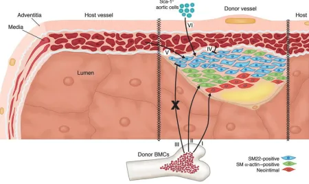
Two seminal studies by Hu et al. provide new insights into the origin of cells popu-lating the developing neointima in a mouse model of TA (8, 9). Using a combination of bone marrow reconstitution and vein allografts (Figure 1), they demonstrated that donor BMCs give rise to neointimal
cells and smooth muscle (SM) α -actin–posi-tive cells (2, 3), although evidence for this is somewhat controversial due to the lack of high-resolution confocal microscopic anal-yses showing definitive colocalization of bone marrow lineage marker genes and SM
α-actin immunostaining (6). However, bone marrow reconstitution studies using donor bone marrow from SM22 promoter-LacZ– transgenic mice have shown that donor BMCs do not give rise to SM22-expressing neointimal cells (2, 3) — a surprising result, since SM22 is an early SMC differentiation marker gene (6). Donor vein graft cells also do not contribute SM22-positive neointi-mal cells, but host vessel–derived cells can give rise to the population of SM22-positive graft neointimal cells. However, it must be noted that the failure of donor vein graft cells to contribute to the intima appears to be unique to this allograft model, wherein there is virtual destruction of all donor vessel cells, including medial SMCs. Thus, neointimal cells that express SM22 do not appear to be bone marrow–derived but rath-er to have some othrath-er host-based source.
In this issue of the JCI, Hu and col-leagues hypothesize that in addition to circulating progenitor cells, Sca-1+
progeni-tor cells that reside in the adventitia may transdifferentiate into SMC-like neointi-mal cells (10). These results provide fresh arguments in a key controversy in adult stem cell and progenitor cell research — i.e., that of defining the relative roles of cell fusion versus transdifferentiation.
The role of adult stem cell contribution to tissues has recently undergone a revolution and counterrevolution. Initial optimism about transdifferentiation of neuronal stem cells (11), hematopoietic stem cells
(HSCs) (12), and other adult stem cell types has diminished in the face of reports of cell fusion (13) and minimal adult stem cell plasticity (14). Surprisingly, the pair of papers that began the furor over cell fusion throughout the adult stem cell field had lit-tle relevance to most cell types being studied. Terada et al. and Ying et al. demonstrated rare cell fusion events when embryonic stem cells are extensively cocultured with BMCs or neural cells, respectively (15, 16). These results, however, did lead to a healthy skep-ticism about the transdifferentiation poten-tial of adult stem cells. Ianus et al. devel-oped a Cre-lox model to specifically test for fusion in their cell systems, and found none, despite the apparent transdifferentiation of BMCs into insulin-secreting pancreatic
β cells (17). Some groups examining BMC transdifferentiation into hepatocytes found that their previous results were mainly due to cell fusion (13). Still others, alerted to the fusion problem, found cell fusion occurred in their systems, but at too low a frequency to account for their transdifferentiation results (18, 19).
Thus, a confusing picture has emerged for most scientists curious about the role of adult stem cells in human dis-ease. We are left with several questions: Is transdifferentiation real or just an arti-fact? Is fusion a problem or is it a potential physiological mechanism to exploit? Are some tissues more susceptible to fusion than others? The liver, for instance, has proven to be highly susceptible to fusion, but, interestingly, in the fumarylacetoacetate hydrolase–/–mouse models studied, fusion
of HSCs to hepatocytes corrects the enzy-matic deficiency, raising the possibility that this phenomenon has therapeutic applica-tions (12). Of note in this issue of the JCI is the work of Camargo et al., who explored the fusibility of hepatocytes with HSCs and discovered that it is specifically the myeloid lineage of these cells, rather than the stem cells themselves, that is responsible for fusion, thus raising the intriguing possibil-ity of “fusion therapy” (20). These ground-breaking new results also emphasize the importance of understanding fusion and transdifferentiation, as they appear to play a potentially important role in several
dis-Nonstandard abbreviations used: bone marrow cell (BMC); hematopoietic stem cell (HSC); smooth muscle (SM); SM cell (SMC); transplant arteriosclerosis (TA).
Conflict of interest: The authors have declared that no conflict of interest exists.
commentaries
ease states that biomedical scientists have been trying to understand for decades.
Adding to the controversy over the true transdifferentiation potential of adult stem cells are the exaggerated claims made by both the scientific and the lay press regarding the relative merits of adult ver-sus embryonic stem cells and their poten-tial for therapeutic applications. Both ave-nues of research would be better served by more accurate portrayals of their science in the press, which might help to lessen the impact of partisan politics on serious con-sideration of the merits of either stem cell type and their potential contributions to science and human therapeutics.
In this issue of the JCI, Hu et al. provide novel evidence that may result in a more coherent picture of different cell contribu-tions to the lesions of TA. Of major inter-est is the fact that Hu et al. isolated a
puta-tive SMC progenitor population from the adventitia of the aortic root with a surface phenotype of Sca-1+/c-kit+/lin–
character-istic of HSCs. Further, through a series of lineage tracing studies, they demonstrated that these cells were not of hematopoietic origin, suggesting they are not HSCs that have lodged or fused in the adventitia. In culture, PDGF-BB induces expression of multiple markers of SMC differentiation, and these cells, when purified from an SM22-LacZ/ApoE–/– transgenic mouse and
applied to the outside of an irradiated vein allograft, migrate to the neointima of the vessel and appear to activate de novo SM22 transcription in the neointima (Figure 1).
These results are exciting and significant in that they identify a potential new origin of cells that contribute to neointimal for-mation in TA. However, caution must be taken, as critical experiments are required
to identify the physiological relevance of these cells and eliminate two limitations of this study. First, application of exoge-nous Sca-1+ cells to an irradiated vein graft
represents an artificial situation in which migration may have been an experimental artifact due to lack of medial SMC contri-bution. To determine if such migration is physiologically relevant, a Sca-1
promoter-Cre mouse could be crossed to a ROSA-stoplox-LacZ/ApoE–/– mouse to determine
if Sca-1 was ever activated in neointimal SMCs. Second, fusion of Sca-1+ cells with
cells from another source, such as irradiat-ed SMCs of the mirradiat-edia, must be eliminatirradiat-ed, although other groups suggest that this is an unlikely eventuality (19). A Cre-lox fusion detection system (17) could be used to quell concerns that Sca-1+ cells adopt the
[image:3.585.65.515.87.355.2]SMC phenotype by fusing to other SMCs rather than by transdifferentiating. Figure 1
From all studies so far we are still left with the fundamental question: Do non-SMCs from the adventitia, host vessel, or bone marrow transdifferentiate to become bona fide SMCs? It is possible that these cells are expressing only a few SMC differentiation markers and/or still expressing non-SMC genes. Finally, and most importantly, we ask, are the results in these animal models relevant to the development of atheroscle-rosis in humans? Can these same cells be identified in humans by Sca-1 antigen? The novel and exciting studies by Hu et al. in this issue of the JCI implicate potential contributions by a putative adventitial pro-genitor cell population in TA and might open yet another chapter in the long quest to define origins of cells in intimal lesions and their mechanistic contributions to the pathogenesis of atherosclerosis.
Mark H. Hoofnagle and Brian R. Wamhoff contributed equally to this work.
Address correspondence to: Gary K. Owens, Department of Molecular Physiology and Biological Physics, University of Virginia, MR5 Room 1220, 415 Lane Road, PO Box 801394, Charlottesville, Virginia 22908,
USA. Phone: (434) 924-2652; Fax: (434) 982-0055; E-mail: [email protected].
1. Ross, R., and Glomset, J.A. 1973. Atherosclerosis and the arterial smooth muscle cell: proliferation of smooth muscle is a key event in the genesis of the lesions of atherosclerosis. Science.180:1332–1339. 2. Sata, M., et al. 2002. Hematopoietic stem cells
dif-ferentiate into vascular cells that participate in the pathogenesis of atherosclerosis. Nat. Med.8:403–409. 3. Shimizu, K., et al. 2001. Host bone-marrow cells are a source of donor intimal smooth-muscle-like cells in murine aortic transplant arteriopathy. Nat. Med.
7:738–741.
4. Glaser, R., Lu, M.M., Narula, N., and Epstein, J.A. 2002. Smooth muscle cells, but not myocytes, of host origin in transplanted human hearts. Circula-tion.106:17–19.
5. Simper, D., Stalboerger, P.G., Panetta, C.J., Wang, S., and Caplice, N.M. 2002. Smooth muscle progenitor cells in human blood. Circulation.106:1199–1204. 6. Owens, G.K., Kumar, M.S., and Wamhoff, B.R.
2004. Molecular regulation of vascular smooth muscle cell differentiation in development and disease. Physiol. Rev. In press.
7. Campbell, J.H., Han, C.L., and Campbell, G.R. 2001. Neointimal formation by circulating bone marrow cells. Ann. N. Y. Acad. Sci.947:18–24.
8. Hu, Y., et al. 2002. Both donor and recipient origins of smooth muscle cells in vein graft atherosclerotic lesions. Circ. Res.91:13e–20e.
9. Hu, Y., et al. 2002. Smooth muscle cells in trans-plant atherosclerotic lesions are originated from recipients, but not bone marrow progenitor cells.
Circulation.106:1834–1839.
10. Hu, Y., et al. 2004. Abundant progenitor cells in the adventitia contribute to atherosclerosis of
vein grafts in ApoE-deficient mice. J. Clin. Invest.
113:1258–1265. doi:10.1172/JCI200419628. 11. Bjornson, C.R., Rietze, R.L., Reynolds, B.A., Magli,
M.C., and Vescovi, A.L. 1999. Turning brain into blood: a hematopoietic fate adopted by adult neu-ral stem cells in vivo. Science.283:534–537. 12. Lagasse, E., et al. 2000. Purified hematopoietic stem
cells can differentiate into hepatocytes in vivo. Nat. Med.6:1229–1234.
13. Wang, X., et al. 2003. Cell fusion is the principal source of bone-marrow-derived hepatocytes.
Nature.422:897–901.
14. Wagers, A.J., Sherwood, R.I., Christensen, J.L., and Weissman, I.L. 2002. Little evidence for develop-mental plasticity of adult hematopoietic stem cells.
Science.297:2256–2259.
15. Terada, N., et al. 2002. Bone marrow cells adopt the phenotype of other cells by spontaneous cell fusion. Nature.416:542–545.
16. Ying, Q.L., Nichols, J., Evans, E.P., and Smith, A.G. 2002. Changing potency by spontaneous fusion.
Nature.416:545–548.
17. Ianus, A., Holz, G.G., Theise, N.D., and Hussain, M.A. 2003. In vivo derivation of glucose-compe-tent pancreatic endocrine cells from bone mar-row without evidence of cell fusion. J. Clin. Invest.
111:843–850. doi:10.1172/JCI200316502. 18. Spees, J.L., et al. 2003. Differentiation, cell fusion,
and nuclear fusion during ex vivo repair of epithe-lium by human adult stem cells from bone marrow stroma. Proc. Natl. Acad. Sci. U. S. A. 100:2397–2402. 19. Saiura, A., et al. 2003. Little evidence for cell fusion
between recipient and donor-derived cells. J. Surg. Res. 113:222–227.
20. Camargo, F.D., Finegold, M., and Goodell, M.A. 2004. Hematopoietic myelomonocytic cells are the major source of hepatocyte fusion partners. J. Clin. Invest. 113:1266–1270. doi:10.1172/JCI200421301.
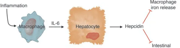
Anemia of inflammation: the cytokine-hepcidin link
Nancy C. AndrewsChildren’s Hospital, Howard Hughes Medical Institute, Harvard Medical School, and Dana-Farber Cancer Institute, Boston, Massachusetts, USA.
The anemia of inflammation, commonly observed in patients with
chronic infections, malignancy, trauma, and inflammatory disorders, is
a well-known clinical entity. Until recently, we understood little about
its pathogenesis. It now appears that the inflammatory cytokine IL-6
induces production of hepcidin, an iron-regulatory hormone that may be
responsible for most or all of the features of this disorder (see the related
article beginning on page 1271).
In 1932, Locke et al. made the important observation that infection was associated with hypoferremia (low serum iron), pro-viding a partial explanation for the com-mon finding of anemia in patients with chronic infections (1). Cartwright and Wintrobe went on to show that the ane-mia associated with infection was indis-tinguishable from the anemia of
inflam-mation, and established that hypoferremia resulted from reticuloendothelial seques-tration of iron and interruption of intesti-nal iron absorption (2, 3). Cartwright and Lee recognized that similar findings could be induced in mice by exposure to bacte-rial endotoxin (4). Others correlated the anemia of inflammation with elaboration of inflammatory cytokines, and ascribed changes in iron metabolism to the effects of these cytokines (5). Cytokines have been shown to modulate the expression of iron transport and storage proteins (6), but it was not clear that these changes accounted
for the abnormalities of iron homeostasis observed in the anemia of inflammation.
The roles of hepcidin and IL-6 Over the past two years, a variety of experi-ments have converged to establish a role for hepcidin, a liver-derived peptide regula-tor of iron homeostasis, as a key mediaregula-tor of hypoferremia in inflammation (7–9). In an elegant report in this issue of the JCI, Nemeth, Rivera, and colleagues have eluci-dated an important link between inflamma-tory cytokines and hepcidin (10). Using both mice and humans as experimental models, they have shown that IL-6 acts directly on hepatocytes to stimulate hepcidin produc-tion. Hepcidin, in turn, acts as a negative regulator of intestinal iron absorption and macrophage iron release.
In previous work Nemeth, Rivera, and colleagues showed that IL-6 induced hepcidin expression in hepatic cells (9).
Conflict of interest: The author has declared that no conflict of interest exists.
commentaries
Here, they have replicated this effect using conditioned medium from endotoxin-treated macrophages and shown that a neutralizing antibody against IL-6 blocked hepcidin induction (10). Other inflamma-tory cytokines did not stimulate hepcidin production; in fact, TNF-α inhibited it.
Cytokines have all sorts of effects on cul-tured cells, and it was important to show that IL-6 induction of hepcidin occurred in vivo and triggered hypoferremia, as pre-dicted. First, Nemeth, Rivera, et al. used turpentine injection to cause inflammato-ry abscesses in wild-type and IL-6 knockout mice and analyzed the responses (10). Wild-type mice had increased hepcidin expres-sion and a substantial decrease in serum iron levels. In contrast, IL-6 knockout mice had no increase in hepcidin expression and no decrease in serum iron. A complemen-tary experiment carried out in human vol-unteers showed that IL-6 infusion stimu-lated urinary hepcidin excretion within 2 hours and induced hypoferremia. Taken together, these data provide strong sup-port for the conclusions that IL-6 is a pri-mary inducer of hepcidin expression and that increased hepcidin expression results in hypoferremia (Figure 1). This is gratify-ingly consistent with clinical observations that hypoferremia occurs very quickly after the onset of inflammation.
Earlier studies had shown that rodents with induced iron overload also had increased hepcidin expression (11, 12), presumably to try to compensate for iron excess. However, the signal to increase hepcidin expression was unknown. Here, Nemeth, Rivera, and colleagues have shown that IL-6 is not involved in the regulation of hepcidin in response to iron (10). Furthermore, their data suggest that hepcidin levels are not simply responding to increased iron stores. In human volun-teers, urinary hepcidin levels were boosted soon after a single dose of oral iron, which should have no significant effect on iron stores. Perhaps the serum iron level, known to increase transiently after iron
inges-tion, might itself be the signal to induce hepcidin expression. Alternatively, the sig-nal might relate to the degree of iron satu-ration of serum transferrin.
However, if transferrin iron saturation modulates hepcidin expression, other sig-nals can clearly override its effects. The IL-6–mediated inflammatory induction of hepcidin does not appear to be offset by the hypoferremia it causes, at least in the short term. And mice with thalassemia interme-dia (which presumably have elevated serum iron) have decreased hepcidin expression (13), as occurs in other mouse models with increased erythroid iron demand (7, 8).
In my opinion, this report from Nemeth, Rivera, et al. (10) leaves little room for doubt about the importance of hepcidin in the pathogenesis of anemia of inflam-mation. This was challenged by a recent report that concluded that elevated serum hepcidin levels were not useful in the diag-nosis of the anemia of inflammation (14). However, that study did not provide data to support the authors’ contention that they had developed a sensitive, specific test for serum hepcidin. Furthermore, as they also pointed out, it was not clear that serum measurements were as useful as uri-nary hepcidin measurements. Hepcidin gene expression seems to be exquisitely sensitive to regulation, and the circulating peptide is small enough to be quantitative-ly filtered by the kidneys. Urine samples probably provide a better indication of recent hepcidin expression than individual serum samples.
A possible treatment?
If inflammatory induction of hepcidin causes hypoferremia, it is logical to pre-dict that inhibition of hepcidin expression or activity would ameliorate the anemia of inflammation. Would that be advanta-geous? Perhaps, particularly in noninfec-tious inflammatory disorders. We know that patients (15) and mice (16) lacking hepcidin have increased intestinal iron absorption and increased serum iron, but
this is unlikely to be harmful in the short term. However, there may be more cause for concern in patients with infections or malig-nancy. Decreased serum iron is believed to contribute to host defense against invad-ing pathogens and tumor cells (17), and hepcidin itself has antimicrobial properties of uncertain importance (18). If hepcidin antagonists become available, careful clini-cal trials will be required to define appropri-ate indications for their use.
Address correspondence to: Nancy C. Andrews, Children’s Hospital, Howard Hughes Medical Institute, and Harvard Medical School, 300 Longwood Avenue, Boston, Massachusetts 02115-5737, USA. Phone: (617) 919-2116; Fax: (617) 730-0934; E-mail: [email protected].
1. Locke, A., Main, E.R., and Rosbach, D.O. 1932. The copper and non-hemoglobinous iron con-tents of the blood serum in disease. J. Clin. Invest.
11:527–542.
2. Cartwright, G.E., and Wintrobe, M.M. 1952. The anemia of infection. XVII. A review. Adv. Intern. Med.5:165–226.
3. Cartwright, G.E. 1966. The anemia of chronic dis-orders. Semin. Hematol.3:351–375.
4. Cartwright, G.E., and Lee, G.R. 1971. The anaemia of chronic disorders. Br. J. Haematol.21:147–152. 5. Means, R.T. 1995. Pathogenesis of the anemia of
chronic disease: a cytokine-mediated anemia. Stem Cells.13:32–37.
6. Ludwiczek, S., Aigner, E., Theurl, I., and Weiss, G. 2003. Cytokine-mediated regulation of iron transport in human monocytic cells. Blood.
101:4148–4154.
7. Weinstein, D.A., et al. 2002. Inappropriate expres-sion of hepcidin is associated with iron refractory anemia: implications for the anemia of chronic dis-ease. Blood.100:3776–3781.
8. Nicolas, G., et al. 2002. The gene encoding the iron regulatory peptide hepcidin is regulated by anemia, hypoxia, and inflammation. J. Clin. Invest.
110:1037–1044. doi:10.1172/JCI200215686. 9. Nemeth, E., et al. 2003. Hepcidin, a putative
media-tor of anemia of inflammation, is a type II acute-phase protein. Blood.101:2461–2463.
10. Nemeth, E., et al. 2004. IL-6 mediates hypoferremia of inflammation by inducing the synthesis of the iron regulatory hormone hepcidin. J. Clin. Invest.
113:1271–1276. doi:10.1172/JCI200420945. 11. Pigeon, C., et al. 2001. A new mouse liver-specific
[image:5.585.55.360.85.167.2]gene, encoding a protein homologous to human antimicrobial peptide hepcidin, is overexpressed during iron overload. J. Biol. Chem.276:7811–7819. 12. Muckenthaler, M., et al. 2003. Regulatory defects
Figure 1
in liver and intestine implicate abnormal hepcidin and Cybrd1 expression in mouse hemochromato-sis. Nat. Genet.34:102–107.
13. Adamsky, K., et al. 2004. Decreased hepcidin mRNA expression in thalassemic mice. Br. J. Hae-matol.124:123–124.
14. Dallalio, G., Fleury, T., and Means, R.T. 2003.
Serum hepcidin in clinical specimens. Br. J. Haema-tol.122:996–1000.
15. Roetto, A., et al. 2003. Mutant antimicrobial peptide hepcidin is associated with severe juvenile hemochromatosis. Nat. Genet. 33:21–22. 16. Nicolas, G., et al. 2001. Lack of hepcidin gene
expression and severe tissue iron overload in
upstream stimulatory factor 2 (USF2) knockout mice. Proc. Natl. Acad. Sci. U. S. A.98:8780–8785. 17. Weinberg, E.D. 1986. Iron, infection and neoplasia.
Clin. Physiol. Biochem.4:50–60.
18. Park, C.H., Valore, E.V., Waring, A.J., and Ganz, T. 2001. Hepcidin, a urinary antimicrobial peptide syn-thesized in the liver. J. Biol. Chem.276:7806–7810.
Hold the antioxidants and improve plasma lipids?
Ronald M. KraussChildren’s Hospital Oakland Research Institute, Oakland, California, USA.
Intrahepatic proteolysis is a major determinant of secretion of
ApoB-con-taining lipoproteins into plasma. Stimulation of post-ER presecretory
proteolysis (PERPP) of ApoB by n-3 polyunsaturated fatty acids has been
found to result in reduced secretion of VLDL particles by hepatocytes. A new
study has shown that this stimulation is promoted by pro-oxidant
condi-tions that result in increased hepatic lipid hydroperoxide content (see the
related article beginning on page 1277). Conversely, PERPP is suppressed by
antioxidants and by saturated fatty acids, which are not susceptible to lipid
peroxidation. Hence reduction of oxidative stress may have the unexpected
side effect of increasing plasma lipid levels.
Nonstandard abbreviations used: ER-associated deg-radation (ERAD); microsomal triglyceride transfer pro-tein (MTP); post-ER presecretory proteolysis (PERPP); thiobarbituric acid–reactive substance (TBARS).
Conflict of interest: The author has declared that no conflict of interest exists.
Citation for this article:J. Clin. Invest.113:1253–1255 (2004). doi:10.1172/JCI200421637.
Dietary fats with differing fatty acid com-position can influence plasma lipid levels by modulating hepatic production and clearance of lipoproteins (1), as well as by altering activity of cholesteryl ester transfer protein (2). Suppression and stimulation of hepatic LDL receptor activity are major determinants, respectively, of the effects of saturated and polyunsaturated fatty acids on plasma LDL clearance (1), but the mech-anisms for effects of specific fatty acids on hepatic lipoprotein production are less well understood. This is in large part due to the multiple influences of fatty acids on pro-cesses that regulate hepatic lipid produc-tion and storage, and processing of ApoB in conjunction with lipoprotein synthesis and secretion (3).
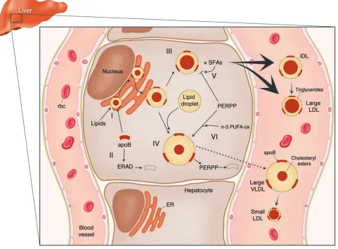
Fatty acids are critically involved in hepatic lipoprotein production pathways that help maintain hepatic cholesterol homeostasis and the ability to respond to energy and other metabolic needs. Tri-glycerides influence a critical early step in secretion of ApoB-containing lipoproteins, namely the cotranslational binding of lip-ids to ApoB in the ER mediated by
micro-somal triglyceride transfer protein (MTP). The resulting protection of specific ApoB domains from proteolysis, termed ER-asso-ciated degradation (ERAD), leads either to secretion of a relatively lipid-depleted par-ticle or to further, posttranslational lipida-tion (3) (Figure 1). The latter may occur in a graded manner in the ER, the vesicular tubular complex, and/or the Golgi appa-ratus, or by fusion with a preformed lipid droplet in the ER by a process that is not dependent on MTP.
Post-ER presecretory proteolysis and hepatic lipid hydroperoxide content Recently, Fisher et al. have identified another degradative process that can mod-ulate hepatic secretion of more mature ApoB-containing lipoproteins (4). They have found that inhibition of hepatic ApoB secretion by n-3 polyunsaturated fatty acids occurs via activation of this process, which has been designated post-ER presecretory proteolysis (Ppost-ERPP) (4). In this issue of the JCI, Pan, Fisher, and colleagues have now shown, using several lines of evidence, that this effect is medi-ated by fatty acid peroxidation, and that it also occurs with n-6 polyunsaturated fatty acids (5). Moreover, the finding that ApoB degradation is stimulated by pro-oxidant conditions and inhibited by antioxidants raises the question of whether variation in intrahepatic oxidative stress contributes to physiologic and/or pathologic modulation
of ApoB-containing lipoprotein metabo-lism. There is indeed abundant evidence that consumption of supplements of lon-ger-chain n-3 marine-derived polyunsatu-rated fatty acids (eicosapentaenoic and docosahexanoic acids) can lower plasma triglyceride and VLDL levels in humans (6). Recently, it has been shown that popu-lation variation in dietary intake of a plant-derived n-3 polyunsaturated fatty acid, linolenic acid, is significantly associated with plasma triglyceride levels, indepen-dent of other nutrients, including longer-chain n-3 polyunsaturated fatty acids (7). As Pan et al. point out, however, in their study linolenic acid suppressed ApoB secre-tion from rat hepatoma cells to a greater extent than could be accounted for by the relation of secretion to intrahepatic lipid hydroperoxide content as assessed by levels of thiobarbituric acid–reactive substances (TBARSs) (5). It should be noted, howev-er, that TBARSs do not represent the full spectrum of products of lipid peroxidation, such as F2-isoprostanes. Moreover, despite the capacity for peroxidation of both n-3 and n-6 polyunsaturated fatty acids, the latter have minimal and generally non-significant effects on plasma triglyceride levels in humans (8). Hence increased lipid peroxidation of n-3 fatty acids does not fully explain the effects of these fatty acids on hepatic ApoB-containing lipoprotein secretion. Other effects, such as reduced lipid synthesis, may also play a role.
Oxidative stress and modulation of pathways for hepatic
lipoprotein secretion
commentaries
lipoproteins from liver cells in conjunc-tion with protecconjunc-tion from a proteolytic process consistent with PERPP (9). Inter-estingly, the secreted lipoproteins under these conditions were denser than VLDLs and were more triglyceride-depleted than those secreted in the presence of albumin or oleic acid. This suggests that differences in conformation or composition of newly synthesized ApoB-containing lipoproteins can alter their exposure to PERPP in such a way that smaller, triglyceride-depleted par-ticles formed under the influence of satu-rated fatty acids tend to escape this process, while larger, triglyceride-rich VLDLs are more susceptible. However, the results of Pan et al. (5) indicate that the stimulation of lipoprotein secretion by myristic acid
is disproportionately greater than would be predicted by reduced hepatic TBARS content, suggesting, as was the case with linolenic acid, that effects other than those related to lipid peroxidation are involved.
[image:7.585.56.537.91.445.2]Many questions remain as to the nature and physiologic importance of the PERPP system, the properties of lipoproteins that affect their susceptibility to PERPP, the mechanisms by which dietary fatty acids can affect this susceptibility, and the means by which oxidation products can suppress PERPP. It will be particularly challenging to assess the extent to which these mecha-nisms may influence metabolism of ApoB-containing lipoproteins in humans. How-ever, clues may be found in evidence that levels and distribution of LDL subspecies Figure 1
Scheme for the roles of intracellular proteolytic processes in regulating pathways for hepatic secretion of ApoB-containing lipoproteins and the modulating effects of saturated and n-3 fatty acids on these processes. Cotranslational lipidation of ApoB protects nascent particles from ERAD. Maturing particles can acquire choleteryl ester and be secreted as IDLs and larger LDLs or can fuse with a preformed lipid droplet to form larger VLDLs. Saturated fatty acids (SFAs) protect the smaller particles from PERPP, leading to increased secretion. Peroxidation products of n-3 poly-unsaturated fatty acids (n-3 PUFA-ox), and perhaps also n-6 polypoly-unsaturated fatty acids (not shown), increase PERPP, leading to decreased secretion of larger VLDLs and hence lower levels of their catabolic products, including small LDL particles.
can serve as markers for intrahepatic path-ways that result in formation of different hepatic secretory products (10) (Figure 1).
evi-dence cited above that such secretion may be facilitated by protection from PERPP. Similarly, increased intake of ω-3 fatty acids has been shown to result in a shift from small, dense LDLs to large, buoy-ant LDLs (14). This effect is likely to be multifactorial, with an important role for reduced cholesteryl ester transfer protein– mediated transfer of triglyceride to LDLs and subsequent lipolysis (10, 13). However, it is also consistent with evidence that larg-er, triglyceride-rich VLDLs are metabolic precursors of smaller LDL particles (10), and with the findings of Pan et al. (5) that increased PERPP reduces hepatic output of particles in this pathway (Figure 1).
A possible role for oxidation products in modulating hepatic lipoprotein secretion in humans can be assessed by determina-tion of whether antioxidant treatment increases plasma transport or concentra-tions of ApoB-containing lipoproteins. In a randomized placebo-controlled study of 20,536 adults with pre-existing vascular disease or diabetes, daily supplementation with 600 mg vitamin E, 250 mg vitamin C, and 20 mg β-carotene resulted in small but statistically significant increases in triglyc-eride (11%), ApoB (5%), and LDL cholesterol (3%), along with a small reduction in HDL cholesterol (15). No changes in triglyceride or LDL levels following antioxidant supple-mentation were found in a much smaller trial, but there was a reduction in the HDL2 cholesterol fraction, which was associated
with reduced benefit on coronary disease endpoints when antioxidants were com-bined with simvastatin plus niacin therapy (16). Thus, although there is considerable evidence for the involvement of oxidative stress in many disease processes, including atherosclerosis, the potential for unintend-ed outcomes of antioxidant therapy should serve as a warning against proceeding with such treatment in the absence of clinical-trial evidence for benefit and safety.
Address correspondence to: Ronald M. Krauss, Children’s Hospital Oakland Research Institute, 5700 Martin Luther King, Jr., Way, Oakland, California 94609, USA. Phone: (510) 450-7908; Fax: (510) 450-7909; E-mail: [email protected].
1. Woollett, L.A., Spady, D.K., and Dietschy, J.M. 1992. Saturated and unsaturated fatty acids inde-pendently regulate low density lipoprotein receptor activity and production rate. J. Lipid Res.33:77–88. 2. Kurushima, H., et al. 1995. Opposite effects on
cho-lesterol metabolism and their mechanisms induced by dietary oleic acid and palmitic acid in hamsters.
Biochim. Biophys. Acta.1258:251–256.
3. Fisher, E.A., and Ginsberg, H.N. 2002. Complexity in the secretory pathway: the assembly and secre-tion of apolipoprotein B-containing lipoproteins.
J. Biol. Chem.277:17377–17380.
4. Fisher, E.A., et al. 2001. The triple threat to nascent apolipoprotein B. Evidence for mul-tiple, distinct degradative pathways. J. Biol. Chem.
276:27855–27863.
5. Pan, M., et al. 2004. Lipid peroxidation and oxi-dant stress regulate hepatic apolipoprotein B degradation and VLDL production. J. Clin. Invest.
113:1277–1287. doi:10.1172/200419197. 6. Kris-Etherton, P.M., Harris, W.S., and Appel, L.J.
2003. Fish consumption, fish oil, omega-3 fatty acids, and cardiovascular disease. Arterioscler. Thromb. Vasc. Biol.23:e20–e30.
7. Djousse, L., et al. 2003. Dietary linolenic acid is inversely associated with plasma triacylglycerol: the National Heart, Lung, and Blood Institute Family Heart Study. Am. J. Clin. Nutr.78:1098–1102. 8. Mensink, R.P., Zock, P.L., Kester, A.D., and Katan,
M.B. 2003. Effects of dietary fatty acids and car-bohydrates on the ratio of serum total to HDL cholesterol and on serum lipids and lipoproteins: a meta-analysis of 60 controlled trials. Am. J. Clin. Nutr.77:1146–1155.
9. Kummrow, E., Hussain, M.M., Pan, M., Marsh, J.B., and Fisher, E.A. 2002. Myristic acid increases dense lipoprotein secretion by inhibiting apoB deg-radation and triglyceride recruitment. J. Lipid Res.
43:2155–2163.
10. Berneis, K.K., and Krauss, R.M. 2002. Metabolic origins and clinical significance of LDL heteroge-neity. J. Lipid Res.43:1363–1379.
11. Dreon, D.M., et al. 1998. Change in dietary satu-rated fat intake is correlated with change in mass of large low-density-lipoprotein particles in men.
Am. J. Clin. Nutr.67:828–836.
12. Krauss, R.M., Lindgren, F.T., and Ray, R.M. 1980. Interrelationships among subgroups of serum lipoproteins in normal human subjects. Clin. Chim. Acta.104:275–290.
13. Packard, C.J., et al. 2000. Apolipoprotein B metabo-lism and the distribution of VLDL and LDL sub-fractions. J. Lipid Res.41:305–318.
14. Calabresi, L., Donati, D., Pazzucconi, F., Sirtori, C.R., and Franceschini, G. 2000. Omacor in famil-ial combined hyperlipidemia: effects on lipids and low density lipoprotein subclasses. Atherosclerosis.
148:387–396.
15. Heart Protection Study Collaborative Group. 2002. MRC/BHF Heart Protection Study of antioxidant vitamin supplementation in 20,536 high-risk indi-viduals: a randomised placebo-controlled trial. Lan-cet.360:23–33.
16. Brown, B.G., et al. 2001. Simvastatin and niacin, antioxidant vitamins, or the combination for the prevention of coronary disease. N. Engl. J. Med.
345:1583–1592.
Dysbindin-1 and schizophrenia:
from genetics to neuropathology
Michael J. Owen, Nigel M. Williams, and Michael C. O’Donovan
Department of Psychological Medicine, University of Wales College of Medicine, Cardiff, United Kingdom.
The gene encoding dysbindin-1 has recently been implicated in
suscep-tibility to schizophrenia. In this issue of the
JCI
, Talbot et al. show that,
contrary to expectations, dysbindin-1 is located presynaptically in
gluta-matergic neurons and is reduced at these locations in schizophrenia (see
the related article beginning on page 1353). Further studies of dysbindin-1
and the proteins with which it interacts can be expected to throw light on
the pathogenesis of schizophrenia.
Nonstandard abbreviations used: hippocampal formation (HF).
Conflict of interest: The authors have declared that no conflict of interest exists.
Citation for this article:J. Clin. Invest.113:1255–1257 (2004). doi:10.1172/JCI200421470.
Schizophrenia is a common, severely dis-abling, mental disorder (1), and understand-ing its etiology and pathogenesis is one of the most important challenges facing psy-chiatry. Despite great endeavor, achieving this has proven difficult given the absence
commentaries
neurotransmission in addition to classical hyperdopaminergic explanations (4).
The most robust body of evidence regard-ing etiology comes from genetic epidemio-logical studies, which show that individual differences in liability are predominantly genetic, with heritability estimates around 80% (5). The most common mode of trans-mission is probably oligogenic, polygenic, or a mixture of the two (5), but the number of loci, the disease risk conferred by each, the extent of genetic heterogeneity, and the degree of interaction among loci are unknown. As for other common genetic disorders, the small effect on susceptibility conferred by any given locus makes iden-tifying susceptibility genes by positional
genetics difficult. However, as sample sizes and hence power have increased, convinc-ing linkages to a number of chromosomal regions have emerged (6). Moreover, sys-tematic examinations of several of these linkage regions have produced replicated evidence implicating specific schizophre-nia susceptibility genes (6, 7).
Evidence that DTNBP1 is a
susceptibility gene for schizophrenia Currently, the best-supported susceptibil-ity gene is DTNBP1 (e.g., refs. 8–10),which encodes dysbindin-1 and is located within chromosome 6p22.3. Dysbindin-1 is a 40–50 kDa protein that binds both α- and β -brevin, which are components of the
phin glycoprotein complex (11). The dystro-phin complex is found in the sarcolemma of muscle (11) but is also located in postsynap-tic densities in a number of brain areas (12). Although its functions are largely unknown, its location initially suggested that genetic variation in DTNBP1 might confer risk of schizophrenia by mediating effects on post-synaptic structure and function (8).
Despite the strong evidence implicating
DTNBP1, in the absence of schizophrenia-associated changes that alter the amino acid sequence of dysbindin-1, the actual susceptibility variants remain unknown. It even remains formally possible that these lie within an adjacent gene, but it is more likely that variation within DTNBP1 affects mRNA expression or processing. The latter possibility is indirectly supported by evi-dence for as yet unknown cis-acting poly-morphisms affecting DTNBP1 expression in human brain (13).
Presynaptic reductions of dysbindin-1 in schizophrenia
In this issue of the JCI, Talbot and col-leagues have tested this hypothesis by examining dysbindin-1 protein levels in human brain (14). Their findings provide several novel and important insights. First, while they confirmed fairly widespread neuronal distribution of dysbindin-1 and
β-dystrobrevin, they also showed presyn-aptic localization of dysbindin-1 but not
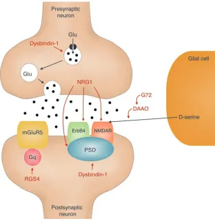
β-dystrobrevin, suggesting that dysbindin-1 plays a presynaptic role in the HF that is independent of β-dystrobrevin and the dystrophin glycoprotein complex. Second, they found high levels of dysbindin-1 in the cells providing the intrinsic glutamatergic pathways of the HF and an inverse corre-lation with VGlutT-1, the main vesicular glutamate transporter present in the HF. This supports a relationship between dys-bindin-1 and glutamate neurotransmission and suggests an effect on VGlutT-1 expres-sion, synthesis, or degradation in the HF. Third, they found a significant reduction in dysbindin-1 expression at these hippo-campal presynaptic sites in two separate populations of patients with schizophre-nia. If correct, these findings suggest that, in the HF at least, dysbindin-1 may influ-ence schizophrenia risk through presynap-tic mechanisms that are independent of the dystrophin glycoprotein complex.
[image:9.585.52.364.82.403.2]The authors have tried hard, by careful matching of cases and controls, to mini-mize the potential impact of many of the possible confounding variables that bedevil Figure 1
studies of postmortem brain. Most patients with schizophrenia have taken psychotro-pic medication for many years, and post-mortem material from drug-naive patients is extremely difficult to obtain. Talbot and colleagues showed that dysbindin-1 and VGlutT-1 levels were not correlated with antipsychotic drug dose in the month before death and provide evidence against an effect of chronic drug treatment from studies of chronic haloperidol administra-tion to mice (14). However, as the authors acknowledge, the validity of drug-treated mice as a control is open to question, and also no mention is made of matching of cases and controls for agonal state. Given the difficulties confirming previous post-mortemfindings in schizophrenia, it is now imperative that multiple independent replications be obtained. From the genetic perspective, it would also strengthen the mechanistic case of Talbot and colleagues if disease-associated single nucleotide poly-morphisms or haplotypes can be linked to effects on gene expression. Here, ethnic variation in risk haplotypes and possible allelic heterogeneity within populations might prove problematic.
Dysbindin-1 and the pathogenesis of schizophrenia
These findings raise a number of impor-tant issues. First, what is the presynaptic function of dysbindin-1? A possible clue comes from recent evidence implicat-ing it as a member of a protein complex involved in the trafficking of lysosome-related organelles in association with the proteins muted, pallidin, and cappuccino (15). As Talbot et al. point out, lysosomes are found presynaptically in the HF, and reduced lysosomal trafficking might result in elevated VGlutT-1 (14). However, further studies are required to identify dysbindin-1 interactors and to understand the control of DTNBP1 expression in brain. In addi-tion, analysis of the mouse mutant sandy (sdy), which, in its homozygous form, expresses no dysbindin-1 protein due to a
large deletion in Dtnbp1, and of conditional knockouts might also be expected to throw light upon the function of dysbindin-1 in brain. Second, Talbot and colleagues stud-ied a restricted number of brain regions, particularly the HF. Dysbindin-1 is widely expressed in brain (11), and many lines of evidence suggest widespread involvement of different brain areas in the structural and functional abnormalities of schizo-phrenia (3). There is now a need to deter-mine whether reduced dysbindin-1 levels are found in other brain areas and whether the association with glutamate is general-ized or specific to the relatively restricted population of hippocampal regions impli-cated by Talbot and colleagues. Interest-ingly, Talbot and colleagues report region-al variation in the distribution of the two dysbindin isoforms studied: the HF and cerebral cortex contained both 50 and 40 kDa isoforms while the cerebellar cortex contained only the 50 kDa variant, imply-ing that there are differences in their tran-scriptional control.
The identification of a potential role for dysbindin-1 in glutamate transmission is of considerable interest. Evidence for glu-tamate dysfunction in schizophrenia is accumulating from both basic and clinical research (4). Moreover, a case can be made that other susceptibility genes identified on the basis of positional genetic studies, such as those encoding Neuregulin 1, G72, D-amino acid oxidase, and regulator of G protein signalling 4, may have convergent effects upon glutamate synapses (Figure 1), although the links are speculative and several caveats remain (7).
Finally, if these findings are confirmed, they will indicate the potential power of positional genetics to home in on novel mechanisms. There are other linkages in schizophrenia that are as strong as that to 6p22.3, indicating the location of other susceptibility genes (6). On the basis of Talbot and colleagues’ work (14) we can expect their identification to yield further crucial advances.
Address correspondence to: Michael J. Owen, Department of Psychological Medi-cine, Henry Wellcome Building, Univer-sity of Wales College of Medicine, Heath Park, Cardiff CF14 4XN, United Kingdom. Phone: 44-920 74 32 48; Fax: 44-920 74 65 54; E-mail: [email protected].
1. Gottesman, I.I. 1991. Schizophrenia genesis: the origins of madness. Freeman. New York, New York, USA. 296 pp. 2. Weinberger, D.R. 1995. From neuropathology to
neurodevelopment. Lancet. 346:552–557. 3. Harrison, P.J. 1999. The neuropathology of
schizo-phrenia. A critical review of the data and their inter-pretation. Brain. 122:593–624.
4. Moghaddam, B. 2003. Bringing order to the gluta-mate chaos in schizophrenia. Neuron. 40:881–884. 5. Owen, M.J., O’Donovan, M.C., and Gottesman, I.I.
2002. Schizophrenia. In Psychiatric genetics and genom-ics. P. McGuffin, M.J. Owen, and I.I. Gottesman, edi-tors. Oxford University Press. New York, New York, USA. 247–266.
6. O’Donovan, M.C., Williams, N.M., and Owen, M.J. 2003. Recent advances in the genetics of schizophre-nia. Hum. Mol. Genet. 12:R125–R133.
7. Harrison, P.J., and Owen, M.J. 2003. Genes for schizophrenia? Recent findings and their pathologi-cal implications. Lancet. 361:417–419.
8. Straub, R.E., et al. 2002. Genetic variation in the 6p22.3 gene DTNBP1, the human ortholog of the mouse dysbindin gene, is associated with schizo-phrenia. Am. J. Hum. Genet. 71:337–348.
9. Schwab, S.G., et al. 2003. Support for association of schizophrenia with genetic variation in the 6p22.3 gene, dysbindin, in sib-pair families with linkage and in an additional sample of triad families. Am. J. Hum. Genet.72:185–190.
10. Williams, N.M., et al. 2004. Identification in two independent samples of a novel schizophrenia risk haplotype of the dystrobrevin binding protein gene (DTNBP1). Arch. Gen. Psychiatry. 61:336–344. 11. Benson, M.A., Newey, S.E., Martin-Rendon, E.,
Hawkes, R., and Blake, D.J. 2001. Dysbindin, a novel coiled-coil-containing protein that interacts with the dystrobrevins in muscle and brain. J. Biol. Chem.
276:24232–24241.
12. Blake, D.J., Hawkes, R., Benson, M.A., and Beesley, P.W. 1999. Different dystrophin-like complexes are expressed in neurons and glia. J. Cell Biol.
147:645–657.
13. Bray, N.J., Buckland, P.R., Owen, M.J., and O’Donovan, M.C. 2003. Cis-acting variation in the expression of a high proportion of genes in human brain. Human Genet. 113:149–153.
14. Talbot, K., et al. 2004. Dysbindin-1 is reduced in intrinsic, glutamatergic terminals of the hippo-campal formation in schizophrenia. J. Clin. Invest.
113:1353–1363. doi:10.1172/JCI200420425. 15. Li, W., et al.2003. Hermansky-Pudlak syndrome type