0095-1137/96/$04.0010
Copyrightq1996, American Society for Microbiology
Immunomagnetic Separation and Solid-Phase Detection
of Bordetella pertussis
MALIN STARK,
1ELISABET REIZENSTEIN,
2MATHIAS UHLE
´ N,
1AND
JOAKIM LUNDEBERG
1*
Department of Biochemistry, KTH, Royal Institute of Technology,
1and Swedish Institute for Infectious
Disease Control,
2Stockholm, Sweden
Received 18 August 1995/Returned for modification 26 October 1995/Accepted 27 December 1995
In the present study, novel solid-phase methods were used for both sample preparation and PCR detection
of
Bordetella pertussis
. The sample preparation was performed by immunomagnetic separation with
paramag-netic beads coated with polyclonal antibodies directed toward the surface antigens of the bacteria. The
precoated immunobeads were directly used on nasopharyngeal aspirates to capture the bacteria on the solid
support and were subsequently transferred to the PCR tube with no further manipulations. The region
encompassing the pertussis toxin promoter was analyzed to allow direct discrimination between the three
major
Bordetella
species (
B. pertussis
,
B. parapertussis
, and
B. bronchiseptica
). The resulting amplicons were
captured on a second magnetic solid phase, allowing detection and restriction analysis of the target sequence.
A colorimetric detection system based on a DNA binding fusion protein enabled the use of standardized
enzyme-linked immunosorbent format tests both for the detection of
Bordetella
spp. and for species evaluation.
When the optimized system was evaluated on 55 clinical aspirate samples, 21 of 22 (95%) culture-positive
samples were positive by the system that we developed. In addition, two samples were positive by the
PCR-based assay, while the culture assay was negative. The implications of these results are discussed.
The bacterium Bordetella pertussis is the causative agent of
whooping cough, which is an infectious disease that occurs
worldwide with a high incidence among young, unvaccinated
infants and occasional mortality. Related species are B.
para-pertussis, which causes a milder respiratory infection, and B.
bronchiseptica, which primarily infects animals. Traditionally,
the laboratory diagnosis of pertussis infection is performed by
culture, and this is sometimes combined with serology (11, 26).
Culture, whose use is possible only in the initial phase of the
infection, is highly specific when the colonies are verified by
agglutination and biochemical tests. However, the sensitivity is
poor and decreases with the duration of time after onset, and
under optimal conditions sensitivity is often only 50%
com-pared with that of serology (12, 26). The sensitivity is further
decreased by prolonged sample transport, vaccination, and
antibiotic treatment of the patient. In addition, culture takes 3
to 7 days to complete (26). Serology is considered highly
sen-sitive (26), but there are two major drawbacks; it cannot be
used in the acute phase of the disease, and it can be difficult to
distinguish between vaccine effects and a pertussis infection.
Moreover, the method is considered less reliable in infants.
Thus, there is a need for more rapid diagnostic methods with
high degrees of specificity and sensitivity, preferably for use
early in infection.
Several DNA amplification-based methods for the detection
of Bordetella spp. have been developed; these methods amplify
insertion sequences (1, 7, 13, 16, 21, 33, 37), the adenylate
cyclase gene (6), or the porin gene (20). Amplification of
in-sertion sequences is usually specific for B. pertussis, but it
sometimes also amplifies B. bronchiseptica DNA, and
amplifi-cation of the adenylate cyclase gene cannot distinguish
be-tween the three Bordetella species, while use of the porin gene
can differentiate B. pertussis from B. parapertussis or B.
bron-chiseptica. The pertussis toxin promoter region has also been
used in B. pertussis-specific PCR-based assays (2, 8, 13, 16, 30)
or to differentiate between B. pertussis, B. parapertussis, and B.
bronchiseptica (27, 28). The amplification of pathogen-derived
DNA in clinical specimens often requires initial sample
prep-aration both to concentrate the sample in a small volume
suitable for PCR and to remove polymerase inhibitors present
in crude samples. Immunomagnetic separation (IMS) has been
shown to be effective in recovering pathogens from urine,
fe-ces, blood, and food (14, 15, 19, 25, 31, 32).
In the study described here we investigated the possibility of
using IMS for the isolation of Bordetella spp. from
nasopha-ryngeal aspirates prior to PCR amplification. The diagnostic
procedure includes a colorimetric detection system, which
avoids the need for electrophoresis. The integrated concept
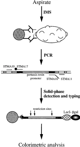
(Fig. 1) consists of (i) IMS, (ii) PCR, and (iii) detection of
immobilized amplified nucleic acid (DIANA) (34) in an
en-zyme-linked immunosorbent assay (ELISA) format together
with (iv) typing of bacteria by restriction enzyme analysis (27).
In the present study, only B. pertussis and B. parapertussis were
detected, since the detection of B. bronchiseptica is of minor
importance for the diagnosis of disease in humans (35). Here
we show that the integrated system can be used to capture and
detect Bordetella bacteria in clinical samples. The amplification
of single Bordetella genomes in a homogeneous lysate is
dem-onstrated, as is the isolation of bacteria by IMS from bacterial
cultures. Fifty-five nasopharyngeal aspirates were investigated
by the described approach (see Table 1).
MATERIALS AND METHODS
Patient material.Fifty-five frozen nasopharyngeal aspirates from patients par-ticipating in a phase III pertussis vaccine efficacy trial (10) and with suspected pertussis infection were obtained from The Swedish Institute for Infectious Disease Control. Twenty-two of them were positive by culture (Table 1). The numbers of colonies on the plates were estimated in four orders of magnitude: 1 to 10, 10 to 100,.100, and confluent growth. A negative aspirate sample was divided and spiked with known amounts of B. pertussis and B. parapertussis bacteria to analyze the efficiency of capture by IMS in more viscous samples compared with that in pure bacterial cultures.
* Corresponding author. Mailing address: Department of Biochem-istry, KTH, Royal Institute of Technology, 100 44 Stockholm, Sweden. Phone: 46-8-790 87 58. Fax: 46-8-24 54 52. Electronic mail address: [email protected].
778
on May 15, 2020 by guest
http://jcm.asm.org/
Bacterial strains and crude lysates.The sensitivity of the PCR was determined by using bacterial strains (B. pertussis patient isolate 324 and B. parapertussis ATCC 15237) serially diluted (1:10) in phosphate-buffered saline (PBS) buffer (10 mM phosphate buffer [pH 7.5], 150 mM NaCl), and the bacteria were quantified by determining the viable counts. These pure cultures were also used for optimization of the IMS procedure and as a positive PCR control. The template used during optimization of PCR conditions was a serially diluted B.
pertussis ATCC 9797 lysate (cultures and lysate were also obtained from The
Swedish Institute for Infectious Disease Control), which was quantified as de-scribed above. This also gave an estimation of the detection limit (number of CFU) of the PCR step.
Oligonucleotide synthesis.Two sets of PCR oligonucleotides (outer oligonu-cleotides, STMA10-STMA11; inner oligonuoligonu-cleotides, STMA17-STMA18 [see Fig. 2]) were synthesized by phosphoramidite chemistry on an automated DNA synthesis machine (Gene Assembler Plus; Pharmacia Biotech, Uppsala, Sweden) according to the manufacturer’s manual. Primer STMA17 was synthesized with a biotin amidite (Biodite; Pharmacia Biotech) at its 59end. All primers were purified by fast-performance liquid chromatography on a 5/5 pepRPC column (Pharmacia Biotech). The four primers are complementary to sequences in the promoter region of the pertussis toxin operon, which contains nucleotide varia-tions in B. parapertussis and B. bronchiseptica, resulting in different recognition sites for restriction enzymes in the three species (22).
IMS.Two polyclonal serum samples, rabbit anti-B. pertussis (Difco Laborato-ries, Detroit, Mich.) and rabbit anti-B. parapertussis (Difco Laboratories), were separately mixed with precoated magnetic beads (Dynabeads M-280 sheep anti-rabbit immunoglobulin G; Dynal AS, Oslo, Norway) and were incubated in PBS with gentle rotation for 30 min at room temperature. The amounts of precoated beads and polyclonal sera were 100mg (10ml) and 50ml of a 1:100 dilution, respectively, per sample for each serum sample. The excess of unbound antibody was removed by washing the bead-antibody complexes with PBS. The previously washed bead-antibody complexes were then mixed and incubated with the bac-terial culture in PBS in a total volume of 100ml. When IMS was performed on nasopharyngeal aspirates, the volume was larger (up to 800ml). The beads with the immunocaptured bacteria were washed twice with PBS and were resus-pended in 10ml of 10 mM Tris-HCl (pH 8.3). Occasionally, the resuspended beads were frozen before analysis by PCR.
In vitro amplification.A 300-bp fragment of the pertussis toxin promoter region was amplified with two sets of oligonucleotide primers in a nested pro-cedure (see Fig. 2). The PCR mixture in both the inner and the outer amplifi-cations consisted of 10 mM Tris-HCl (pH 8.3), 3 mM MgCl2, 50 mM KCl, 0.1%
Tween 20, 0.2 mM (each) deoxynucleoside triphosphates, 0.1mM (each) primer, and 1 U of AmpliTaq DNA polymerase (Perkin-Elmer, Norwalk, Conn.) in total volumes of 50ml (outer) and 75ml (inner). The mixture was covered with two drops of light white mineral oil (Sigma Chemical Co., St. Louis, Mo.) as a barrier to prevent contamination, and 10ml of resuspended beads was added through the layer of mineral oil. The PCR was performed with a PE9600 instrument (Perkin-Elmer). The first amplification step was run with the outer primer pair (STMA10-STMA11). The cycle conditions for the outer amplification were per-formed at 948C for 5 min; this was followed by 35 cycles of 968C for 30 s, 648C for 30 s, and 728C for 1 min, with a final extension at 728C for 10 min. A total of 5ml of the outer amplification product was used as template in the subsequent inner amplification step with the inner primer pair (STMA17-STMA18), gener-ating the specific 300-bp inner fragment. The cycle conditions used were the same as those used during the outer amplification, except that an annealing temperature of 688C was used. Water and B. pertussis lysate were used as negative and positive PCR controls, respectively. The amplified product (5ml) was analyzed by agarose gel electrophoresis and by the DIANA system (15ml). To avoid cross-contamination, three separate rooms were used for mixing the reagents, the addition of template, and post-PCR work. Separate pipettes were used, together with aerosol-resistant tips (Molecular Bio-Products Inc., San Diego, Calif.). Two types of negative controls were used: one was processed in parallel with the samples to check cross-contamination during immunomagnetic sample preparation and one was processed to check the PCR solutions. Ampli-fication of clinical samples was performed blinded, without knowledge of the culture results.
DIANA.The solid phase used for analysis in the DIANA consisted of magnetic beads with covalently coupled streptavidin (Dynabeads M-280 Streptavidin; Dy-nal AS). The magnetic beads (30ml; 300mg) were mixed with 15ml of the PCR mixture and 50ml of LacI–b-galactosidase fusion protein (Dynal AS) in a mi-crotiter plate (Sero-Wel; Bibby Sterilin Ltd., Stone, United Kingdom), and the mixture was incubated for 15 min at room temperature. By applying a microtiter plate magnet (MPC 96; Dynal AS), the bead-DNA-fusion protein complexes were separated and washed four times with 150ml of DIANA buffer (0.1 M Tris-HCl [pH 7.5], 0.15 M NaCl, 0.1% Tween 20, 1 mM MgCl2, 10 mMb
-mer-captoethanol). After the last washing step, the beads were resuspended in 50ml of DIANA buffer, and 50ml of substrate solution (o-nitrophenyl-b-D-galactoside [ONPG]; 1.25 mg/ml) was added. After 5 min, 90ml of the reaction mixture was transferred to a well containing 100ml of stop solution (1 M Na2CO3[pH 12])
and the change in the A405at room temperature was measured in an ELISA plate
reader (EAR 340 AT; SLT Instruments, Salzburg, Austria).
Colorimetric restriction enzyme analysis.Samples positive by DIANA (or by agarose gel electrophoresis) can be treated with restriction enzymes to identify
individual species (27). This was performed by dividing into aliquots 45ml of amplified product immobilized onto the streptavidin-coated magnetic beads and placing the aliquots into three vials for treatment with TaqI (Gibco, Life Tech-nologies), which cleaves only the amplified sequences from B. pertussis and B.
bronchiseptica; with BglI (Pharmacia), which cleaves only those from B. para-pertussis; and with HaeII (Boehringer, Mannheim, Germany), which cleaves B. parapertussis and B. bronchiseptica amplicons (see Fig. 2). The DNA immobilized
onto the beads was restricted according to the manufacturer’s recommendations. The beads were then mixed with the LacI–b-galactosidase fusion protein, and the DIANA analysis was continued as described above.
Cloning and expression of a LacI–b-galactosidase fusion protein.In the initial experiments, a previously reported LacI–b-galactosidase fusion protein (34) was used. The fusion protein was based on a genomic fusion between the Escherichia
coli lacI and lacZ genes in strain XA90/D14 (5). To improve protein production, a plasmid construct encoding recombinant LacI–b-galactosidase was created.
The gene fusion was assembled between the lacI gene encoding the DNA-binding lac repressor molecule and the lacZ gene encoding the enzymeb -galac-tosidase. E. coli RR1DM15 (29) and LG90 (9) were used as bacterial hosts, and the plasmid vectors used were pRIT28 (18), pRIT34 (23), and pSKS106 (4). First, the lacI gene was isolated from pRIT34 by PCR cloning with the oligonu-cleotides STMA4 (59-GCT AAG CTT GAA ACC AGT AAC GTT ATA CGA TG-39) and LUJO (59-ATT CCC GGG GAT CCT CTG CCC GCT TTC CAG-39), omitting the stop codon of lacI. The lacI gene was cloned into pRIT28, and its sequence was verified by solid-phase sequencing (17). The gene was then released with HindIII and BamHI and was inserted into similarly digested pSKS106, in frame with its lacZ gene. The resulting construction, pRIT35, encodes the lacI-lacZ fusion gene of 4,185 bp.
E. coli LG90 (lac mutant) harboring plasmid pRIT35 was grown in baffled
[image:2.612.348.514.74.376.2]Erlenmeyer flasks containing tryptic soy broth (30 g/liter; Difco) to which yeast extract (7 g/liter; Difco) and ampicillin (100mg/ml) were added. At an optical density at 600 nm of 1, the culture was divided into two parallel batches; one was induced with 1 mM n-isopropyl-b-D-thiogalactopyranoside (IPTG) and the other was incubated without IPTG. The cells were harvested 2 h after induction. After washing of the cell pellets in TS buffer (0.1 M Tris-HCl [pH 7.5], 0.15 M NaCl), the cells were disrupted by sonication in TST buffer (25 mM Tris-HCl [pH 8.0], 0.2 M NaCl, 0.05% Tween 20, 1 mM EDTA) containing 10 mMb -mercapto-ethanol. A comparison of the plasmid-encoded LacI–b-galactosidase and the previously used protein produced by the mutant strain XA90/D14 was per-formed.
FIG. 1. Principle of the IMS-PCR-DIANA method.bgal,b-galactosidase.
on May 15, 2020 by guest
http://jcm.asm.org/
Theb-galactosidase activity was determined in the supernatant after sonica-tion by a colorimetric procedure with ONPG as the substrate. In addisonica-tion, the DNA binding property of the fusion protein was tested by incubating the super-natants with streptavidin-coated magnetic beads with immobilized DNA con-taining the lac operator sequence, and after washing away unbound proteins, ONPG was added and the change in the A405was measured.
RESULTS
Sample preparation and PCR analysis.
The approach used
for the detection of Bordetella bacteria in nasopharyngeal
as-pirates (outlined in Fig. 1) was based on the enrichment and
purification of bacteria through IMS (14, 15, 19, 25, 31, 32).
Two polyclonal rabbit sera directed against B. pertussis and B.
parapertussis were used as primary antibodies. The antibodies
were bound to a secondary anti-rabbit antibody, which was
covalently coupled to the magnetic bead. The
immunocap-tured bacteria were subsequently used, without further
purifi-cation, in a nested PCR. The outer primers (primers STMA10
and STMA11) were completely complementary to sequences
found in the promoter region of the pertussis toxin operon,
while one of the inner primers (primer STMA17) was labelled
with biotin, and the other inner primer (primer STMA18)
contained the lac operator as a handle sequence (Fig. 2).
Fol-lowing PCR amplification, simultaneous binding of the PCR
product onto streptavidin-coated magnetic beads and binding
of the recombinant lac repressor–
b
-galactosidase fusion
pro-tein was performed. Positive samples, with the lac operator
sequence incorporated by PCR, were colorimetrically detected
after the addition of the chromogenic
b
-galactosidase
sub-strate ONPG (see Fig. 5). Furthermore, this assay was
com-bined with species identification on the basis of restriction
enzyme analysis of PCR products immobilized onto the solid
support, allowing a colorimetric determination of Bordetella
species in a given sample (Fig. 3B).
Performance sensitivity and specificity of the PCR step.
The
nested PCR primers were designed to be complementary to
sequences in the promoter region of the pertussis toxin gene
(Fig. 2). This gene has a high degree of homology between the
different Bordetella species analyzed and has previously been
successfully used for the detection of Bordetella species (2, 8,
13, 16, 27, 28, 30). The specificities of the PCR primers were
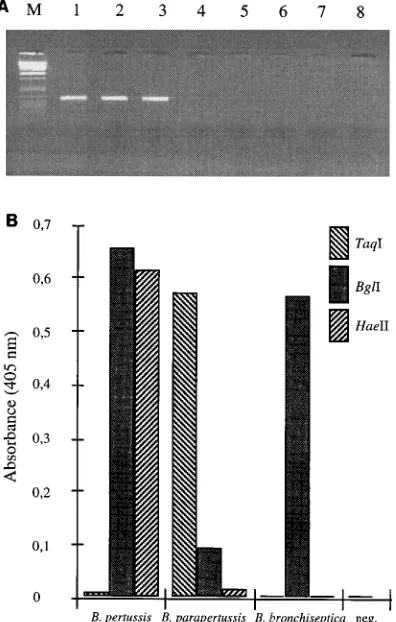
tested by using lysates from the three Bordetella species (Fig.
3A, lanes 1 to 3) and three frequently occurring non-Bordetella
species in nasopharyngeal aspirates: Moraxella catarrhalis,
Hae-mophilus influenzae, and Streptococcus pneumoniae (Fig. 3A,
lanes 4 to 6, respectively). Figure 3A shows a distinct fragment
of 300 bp for the Bordetella species, with no amplification for
the other strains. This is in agreement with the results of
Reizenstein et al. (27, 28), who have tested several more
spe-cies by also amplifying a part of the pertussis toxin promoter.
The detection limit of the nested PCR was analyzed with a
lysed B. pertussis culture. The culture was quantified by viable
counting. A limiting dilution experiment was performed by
serial dilutions of the lysate, enabling an estimate of the
num-ber of targets (3). The results of the corresponding gel
elec-trophoresis analysis after outer and inner amplification are
shown in Fig. 4. Triplicate samples of a B. pertussis lysate were
diluted 10-fold. After outer amplification, the intensity of the
fragment decreased with increasing dilution factors. A weak
fragment could still be seen at the dilution corresponding to 6
FIG. 2. Locations of the primers in the pertussis toxin promoter region. Nucleotides are numbered as described by Locht and Keith (22). The amplified sequences in the outer and the inner PCRs are 499 and 300 bp, respectively. Primer nucleotides in italics correspond to the lac operator sequence. The different restriction enzyme sites for the three species are marked by arrows.
on May 15, 2020 by guest
http://jcm.asm.org/
CFU. After inner amplification the last dilution step amplified
corresponded to 0.6 CFU. Note that the nested amplification
results in similar intensities for all dilution steps, and therefore,
the endpoint can easily be determined. The longer fragments
generated by more concentrated bacterial lysates correspond
to the outer PCR product. Importantly, to achieve good
re-producibility in the end dilution experiments, the bacteria had
to be lysed prior to dilution. Results from similar PCR end
dilutions directly on bacterial suspensions showed both
intra-and interassay variations intra-and could not be correlated with the
results from experiments with lysed cells. Gaps also appeared
in the dilution series for amounts of
,
100 CFU (data not
shown). We interpret this low level of reproducibility as an
artifact of the capsule covering the bacteria, causing them to
aggregate.
Isolation of bacteria by IMS.
Capture of the bacteria by IMS
was investigated by PCR, despite the difficulties in making
representative end dilutions of bacterial cultures for the
deter-mination of the absolute concentration (see above). We chose
to test the IMS step by coating polyclonal rabbit anti-B.
per-tussis and anti-B. paraperper-tussis antisera onto sheep anti-rabbit
immunoglobulin G magnetic beads and incubating the
immu-nobeads with different amounts of B. pertussis and B.
paraper-tussis in an end dilution scheme. The magnetic beads captured
intact fresh or frozen bacteria on the bead surface and enabled
the efficient removal of potent PCR inhibitors by magnetic
separation. A precise determination was not possible, but
ap-proximately 20 CFU could be detected after IMS and PCR
(data not shown). However, the subsequent clinical study,
which did not involve serial dilutions, may indicate that the
system enables few viable bacteria to be detected (see below).
In addition, a negative nasopharyngeal aspirate was divided
and spiked with known amounts of both B. pertussis and B.
parapertussis organisms in cultures to estimate the efficiency of
the IMS in more crude and viscous samples. The sensitivity in
the spiked aspirate was the same as that in the pure cultures
tested in parallel (data not shown).
Species identification.
Amplification of the pertussis toxin
promoter region makes it possible to distinguish between the
different Bordetella species by restriction enzyme analysis of
the amplified sequences (27), since the nucleotide variations
found in B. parapertussis and B. bronchiseptica result in
differ-FIG. 3. (A) Specificities of the PCR primers. The specificities of the PCR primers was tested on lysates from three Bordetella species and three common contaminating species present in nasopharyngeal aspirate samples. Lanes 1 to 3, amplified products of B. pertussis, B. parapertussis, and B. bronchiseptica, respec-tively; there was no amplification of M. catarrhalis, H. influenzae, or S.
pneu-moniae (lanes 4 to 6 respectively). Lanes 7 and 8, negative PCR controls.
[image:4.612.74.272.76.387.2]Bac-teriophagelDNA restricted with PstI was used as a marker (lane M). (B) Colorimetric typing of Bordetella species by DIANA combined with restriction enzyme analysis. The amplified fragments of the three species were immobilized onto streptavidin-coated beads, treated with restriction enzymes, and analyzed colorimetrically to identify species (see text for details).
FIG. 4. Detection limit of the nested PCR, evaluated on a bacterial lysate. Lanes 1 to 3, 600 CFU of bacteria; lanes 4 to 6, 60 CFU; lanes 7 to 9, 6 CFU; lanes 10 to 12, 0.6 CFU; lanes 13 to 15, 0.06 CFU; lanes 16 and 17, negative PCR controls.
on May 15, 2020 by guest
http://jcm.asm.org/
ent recognition sites for restriction enzymes for the three
spe-cies (22). To avoid analysis by gel electrophoresis, we adopted
a colorimetric procedure allowing for detection (DIANA) and
analysis of species. The labelled inner PCR amplicons were
detected with the detection reagent LacI–
b
-galactosidase and
a chromogenic substrate. Solid-phase identification of positive
samples was performed by restriction cleavage (Fig. 2) of
ad-ditional immobilized PCR products and then the addition of
the detection reagent (LacI–
b
-galactosidase). The restricted
fragments resulted in little or no signal, since the lac operator
was removed by magnetic separation, and the intact fragments
resulted in a strong colorimetric response (Fig. 3B). When the
three reference Bordetella species were treated, the results
showed that the different enzymes can be used to cleave PCR
products on a solid support.
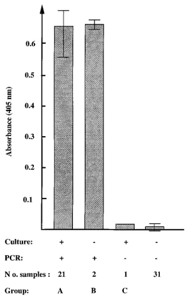
Clinical samples.
Fifty-five frozen nasopharyngeal aspirates
from patients with suspected pertussis infection already tested
by culture were analyzed by the IMS-PCR-DIANA approach
in a blinded fashion. Thirty-one of the samples were negative
by both culture and the IMS-PCR-DIANA method that we
evaluated. Table 1 summarizes the results for 24 samples,
placed into three groups, which were positive by at least one
analysis technique. Among 22 samples from which B. pertussis
was isolated, 21 (group A) were positive by IMS-PCR-DIANA
(95%). The growth estimates showed that this group contained
samples with both few and many viable bacteria. Two samples
(group B) were negative by culture, but they were shown to be
positive when they were analyzed by the PCR-based assay. The
two patients had durations of paroxysmal cough of 29 and 55
days, respectively, which should be compared with the minimal
criterion of
$
21 days for pertussis, which was used within the
trial, in accordance with a consensus meeting of the World
Health Organization (10, 36). Unfortunately, serology results
were missing for both patients, but one of the patients had a
serology-positive sibling (data not shown). The third group
(group C) consisted of one culture-positive sample which was
negative by PCR. The patient who provided this sample was
also positive for B. pertussis by serology but had no paroxysmal
cough. The sample contained few bacteria, corresponding to 3
CFU. None of the patients in groups B and C had been treated
with antibiotics. The colorimetric signal distribution among the
samples tested showed high values for the 23 PCR-positive
samples, with an average A
405of 0.654 (standard deviation
5
0.034), while the signals for the 32 negative samples were close
to background, with an average A
405of 0.007 (standard
devi-ation
5
0.006), making it easy to determine a positive sample
(Fig. 5). Restriction enzyme typing of amplicons showed that
all samples from patients in groups A and B were of B. pertussis
in origin, and no mixed infections were observed. This was in
accordance with the culture results.
DISCUSSION
[image:5.612.61.556.83.177.2]Whooping cough is a highly contagious disease caused by B.
pertussis. The diagnosis of B. pertussis by culture is a highly
specific method, but it has a low degree of sensitivity (26), and
the bacterium is fastidious, but culture is the ‘‘gold standard’’
in many laboratories. However, in the future the gold standard
may be a DNA-based system. PCR detection has been proven
to be a very promising tool for Bordetella detection, but it has
some limitations such as robustness and the fact that it cannot
distinguish viable from nonviable bacteria. Also, the sample
preparation method is critical for the diagnostic sensitivity of
the PCR, the two main factors being the yield of bacterial
DNA from the sample and the occurrence of Taq DNA
poly-merase-inhibiting factors in clinical samples. To address these
issues, we have developed a sample preparation system that
FIG. 5. Distribution of the absorbance values. The average A405values for
[image:5.612.341.525.412.700.2]the respective groups (a total of 55 clinical samples) are shown. The maximum and minimum values are also indicated.
TABLE 1. Summary of results for the 24 samples which were positive by the PCR-based assay and/or the culture assay
Group Specimen Culture result (CFU) IMS-PCR-DIANA result
A 47 B. pertussis (1–10) B. pertussis
11, 22, 29, 40, 43, 50a B. pertussis (10–100) B. pertussis
9, 10, 27 B. pertussis (.100) B. pertussis
6, 7, 8, 30a, 31, 42, 45, 49 B. pertussis (confluent growth) B. pertussis
17, 19, 20 B. pertussis (not determined) B. pertussis
B 39, 54 Negative B. pertussis
C 18 B. pertussis (1–10) Negative
aSamples 30 and 50 were first negative by PCR but were positive when they were retested after dilution of the samples with PBS and mixed on a Vortex mixer to
achieve more homogeneous samples.
on May 15, 2020 by guest
http://jcm.asm.org/
enables us to capture bacteria in crude samples on a magnetic
support by antibody-antigen binding. The paramagnetic
sup-port allows for the rapid handling and the complete removal of
inhibitors by magnetic separation. Importantly, the handling of
the specimen is also simplified so that the risk of false-positive
results because of laboratory contamination is lowered.
An-other important advantage is that intact bacterial cells can be
used as the sample, since the beads with the immunocomplex
are transferred directly to the PCR tube without any phenol
extractions, which are generally applied to remove inhibitory
substances (24, 33). The cells are simply lysed by an extended
(5-min) initial heat denaturation cycle during the PCR
pro-gram.
The nested primer approach has previously been used for
the detection of B. pertussis, showing an increased robustness
for complex nasopharyngeal samples (1, 21, 28). Here, we show
an improved robustness by the signal distribution and the high
signal-to-noise ratio among the positive samples (Fig. 5).
Fur-thermore, the robustness of the assay is also indicated by the
strong intensity of the PCR fragments in the last steps of the
end dilution series (Fig. 4). Several alternatives that allow for
the simultaneous typing of the different Bordetella species on
the basis of species-specific primer sets or shared primers have
been published (20, 37). In the present study we chose the
pertussis toxin promoter, which enables one and the same
primer set to be used without bias for the identification of the
three investigated species (27). Note that sensitivity is not a
major issue with this nonrepeated gene, since we detected
single targets by the nested approach (Fig. 4). The attractive
feature with the chosen target gene is that a non-gel-based,
colorimetric system can be used to detect and distinguish the
species involved (Fig. 3B). Restriction enzyme typing was
per-formed on the DNA immobilized onto the bead surface, with
subsequent evaluation performed by using the reporter fusion
protein LacI–
b
-galactosidase. This also constitutes an extra
verification step. The nature of the colorimetric assay also
allows for semiquantitative analysis of samples containing
more than one species. Thus, both sample preparation and
detection were based on solid supports, which opens the
pos-sibility of automating the handling and analysis procedures and
thereby increasing the robustness and throughput for the
di-agnosis of Bordetella species.
The present approach was evaluated with 55 clinical
naso-pharyngeal aspirates, and the results were compared with those
obtained by culture. As shown in Table 1, a good correlation
was shown for 21 samples containing different amounts of
bacteria. Two more positive samples were found by PCR, while
one sample with only 3 CFU on the plate was missed. This
unexpectedly missed sample stresses the fact that
homoge-neous samples are difficult to achieve, making these
compari-sons somewhat difficult. Possible means of reducing viscosity
could be the use of Sputolysine or similar compounds in
par-allel with immunomagnetic capture. Also, in order to further
control the IMS step, the polyclonal antibodies, which were
commercial antisera in the present study, should be exchanged
for characterized monoclonal antibodies, which can be directly
coupled to a solid support.
In conclusion, we have developed a reliable sample
prepa-ration step for PCR analysis based on intact (fresh or frozen)
Bordetella bacteria. Detection and typing was performed in an
ELISA-like formate, enabling automated methods to be
de-veloped. The present system allows for the rapid and specific
diagnosis of Bordetella infections and increases the possibility
of the more rapidly initiation of antibiotic treatment.
ACKNOWLEDGMENTS
This work was supported by the Go¨ran Gustafsson Foundation for Research in Natural Sciences and Medicine and by contract NO1-A1-15125 from the U.S. National Institute of Allergy and Infectious Dis-eases.
We thank Lena Lindberg for technical assistance and Deirdre O’Meara and Hans Hallander for critical comments.
REFERENCES
1. Ba¨ckman, A., B. Johansson, and P. Olce´n.1994. Nested PCR optimized for detection of Bordetella pertussis in clinical nasopharyngeal samples. J. Clin. Microbiol. 32:2544–2548.
2. Birkebæk, N. H., I. Heron, and K. Skjødt. 1994. Bordetella pertussis diag-nosed by polymerase chain reaction. APMIS 102:291–294.
3. Brinchmann, J. E., J. Albert, and F. Vartdal. 1991. Few infected CD1T cells but a high proportion of replication-competent provirus copies in asymp-tomatic human immunodeficiency virus type 1 infection. J. Virol. 65:2019– 2023.
4. Casadaban, M. J., A. Martinez-Aries, S. K. Shapira, and J. Chou. 1983. Beta-galactosidase gene fusions for analyzing gene expression in Escherichia
coli and yeast. Methods Enzymol. 100:293–308.
5. Coulondre, C., and J. H. Miller. 1977. Genetic studies of the lac repressor. III. Additional correlation of mutational sites with specific amino acid resi-dues. J. Mol. Biol. 117:59–71.
6. Douglas, E., J. G. Coote, R. Parton, and W. McPheat. 1993. Identification of
Bordetella pertussis in nasopharyngeal swabs by PCR amplification of a
re-gion of the adenylate cyclase gene. J. Med. Microbiol. 38:140–144. 7. Glare, E. M., J. C. Paton, R. R. Premier, A. J. Lawrence, and I. T. Nisbet.
1990. Analysis of a repetitive DNA sequence from Bordetella pertussis and its application to the diagnosis of pertussis using the polymerase chain reaction. J. Clin. Microbiol. 28:1982–1987.
8. Grimprel, E., P. Be´gue´, I. Anjak, F. Betsou, and N. Guiso. 1993. Comparison of polymerase chain reaction, culture, and Western immunoblot serology for diagnosis of Bordetella pertussis infection. J. Clin. Microbiol. 31:2745–2750. 9. Guarente, L., G. Lauer, T. M. Roberts, and M. Ptashne. 1980. Improved methods for maximizing expression of a cloned gene: a bacterium that synthesizes rabbit beta-globin. Cell 20:543–553.
10. Gustafsson, L., H. O. Hallander, P. Olin, E. Reizenstein, and J. Storsaeter. 1996. A controlled trial of a two-component acellular, and a five-component acellular and a whole-cell pertussis vaccine. N. Engl. J. Med. 334:349–355. 11. Hallander, H. O., E. Reizenstein, B. Renemar, G. Rasmuson, L. Mardin, and
P. Olin.1993. Comparison of nasopharyngeal aspirates with swabs for cul-ture of Bordetella pertussis. J. Clin. Microbiol. 31:50–52.
12. Hallander, H. O., J. Storsæter, and Roland Mo¨llby.1991. Evaluation of serology and nasopharyngeal cultures for diagnosis of pertussis in a vaccine efficacy trial. J. Infect. Dis. 163:1046–1054.
13. He, Q., J. Mertsola, H. Soini, and M. K. Viljanen. 1994. Sensitive and specific polymerase chain reaction assays for detection of Bordetella pertussis in nasopharyngeal specimens. J. Pediatr. 124:421–426.
14. Hedrum, A., J. Lundeberg, C. Påhlson, and M. Uhle´n. 1992. Immunomag-netic recovery of Chlamydia trachomatis from urine with subsequent color-imetric DNA detection. PCR Methods Appl. 2:167–171.
15. Hornes, E., Y. Wasteson, and Ø. Olsvik. 1991. Detection of Escherichia coli heat-stable enterotoxin genes in pig stool specimens by an immobilised, colorimetric nested polymerase chain reaction. J. Clin. Microbiol. 29:201– 208.
16. Houard, S., C. Hackel, A. Herzog, and A. Bollen. 1989. Specific identification of Bordetella pertussis by the polymerase chain reaction. Res. Microbiol.
140:477–487.
17. Hultman, T., S. Berg, T. Moks, and M. Uhle´n. 1991. Bidirectional solid phase sequencing of in vitro amplified plasmid DNA. BioTechniques 10:84–93. 18. Hultman, T., S. Ståhl, T. Moks, and M. Uhle´n. 1988. Approaches to solid
phase DNA sequencing. Nucleosides Nucleotides 7:629–638.
19. Islam, D., and A. A. Lindberg. 1992. Detection of Shigella dysenteriae type 1 and Shigella flexneri in feces by immunomagnetic isolation and polymerase chain reaction. J. Clin. Microbiol. 30:2801–2806.
20. Li, Z., D. L. Jansen, T. M. Finn, S. A. Halperin, A. Kasina, S. P. O’Connor,
T. Aoyama, C. R. Manclark, and M. J. Brennan.1994. Identification of
Bordetella pertussis infection by shared-primer PCR. J. Clin. Microbiol. 32:
783–789.
21. Lichtinghagen, R., R. Diedrich-Glaubitz, and B. von Ho¨rsten.1994. Identi-fication of Bordetella pertussis in nasopharyngeal swabs using the polymerase chain reaction: evaluation of detection methods. Eur. J. Clin. Chem. Clin. Biochem. 32:161–167.
22. Locht, C., and J. M. Keith. 1986. Pertussis toxin gene: nucleotide sequence and genetic organization. Science 232:1258–1264.
23. Lundeberg, J., J. Wahlberg, and M. Uhle´n. 1990. Affinity purification of specific DNA fragments using a lac repressor protein. Genet. Anal. Techn. Appl. 7:47–52.
24. Meade, B. D., and A. Bollen. 1994. Recommendations for use of the poly-merase chain reaction in the diagnosis of Bordetella pertussis infections. J.
on May 15, 2020 by guest
http://jcm.asm.org/
Med. Microbiol. 41:51–55.
25. Olsvik, Ø., T. Popovic, E. Skjerve, K. F. Cudjoe, E. Hornes, J. Ugelstad, and
M. Uhle´n.1994. Magnetic separation techniques in diagnostic microbiology. Clin. Microbiol. Rev. 7:43–54.
26. Onorato, I. M., and S. G. F. Wassilak. 1987. Laboratory diagnosis of per-tussis: the state of the art. Pediatr. Infect. Dis. J. 6:145–151.
27. Reizenstein, E., B. Johansson, L. Mardin, J. Abens, R. Mo¨llby, and H. Hallander.1993. Diagnostic evaluation of polymerase chain reaction dis-criminative for Bordetella pertussis, B. parapertussis, and B. bronchiseptica. Diagn. Microbiol. Infect. Dis. 17:185–191.
28. Reizenstein, E., L. Lindberg, R. Mo¨llby, and H. O. Hallander.1996. Vali-dation of nested Bordetella PCR in pertussis vaccine trial. J. Clin. Microbiol.
34:810–815.
29. Ru¨ter, U.1982. pUR 250 allows rapid chemical sequencing of both DNA strands of its insert. Nucleic Acids Res. 10:5765–5772.
30. Schla¨pfer, G., J. D. Cherry, U. Heininger, M. U¨ berall, S. Schmitt-Grohe, S. Laussucq, M. Just, and K. Stehr.1995. Polymerase chain reaction identifi-cation of Bordetella pertussis infections in vaccinees and family members in a pertussis vaccine efficacy trial in Germany. Pediatr. Infect. Dis. J. 14:209–214. 31. Seesod, N., J. Lundeberg, A. Hedrum, L. Åslund, A. Holder, S. Thaithong,
and M. Uhle´n.1993. Immunomagnetic purification to facilitate DNA
diag-nosis of Plasmodium falciparum. J. Clin. Microbiol. 31:2715–2719. 32. Ugelstad, J., A. Berge, T. Ellingsen, R. Schmid, T.-N. Nilsen, P. C. Mørk, E.
Hornes, and Ø. Olsvik.1992. Preparation and application of new monosized polymer particles. Prog. Polymer Sci. 17:87–161.
33. Wadowsky, R. M., S. Laus, T. Libert, S. J. States, and G. D. Ehrlich. 1994. Inhibition of PCR-based assay for Bordetella pertussis by using calcium alg-inate fiber and aluminum shaft components of a nasopharyngeal swab. J. Clin. Microbiol. 32:1054–1057.
34. Wahlberg, J., J. Lundeberg, T. Hultman, and M. Uhle´n. 1990. General colorimetric method for DNA diagnostics allowing direct solid phase genomic sequencing of the positive samples. Proc. Natl. Acad. Sci. USA
87:6569–6573.
35. Woolfrey, B. F., and J. A. Moody. 1991. Human infections associated with
Bordetella bronchiseptica. Clin. Microbiol. Rev. 4:243–255.
36. World Health Organization. 1991. WHO meeting on case definition of pertussis, p. 4–5. Report MIN/EPI/PERT/91.1. World Health Organization, Geneva.
37. van der Zee, A., C. Agterberg, M. Peeters, J. Schellekens, and F. R. Mooi. 1993. Polymerase chain reaction assay for pertussis: simultaneous detection and discrimination of Bordetella pertussis and Bordetella parapertussis. J. Clin. Microbiol. 31:2134–2140.