ScholarWorks @ Georgia State University
ScholarWorks @ Georgia State University
Biology Dissertations Department of Biology
12-18-2014
Characterization of DrrAB Complex from Streptomyces Peucetius
Characterization of DrrAB Complex from Streptomyces Peucetius
as A Multidrug Transporter
as A Multidrug Transporter
Wen Li
Follow this and additional works at: https://scholarworks.gsu.edu/biology_diss
Recommended Citation Recommended Citation
Li, Wen, "Characterization of DrrAB Complex from Streptomyces Peucetius as A Multidrug Transporter." Dissertation, Georgia State University, 2014.
https://scholarworks.gsu.edu/biology_diss/150
A MULTIDRUG TRANSPORTER
by
WEN LI
Under the Direction of Parjit Kaur, PhD
ABSTRACT
The soil bacterium Streptomyces peucetius produces two widely used anticancer
antibiotics doxorubicin and daunorubicin. Present within the biosynthesis gene cluster in S.
peucetius is the drrAB operon which codes for a dedicated ATP-binding cassette type transporter
for the export of these two closely related antibiotics. DrrAB system was believed to be the
single-drug transporter due to its dedicated nature; however, our study demonstrated under both
in vivo and in vitro conditions that DrrAB system can transport not only doxorubicin but also
Hoechst 33342 and ethidium bromide. Moreover, many other well-studied multi-drug resistance
proteins substrates (including verapamil, vinblastine and rifampicin) inhibit DrrAB-mediated
competitive inhibition by verapamil, suggesting the possibility of more than one drug binding
site in the DrrAB system. This is the first in-depth study of a drug resistance system from a
producer organism, and it shows that a dedicated efflux system like DrrAB contains specificity
for multiple drugs.
Our study also provides the first direct evidence for the dual role of the metalloprotease
FtsH in the biogenesis of membrane proteins. We found that FtsH is not only responsible for
proteolysis of unassembled DrrB protein but it also plays a much broader role in biogenesis of
the DrrAB complex. DrrA and DrrB proteins expressed together in a temperature sensitive ftsH
mutant strain were found to be non-functional due to their incorrect assembly. Simultaneous
expression of wild-type FtsH in trans resulted in normal doxorubicin efflux. Strikingly,
doxorubicin efflux could be restored in mutant cells irrespective of whether FtsH was expressed
simultaneously with DrrAB or expressed after these proteins had already accumulated in an
inactive conformation, thus providing crucial evidence for the ability of FtsH to refold the
misassembled proteins. Complementation experiments also showed that the catalytic ATP
binding domain of FtsH contains a chaperone-like activity, however both the catalytic and the
proteolytic domains of FtsH are required to be present and work coordinately to participate in
biogenesis of DrrAB complex in the membrane.
A MULTIDRUG TRANSPORTER
by
WEN LI
A Dissertation Submitted in Partial Fulfillment of the Requirements for the Degree of
Doctor of Philosophy
in the College of Arts and Sciences
Georgia State University
Copyright by Wen Li
A MULTIDRUG TRANSPORTER
by
WEN LI
Committee Chair: Parjit Kaur
Committee: Parjit Kaur
Phang C.Tai
Casonya Johnson
Electronic Version Approved:
Office of Graduate Studies
College of Arts and Sciences
Georgia State University
ACKNOWLEDGEMENTS
I owe a great many thanks to a great many people who helped me and supported me
during the study of my Ph.D. I truly apologize if I have forgotten to mention someone.
First, I would like to express my deepest appreciation to my advisor Dr.Parjit Kaur, who
gave me guidance, encouragement, caring and support during my Ph.D. I would never have
been able to get these achievements without your help. You patiently guided me and inspired me
during the hardest time. Thank you for all these special moments for my life.
I would also like to thank my committee members, Dr. Phang C. Tai and Dr. Casonya
Johnsonfor their critical comments and suggestions for my research. I am very thankful to my
fellow lab members, who listen to me, help me and support me all the time. Special thanks to
Dr.Han Zhang and Sadia Jannath, who are my good friends, accompanies and excellent
researchers. I would also like to express my gratitude to members in the core facility, the other
professors in the biology department and staff in the biology department.
I would like to acknowledge all my friends who cares me sometimes more than myself.
They make my life more colorful and meaningful.
Finally, there is never enough thanks to my family, they are part of my life and they
complete me. My parents brought me up so many years; I owe a lot to them. My husband, Mr.
Ning Song, is my strongest support during these years, thanks for holding my hands all the time
during the good and bad times. Even though my son, Eric Song, is too small to contribute to my
dissertation, his smile and love make me stronger and my life more meaningful. A special
TABLE OF CONTENTS
ACKNOWLEDGEMENTS ... iv
LIST OF TABLES ... vii
LIST OF FIGURES ... viii
1 GENERAL INTRODUCTION ... 1
1.1 Multidrug Resistance and ABC Transporters... 1
1.2 Characterization of P-glycoprotein ... 2
1.3 P-glycoprotein is a multidrug transporter ... 3
1.4 Mechanism of drug transport ... 3
1.5 Drug binding sites on Pgp ... 4
1.6 DrrAB is a multidrug transporter ... 5
1.7 Quality control of MDR proteins in membranes... 6
2 DUAL ROLE OF THE METALLOPROTEASE FTSH IN BIOGENESIS OF THE DRRAB DRUG TRANSPORTER ... 8
2.1 Introduction ... 8
2.2 Materials and Methods ... 11
2.3 Results ... 14
2.4 Discussion ... 23
3.1 Introduction ... 39
3.2 Materials and Methods ... 43
3.3 Results ... 47
3.4 Discussion ... 57
4 GENERAL DISCUSSION ... 73
LIST OF TABLES
Table 2.1 Baterial strains, plasmids and anti-sera ... 28
Table 2.2 The ATPase activity of purified FtsH and its variants... 28
LIST OF FIGURES
Figure 2.1 Role of DrrA and FtsH in stable maintenance of DrrB ... 29
Figure 2.2 Western blot analysis of the cytosol, membrane, and inclusion body fractions of E. coli 796 or 797 cells expressing DrrAB at 30 °C or 42 °C. ... 30
Figure 2.3 Growth inhibition resulting from expression of DrrB alone or DrrAB together in E. coli 797 cells can be relieved by the overexpression of FtsH or GroESL... 31
Figure 2.4 FtsH preferentially degrades misfolded DrrAB. ... 33
Figure 2.5 FtsH promotes assembly of the DrrAB complex ... 34
Figure 2.6 In vitro digestion of α-casein by purified FtsH or FtsH(HEH) ... 35
Figure 2.7 The AAA domain of FtsH is sufficient to complement the growth defect resulting from the expression of DrrAB in 797 cells. ... 36
Figure 2.8 DrrAB-mediated Dox efflux. ... 37
Figure 3.1 Characterization of the DrrAB-mediated Dox efflux under in vivo and in vitro conditions. ... 63
Figure 3.2 Effect of point mutations in the nucleotide binding domain of DrrA on DrrAB-mediated Dox efflux in IOVs. ... 66
Figure 3.3 Inhibition of DrrAB-mediated Dox efflux by known MDR substrates. ... 68
Figure 3.4 Kinetic characterization of the inhibition of DrrAB-mediated Dox efflux by Hoechst 33342, verapamil or rifampicin ... 70
1 GENERAL INTRODUCTION
1.1 Multidrug Resistance and ABC Transporters
In the chemotherapies of cancer and infectious diseases, multidrug resistance (MDR) is
seen to develop in more than 90% of the patients. This resistance can be caused by the decreased
uptake of drugs, altered cell cycle checkpoint, altered drug target or the increased efflux of drugs
by drug transporter (1). In tumor cells, the major reason for multidrug resistance is the over
efflux of anti-cancer drugs. The first and the most well-characterized multidrug transporter in
human cells is P-glycoprotein (P-gp), which belongs to the ATP-binding cassette (ABC)
superfamily. Proteins in this family share highly conserved domains and functions, including
nucleotide binding domains (NBDs) and transmembrane domains (TMDs). Members of the
ABC superfamily range from prokaryotes to eukaryotes. In eukaryotes, most ABC transporters
only carry out drug efflux, while in prokaryotes, ABC proteins include both importers and
exporters (2).
Most members of the ABC superfamily mediate the transport of highly specific
substrates, following the “lock-key” hypothesis (3); however, some transporters and are
categorized as multidrug transporters. Well-characterized multidrug transporters in eukaryotic or
prokaryotic cells include P-gp, MsbA, LmrA and Sav1866, etc. (4-7). This study focuses on the
prototype drug transporter DrrAB found in Streptomyces peucetius, a soil organism that produces
anticancer drugs doxorubicin (Dox) and daunorubicin (Dnr) (8). The DrrAB system shows
1.2 Characterization of P-glycoprotein
P-gp is known as multidrug resistance protein 1 (MDR1) or ATP-binding cassette
sub-family B member 1 (ABCB1). It was first discovered by Biedler and coworkers, who found that
Chinese Hamster cells, which are resistant to actinomycin D, are also cross-resistant to other
several antibiotics (11).
P-gp is encoded by a single polypeptide chain with 1280 amino acids and assembled as
two homologous halves in the plasma membrane (12). These two halves are not identical, but
share 43% sequence identity and 78% similarity (12). They are connected by a central linker.
Each half contains one TMD (with six transmembrane helices (TMH)) and one cytoplasmic ATP
binding domain or NBD (13). The two TMDs from each halve form a large drug transport
channel with 12 TMHs connected by loops, while the two ATP binding domains constitute the
cytosolic ATP binding pockets. The NBD contains conserved motifs shared by other proteins in
the ABC transporter family, such as Walker A (P-loop), Walker B, ABC signature, Q-loop and
switch region (H-loop) (14). These conserved motifs are involved in ATP binding and
hydrolysis, energy transduction, and the cross-talk between NBD and TMD. The NBDs of most
ABC transporters share high homology irrespective of their substrate specificities while the
sequence and structure of TMDs varies significantly (15).
P-gp is expressed in tumor cells and normal tissue cells, its main physiological function is
to protect sensitive organs, such as intestine, brainor placenta by pumping the drugs or toxic
agents into bile, urine or lumen of gastrointestinal tract (16). In tumor cells, P-gp is
1.3 P-glycoprotein is a multidrug transporter
P-gp can interact with a wide range of structurally unrelated chemical compounds,
including natural products, anticancer drugs, steroids, fluorescent dyes, linear and cyclic
peptides, etc. According to the type of interactions, these drugs are classified as substrates or
modulators. Substrates that are transported by P-gp through the membrane, such as
anthracyclines (doxorubicin, daunomycin), and vinca alkaloids (vincristine, vinblastine) (17).
P-gp can also translocate fluorescent phospholipids such as C6-NBD-diacylglycerol (18) or
lipid-based drugs. Modulators can block the function of the transporter and generate a drug
concentration gradient; some of them can also be effluxed by the transporter, such as verapamil
and cyclosporine A (19). A modulator has very significant clinical application by reversing the
multidrug resistance problem during cancer chemotherapy. A combination of anti-cancer drugs
and modulators will greatly improve the effect of cancer cells treatment (20).
1.4 Mechanism of drug transport
Gottesman and co-workers had proposed the hydrophobic vacuum cleaner model (4,17),
which now has been extended to many other MDR proteins (21). In this model, substrate first
interacts with the lipids phase. With the energy provided by ATP hydrolysis, P-gp detects the
hydrophobic substrate from the lipid bilayer and extracts the substrates from inner leaflet to the
extracellular medium (22-26). Strong evidences to support this hypothesis include the early
studies by Victor Ling’s group, which suggested that P-glycoprotein transports Hoechst 33342 or
LDS-751 from the cytoplasmic leaflet of the plasma membrane directly to the extracellular
medium (22,27,28). LmrA, the homologue of Pgp, may also transport lipophilic substrate by a
The complete process of drug efflux requires a series of conformational changes of the
drug transporter, which can be divided into four steps (30,31).
(1) Initial binding of substrate to the drug binding pocket;
(2) ATP binding and dimerization of two NBDs;
(3) Hydrolysis of ATP resulting in the efflux of the drugs, which causes drug binding affinity
to switch from high to low;
(4) Resetting of the transport back to the original state after the reaction cycle.
1.5 Drug binding sites on Pgp
During the last two decades, the studies of P-gp drug transporter have revealed specific
drug binding locations, the kinetic interactions of multiple drugs and the energy coupling
between ATP hydrolysis and drug efflux.
As P-gp can interact with unrelated chemical compounds, the question whether Pgp
contains one single flexible and open drug binding site or multiple drug binding sites with
different binding affinities is still not clear. The presence of multiple drug interaction sites has
been demonstrated by a number of groups. For example, in 1997, Shapiro and Ling’s group
identified two drug binding sites on P-gp. The H-site preferentially transports Hoechst 33342
and the R-site preferentially transports rhodamine 123 (32). These two sites interact in a
cooperative manner. Later, they further discovered the third drug-binding site, which has a
positively allosteric effect on the drug transport through H and R sites (33). In 2000, Richard
Callaghan found the presence of at least four distinct drug interaction sties on P-gp using
equilibrium and kinetic radioligand binding assays (34). Specific residues potentially involved
in the drug binding were subsequently identified by Loo and Clark via a combination of cysteine
the drug substrate dibromobimane (dBBn) (35-37) and the drug substrate analogues
methanethiosulfonate (MTS)-verapamil (38) or MTS rhodamine (39). First, single cysteine
substitution mutation of every residue in the TMDs was generated; the expression level and
function of these mutations were confirmed. The residues protected by drug substrates from
being labeled by thiol-reactive analogs were identified as potential drug binding sites. This work
showed that residues in TMHs 4-6 in TMD1and TMHs 9-12 in TMD2 of P-gp are involved in
the drug binding process.
P-gp has been proven to contain multiple drug binding sites by various techniques;
however, other studies suggest that P-gp contains a large single drug binding pocket . Loo and
Clark showed that the P-gp drug binding pocket can bind verapamil and TMEA simultaneously,
and these two drugs may occupy different regions of the shared drug-binding pocket (40). This
phenotype is also supported by low-resolution cryo-election microscopy structure of P-gp, which
present the existence of a large, 5 nm diameter, central chamber for drug binding inside of P-gp
(41). A much clearer crystal structure was not obtained until 2009 by Chang’s group. The
apo-P-gp structure contains an internal cavity of ~6,000 Å3 with a 30 Å separation of two NBDs (42).
This large internal cavity could fit at least two substrates simultaneously and the drug binding
pocket is mainly occupied by hydrophobic and aromatic residues (43-46).
1.6 DrrAB is a multidrug transporter
Streptomyces peucetius produces two anti-cancer drugs, Dox and Dnr, and is
self-resistant to these drugs due to the efflux action of membrane transporter DrrAB (47). DrrA and
DrrB proteins are coded by the drrAB operon present within the Dox biosynthesis gene cluster
(47). DrrA is the catalytic subunit (36 kDa). It belongs to the ABC family of proteins and binds
eight transmembrane α-helices and belongs to the ABC-2 subfamily (3). Two subunits each of
DrrA and DrrB interact together to form a functional tetrameric complex (49). Both DrrAB and
P-gp belong to the ABC (ATP-binding cassette) superfamily of proteins. The DrrAB system
belongs to the Class III, DRA family, and DRR subfamily of ABC proteins (50). The NBD of
DrrA shares 29% identity and 47% similarity to NBD1 and NBD2 of Pgp (48). Thus the
bacterial DrrAB system could serve as a prototype model system for understanding the function
and evolution of multidrug transporters in general.
Due to it dedicated nature, the DrrAB system was previously believed to form a
single-drug-transporter. However, in our recent studies, it was shown that DrrAB is a multidrug
transporter that can carry out efflux of at least 3 different drugs: Dox, Hoechst 33342 and
ethidium bromide (8). Inhibition studies also suggest that DrrAB can interact with a number of
other well-characterized MDR substrates, such as verapamil, rifampicin and colchicine. The
interactions between two different drugs in DrrAB were also studied by kinetics analysis, which
revealed competitive inhibition of Dox efflux by Hoechst 33342 and rifampicin but
non-competitive inhibition of Dox efflux by verapamil. These findings indicate that DrrAB is a
multidrug transporter with at least two drug binding sites. This work also highlights overlaps of
the substrate specificity of the DrrAB system and Pgp as well as other bacterial MDR systems
and points to a common mechanism, and perhaps origin, for most MDR proteins.
1.7 Quality control of MDR proteins in membranes
One of the major causes of MDR is the over-expression of the drug transporters, which
results in efflux of anti-cancer drugs out of the cancer cells. Our previous studies have indicated
that DrrA and DrrB proteins need to co-assemble in the membrane to maintain the normal
undetectable due to its proteolysis by FtsH on the membrane. FtsH, also defined as HflB, is an
inner membrane-embedded, ATP and Zn2+-dependent metalloprotease (52). It has been shown
to play a role in quality control of soluble proteins, such as σ32, as well as membrane proteins
including SecY and YccA, etc. (53-55). FtsH protein consists of two domains: the N-terminal
AAA domain which carries out hydrolysis of ATP, and the C-terminal proteolytic domain (52).
The degradation process of FtsH is triggered by the hydrolysis of ATP and conformational
changes of the hexameric ring structure of FtsH (52). Once the peptide tail is bound by FtsH, the
peptide can be unfolded to an open structure, extracted from the membrane lipid bilayer and
translocated through the central pore into the protease chamber for further digestion (56). As
FtsH has relatively low unfolding activity, it preferentially degrades proteins with natural
structure or low thermostabilities (57).
In our studies, we have demonstrated that FtsH plays dual roles in the quality control of
the DrrAB complex; it is not only involved in proteolysis of the unassembled DrrB protein, but
also facilitates assembly of the DrrAB complex (58). We showed that the DrrAB complex is
improperly assembled in an FtsH-deficient cell and loses its drug efflux function. However,
when FtsH is co-expressed in trans in FtsH-deficient cells, the function of the DrrAB complex is
fully restored. Interestingly, the Dox efflux activity could be restored in FtsH-deficient cells
irrespective of whether FtsH was expressed simultaneously with DrrAB or expressed after these
proteins had already accumulated in a misfolded conformation. To facilitate this assembly
function, both the AAA domain and proteolytic domain need to be present and work
2 DUAL ROLE OF THE METALLOPROTEASE FTSH IN BIOGENESIS OF THE
DRRAB DRUG TRANSPORTER
2.1 Introduction
Membrane proteins play essential roles in cell physiology. They carry out import of
nutrients, export of toxins, antibiotics and drugs, and play important roles in energy and signal
transduction. Improper assembly of membrane proteins is known to result in various diseases.
However, because of the complexity of the assembly process and the diverse array of factors
involved, understanding mechanisms of membrane protein assembly poses a serious challenge.
The bacterial DrrAB (doxorubicin resistance proteins A and B) system is an attractive model for
studying assembly of ABC (ATP-Binding Cassette) transporters. In this system, the catalytic
function (DrrA) and the transport function (DrrB) are present on separate subunits (59), which
together form a tetrameric complex in the membrane (60) and carry out ATP-dependent efflux of
the anti-cancer drugs doxorubicin (Dox) and daunorubicin (61). Previous studies from this lab
suggested that for proper function of the DrrAB complex the DrrA and DrrB proteins may be
required to co-assemble (51). It was also shown that interaction between DrrA and DrrB is
essential for stable maintenance of DrrB in the membrane so that the expression of DrrB is
undetectable in the absence of a simultaneous expression of DrrA (60). Co-expression of DrrA
in cis or trans restores the wild-type levels of DrrB expression, therefore suggesting that DrrA
protects DrrB from proteolysis by a cellular protease. The nature of the protease and whether it
plays a specific role in quality control and biogenesis of the DrrAB complex has so far remained
uncharacterized. In this study, we examine the role of FtsH (filament temperature-sensitive
FtsH is a zinc-dependent metalloprotease which belongs to the AAA (ATPases
Associated with Diverse Cellular Activities) family of proteins. Along with other proteases,
such as ClpAP, ClpXP, HsIUV, and Lon, these proteins form the large AAA+ superfamily of
proteins, members of which share a similar AAA-ATPase domain (62). FtsH is evolutionarily
conserved with more than 40% sequence identity observed between bacterial, yeast and human
homologs (63). E. coli FtsH is the best studied of all known members, and it has been shown to
be the only growth-essential protease in E. coli. Yeast cells lacking the three FtsH orthologs
(two m-AAA and one i-AAA) were also found to be non-viable, demonstrating the essential
function of this enzyme in eukaryotic cells (63,64). FtsH is unique in being embedded in the cell
membrane in E. coli, and in the mitochondrial inner membrane in eukaryotes (65), where it
forms homohexameric ring-like structures. The major role of FtsH is believed to be in the
quality control of specific membrane proteins, such as degradation of the unassembled SecY and
subunit ‘a’ of F0 sector of the ATP synthase, in addition to modulating levels of some soluble
regulatory proteins (65-67).
FtsH contains two transmembrane helices at the N terminus, followed by a cytoplasmic
domain containing the catalytic AAA domain in the middle and the proteolytic domain at the C
terminus. The AAA domain (residues 144-398) consists of the conserved Walker A, Walker B
and SRH (second region of homology) motifs, which are essential for ATP binding and
hydrolysis (65). The proteolytic domain contains the conserved Zn2+ binding motif
H417EXXH421, the third Zn2+-ligand residue E479, and the coiled-coil leucine-zipper sequence
(65). FtsH carries out proteolysis of polypeptides in an ATP and Zn2+-dependent manner, while
other AAA+ proteases, such as Lon and ClpA/P, are serine peptidases (68). To initiate
domain of FtsH is believed to capture the cytoplasmic tail (either at the N or the C terminus) of
the membrane substrate, followed by dislocation and processive unfolding of the protein to an
open structure (65). Therefore, both the catalytic AAA domain and the proteolytic domain are
required for proteolysis.
Although the major function of the AAA+ proteases is in proteolysis, they also exhibit
chaperone-like activities, which allow them to monitor the folding status of a protein, promote
disassembly or unfolding (69), and specifically degrade non-native proteins (70). For a long
time, it has been speculated that the AAA+ proteases may also have the ability to refold their
substrate proteins (71), which remains an open question till date (72). FtsH was originally
identified by Ito and co-workers in a screen to isolate factors which may assist in membrane
protein assembly (73). They used SecY-PhoA fusion to screen for stop-transfer defect (Std)
mutations, and found that such a mutation lies in the ftsH gene. Depletion of FtsH also resulted
in significant export defects of β-lactamase and OmpA in E. coli in addition to causing a strong
Std phenotype. Together, these studies indicated that FtsH may be involved in protein assembly
into and through the membrane and may play a role in determining orientation of membrane
proteins (73). In another study, FtsH orthologs Yta10 and Yta12 in yeast mitochondria were
shown to be required for the formation of a 48 kDa assembly intermediate of the F0 subunit 9
(74). Finally, in vitro studies showed that the purified AAA domain of Yme1, a yeast
mitochondrial homolog of FtsH, suppresses aggregation of a model polypeptide (75). Despite
these observations, however, no direct evidence for the role of FtsH or its homologs in functional
assembly of membrane proteins has been obtained so far.
In this study, we provide the first direct evidence that FtsH is a dual function enzyme
for removal of the unassembled DrrB but that it is actually able to refold previously
misassembled DrrAB proteins and restore Dox efflux function of the complex. Our results also
show that while the AAA domain of FtsH provides recognition and specificity for binding of the
substrate, both ATP hydrolysis and the proteolytic functions of FtsH are used concurrently for
refolding of DrrAB and restoration of function. Our studies, therefore, not only shed light on the
mechanism of assembly of the DrrAB complex but also further elucidate the function of the FtsH
protease.
2.2 Materials and Methods
Strains and plasmids– The E. coli strains and plasmids are described in Table 1.
Media and growth conditions – E. coli cells were normally grown in LB medium at 30°C or 37
°C, unless indicated otherwise. Chloramphenicol, kanamycin, or ampicillin was added to a final
concentration of 20, 30 or 100 µg/ml, respectively, where indicated. E. coli cells used for the
Dox efflux assay were grown in TEA medium (61).
Site-directed mutagenesis of ftsH – Site-directed mutagenesis of the ftsH gene was carried out
by a Strategene QuickChange Multisite-directed mutagenesis kit (La Jolla, CA). Using
pUC18/ftsH plasmid as the template, Lys198, located in the conserved Walker A motif of the
AAA domain, was changed to aspargine. The resulting plasmid was named pUCftsH(K198N).
Another mutant, named ftsH(HEH), was obtained by substituting His417Glu418His421 in the
conserved HEAGH motif present in the proteolytic domain to Ala417Gln418Ala421. The primers
for both substitutions are shown in Table S1.
Subcloning of the AAA domain of ftsH into pUC18 vector– To completely remove the
proteolytic domain, a fragment of ftsH corresponding to the first 1194 base pairs was
resulting in pUC18(AAA). This construct is referred to as ftsH(AAA) in this article. The primers
are shown in Table S1.
Subcloning of ftsH and the groESL genes – Wild-type ftsH, ftsH(K198N), ftsH(HEH) and
ftsH(AAA) genes were subcloned into pET28a vector using SnaBI and HindIII restriction
enzymes. The resulting plasmids were named pETftsH, pETftsH(K198N), pETftsH(HEH) and
pETftsH(AAA), respectively. The groES/L genes were subcloned from pKY326 (76) into
pUC18 vector using EcoRI and SmaI restriction enzymes resulting in pUCgroESL
Cloning of ftsH into the pBAD vector– Using pBAD/HisA (Invitrogen) as a template, a 3.9kb
fragment between NcoI and HindIII sites was amplified. The NcoI site was substituted with the
XhoI site in the primers used for amplification (Table S1). This resulted in deletion of the
multiple cloning sites and the polyhistidine region. The ftsH gene was amplifed from pUCftsH
using primers containing XhoI and HindIII sites and ligated to the above fragment.
Growth and Protein expression–E. coli TG1, AR796, AR797, E. coli SG1110, and E. coli
SG1126 cells containing the indicated plasmids were grown at 30 °C or 37 °C to mid-log phase
(A600nm=0.6). The proteins were induced by addition of 0.1 or 0.25 mM IPTG, and the
incubation was continued at 30 °C, 37 °C, or 42 °C for 3 h. The cells were spun down,
resuspended in 5 ml lysis buffer (2 mM Tris-Cl, pH 7.5, 20% glycerol, 2mM EDTA, 1 mM
DTT) and lysed by a single passage through a French pressure cell at 20,000 p.s.i. After
centrifugation at 10,000 X g for 15 min, the pellet represented the inclusion body fraction. The
supernatant was centrifuged at 100,000 X g for 1 h to separate the supernatant (cytosolic
fraction) and the pellet (membrane fraction). The membrane, cytosol and the inclusion body
fractions were analyzed by 12% SDS-PAGE, followed by Western-blot analysis using anti-DrrA
Purification of the FtsH protein – FtsH protein was purified according to published protocols
(66) with modifications. E. coli HMS174(DE3) cells containing the pETftsH plasmid or its
variants were inoculated in 1liter LB medium supplemented with 30 µg/ml kanamycin at 37 °C.
The cells were grown to mid-log phase and induced with 0.25 mM IPTG at 20 °C overnight.
The membrane fraction was prepared as described earlier(60). 5 mg of the membrane fraction
was solubilized with 5 ml solubilization buffer (50 mM Tris-Cl, pH7.5, 500 mM KCl, 0.5 %
(w/v) Nonidet P-40, 15% (w/v) Glycerol, 2.9 mM 2-mercaptoethanol). The solubilized protein
was purified using Ni2+ -NTA-agarose column and eluted with a gradient of 50 to 500 mM
imidazole. Fractions containing FtsH were collected and dialyzed against 2 liters of the dialysis
buffer (10 mM Tris-Cl, pH7.5, 15% (w/v) glycerol, 50 mM KCl, 0.5% (w/v) Nonidet P-40, 5mM
MgCl2, 1 mM dithiothreitol) for 12 hours. The protein was stored at -80 °C until used.
ATPase activity assay – The ATPase activity of FtsH was detected by the malachite
green-ammonium molybdate colorimetric assay (66).
In vivo FtsH Proteolytic assay - Membrane-bound DrrAB proteins were used as a substrate to
determine the proteolytic activity of FtsH. pBAD vector or the pBADftsH plasmid was
transformed into AR797 cells containing pDX101 (pSU2718drrAB). The cells were grown in
LB medium to mid-log phase and the DrrAB proteins were induced with 0.25 mM IPTG at 42
°C for 1 h. To terminate the synthesis of DrrAB, 1000 µg/ml chloramphenicol was added to the
cell culture and incubated at 42 °C for 30 min. The cells were washed extensively to remove
chloramphenicol and IPTG. After resuspending the cells in fresh medium, synthesis of FtsH
from the pBADftsH plasmid was induced by addition of 0.2% arabinose at 42 °C. An aliquot of
as described above, and 20 µg total membrane protein was loaded onto 12% SDS-PAGE,
followed by Western-blot analysis using anti-DrrA, anti-DrrB or anti-FtsH antibodies.
In vitro FtsH Proteolytic assay - α-Casein was used as a substrate in vitro to demonstrate the
proteolytic activity of purified FtsH and the FtsH(HEH) protein (77). 40 µg of α-casein (Sigma
Aldrich) was mixed with 40 µg purified FtsH or FtsH (HEH) in 150 µl protease buffer (50 mM
Tris-Cl, pH8.0, 20 mM KCl, 5 mM MgCl2, 12.5 µM Zn(OAc)2, 0.5 % (w/v) NP-40, 10 % (w/v)
glycerol, 1 mM dithiothreitol) at 42 °C. The reaction was initiated by adding 8 mM ATP, and an
aliquot (20 µl) of the sample was taken out at the indicated time points. The reaction was
terminated by adding 7 µl of 4×SDS sample buffer. The samples were analyzed by 12%
SDS-PAGE, followed by Coomassie Brilliant blue staining.
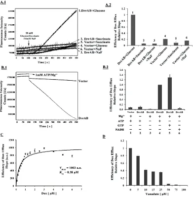
Whole cell Dox efflux assay – The whole cell Dox efflux assay was carried out according to the
protocol published previously (61). The fluorescence spectra were recorded on an Alphascan-2
spectrofluorometer (Photon Technology International, London, Ontario, Canada). The slope of
the Dox efflux curve of the positive control (the first sample) in each panel was designated as
1.0. The efficiency of Dox efflux of each sample within one panel was calculated by dividing
the slope of the efflux curve by the slope of sample 1. The average data obtained from three
independent experiments were plotted in the histograms.
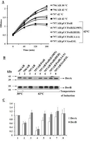
2.3 Results
FtsH is responsible for the proteolysis of unassembled DrrB – Previous studies from this
laboratory showed that DrrB is undetectable in wild-type E. coli membranes in the absence of
simultaneous expression of DrrA ((60), Fig 1A, lane 2), however stable expression of DrrB is
seen when both DrrA and DrrB are expressed together (lane 1). These results suggest that DrrB
that proteins of the AAA+ family, especially FtsH, may be involved in the quality control of
membrane proteins (70). To determine if this is true for DrrB, three proteases, including Lon,
ClpA/ClpP and FtsH, were investigated. If any of these proteases is responsible for degradation
of unassembled DrrB, stable expression of DrrB will be observed in cells deficient in that
protease as compared to the wild-type cells. The FtsH-deficient E. coli AR797 strain (Table 1)
used in this study contains a temperature sensitive mutation in the ftsH gene, therefore it was
grown at 30 °C but the temperature was switched to 42 °C to inactivate FtsH. The Lon
-(SG1110) and the ClpA- (SG1126) cells were grown normally at 30 °C or 37 °C. The isogenic
E. coli AR796 parent strain was used as a control. Of the 3 protease-deficient strains tested, only
the FtsH-deficient E. coli cells showed stable expression of DrrB in the absence of DrrA (Fig.
1B, lanes 1-3) (Fig. 1C, lane 2), while the Lon- and the ClpA- cells showed no effect on the
stability of DrrB (Fig. 1C, lanes 3-4). Since DrrB was not seen in AR796 (wild-type) cells at
either 30 °C or 42 °C (Fig. 1B, lanes 1 and 2), but it was stably expressed in the 797 (FtsHts)
cells at 42 °C (lane 3), these results show that FtsH degrades unassembled DrrB. In contrast to
the expression of DrrB alone, when both DrrA and DrrB were expressed together in wild-type
796 cells at 30 °C or 42 °C, stable expression of DrrB was seen (Fig. 1B, lanes 4 and 5),
confirming that DrrA protects DrrB against FtsH proteolysis. Note that the amount of DrrA and
DrrB in the wild-type cells was less at 42 °C as compared to at 30 °C (Fig. 1B, lanes 4-5),
suggesting that the DrrAB complex acquires a more open conformation at higher temperature
resulting in partial proteolysis by endogenous FtsH. Protection of DrrB from FtsH proteolysis by
DrrA was also seen in the ClpA- and Lon- backgrounds (Fig. 1C, lanes 5-6). Together, the data
in Fig. 1 show that FtsH plays an important role in quality control of the DrrB protein when
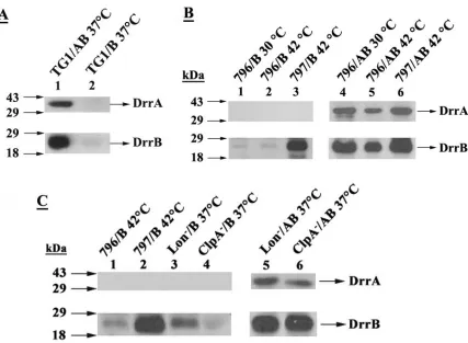
expression is induced at 42 °C, the membrane, cytosol, and inclusion body fractions were
prepared from both wild type and FtsHts cells and analyzed by Western blotting using anti-DrrA
and anti-DrrB antibodies. The data in Fig. 2 show that although some DrrB protein is present in
the inclusion body fraction in both wild type (lower panel, lane 3) and the FtsHts cells (lane 9) at
30 °C, the induction of either strain at 42 °C did not result in any increase in the amount of
inclusion body formation (lower panel, lanes 6 and 12). Moreover, no aggregated DrrAB
proteins were seen in the stacking region of the gel in any of the fractions of wild type or FtsHts
cells, indicating absence of any significant aggregation under the conditions used in these
experiments. Note that the anti-DrrB antibody is an anti-peptide antibody, therefore it shows
some cross-reactivity with epitopes in some other E. coli proteins, as explained in a previous
publication (1).
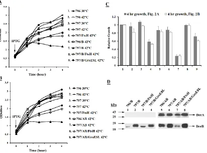
Expression of DrrB alone or DrrAB together in FtsH-deficient cells results in growth
inhibition- The growth analysis of the wild type and FtsHts cells expressing DrrB or DrrAB at
30 °C or 42 °C was carried out (Figs. 3A and 3B). The relative growth of various strains at the
four hour time point was plotted in a histogram (Fig. 3C). The data in Fig. 3A show that the
growth of mutant FtsHts cells is compromised at 42 °C (Fig. 3A; 797, open diamonds; Fig. 3C,
Column 4) as compared to the growth of wild-type cells under similar conditions (Figs. 3A, 796,
lines; Fig. 3C, Column 2). This result is not surprising due to the essential nature of E. coli FtsH.
Interestingly, the expression of DrrB alone in FtsHts cells at 42 °C further inhibited the growth of
these cells (Fig. 3A, compare 797, open diamonds with 797/B, filled triangles; Fig. 3C, Column
7). However, growth inhibition was not seen when DrrB was expressed in wild-type cells at 42
°C (compare 796, lines with 796/B, open rectangles; Fig. 3C, column 6). Since DrrB
conclude that the growth defect seen in mutant cells is caused by the accumulation of
unassembled DrrB protein. Surprisingly, however, growth inhibition was also seen in the FtsHts
cells expressing DrrA and DrrB together at 42 °C (Fig. 3B, 797/AB, filled triangles; Fig. 3C,
Column 7). This effect was unexpected because DrrA and DrrB can be expressed together in the
wild-type cells at 42 °C without any negative effect on their growth (Fig. 3B, 796/AB, open
rectangles; Fig. 3C, Column 6). These data indicate that the DrrAB proteins expressed in FtsHts
cells at 42 °C may be misfolded, and the accumulation of misfolded membrane proteins results
in growth inhibition. It was also observed that if the FtsHts cells initially induced at 42 °C were
shifted down to 30 °C, the cell growth resumed, albeit slowly. After 43 hours of temperature
shift-down, the final growth was about half as compared to the cells induced and maintained at
30 °C (data not shown), indicating that the growth inhibition of these cells is quite severe. In
summary, the data in Fig. 3 suggest that FtsH is not only responsible for removing unassembled
DrrB (in the absence of DrrA) but it may also be critical for proper assembly of the DrrAB
complex in the membrane.
The growth defect of the FtsHts cells expressing DrrB or DrrAB at 42 °C could be
rescued by overexpression of FtsH in trans (Figs. 3A-B, open triangles; Fig. 3C, Column 8),
indicating that the absence of functional FtsH was solely responsible for this defect. The growth
defect in each case was also suppressed by overexpression of the chaperone GroESL (Figs.
3A-B, filled circles; Fig. 3C, Column 9) but not to the same extent as seen with FtsH. Western blot
analysis of the membrane fractions (prepared from the four hour cultures of samples # 6-9 in
Figs. 3A-B) showed that while FtsH overexpression resulted in significant proteolysis of DrrAB
that FtsH restores growth by simply removing misfolded DrrAB proteins, while GroESL is able
to alter their conformation, thus alleviating growth inhibition.
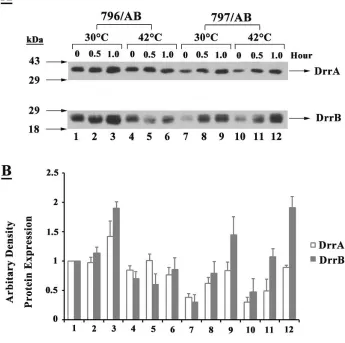
FtsH preferentially proteolyzes misfolded DrrAB - To determine if FtsH discriminates
between properly assembled and misfolded DrrAB, the effect of overexpression of FtsH
proteolysis was compared in wild type or FtsHts cells at 30 °C or 42 °C. Interestingly,
overexpression of FtsH in either wild type (Figs. 4A-B, compare lanes 1 and 3) or FtsHts cells
(lanes 2 and 4) produced no significant proteolysis of DrrA and DrrB expressed at 30 °C,
showing that FtsH does not proteolyze properly assembled DrrAB. However, when the DrrAB
proteins were expressed in wild-type cells at 42 °C, simultaneous overexpression of FtsH
resulted in significant proteolysis (Figs. 4A-B, compare lanes 3 and 7). These data suggest that
the DrrAB proteins must acquire a partially unfolded conformation at a higher temperature (as
also seen in Fig. 1B), thus making them more susceptible to proteolysis by over-expressed FtsH.
As expected, overexpression of FtsH in FtsHts cells also showed significant proteolysis of DrrAB
expressed at 42 °C (Figs. 4A-B, lanes 4 and 8). This is consistent with the data in Fig. 3, which
suggested that the DrrAB proteins expressed at 42 °C in FtsHts cells are misfolded. (Please note
that the conformation of the DrrAB proteins in wild-type cells at 42 °C is completely different
from the DrrAB expressed in mutant FtsHts cells at 42 °C even though they are both sensitive to
overexpressed FtsH. In a later experiment in Fig. 8, it is shown that the DrrAB proteins
expressed in wild type cells at 42 °C retain normal function, while the DrrAB expressed in FtsHts
cells are inactive due to their misfolding)
The rate of proteolysis of misfolded DrrAB by FtsH was analyzed in a separate
time-course experiment. The misfolded DrrAB proteins were first allowed to accumulate in the
added to stop further synthesis, as described under Methods. The synthesis of FtsH from
pBADftsH was induced by addition of arabinose, and the proteolysis of DrrA and DrrB by FtsH
was determined by Western blot analysis. The data in Figs. 4C and 4D (filled circles) show that
synthesis of FtsH (Fig. 4E, lower panel) resulted in increasing proteolysis of misfolded DrrA and
DrrB from the membrane. At 120 minutes after addition of arabinose, about 75-80% of DrrA
and DrrB were removed from the membrane. These observations are in agreement with the
dislocation model proposed previously for the activity of FtsH (65). No significant proteolysis
of DrrAB was seen in the absence of FtsH synthesis (Figs. 4C and 4D, filled rectangles).
The AAA domain of FtsH contains a chaperone-like activity, but it is not sufficient by itself
to restore the Dox efflux function - The growth experiment in Fig. 3B suggested that FtsH may
be critical for the assembly of the DrrAB complex. Further support for this idea was obtained by
comparing the rate of assembly of the DrrAB complex in wild type and FtsHts cells. The data in
Fig. 5 show that the assembly of DrrA and DrrB in the membrane of FtsHts cells is significantly
compromised already at 30 °C as compared to in the wild type cells. A significant difference in
the amounts of DrrAB in the membrane of wild type and mutant cells was seen at all time points
tested (Fig. 5, compare lanes 1-3 with 7-9). However, this difference is most evident at the early
time points, which suggests that the rate of assembly of DrrAB is affected by FtsH. This is most
likely due to the partial defect of FtsH function in FtsHts cells already at 30 °C, leading to the
low efficiency of the DrrAB complex formation.
To determine if the ability to promote assembly of DrrAB resides in the AAA domain of
FtsH, variants containing mutations in the Walker A motif of the AAA domain (K198N
mutation) or the conserved amino acids in the proteolytic domain (the HEH mutation and the
defective AAA domain, while the HEH mutant and the AAA subclone contained an intact AAA
domain. As expected, the K198N mutation resulted in a significantly reduced ATPase activity,
however the HEH mutant and the AAA subclone were unaffected (Table 2). The in vitro
proteolytic activity assay showed that while the wild-type FtsH completely proteolyzed α-casein
in one hour, no significant reduction in the α-casein level was seen with the HEH mutant even
after two hours of incubation (Fig. 6). These analyses confirmed that the AAA and the
proteolytic domain mutants behave as expected. Therefore, they were used in two different
complementation experiments (described below) to determine if the AAA domain by itself is
sufficient for promoting assembly of the DrrAB complex.
The expression of the DrrAB proteins in FtsHts cells was previously shown to result in
severe growth inhibition (Fig. 3B, filled triangles). This inhibition was reversed by simultaneous
expression of FtsH (Fig. 3B, open triangles). In the next experiment, we asked whether
co-expression of the HEH allele or the AAA subclone can rescue FtsHts cells from the growth
inhibition resulting from DrrAB expression. The data in Fig. 7 show that the simultaneous
expression of either the HEH mutant (Fig. 7A, open circles) or the AAA subclone (Fig. 7A, filled
triangles) with DrrAB can complement the growth defect of FtsHts cells, indicating that the AAA
domain of FtsH indeed contains a chaperone-like activity. Interestingly, the HEH mutant
showed much better complementation of the growth defect as compared to the AAA subclone,
perhaps due to a more native conformation of the full-length HEH protein as compared to the
AAA subclone. The K198N mutation, on the other hand, showed no growth complementation
effect (Fig. 7A, open triangles), showing that the ATPase activity associated with the AAA
domain is important for the chaperone function of FtsH. Western blot analysis showed that the
(K198N)-containing strains (Figs. 7B and 7C, lanes 5-7), and the amounts of DrrA and DrrB in these cells
were comparable to the levels in their absence (lane 3). Therefore the restoration of growth by
the HEH and AAA clones must result from a change in conformation of the DrrAB proteins
brought about by the functional AAA domain present in these two clones. Whether the HEH and
AAA variants of FtsH can also restore function of the DrrAB complex is addressed in the next
experiment.
We previously showed that the wild-type DrrA and DrrB proteins together carry out
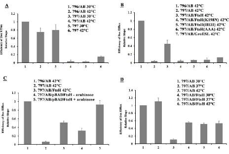
ATP-dependent efflux of the anticancer drug doxorubicin (61). Here, we investigated whether
co-expression of the AAA subclone or the HEH allele can restore the Dox efflux function of the
misassembled DrrAB proteins expressed in FtsHts cells at 42 °C (Fig. 8). The data in Fig 8A
indicate that the rate of DrrAB-mediated Dox efflux at 30 °C in the wild-type 796 and FtsHts 797
cells is comparable (Columns 1 and 3). Induction of wild-type cells at 42 °C showed only a
slight reduction in Dox efflux (Column 2), which is likely due to the destabilization effect
produced by high temperature on DrrAB, as seen earlier in Figs. 1 and 4. By contrast, the FtsHts
cells induced at 42 °C showed very little or no DrrAB-mediated Dox efflux (Fig. 8A, column 4),
which was comparable to the efflux seen with control cells containing empty vector (Fig. 8A,
Columns 5 and 6). These data confirm that the DrrAB proteins are misfolded in the absence of
functional FtsH. Simultaneous overexpression of the FtsH variants, K198N, HEH or the AAA
subclone, in FtsHts cells did not restore function of misassembled DrrAB (Fig. 8B, columns 4-6),
even though restoration of growth by HEH and the AAA subclone was earlier seen in Fig. 7A.
Similarly, overexpression of GroESL also did not complement the DrrAB-mediated Dox efflux
in FtsHts cells (Fig. 8B, column 7). One possible explanation for these data could be that even
proteins and relieve growth inhibition, it is not sufficient by itself to restore proper conformation
required for full function of the complex.
Wild-type FtsH can refold previously misassembled DrrAB and restore function
-Surprisingly, co-expression of wild-type FtsH restored the Dox efflux function of the
misassembled proteins expressed in FtsHts cells at 42 °C resulting in a significant recovery
(about 45%) of Dox efflux by the DrrAB complex (Fig. 8B; compare columns 2 and 3). Since
this could only have resulted if FtsH expressed in trans facilitated assembly of the complex,
these results imply that the AAA and the proteolytic domains of FtsH must work hand-in-hand to
bring about functional assembly of the DrrAB complex. In this experiment, however, FtsH and
the DrrAB proteins were expressed simultaneously by IPTG induction, therefore it was not
possible to determine if FtsH assists only the newly synthesized DrrAB to achieve proper
conformation, or if it can also bring about refolding of the DrrAB proteins that have already been
misfolded.
Therefore, in the next experiment, DrrAB and wild-type FtsH were expressed in FtsHts
cells in a sequential manner. The ftsH gene subcloned under the control of the araBAD
promoter was induced by arabinose, while the drrAB genes remained under the control of the lac
promoter induced by IPTG. The FtsHts cells containing both the plasmids (797/AB/pBADftsH)
were grown at 30 °C to mid-log phase. The DrrAB proteins were first induced with IPTG at 42
°C for 1 hour (this condition inactivates FtsH and renders DrrAB inactive as seen in Figs. 8A and
8B). The cells were then washed several times to remove extracellular IPTG and stop further
synthesis of DrrAB. The expression of FtsH was induced by arabinose for 1 hour at 42 °C, and
the cells were subjected to the Dox efflux assay. To maintain the chromosomally-encoded FtsH
the experiment. As previously seen in Fig. 8B, simultaneous expression of DrrAB and FtsH
resulted in restoration of Dox efflux (Fig. 8C; 797/AB/pUCftsH, column 3). More interestingly,
however, even greater restoration of the DrrAB-mediated Dox efflux was seen when FtsH was
induced after DrrAB proteins had been pre-synthesized in these cells (Fig. 8C,
797/AB/pBADftsH+ara, column 5). In the absence of arabinose induction of FtsH, much lower
restoration of Dox efflux was seen (Fig. 8C, 797/AB/pBADftsH – ara, column 4). These data,
therefore, show that the sequential expression of DrrAB and FtsH can still restore the function of
the previously misfolded DrrAB proteins to the same (or even higher) extent as seen with
simultaneous expression.
Finally, the effect of over-expression of FtsH on Dox efflux function of DrrAB expressed
at different temperatures was investigated. Irrespective of whether the DrrAB proteins were
induced at 30 °C, 37 °C, or 42 °C in FtsHts cells, simultaneous expression of FtsH resulted in a
very similar final Dox efflux efficiency (Fig. 8D). At 42 °C, co- expression of FtsH enhanced
Dox efflux of misfolded DrrAB 5-fold (compare columns 3 and 6), yielding about 45% Dox
efflux efficiency. Interestingly, over-expression of FtsH at 30 °C or 37 °C reduced the efficiency
by about half (compare columns 1-2 with 4-5), once again yielding final Dox efflux efficiency of
about 45%. These results imply that FtsH produces an optimal level of functional complexes in
the membrane perhaps by exerting both proteolytic and refolding effects concurrently.
2.4 Discussion
Non-native proteins, especially unassembled membrane proteins, interfere with cellular
processes and are known to become toxic to the cells. Therefore, quality control systems,
consisting of chaperones and proteases, play essential roles by monitoring their folding and
proteins provide classical examples of ATP-dependent chaperones which prevent aggregation of
newly translated proteins and promote their refolding (79). A special class of chaperones (e.g.
ClpB in bacteria and its homologs Hsp78 and Hsp104 in eukaryotes) is known to resolubilize
protein aggregates and, in cooperation with the Hsp70 chaperones (specifically DnaKJE), can
result in regaining function of the affected protein (72). On the other end of the spectrum are
proteins classically defined as proteases, for example Lon, ClpA/P, ClpX/P and FtsH, whose
major function is considered to be removal of irreversibly damaged proteins from the cell
(63,71,81). Despite their differences, however, both classical chaperones and proteases share
common features. For example, both have the ability to recognize and bind non-native
polypeptides and both bring about unfolding of their substrates, which are subsequently refolded
(by a chaperone) or degraded (by a protease)(69,79,82). Because of the ATP-dependent
unfolding function of AAA+ proteases, it has been speculated that they may also have the ability
to refold substrate proteins and may participate in protein biogenesis. However, very little direct
evidence is available for the role of FtsH or other AAA+ proteases in biogenesis, especially of
membrane protein complexes. In this study, we provide clear evidence that the E. coli FtsH is
able to both degrade and refold misassembled DrrAB proteins, resulting in regaining the Dox
efflux function of the membrane complex.
We show that in the absence of the DrrA protein, DrrB acquires an FtsH-sensitive
conformation and is completely proteolyzed. However, in the absence of functional FtsH, the
DrrB protein accumulates even in the absence of DrrA confirming that FtsH monitors the folding
status of DrrB and removes it if it is improperly assembled. The molecular details of proteolysis
of DrrB by FtsH are currently unknown, however based on the prevalent model for its action
end (both of which are found in the cytoplasm, (84)). Cross-linking studies previously showed
that the N terminus of DrrB is the major site of interaction with DrrA (61,84); therefore we
propose that proteolysis of DrrB initiates at its N- terminal tail, and binding of DrrA to this
region of DrrB protects it from proteolysis by FtsH.
Interestingly, we found that the function of FtsH is not limited to proteolysis of
unassembled DrrB, but it also plays an essential role in folding and assembly of the DrrAB
complex. This conclusion is supported by several lines of evidence presented in this paper.
First, the expression of DrrA and DrrB together in FtsHts cells at 42 °C (which results in
inactivation of FtsH) was found to be growth inhibitory (Fig. 3B)suggesting that the complex is
improperly assembled in the absence of a functional FtsH. Second, the rate of assembly of the
DrrAB complex in the FtsHts cells at 30 °C was found to be significantly reduced as compared to
the wild-type cells (Fig. 5). Third, functional analysis showed complete absence of the
DrrAB-mediated Dox efflux in FtsHts cells under conditions of FtsH inactivation, suggesting that most
or all of the DrrAB proteins expressed in these cells at 42 °C are misassembled. By contrast, the
DrrAB proteins expressed in wild-type cells at 42 °C retained on average 85-90% of the Dox
efflux activity (Fig. 8A). Finally, co-expression of FtsH in trans in FtsHts cells restored the
ability of DrrAB to carry out Dox efflux, confirming that FtsH facilitates assembly of the DrrAB
complex (Fig. 8B). Nevertheless, this result was surprising because FtsH contains a functional
proteolytic domain. It’s overexpression in FtsHts
cells results in proteolysis of the misfolded
DrrAB proteins (as seen in Fig. 4), however the data in Fig. 8B show that FtsH also facilitated
some folding resulting in about 45% recovery of the Dox efflux activity. Either the AAA
domain by itself or the GroESL chaperone was unable to complement the Dox efflux function of
together these data suggest that both the AAA and the protease domains of FtsH are essential for
promoting functional assembly of DrrAB.
The most crucial evidence for the refolding function of FtsH, however, came from the
sequential expression studies. Irrespective of whether FtsH was expressed simultaneously with
DrrAB or expressed after the non-functional DrrA and DrrB proteins had already accumulated in
FtsHts cells, it was able to restore the function of the complex (Fig. 8C), thus showing
conclusively that FtsH not only facilitates assembly of the DrrAB complex but it is also actively
involved in refolding previously misassembled DrrAB proteins. Interestingly, we also found that
the sequential expression of DrrAB and FtsH resulted in a significantly higher recovery of the
Dox efflux function of DrrAB as compared to simultaneous expression (Fig. 8C). This finding
suggests that FtsH treats its substrate differently during its synthesis as compared to after it has
already been synthesized.
In summary, our studies confirm that the AAA domain of FtsH can recognize and bind
substrates and change their conformation, which is in agreement with the previous studies (75).
However, we also show that the two activities (ATPase and proteolytic) of FtsH must be present
simultaneously and occur in a coordinated manner to facilitate assembly and refolding of DrrAB.
Much more extensive analysis will be required in the future to understand the nature of the
molecular processes involved in refolding of DrrA and DrrB and to determine if other factors
also play a role in the assembly of the DrrAB complex. Further studies will also provide clues
about how degradation and assembly of multi-subunit complexes are regulated, and whether
other AAA+ proteases may also contain chaperone activity. This study raises intriguing
questions about the distinction between classical chaperones like GroESL (that can prevent
proteolysis but also actively participate in refolding of their specific substrates, as shown in this
study. Bukau and Mogk previously (79) coined four terms to describe the various activities of
chaperones and proteases: Holders (small heat shock proteins, Hsps), Folders (GroESL and
DnaK), Unfolders (ClpA, ClpX, and ClpB), and Proteases (Lon, ClpP, and FtsH). In light of the
findings reported in this article, we propose a new term ‘Specific Refolder’ to describe the
function of FtsH and possibly other AAA+ proteases that may be shown in the future to contain
Table 2.1 Baterial strains, plasmids and anti-sera
Table 2.2 The ATPase activity of purified FtsH and its variants
Name Description Reference or
Source Bacterial Strains
TG1 K-12 Δ(lac-pro) supE thi hsdD5/F’tra36 proA+B+ lacIq
lacZΔM15
(85) HMS174(DE
3)
F- recA1 hsdR (rK12- mK12+)(DE3)(Rif R) (85)
AR796 F-, zhd-33::Tn10, araD139, (argF-lac)U169, rpsL150, rpsE,
relA1, f1bB5301, deoC1, ptsF25, rbsR, zgj3198::Tn10kan.
(54) AR797 F-, zhd-33::Tn10, araD139, (argF-lac)U169, rpsL150, rpsE,
relA1, f1bB5301, deoC1, ptsF25, rbsR, zgj3198::Tn10kan.
ftsH1
(54)
SG1110 Δlon-510, zba-1091:: ΔTn10(λ) (86)
SG1126 Δarg, clpA319:: Δkan (86)
Plasmids
pSU2718 Cloning vector, pACYC184 derivative, Cmr (59)
pKY326 groES/groEL in pHSG575, Cmr (76)
pDX101 drrAB in pSU2718, Cmr (59)
pDX103 drrB in pSU2718, Cmr (59)
pUCftsH ftsH in pUC18, Ampr, restriction sites: EcoRI, HindIII This study pUCftsH(K19
8N)
pUC18 containing ftsH with mutation of Lys198 to Asn, Ampr This study pUCftsH(HE
H)
pUC18 containing ftsH with mutations of His417Glu418His421
to Ala417Gln418Ala421, Ampr
This study pUCftsH(AA
A)
pUC18 containing ftsH with deletion of proteolytic domain,
Ampr
This study pUCgroESL groES/groEL in pUC18, Ampr, restriction sites: NdeI,
HindIII
This study
pETftsH ftsH in pET28a with C-terminal histag, Kanr This study
pETFftsH(K1
98N)
ftsH(K198N) in pET28a with C-terminal histag, Kanr This study
pETftsH(HEH
)
ftsH(HEH) in pET28a with C-terminal histag, Kanr This study
pETftsH(AAA
)
ftsH(AAA) in pET28a with C-terminal histag, Kanr This study
pBADftsH ftsH in pBAD/HisA, Ampr This study
FtsH Variant ATPase activity (nmol Pi/min/mg)
Wild-type FtsH 139.5
FtsH(K198N) 18.1
FtsH(HEH) 147.8
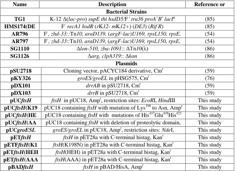
[image:39.612.64.499.521.595.2]Figure 2.1 Role of DrrA and FtsH in stable maintenance of DrrB
FIGURE 2.1 Role of DrrA and FtsH in stable maintenance of DrrB. (A) DrrA is required
for stable expression of DrrB. Wild-type E.coli (TG1) cells expressing DrrAB (pDX101) or
DrrB alone (pDX103) were grown at 37° C to mid-log phase (A600nm=0.6) and induced with 0.25
mM IPTG for 3 h. 30 µg membrane proteins were loaded onto 12% SDS-PAGE gels, followed
by Western blotting against anti-DrrA (upper panel) and anti-DrrB (lower panel) antibodies. (B)
FtsH is responsible for proteolysis of unassembled DrrB. Wild-type E. coli (796) or the E.
coli 797 (ftsHts) cells expressing DrrAB (pDX101) or DrrB alone (pDX103) were grown at 30 °C
to mid-log phase and induced with 0.25 mM IPTG at either 30 °C or 42 °C for 3 h. Western blot
protease is not involved in quality control of DrrB.Wild-type E. coli (796), E. coli 797
(ftsHts), E. coli SG1110 (Lon-), and E. coli SG1126 (ClpA-) cells expressing DrrAB (pDX101) or
DrrB alone (pDX103) were grown at 30 °C or 37 °C to mid-log phase and induced with 0.25
mM IPTG at either 42 °C or 37 °C for 3 h, as indicated. Analysis was carried as described in (B)
[image:41.612.148.502.234.622.2]above.
FIGURE 2.2 Western blot analysis of the cytosol, membrane, and inclusion body fractions
of E. coli 796 or 797 cells expressing DrrAB at 30 °C or 42 °C. Wild-type E. coli 796 or the
E. coli 797 (ftsHts) cells expressing DrrAB (pDX101) were grown at 30 °C to mid-log phase and
induced with 0.25 mM IPTG at either 30 °C or 42 °C for 3 h. Cell fractions were prepared, as
described under Methods. 20 µg of each fraction was loaded onto 12% SDS-PAGE gels,
followed by Western blotting against anti-DrrA (upper panel) and anti-DrrB (lower panel)
antibodies. (m, membrane; c, cytosol; IB, inclusion body). Note that the anti-DrrB antibody is
an anti-peptide antibody, therefore it shows some cross-reactivity with epitopes in other E. coli
[image:42.612.80.492.322.627.2]proteins, as explained in a previous publication (59).
Figure 2.3 Growth inhibition resulting from expression of DrrB alone or DrrAB together in E.
coli 797 cells can be relieved by the overexpression of FtsH or GroESL.
FIGURE 2.3 Growth inhibition resulting from expression of DrrB alone or DrrAB together
in E. coli 797 cells can be relieved by the overexpression of FtsH or GroESL. (A) Effect of
overexpression of FtsH or GroESL on growth of 797 cells expressing DrrB. 797 cells
containing pDX103 (DrrB) and pUCftsH or pUCgroESL were grown at 30 °C and induced at 42
°C, as in Fig. 1B. The growth was monitored at O.D. 600 nm for four hours after induction. (B)
Effect of overexpression of FtsH or GroESL on growth of 797 cells expressing DrrAB. 797
cells containing pDX101 (DrrAB) and pUCftsH or pUCgroESL were anayzed, as described in
(A) above. (C) Quantitation of the growth of 796 or 797 cells expressing DrrB or DrrAB at
4 hours after induction. The growth of the sample #1 at 4 hours in Fig. 3A or 3B was
designated as 1.0. The relative growth of each culture at 4 hours was calculated. The histogram
represents an average of 3 experiments. Columns 1-9 correspond to the cultures 1-9 in Figs 3A
(filled) and 3B (open). (D) Western blot analysis. The membrane fractions were prepared from
sample #s 6-9 collected at the 4 hour time point in Fig. 3A and 3B. Western blot analysis was
Figure 2.4 FtsH preferentially degrades misfolded DrrAB.
FIGURE 2.4 FtsH preferentially degrades misfolded DrrAB. (A) E. coli 796 or 797 cells
containing pDX101(DrrAB) and pUCftsH were grown and analyzed by Western blotting, as in
Fig. 1A. (B) Quantitative analysis of the amounts of DrrA and DrrB. The intensity of the
bands on the nitrocellulose membrane in Fig. 4A was determined by densitometric scanning.
The intensity of DrrA and DrrB in lane 1 was designated as 1. The data represent an average of
3 experiments. Columns 1-8 correspond to the lanes 1-8 in Fig. 4A. (C), (D) and (E) In vivo
FtsH proteolytic assay. The assay was carried out as described under Methods. The membrane
fractions were prepared from the cells collected at the indicated time points after addition of
arabinose. 20 µg total membrane protein was loaded onto 12% SDS-PAGE, followed by
Western blot with anti-DrrA, anti-DrrB, or anti-FtsH antibodies. The intensity of the bands on
the nitrocellulose membrane was determined by densitometric scanning. The intensity of DrrA
membrane at the indicated time points after addition of arabinose. The data shown represent an
average of 3 experiments. (D) Quantitation of DrrB in the membrane. (E) Western blot analysis
showing amount of FtsH present in the membrane at various times after addition of arabinose.
Top panel: cells containing pBAD vector only. Bottom panel: cells containing the plasmid
[image:45.612.132.477.254.597.2]pBADftsH.