C A S E R E P O R T
Open Access
Recurrent biliary dissemination of colon
cancer liver metastasis: a case report
Ichiro Onishi
1*, Masato Kayahara
1, Ryohei Takei
1, Naoki Makita
1, Masayoshi Munemoto
1, Yasumichi Yagi
1and Atsuhiro Kawashima
2Abstract
Background:Most colorectal cancer liver metastases form nodules within the hepatic parenchyma, and hepatectomy is the only radical treatment for synchronous metastases. There is concern about intrabiliary tumor growth which may affect the surgical margin, resulting in local recurrence after hepatectomy for colorectal cancer liver metastasis; however, there has been no report of the dissemination in the bile duct after hepatectomy. Here, we report an unusual case of biliary dissemination of colorectal cancer that caused recurrent intrabiliary growth after hepatectomy, and discuss the management of intrabiliary metastasis of colorectal cancer.
Case presentation:A 69-year-old Japanese man underwent treatment for liver dysfunctions 3 years after aortic valve replacement. Computed tomography revealed an enhanced tumor within the hilar bile duct and dilatation of the left hepatic duct, typical of hilar cholangiocarcinoma. Endoscopic retrograde cholangiopancreatography revealed tumor shadow in his bile duct, and the cytology confirmed malignant cells in the bile. We performed extended left hepatectomy with bile duct resection; his postoperative course remained good without acute
complications. After 3 months postoperatively, he was readmitted for subacute cholangitis and obstructive jaundice. Immediately, percutaneous transhepatic cholangiography drainage was performed, followed by cholangiography that exhibited intrabiliary tumor growth in the remnant liver. On immunohistochemical examination, tumor cells were positive for cytokeratin 20 and CDX2 but negative for cytokeratin 7. Then, computed tomography revealed an enhanced tumor-like lesion at the descending colon. After 3 months, left hemicolectomy was performed.
Meanwhile, the percutaneous transhepatic cholangiography drainage fluid turned bloody, which was considered to be bleeding from a residual bile duct tumor. Accordingly, radiotherapy was initiated to prevent tumor bleeding around the hilar bile duct, but, unfortunately, the effects were short-lived, and cholangitis rebooted after 1 month leading to our patient’s death due to septic liver failure. Autopsy revealed a remnant tumor in the bile duct, but no noticeable nodular metastasis was observed, except for a single small metastasis in the lower lobe of the left lung. Conclusions:The intrabiliary growth of metastatic colorectal cancer mimics cholangiocarcinoma occasionally. To date, as the effect of chemotherapy or radiotherapy remains uncertain, the complete resection of a bile duct tumor is the only method which could result in a better prognosis.
Keywords:Biliary dissemination, Colon cancer, Liver metastasis, Intrabiliary growth
* Correspondence:ionishi@kinbyou.hosp.go.jp
1Department of Surgery, National Hospital Organization Kanazawa Medical
Center, 1-1 Shimoishibiki, Kanazawa 920-8650, Japan
Full list of author information is available at the end of the article
of these, many are case reports, and some are a limited number of case series. Moreover, there has been no report of recurrence in the bile duct inoculation after hepatec-tomy; the actual mechanism of biliary dissemination of CRC is still unknown.
Here we report a rare case of biliary dissemination of CRC which caused recurrent intrabiliary growth after hepatectomy and discuss the management of intrabiliary metastasis of CRC.
Case presentation
A 69-year-old Japanese man underwent treatment for liver dysfunction 3 years after aortic valve replacement. Later, rapid elevation in his serum alkaline phosphatase (ALP) level was recorded and he was readmitted to de-termine the etiology. His body temperature was 36 °C, blood pressure 164/65 mmHg, and pulse rate was 66/ minute. Laboratory data revealed mild anemia and liver-renal injury: white blood cells (WBC) 4600/uL, hemoglobin 9.7 g/dL, platelet 18.9 × 10⁴/dL, C-reactive protein (CRP) 0.29 mg/dL, ALP 1138 U/L, aspartate aminotransferase (AST) 40 U/L, alanine aminotransfer-ase (ALT) 37 U/L, and γ glutamyl transpeptidase (γ-GTP) 298 U/L. His blood urea nitrogen (BUN) was
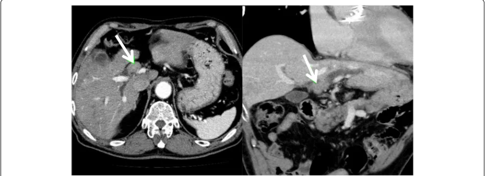
computed tomography (CT) revealed an enhanced tumor within the hilar bile duct and dilatation of the left hepatic duct (Fig.1), which are typical findings for hilar cholangiocarcinoma. In addition, endoscopic retrograde cholangiopancreatography (ERCP) revealed tumor shadow in his bile duct, and the cytology confirmed malignant cells in the bile (Fig.2). As no lymph node and distant metasta-ses were detected, we inserted endoscopic nasobiliary drainage (ENBD) to reduce jaundice as preparation for sur-gery. We performed extended left hepatectomy with resec-tion of his bile duct; his postoperative course was good without severe complications. After 3 months postopera-tively, he was readmitted for subacute cholangitis and ob-structive jaundice. Immediately, percutaneous transhepatic cholangiography drainage (PTCD) was performed, followed by cholangiography that exhibited the intrabiliary tumor growth in the remnant liver.
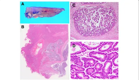
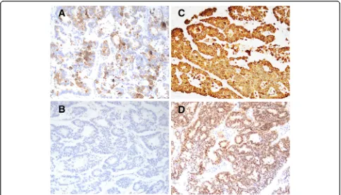
Simultaneously, histological examination of resected specimens revealed tumor growth in the hilar duct across the left hepatic duct (Fig. 3a, b). Microscopic findings at the same site revealed a dilated bile duct filled with well-differentiated tubular adenocarcinoma (Fig. 3c, d). On immunohistochemical examination, tumor cells were positive for cytokeratin (CK) 20 (Fig.4a)
[image:2.595.56.542.527.703.2]but negative for CK7 (Fig. 4b). Furthermore, CK18 (Fig.4c) as control and CDX2 (Fig.4d) were stained. Al-though these findings were not typical of intrahepatic cholangiocarcinoma, hepatic metastasis from another primary lesion was strongly suspected [12,13]. Further-more, CT revealed an enhanced tumor-like lesion at the descending colon, followed by diagnosis of type 2 cancer in total colonography. Then, left hemicolectomy was performed; the immunohistochemical-identified feature matched with an intrabiliary tumor. Meanwhile, the PTCD fluid turned bloody, which was considered to
indicate bleeding from a residual bile duct tumor (Fig.5). Accordingly, we planned chemotherapy with orally ad-ministered capecitabine but our patient experienced a spike fever because of refractory cholangitis. Thus, we abandoned chemotherapy and initiated radiotherapy to stop the tumor bleeding around the hilar bile duct. After completing radiotherapy (total 50 Gy) for approximately 1 month, we observed an improvement in his liver func-tion because of tumor shrinkage. Unfortunately, the effects were short-lived, intrabiliary growth and cholan-gitis rebooted after 1 month leading to his death due to Fig. 2Endoscopic retrograde cholangiopancreatography and bile cytology. Endoscopic retrograde cholangiopancreatography shows the left hepatic duct obstruction and tumor shadow in the common hepatic duct. Malignant cells were observed in the bile duct cytology
Fig. 3Tumor growing in the hilar duct across the left hepatic duct. The dilated bile duct was filled with well-differentiated tubular
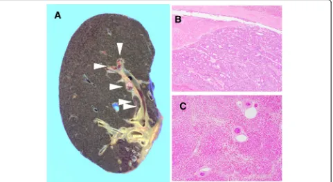
[image:3.595.55.540.88.222.2] [image:3.595.58.538.416.694.2]septic liver failure (Fig. 6). Autopsy revealed a remnant tumor in the bile duct (Fig.7), but no noticeable nodular metastasis was observed, except for a single small metas-tasis in the lower lobe of his left lung.
Discussion
We reported an unusual case of biliary dissemination of CRC that caused recurrent intrabiliary growth after
hepatectomy. Unfortunately, 3 months postoperatively, he was readmitted for subacute cholangitis and obstruct-ive jaundice due to intrabiliary tumor growth in the remnant liver. Eventually he died of septic liver failure caused by cholangitis. An autopsy revealed a remnant tumor in the bile duct, but no noticeable nodular metas-tasis was observed. Intrabiliary metasmetas-tasis of CRC is not typically reported in both Eastern and Western countries Fig. 4On immunohistochemical examination, tumor cells are positive for cytokeratin 20 (a) but negative for cytokeratin 7 (b). Cytokeratin 18 (c) as control and CDX2 (d) were also stained; × 100
[image:4.595.56.540.87.364.2] [image:4.595.57.539.523.702.2][9]. Owing to the rarity of the disease, a majority of pub-lished literature comprises either case reports or studies of small case series [5, 7–9, 11, 12]. Reportedly, the prevalence of intrabiliary metastasis by CRC ranges from 3.6–10.6% [9]; however, the prognosis of this disease re-mains unclear. In Japan, it might be less aggressive com-pared with usual nodular liver metastasis, and active resection is considered to improve patients’survival [2,
3, 6]. We decided to perform extended left hepatectomy in this case of biliary dissemination of CRC. In contrast,
five cases with poor prognosis have been reported in Korea [11]; these patients did not undergo surgery but palliative procedures, such as PTCD or endoscopic retrograde biliary drainage (ERBD), with a risk of re-peated cholangitis. In our case, refractory cholangitis due to intrabiliary tumor growth in the remnant liver caused liver failure similarly. Interestingly, despite the remnant tumor in the bile duct, no life-threatening dis-tant metastasis occurred until our patient’s death. We anticipate that bile duct metastasis is, basically, a sign of Fig. 6After 3 months postoperatively, he was readmitted for subacute cholangitis and obstructive jaundice. Immediately, percutaneous
transhepatic cholangiography drainage was performed, followed by cholangiography that exhibited intrabiliary tumor growth in the remnant liver. Radiotherapy was initiated, but the effects were short-lived.PTCDpercutaneous transhepatic cholangio-drainage
[image:5.595.60.540.87.248.2] [image:5.595.60.541.436.700.2]USA reported the highest number of cases and demon-strated that all prevalence estimates for intrabiliary growth were considerably affected by different clinical management styles for metastatic CRC within each insti-tution [9]. However, the effect of chemotherapy and radiotherapy remains unknown. As mentioned in this case, radiotherapy could have resulted in the tumor shrinkage effect, but it could have been temporary. Per-haps, the complete resection of the intrabiliary growth would have improved the prognosis of our patient.
Conclusions
Occasionally, the intrabiliary growth of metastatic CRC mimics cholangiocarcinoma and might cause refractory cholangitis. To date, as the effect of chemotherapy or radiotherapy remains uncertain, the complete resection of bile duct tumors is the only method to prevent post-operative cholangitis and could result in a better prognosis.
Abbreviations
ALP:Alkaline phosphatase; CK: Cytokeratin; CRC: Colorectal cancer; CT: Computed tomography; PTCD: Percutaneous transhepatic cholangio-drainage
Acknowledgements
We thank Kenichi Harada for valuable assistance on pathological diagnosis.
Availability of data and materials
The datasets analyzed during the current study are available from the corresponding author on reasonable request.
Authors’contributions
IO reported the case and wrote the manuscript. IO, MK, and RT performed the surgery and perioperative management of the patient and helped in drafting the manuscript. NM, YY, and AK participated in revising the manuscript critically. All authors read and approved the final manuscript.
Ethics approval and consent to participate
Not applicable.
Consent for publication
Written informed consent was obtained from the patient's daughter for publication of this case report and any accompanying images. A copy of the written consent is available for review by the Editor-in-Chief of this journal.
Competing interests
The authors declare that they have no competing interests.
1. Adam R, de Gramont A, Figueras J, Kokudo N, Kunstlinger F, Loyer E,et al. Managing synchronous liver metastases from colorectal cancer: a multidisciplinary international consensus. Cancer Treat Rev. 2015;41:729–41. 2. Okano K, Yamamoto J, Moriya Y, Akasu T, Kosuge T, Sakamoto M,et al.
Macroscopic intrabiliary growth of liver metastasis from colorectal cancer. Surgery. 1999;126:829–34.
3. Kubo M, Sakamoto M, Fukushima N,et al.Less aggressive features of colorectal cancer with liver metastases showing macroscopic intrabiliary extension. Pathol Int. 2002;52:514–8.
4. Povoski SP, Klimstra DS, Brown KT, Schwartz LH, Kurtz RC, Jarnagin WR,et al. Recognition of intrabiliary hepatic metastases from colorectal
adenocarcinoma. HPB Surg. 2000;11:383–91.
5. Tokai H, Kawashita Y, Eguchi S, Kamohara Y, Takatsuki M, Okudaira S,et al.A case of mucin producing liver metastases with intrabiliary extension. World J Gastroenterol. 2006;12:4918–21.
6. Sugiura T, Nagino M, Oda K, Ebata T, Nishio H, Arai T,et al.Hepatectomy for colorectal liver metastases with macroscopic intrabiliary tumor growth. World J Surg. 2006;30:1902–8.
7. Kayashima H, Taketomi A, Yamashita Y, Kuroda Y, Kitagawa D, Harimoto N, et al.Liver metastasis with intraductal invasion originating from rectal cancer: report of a case. Surg Today. 2008;38:765–8.
8. Ghittoni G, Caturelli E, Viera FT. Intrabile duct metastasis from colonic adenocarcinoma without liver parenchyma involvement: contrast enhanced ultrasonography detection. Abdom Imaging. 2010;35:346–8.
9. Nakamura T, Harada S, Nakao T, Masuda K, Wilkinson G, Sako H,et al. Successful liver resection for the unusual case of peripheral intra bile duct growth of liver metastasis from colorectal carcinoma. J Surg Case Rep. 2013; https://doi.org/10.1093/jscr/rjt055.
10. Estrella JS, Othman ML, Taggart MW, Hamilton SR, Curley SA, Rashid A,et al. Intrabiliary growth of liver metastases: clinicopathologic features, prevalence, and outcome. Am J Surg Pathol. 2013;37:1571–9. 11. Kawakatsu S, Kaneoka Y, Maeda A, Takayama Y, Fukami Y, Onoe S,et al.
Intrapancreatic bile duct metastasis from colon cancer after resection of liver metastasis with intrabiliary growth: a case report. World J Surg Oncol. 2015;13:254.
12. Koh FH, Shi W, Tan KK. Biliary metastasis in colorectal cancer confers a poor prognosis: case study of 5 consecutive patients. Ann Hepatobiliary Pancreat Surg. 2017;21:57.
13. Rullier A, Le Bail B, Fawaz R, Blanc JF, Saric J, Bioulac-Sage P. Cytokeratin 7 and 20 expression in cholangiocarcinomas varies along the biliary tract but still differs from that in colorectal carcinoma metastasis. Am J Surg Pathol. 2000;24:870–6.
14. Werling RW, Yaziji H, Bacchi CE, Gown AM. CDX2, a highly sensitive and specific marker of adenocarcinomas of intestinal origin: an