Diagnostic Accuracy of PCR Alone and Compared to Urinary Antigen
Testing for Detection of
Legionella
spp.: a Systematic Review
Tomer Avni,a Amir Bieber,aHefziba Green,aTali Steinmetz,aLeonard Leibovici,aMical Paulb
Medicine E, Beilinson Hospital and Sackler Faculty of Medicine, Tel-Aviv University, Israela; Infectious Diseases Unit, Rambam Medical Center and Rappaport Faculty of
Medicine, Tehnion, Israel Institute of Technology, Haifa, Israelb
The diagnosis of Legionnaires’ disease (LD) is based on the isolation ofLegionellaspp., a 4-fold rise in antibodies, a positive uri-nary antigen (UA), or direct immunofluorescence tests. PCR is not accepted as a diagnostic tool for LD. This systematic review assesses the diagnostic accuracy of PCR in various clinical samples with a direct comparison versus UA. We included prospective or retrospective cohort and case-control studies. Studies were included if they used the Centers for Disease Control and Preven-tion consensus definiPreven-tion criteria of LD or a similar one, assessed only patients with clinical pneumonia, and reported data for all true-positive, false-positive, true-negative, and false-negative results. Two reviewers abstracted data independently. Risk of bias was assessed using Quadas-2. Summary sensitivity and specificity values were estimated using a bivariate model and reported with a 95% confidence interval (CI). Thirty-eight studies were included. A total of 653 patients had confirmed LD, and 3,593 pa-tients had pneumonia due to other pathogens. The methodological quality of the studies as assessed by the Quadas-2 tool was poor to fair. The summary sensitivity and specificity values for diagnosis of LD in respiratory samples were 97.4% (95% CI, 91.1% to 99.2%) and 98.6% (95% CI, 97.4% to 99.3%), respectively. These results were mainly unchanged by any covariates tested and subgroup analysis. The diagnostic performance of PCR in respiratory samples was much better than that of UA. Com-pared to UA, PCR in respiratory samples (especially in sputum samples or swabs) revealed a significant advantage in sensitivity and an additional diagnosis of 18% to 30% of LD cases. The diagnostic performance of PCR in respiratory samples was excellent and preferable to that of the UA. Results were independent on the covariate tested. PCR in respiratory samples should be re-garded as a valid tool for the diagnosis of LD.
P
neumonia caused by Legionella spp. (Legionnaires’ disease[LD]) is a life-threatening pulmonary infection. The most
common species causing clinical disease in humans isLegionella
pneumophila(1). In addition toL. pneumophila, 19 species are documented as human pathogens on the basis of their isolation
from clinical specimens (2). LD can affect people both in the
com-munity (3) and in the hospital and, in both settings, can occur in
outbreaks (4,5). The true incidence of LD is difficult to assess,
because the bacterial etiology for community-acquired pneumo-nia (CAP) is generally not documented in clinical practice. LD cannot be differentiated clinically or radiographically from CAP
caused by other bacterial pathogens (6). As Legionellaspp. are
obligatory intracellular bacteria, they are unaffected by beta-lac-tam antibiotics and require specific treatment with high-dose
quinolones or macrolides (7). Treatment providing coverage
againstLegionellaspp. has been shown to improve clinical success
(8). Thus, early diagnosis of LD is important and can have an effect
on both public health and management in hospitals (9,10).
Conventional methods for the diagnosis of LD consist of cul-ture, antigen detection in urine (i.e., urine antigen [UA]), serolog-ical testing, and direct fluorescent antibody (DFA) staining or immunohistochemistry (IHC). PCR-based methods for the
diag-nosis ofLegionellaspp. are usually based on conserved regions of
rRNA sequences for amplification; these regions are not specific
and, hence, can be used for detection of anyLegionellasubspecies.
Real-time PCR methods, on the other hand, frequently use the
mac-rophage infectivity potentiator gene (MIP) as a target for the specific
detection ofL. pneumophila; hence, they are used for the detection
ofL. pneumophila only. PCR enables specific amplification of
minute amounts of LegionellaDNA, provides results within a
short time frame, and has the potential to detect infections caused
byLegionellaspp. We systematically reviewed all studies assessing PCR in clinical samples for the diagnosis of LD. We also compared and assessed the value of PCR compared to, and combined with, UA.
MATERIALS AND METHODS
Inclusion criteria.We included prospective or retrospective cohort stud-ies and case-control studstud-ies. Participants (both cases and controls) were patients with pneumonia, either CAP or hospital acquired, as defined by radiological signs and clinical symptoms and signs (i.e., target condition). Case-control studies in which controls were healthy people were analyzed separately.
The index test was PCR forLegionellaspp. performed on any clinical sample (sputum, bronchoalveolar lavage [BAL] sample, serum, urine, sterile fluids, and tissues). Analyses were made separately for each clinical sample. Any PCR test was acceptable, including standard PCR or real-time, nested, multiplex, or other PCR, and the test could target any
Legio-nellaspp. genes. We primarily used the sample taken at the time closest to
Received24 October 2015Returned for modification6 November 2015 Accepted24 November 2015
Accepted manuscript posted online9 December 2015
CitationAvni T, Bieber A, Green H, Steinmetz T, Leibovici L, Paul M. 2016. Diagnostic accuracy of PCR alone and compared to urinary antigen testing for detection ofLegionellaspp.: a systematic review. J Clin Microbiol 54:401–411.
doi:10.1128/JCM.02675-15.
Editor:B. A. Forbes
Address correspondence to Tomer Avni, [email protected].
Supplemental material for this article may be found athttp://dx.doi.org/10.1128
/JCM.02675-15.
Copyright © 2016, American Society for Microbiology. All Rights Reserved.
on May 16, 2020 by guest
http://jcm.asm.org/
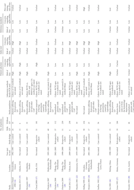
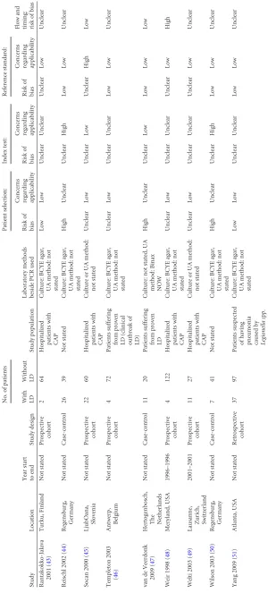
TABLE 1 Study characteristics and the Quadas-2 risk of bias assessment and applicability criteria Study Location Year start to end Study design No. of patients Study population Laboratory methods beside PCR used Patient selection: Index test: Reference standard: Flow and timing: risk of bias With LD Without LD Risk of bias
Concerns regarding applicability
Risk
of
bias
Concerns regarding applicability
Risk
of
bias
Concerns regarding applicability
Alexiou-Daniel 1998 ( 14 ) Thessaloniki, Greece 1992–1997 Case-control 24 10 Patients suffering from proven LD Culture or UA method: not stated High Low Unclear High Low Low Unclear Benitez 2013 ( 15 ) Atlanta, USA Not stated Case-control 15 6 Patients suffering from proven LD Culture or UA method: not stated High High Unclear High Low Low Unclear Bernander 1997 ( 16 ) Stockholm, Sweden Not stated Case-control 25 33 Patients suffering from proven LD and other CAP Culture: BCYE cx, MYW, and BMPAcx aagars, UA method: not stated High Low Unclear Low Low Low Low Cloud 2000 ( 17 ) Utah, USA Not stated Prospective cohort 31 181 Patients suspected of having pneumonia caused by Legionella spp. Culture: BCYE agar, UA method: not stated High High Unclear Unclear Unclear Low Unclear Diederen 2007 ( 18 ) Multicenter, The Netherlands 1995–2005 Case-control 68 36 Patients suffering from proven LD Culture: not stated, UA method: Binax High Low Unclear Low Low Low Low Diederen 2008 ( 19 ) Tilburg, The Netherlands 2002–2005 Retrospective cohort 37 112 Hospitalized patients with CAP Culture: BCYE agar, UA method: not stated Low Low Unclear Unclear Unclear Low Unclear Diederen 2009 ( 20 ) Tilburg, The Netherlands 1998–2000 Prospective cohort 11 230 Hospitalized patients with CAP Culture: not stated,UA method: Binax Low Low Unclear Low Unclear Low Low Fard 2012 ( 21 ) Tehran, Iran 2009–2010 Prospective cohort 4 258 Hospitalized patients with CAP Culture: BCYE agar, UA method: not stated Low Low Unclear High Low Low Unclear Hayden 2001 ( 22 ) Minnesota, USA 1979–1999 Case-control 9 10 Not stated Culture: BCYE agar, UA method: not stated High Low Unclear High Low Low Unclear Helbig 1999 ( 23 ) Dresden, Germany Not stated Prospective cohort 58 224 Not stated Culture or UA method: not stated Low Low Unclear High Low Low High Herpers 2003 ( 24 ) Bilthoven, The Netherlands 2001–2002 Case-control 17 23 Patients suffering from proven LD and other CAP Culture: not stated, UA method: Binax High Low Unclear High Low Low Unclear Jaulhac 1992 ( 25 ) Strasbourg, Lyon, France Not stated Case-control 12 56 Patients suspected of having pneumonia caused by Legionella spp. Culture: BCYE agar, UA method: not stated High Low Unclear High Low Low Unclear Jin 2001 ( 26 ) Beijing, China 1998–1999 Case-control 15 31 Patients suffering from proven LD Culture: BCYE agar, UA method: not stated High Low Unclear Low Unclear Low Low Jonas 1995 ( 27 ) Mainz, Germany Not stated Retrospective cohort 10 246 Patients from intensive care units or from the hematology department Culture: BCYE agar, UA method: not stated Unclear Unclear Unclear Unclear Unclear Low Unclear Kessler 1993 ( 28 ) Graz, Austria Not stated Prospective cohort 6 46 Hospitalized patients with CAP (atypical) Culture or UA method: not stated Unclear Low Unclear Unclear Unclear Low Unclear
on May 16, 2020 by guest
http://jcm.asm.org/
[image:2.585.54.522.60.724.2]Kim 2001 ( 29 ) Seoul, Korea 1997–2000 Prospective cohort 6 425 Hospitalized patients with CAP Culture or UA method: not stated Low Low Unclear High Unclear Low High Koide 2004 ( 31 ) Okinawa, Japan 1997–1999 Case-control 6 17 Patients suffering from proven LD and other CAP Culture: not stated, UA method: Binax, Biotest High Low Unclear High Low Low High Koide 2006 ( 30 ) Okinawa, Japan 1993–2004 Case-control 33 25 Patients suffering from proven LD and other CAP Culture: not stated, UA method: Binax, Biotest, Binax NOW High Low Unclear High Low Low High Lisby 1994 ( 32 ) Herlev, Denmark Not stated Prospective cohort 2 86 Patients suspected of having pneumonia caused by Legionella spp. Culture: BCYE agar, UA method: not stated Low Low Low Unclear Low Low Unclear Loens 2008 ( 33 ) Wilrijk, Belgium 2000–2002 Prospective cohort 4 143 Hospitalized patients with CAP Culture: not stated, UA method: Binax Low Low Unclear Unclear Unclear Low Unclear Matsiota-Bernard 1994 ( 34 ) Garches, France Not stated Case-control 12 17 Hospitalized patients with CAP Culture: BCYE agar, UA method: not stated High Low Unclear High Low Low Unclear Matsiota-Bernard 1997 ( 35 ) Garches, France Not stated Case-control 41 10 Patients suffering from proven LD Culture or UA method: not stated High Low Unclear High Low Low Unclear Maurin 2010 ( 36 ) Grenoble, France 2004–2006 Prospective cohort 19 201 Hospitalized patients with CAP Culture: BCYE agar, UA method: not stated Low Low Unclear Unclear Unclear Low Unclear Mérault 2011 ( 37 ) Multicenter, France 2007–2010 Case-control 22 74 Patients suffering from proven LD and other CAP Culture: BCYE agar, UA method: Binax High Low Unclear High Low Low Low Miyashita 2004 ( 38 ) Multicenter, Japan 1999–2000 Prospective cohort 8 200 Patients who were participants in a multicenter
CAP surveillance study
Culture: BCYE agar, UA method: not stated Low Low Unclear Unclear Unclear Low Unclear Murdoch 1996 ( 39 ) Canterbury, New Zealand 1992–1995 Case-control 28 24 CAP and
nosocomial pneumonia surveillance studies
Culture: BCYE agar, UA method: not stated High Unclear Unclear Low Low Low Low Nomanpour 2012 ( 40 ) Tehran, Iran 2009–2010 Prospective cohort 9 120 Hospitalized patients with CAP Culture: BCYE agar, UA method: not stated Low Low Unclear Unclear Unclear Low Unclear Raggam 2002 ( 41 ) Graz, Austria Not stated Prospective cohort 3 58 Patients suspected of having pneumonia caused by Legionella spp. Culture: BCYE agar, UA method: not stated Unclear Unclear Unclear Unclear Unclear Low Unclear Ramirez 1996 ( 42 ) Louisville, USA Not stated Prospective cohort 6 149 Hospitalized patients with CAP or nosocomial pneumonia Culture: BCYE agar, UA method: not stated Unclear High Unclear Unclear Unclear Low Low (Continued on following page)
on May 16, 2020 by guest
http://jcm.asm.org/
TABLE
1
(Continued)
Study
Location
Year
start
to
end
Study
design
No.
of
patients
Study
population
Laboratory
methods
beside
PCR
used
Patient
selection:
Index
test:
Reference
standard:
Flow
and
timing: risk
of
bias
With LD Without LD
Risk
of
bias
Concerns regarding applicability
Risk
of
bias
Concerns regarding applicability
Risk
of
bias
Concerns regarding applicability
Rantakokko-Jalava
2001
(
43
)
Turku,
Finland
Not
stated
Prospective
cohort
2
64
Hospitalized
patients
with
CAP
Culture:
BCYE
agar,
UA
method:
not
stated
Low
Low
Unclear
Unclear
Unclear
Low
Unclear
Reischl
2002
(
44
)
Regensburg,
Germany
Not
stated
Case-control
26
39
Not
stated
Culture:
BCYE
agar,
UA
method:
not
stated
High
Unclear
Unclear
High
Low
Low
Unclear
Socan
2000
(
45
)
LjubOana,
Slovenia
Not
stated
Prospective
cohort
22
60
Hospitalized
patients
with
CAP
Culture
or
UA
method:
not
stated
Unclear
Low
Unclear
Low
Unclear
High
Low
Templeton
2003
(
46
)
Antwerp,
Belgium
Not
stated
Prospective
cohort
4
72
Patients
suffering
from
proven
LD
(clinical
outbreak
of
LD)
Culture:
BCYE
agar,
UA
method:
not
stated
Unclear
Low
Unclear
Unclear
Low
Low
Unclear
van
de
Veerdonk
2009
(
47
)
Hertogenbosch,
The Netherlands
Not
stated
Case-control
11
20
Patients
suffering
from
proven
LD
Culture:
not
stated,
UA
method:
Binax
NOW
High
Unclear
Unclear
Low
Low
Low
Low
Weir
1998
(
48
)
Meryland,
USA
1996–1996
Prospective
cohort
4
122
Hospitalized
patients
with
CAP
Culture:
BCYE
agar,
UA
method:
not
stated
Unclear
Low
Unclear
Unclear
Unclear
Low
High
Welti
2003
(
49
)
Lausanne,
Zurich, Switzerland
2001–2001
Prospective
cohort
11
27
Hospitalized
patients
with
CAP
Culture
or
UA
method:
not
stated
Unclear
Low
Unclear
Unclear
Unclear
Low
Unclear
Wilson
2003
(
50
)
Regensburg,
Germany
Not
stated
Case-control
7
41
Not
stated
Culture:
BCYE
agar,
UA
method:
not
stated
High
Unclear
Unclear
High
Low
Low
Unclear
Yang
2009
(
51
)
Atlanta,
USA
Not
stated
Retrospective
cohort
37
97
Patients
suspected
of
having
pneumonia caused
by
Legionella
spp
.
Culture:
BCYE
agar,
UA
method:
not
stated
Low
Low
Unclear
Unclear
Low
Low
Unclear
aBCYE,
buffered
charcoal
yeast
extract;
cx,
culture.
on May 16, 2020 by guest
http://jcm.asm.org/
[image:4.585.52.352.66.729.2]the onset of infection. If data were available for more than one test in a study, all results were extracted. We also extracted data on UA in studies reporting both tests separately and together with PCR results. The target condition was pneumonia (either community or hospital acquired). The reference standard included two levels of certainty (11).
We considered a culture positive forLegionellaspp. when a 4-fold increase in serum antibodies forLegionellaspp. occurred if taken 4 to 6 weeks after the clinical episode or when a positive UA confirmed infec-tion. Diagnosis by antigen staining in respiratory secretion lung tissue or in pleural fluid by DFA staining or IHC was regarded as suspected infec-tion (11). We considered all other cases as having no evidence for
Legio-nellainfection. These considerations are compatible with the Centers for
Disease Control and Prevention (CDC) publication for preferred diag-nostic tests for definingLegionellainfection (11).
Electronic searches.We searched MEDLINE, LILACS, and Kore-aMed databases without date or language restrictions, from inception to October 2014, using the following terms and their medical subject head-ings [MESH] (adapted for each database): (PCR or real-time or RT-PCR or reverse-transcription or nested-PCR or PCR) and (legionell* or legion-air* or legionella[MESH] or Legionnaires’ Disease[MESH]). In addition, we searched the European Conference of Clinical Microbiology and In-fectious Diseases and the Interscience Conference on Antimicrobial Agents and Chemotherapy between the years 2010 and 2014 using only the key words forLegionellaor Legionnaires’ and PCR. We scanned the references of all included studies and reviews cited in the included studies. Data collection and risk of bias assessment.Two reviewers indepen-dently selected studies for inclusion and extracted all data from the stud-ies. Risk of bias assessment was conducted using the Quadas-2 tool (12). Statistical analysis and data synthesis.We listed the number of true positives, true negatives, false positives, and false negatives per study, specimen, index test, primer gene used for PCR, and reference standard. We calculated the sensitivity and specificity values and the diagnostic odds ratio (DOR). We used the bivariate model for the data summary. Param-eter estimates from the model were used to obtain hierarchical summary receiver operating curves, with 95% confidence intervals (CIs) and a 95% prediction region. We assessed the effect of the following covariates on results through subgroups analyses: PCR method, study design, primer gene used for PCR, number of LD cases, and the Quadas-2 domains. We compared the test performance of UA and PCR with that of UA alone in LD cases that were not diagnosed by UA alone, compared to the situation where either PCR or UA positivity defined a positive test result. Only direct test comparisons were performed. Studies were included only once in the analysis. Analyses were conducted using Stata 12 and RevMan 5.3 (13).
RESULTS
The search identified 804 references, of which 77 were selected for full-text review (see Fig. S1 in the supplemental material). Thirty-nine studies were excluded. A total of 38 studies that were
pub-lished between the years 1993 and 2013 were included (14-51).
Seven studies reported on the results of PCR in blood or serum, 4 trials reported on PCR in urine, 29 trials reported on PCR in BAL fluid or sputum, 3 trials reported on PCR in pharyngeal swabs, and 3 trials reported on PCR in lung tissue specimens. Five studies performed PCR in several sample types. Thirteen studies reported result for UA separately from results for PCR. Seventeen studies were case-control studies, 3 studies were retrospective cohorts, and the remaining 18 studies were prospective cohort studies. Altogether, 653 patients with confirmed LD, 8 patients with prob-able LD, 3,593 patients with pneumonia caused by pathogens
other thanLegionellaspp., and 296 healthy control patients were
included.
Data from prospective cohorts (not all patients underwent UA and/or culture) showed that cultures were positive in 164 of 2,562
patients with pneumonia (6.4%; 95% CI, 1.4% to 15.7%), UA was positive in 113 patients of 1,445 with pneumonia (7.82%; 95% CI, 2.2% to 15.2%), and PCR was positive in 309 of 3,463 patients with pneumonia (8.9%; 95% CI, 4.5% to 20.2%). Mortality was reported in 4 studies (weighted mean, 5.6%; range, 9.1% to 50%).
Other study characteristics are presented inTable 1.
Risk of bias assessment.The Quadas-2 risk of bias assessment
and applicability criteria are shown in Table 1. Only 12 of 38
studies were at low risk of bias regarding patient selection; 17 of 38 studies were at high risk (all of them were retrospective case-con-trol studies). The remaining 9 of 38 were of unclear risk; among them, 3 studies were also of high risk of bias regarding the appli-cability of the selected population to this review. Concerns regard-ing the applicability of the index test were present in 15 of 38 studies and unclear in another 16 of 38 studies. High risk of bias regarding the flow chart, timing of the index test, and ensuring that all patients received the same tests were present in 5 studies, unclear in 24 studies, and at low risk of bias in 9 studies. Four of 38 studies were from developing nations. In 9 studies, clinical and radiological definitions for pneumonia were presented.
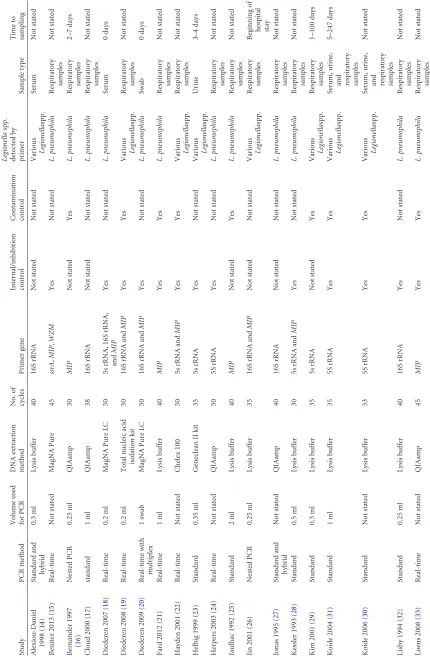
PCR technique.Details of the PCR techniques are presented
Table 2. Standard PCR was used in 12 studies; real-time PCR, in 16
studies; real-time with multiplex PCR, in 4; and nested PCR, in 6
(in 2 studies (43,49), 2 methods of PCR were used). Eight studies
used primers targeting theMIPgene, 7 studies used both 5s rRNA
andMIPgenes, 6 studies used both 16S rRNA andMIPgenes, 7
studies used the 5s rRNA gene, 7 studies used the 16S rRNA gene primers, and 4 studies used other genes (multiple genes were used
in 4 studies). The primers targeted specificallyL. pneumophilain
25 of 38 studies. DNA extraction was performed by the use of
QIAamp kit (n⫽10), MagNA Pure LC DNA isolation kit (n⫽5),
other commercial kits (n⫽10), and phenol-chloroform protocols
(n⫽13). Internal/inhibition controls were described in 27 of 38
studies, and contamination/digestion controls were described in 19 of 38 studies.
Performance of PCR.Details of PCR sensitivity and specificity
for the diagnosis of LD are presented inTable 3. The specificity
was very high regardless of the sample; however, the sensitivity of urine and serum samples were very low (49.7% [95% CI, 26.5% to 73.0%] and 48.9% [95% CI, 38.4% to 59.5%], respectively).
Re-spiratory samples had a high sensitivity for the detection of
Legio-nellaspp. by PCR. The summary sensitivity and specificity values of the bivariate model for all respiratory samples (BAL fluid, spu-tum, pharyngeal swabs, and tissue biopsies) were 97.4% (95% CI, 91.1% to 99.2%) and 98.6% (95% CI, 97.4% to 99.3%), respec-tively. The DOR was 2,826 (95% CI, 738 to 10,815).
Subgroup analysis based on sample type revealed summary sensitivity and specificity values of 97.7% (95% CI, 91.6% to 99.4%) and 98.6% (95% CI, 97.3% to 99.2%), respectively, for BAL fluid (combined occasionally with sputum) and 96.8% (95% CI, 41.2% to 99.9%) and 99.4% (95% CI, 91.7% to 99.9%), re-spectively, for sputum. Analysis based on studies with high
meth-odological qualities (n⫽9) yielded summary sensitivity and
spec-ificity values of 98.6% (95% CI, 57.7% to 99.9%) and 99.0% (95% CI, 96.9% to 99.9%), respectively.
There were no statistically significant differences in the per-formance of real-time, nested, and other PCR types. The use of inhibition control or contamination control did not affect sig-nificantly the performance of PCR. The use of genes specific to L. pneumophilawas associated with increased sensitivity
on May 16, 2020 by guest
http://jcm.asm.org/
TABLE 2 PCR methods Study PCR method Volume used for PCR DNA extraction method No. of cycles Primer gene Internal/inhibition control Contamination control Legionella spp. detected by primer Sample type Time to sampling Alexiou-Daniel 1998 ( 14 ) Standard and hybrid 0.3 ml Lysis buffer 40 16S rRNA Not stated Not stated Various Legionella spp. Serum Not stated Benitez 2013 ( 15 ) Real-time Not stated MagNA Pure 45 ssrA , MIP , WZM Yes Not stated L. pneumophila Respiratory samples Not stated Bernander 1997 ( 16 ) Nested PCR 0.25 ml QIAamp 30 MIP Not stated Yes L. pneumophila Respiratory samples 2–7 days Cloud 2000 ( 17 ) standard 1 ml QIAamp 38 16S rRNA Not stated Not stated L. pneumophila Respiratory samples Not stated Diederen 2007 ( 18 ) Real-time 0.2 ml MagNA Pure LC 50 5s rRNA, 16S rRNA, and MIP Yes Not stated L. pneumophila Serum 0 days Diederen 2008 ( 19 ) Real-time 0.2 ml Total nucleic acid isolation kit 50 16S rRNA and MIP Yes Yes Various Legionella spp. Respiratory samples Not stated Diederen 2009 ( 20 ) Real-time with multiplex 1 swab MagNA Pure LC 50 16S rRNA and MIP Yes Not stated L. pneumophila Swab 0 days Fard 2012 ( 21 ) Real-time 1 ml Lysis buffer 40 MIP Yes Yes L. pneumophila Respiratory samples Not stated Hayden 2001 ( 22 ) Real-time Not stated Chelex 100 50 5s rRNA and MIP Yes Yes Various Legionella spp. Respiratory samples Not stated Helbig 1999 ( 23 ) Standard 0.35 ml Geneclean II kit 35 5s rRNA Yes Not stated Various Legionella spp. Urine 3–4 days Herpers 2003 ( 24 ) Real-time Not stated QIAamp 50 5S rRNA Yes Not stated L. pneumophila Respiratory samples Not stated Jaulhac 1992 ( 25 ) Standard 2 ml Lysis buffer 40 MIP Not stated Yes L. pneumophila Respiratory samples Not stated Jin 2001 ( 26 ) Nested PCR 0.25 ml Lysis buffer 35 16S rRNA and MIP Not stated Not stated Various Legionella spp. Respiratory samples Beginning of hospital stay Jonas 1995 ( 27 ) Standard and hybrid Not stated QIAamp 40 16S rRNA Not stated Not stated L. pneumophila Respiratory samples Not stated Kessler 1993 ( 28 ) Standard 0.5 ml Lysis buffer 30 5s rRNA and MIP Yes Not stated L. pneumophila Respiratory samples Not stated Kim 2001 ( 29 ) Standard 0.3 ml Lysis buffer 35 5s rRNA Not stated Yes Various Legionella spp. Respiratory samples 1–100 days Koide 2004 ( 31 ) Standard 1 ml Lysis buffer 35 5S rRNA Yes Yes Various Legionella spp. Serum, urine,
and respiratory samples
5–247 days Koide 2006 ( 30 ) Standard Not stated Lysis buffer 53 5S rRNA Yes Yes Various Legionella spp. Serum, urine,
and respiratory samples
Not stated Lisby 1994 ( 32 ) Standard 0.25 ml Lysis buffer 40 16S rRNA Yes Not stated L. pneumophila Respiratory samples Not stated Loens 2008 ( 33 ) Real-time Not stated QIAamp 45 MIP Yes Yes L. pneumophila Respiratory samples and swab Not stated
on May 16, 2020 by guest
http://jcm.asm.org/
[image:6.585.53.483.67.730.2]Matsiota-Bernard 1994 ( 34 ) Standard and hybrid 1 ml Lysis buffer 30 5s rRNA and MIP Yes Not stated L. pneumophila Respiratory samples Not stated Matsiota-Bernard 1997 ( 35 ) Standard and hybrid Not stated Lysis buffer Not stated 5s rRNA and MIP Yes Not stated L. pneumophila Serum 2–15 days Maurin 2010 ( 36 ) Real-time Not stated QIAamp Not stated 16S rRNA and MIP Yes Yes L. pneumophila Respiratory samples Not stated Mérault 2011 ( 37 ) Real-time 0.2 ml MagNA Pure LC 50 LPS gene cluster Yes Not stated L. pneumophila Respiratory samples Not stated Miyashita 2004 ( 38 ) Real-time with multiplex 1 swab QIAamp 40 Major outer membrane protein porin Yes Not stated L. pneumophila Swab Not stated Murdoch 1996 ( 39 ) Standard 0.1–0.3 ml Trizol 35 5S rRNA gene Not stated Yes Various Legionella spp. Serum and urine 1–30 days Nomanpour 2012 ( 40 ) Real-time with multiplex Not stated Lysis buffer 35 MIP Yes Yes L. pneumophila Respiratory samples and swab Not stated Raggam 2002 ( 41 ) Real-time 0.1 ml MagNA Pure LC 55 16S rRNA Yes Yes Various Legionella spp. Respiratory samples Not stated Ramirez 1996 ( 42 ) Standard 1 swab Lysis buffer 40 5s rRNA Not stated Yes Various Legionella spp. Swab Not stated Rantakokko -Jalava 2001 ( 43 ) Real-time 0.2 ml High pure PCR template preparation kit 45 16S rRNA Yes Yes L. pneumophila Respiratory samples Not stated Reischl 2002 ( 44 ) Real-time 0.5 ml High pure PCR template preparation kit 50 16S rRNA Yes Yes L. pneumophila Respiratory samples Not stated Socan 2000 ( 45 ) Standard 0.2 ml QIAamp 30 5s rRNA and MIP Yes Not stated L. pneumophila Urine Beginning of hospital stay Templeton 2003 ( 46 ) Real-time 0.2 ml High pure PCR template preparation kit 50 16S rRNA and MIP Yes Yes Various Legionella spp. Respiratory samples and swab Not stated van de Veerdonk 2009 ( 47 ) Real-time 0.2 ml NucliSens easyMAG 45 MIP Yes Not stated L. pneumophila Serum 0 days Weir 1998 ( 48 ) Standard 0.5 ml Lysis buffer Not stated 5s rRNA and MIP Yes Not stated Various Legionella spp. Respiratory samples Not stated Welti 2003 ( 49 ) Real-time with multiplex 1 ml QIAamp 50 16S rRNA and MIP Yes Yes L. pneumophila Respiratory samples Not stated Wilson 2003 ( 50 ) Real-time 0.1 ml QIAamp 45 MIP Yes Yes L. pneumophila Respiratory samples Not stated Yang 2009 ( 51 ) Real-time Not stated KingFisher ML instrument and InviMag kit Not stated 5s rRNA and 23S rRNA Not stated Not stated L. pneumophila Serum,
respiratory samples and
swab
Not
stated
on May 16, 2020 by guest
http://jcm.asm.org/
pared to primers from genes that targeted variousLegionella
spp. (Table 3).
Comparison of PCR to UA.Details of the direct comparison of
PCR and UA are detailed inTable 4. The summary sensitivity and
specificity values of the bivariate model for UA in all studies were 77.0% (95% CI, 55% to 90.0%) and 100% (by definition), respec-tively. The DOR was 7,540 (95% CI, 289 to 19,652). In the direct comparison of PCR in respiratory secretions versus the use of UA,
PCR had higher sensitivity (P⫽0.001).
A subgroup analysis of cases of LD, whena prioriexcluding all
cases of LD that were diagnosed by UA alone, yielded a summary sensitivity of 93.1% (95% CI, 63.9% to 99.0%) for PCR and 51.8% (95% CI, 33.1% to 69.1%) for UA.
Taking into account that UA is easily performed and available for each patient, while performing BAL fluid is invasive, contains certain risks, and is not readily available in all settings, we exam-ined the performance of UA in sputum samples and/or pharyn-geal swabs alone. The summary sensitivity and specificity values of PCR in sputum samples were 97.1% (95% CI, 59.6% to 99.8%)
and 99.7% (95% CI, 91.4% to 99.9%), respectively; those of UA were 52.9% (95% CI, 30.8% to 73.9%) and 100% (by definition), respectively; those of either UA or PCR were 99.9% (95% CI, 99.9% to 99.9%) and 99.7% (95% CI, 90.2% to 99.9%), in 5 stud-ies. In absolute terms, 11 of 61 patients (18%) with LD had a negative UA and a positive sputum PCR and would had been misdiagnosed by conventional methods.
DISCUSSION
We examined the accuracy of PCR alone and in comparison with UA in various clinical samples for the diagnosis of LD among patients with pneumonia, where the reference standard was proven or probable LD according to criteria suggested by the CDC
(11). We demonstrated near perfect specificity values for all
ple types and equally high sensitivity values for all respiratory sam-ples (consisting of BAL fluid, sputum, pharyngeal swabs, tissue biopsy specimens, and other respiratory fluids). Overall, in 35 included studies that used any respiratory sample, the summary sensitivity and specificity estimates were 97.4% and 98.6%,
re-TABLE 3Sensitivity and specificity of PCR in the diagnosis of Legionnaires’ disease
Comparison, no. of studies Sensitivity, % (95% CIa) Specificity, % (95% CI) DORb(95% CI)
PCR in urine samples, 5 49.7 (26.5–73.0) 98.2 (85.6–99.8) 54 (5.7–509)
PCR in blood samples, 7 48.9 (38.4–59.5) 99.8 (59.1–99.9) 889 (1.25–633,052)
PCR in respiratory samples
All, 35 97.4 (91.1–99.2) 98.6 (97.4–99.3) 2,826 (738–10,815)
BALcsample, 29 97.7 (91.6–99.4) 98.6 (97.3–99.2) 3,072 (733–12,786)
Sputum, 9 96.8 (41.2–99.9) 99.4 (91.7–99.9) 5,774 (30–1,110,511)
PCR in respiratory samples
Retrospective studies excluded, 14 99.1 (63.3–99.9) 98.5 (97.1–99.2) 8,335 (127–546,320)
All high ROBdstudies excluded, 9 98.4 (57.7–99.9) 99.0 (96.9–99.6) 6,447 (132–314,848)
Standard PCR, 8 98.8 (47.5–99.9) 97.9 (94.9–99.2) 3,996 (46–346,416)
Nested/hybrid PCR, 5 97.0 (83.4–99.5) 98.4 (92.9–99.6) 2,169 (227–20,697)
Real-time PCR, 17 97.9 (89.1–99.6) 98.7 (96.8–99.4) 3,675 (529–25,509)
L. pneumophilagenes, 20 98.4 (91.4–99.7) 98.3 (96.7–99.2) 3,957 (655–23,875)
VariousLegionellaspp. genes, 9 95.7 (69.4–99.5) 99.1 (96.4–99.8) 2,680 (141–50,611)
No inhibition control, 9 97.0 (73.6–99.7) 98.0 (94.2–99.3) 1,720 (185–15,930)
Inhibition control, 20 98.3 (90.6–99.7) 98.6 (97.1–99.3) 4,435 (622–31,632)
a
CI, confidence interval.
bDOR, diagnostic odds ratio. c
BAL, bronchoalveolar lavage.
[image:8.585.40.548.78.265.2]dROB, risk of bias.
TABLE 4Direct comparisons of PCR in respiratory samples versus UAa
Comparison, no. of studies Sensitivity, % (95% CIb) Specificity, % (95% CI) DORc(95% CI)
UA: all studies, 13 77.0 (55.3–90.0) 99.9 (99.9–99.9) 7,540 (289–196,522)
PCR in respiratory samples vs UAd
PCR, 8 93.1 (63.9–99.0) 99.1 (98.0–99.5) 1,515 (185–12,344)
UA, 8 51.8 (33.6–69.6) 99.9 (99.9–99.9) NAe
UA or PCR, 8 95.6 (68.2–99.5) 99.1 (97.6–99.6) 2,577 (209–31,650)
PCR in sputum samples/swabs vs UAd
PCR, 5 97.1 (59.6–99.8) 99.7 (91.4–99.9) 12,467 (171–907,125)
UA, 5 52.9 (30.8–73.9) 99.9 (99.9–99.9) NA
UA or PCR, 5 99.9 (99.9–99.9) 99.7 (90.2–99.9) NA
aUA, urinary antigen. b
CI, confidence interval.
cDOR, diagnostic odds ratio. d
All cases of LD diagnosed by UA alone were excluded.
eNA, not applicable.
on May 16, 2020 by guest
http://jcm.asm.org/
[image:8.585.42.546.563.678.2]spectively. In studies that used easy-to-obtain samples, such as sputum samples and pharyngeal swabs, the summary sensitivity and specificity estimates were 94.5% and 99.2%, respectively (13 studies). PCR sensitivity of urine and blood samples was low (roughly, 50%), rendering these samples unusable for clinical practice. We explored further the accuracy of PCR through sub-group and sensitivity analyses. We discovered that PCR sensitivity in respiratory samples remains very high after consideration for methodological quality, study design, and various PCR methods. When we compared the results of PCR in respiratory samples to those of UA, we demonstrated improved sensitivity with similar specificity, regardless of the sample type. Furthermore, when cases that were diagnosed only by UA (without positive culture, serol-ogy, or DFA) and all cases that were diagnosed by BAL fluid were excluded, leaving a real-life comparison of PCR of pharyngeal swabs and/or sputum samples and the UA, PCR was considerably more sensitive than the UA and resulted in reclassification of 18% of patients with pneumonia and negative UA to an LD diagnosis. Using the pooled sensitivity and specificity estimates of our review, the negative and positive predictive values (NPV and PPV, respectively) of the test can be calculated, using a defined
preva-lence of disease (52). With a prevalence of LD of 7.5% among
patients with CAP (as observed from the prospective cohort stud-ies in our review), negative PCR in respiratory sample excludes LD in 99.7% of patients, and positive PCR confirms LD in 84.9%. When both PCR on sputum sample/swab and UA are performed and either positive result defines a positive test, the NPV is 99.9%, and the PPV is 96%. Thus, a negative PCR rules out the diagnosis
of LD with a very high probability (ⱖ97%). Performing both tests
increases the probability of ruling in LD without affecting speci-ficity.
When LD is diagnosed, combination therapy directed at
Legio-nellaspp. increases the chances of survival (53) Therefore, the diagnosis of LD among patients hospitalized with CAP, especially when severe, may directly influence prognosis, while other
pa-tients may be treated with beta-lactam monotherapy (54). The
diagnosis of LD today is based on several traditional methods. Culture requires special media, processing, and technical exper-tise, and 3 to 5 days are required to obtain a positive result.
Sero-logical testing forLegionellahas little impact on clinical practice, as
20% to 30% of patients with LD do not develop a detectable
anti-body response if tested too early (55) or at all (56). The most
common method currently used for diagnosing LD in the clinical
setting is UA detection ofL. pneumophilaserogroup 1 (57). In a
previous systematic review, the pooled sensitivity of UA assays for
the detection ofL. pneumophilaserogroup 1 was 74% (95% CI,
68% to 81%), with a pooled specificity of 99% (95% CI, 98% to
99%) (58). Our results are in concordance with this systematic
review (pooled UA sensitivity of 77% and near 100% specificity). However, the antigen is excreted in urine for weeks (and up to a year) after an infectious episode, which weakens its specificity
(59). Furthermore,L. pneumophilaserogroup 1 is the
predom-inantLegionellaspp. that causes LD in the United States and Europe
but not in Asia and Australia (60). LD from non-pneumophila
Legio-nellaspecies is more common in immunocompromised patients, andL. pneumophilaserogroups other than serogroup 1 can cause
nosocomial outbreaks of LD (61,62). In such cases, the UA might
provide false-negative results. Diagnosis is LD among immuno-compromised patients and in the nosocomial setting is critical, and PCR might improve the diagnosis of these cases significantly.
One of the main criticisms against the use of PCR in the diag-nosis of LD, and one of the major limitations of analyzing PCR-based methods, is the lack of standardization in performance and reporting of the PCR methods. The contamination of commercial DNA extraction kits may produce false-positive results with the
lack of a negative control (63). The occurrence of false-positive
testing demonstrates the need for a standardized laboratory pro-tocol for the needed stringent quality control requirements. Vari-able methods of sampling, extraction, and amplification protocols were used in the studies included in our review. We did not ob-serve an effect of each parameter on results, except for improved
sensitivity with primers made from a gene sequence ofL.
pneumo-phila. However, the number of studies included in our review was too small, and reporting was insufficient to assess individually and in combination the large number of variables relating to PCR methods. Moreover, PCR kits are expensive, PCR requires a ded-icated laboratory equipment and personnel, and PCR is not easily interpreted, whereas the UA is relatively inexpensive (around $10 per test in the United States) and requires no special equipment or training.
In summary, we show an excellent sensitivity and specificity of PCR for the diagnosis of LD in any respiratory sample. The NPV given the usual disease prevalence was over 95% regardless of the subgroup examined. The PPV was also above 95%, thus making the PCR an excellent tool for ruling in or out LD. The sensitivity of the PCR in respiratory samples was superior to the UA and may
result in the additional diagnosis of patients withL. pneumophila
serogroup 1 LD and those with non-pneumophila Legionella
spe-cies or non-serogroup 1 LD. We suggest using the PCR especially
when infectdion with non-pneumophila Legionellaspecies is
pos-sible.
REFERENCES
1.Mandell GL, Bennett JB, Dolin R. Mandell, Douglas, and Bennett’s principles and practice of infectious diseases, 7th ed, Churchill Living-stone, London, England.
2.Muder RR, Yu VL.2002. Infection due toLegionellaspecies other thanL.
pneumophila. Clin Infect Dis 35:990 –998. http://dx.doi.org/10.1086
/342884.
3.Roig J, Aguilar X, Ruiz J, Domingo C, Mesalles E, Manterola J, Morera J.1991. Comparative study ofLegionella pneumophilaand other nosoco-mial-acquired pneumonias. Chest99:344 –350.http://dx.doi.org/10.1378 /chest.99.2.344.
4.Tkatch LS, Kusne S, Irish WD, Krystofiak S, Wing E.1998. Epidemi-ology ofLegionellapneumonia and factors associated withLegionella -related mortality at a tertiary care center. Clin Infect Dis27:1479 –1486.
http://dx.doi.org/10.1086/515040.
5.Borella P, Guerrieri E, Marchesi I, Bondi M, Messi P. 2005. Water ecology ofLegionellaand protozoan: environmental and public health perspectives. Biotechnol Annu Rev11:355–380.http://dx.doi.org/10.1016 /S1387-2656(05)11011-4.
6.Stout JE, Yu VL.1997. Legionellosis. N Engl J Med337:682– 687.http: //dx.doi.org/10.1056/NEJM199709043371006.
7.Sabrià M, Pedro-Botet ML, Gomez J, Roig J, Vilaseca B, Sopena N, Banos V.2005. Fluoroquinolones versus macrolides in the treatment of Legionnaires’ disease. Chest 128:1401–1405. http://dx.doi.org/10.1378 /chest.128.3.1401.
8.Robenshtok E, Shefet D, Gafter-Gvili A, Paul M, Vidal L, Leibovici L.
2008. Empiric antibiotic coverage of atypical pathogens for community acquired pneumonia in hospitalized adults. Cochrane Database Syst Rev (1):CD004418.http://dx.doi.org/10.1002/14651858.CD004418.pub3. 9.Postma DF, van Werkhoven CH, van Elden LJ, Thijsen SF, Hoepelman
AI, Kluytmans JA, Boersma WG, Compaijen CJ, van der Wall E, Prins JM, Oosterheert JJ, Bonten MJ, Group C-SS.2015. Antibiotic treatment strategies for community-acquired pneumonia in adults. N Engl J Med
372:1312–1323.http://dx.doi.org/10.1056/NEJMoa1406330.
on May 16, 2020 by guest
http://jcm.asm.org/
10. Garin N, Genne D, Carballo S, Chuard C, Eich G, Hugli O, Lamy O, Nendaz M, Petignat PA, Perneger T, Rutschmann O, Seravalli L, Harbarth S, Perrier A.2014. Beta-lactam monotherapy versus beta-lactam-macrolide combination treatment in moderately severe community-acquired pneumo-nia: a randomized noninferiority trial. JAMA Intern Med174:1894 –1901.
http://dx.doi.org/10.1001/jamainternmed.2014.4887.
11. Centers for Disease Control and Prevention.2005. Strengthening sur-veillance for travel-associated legionellosis and revised case definitions for legionellosis. http://www.cdc.gov/legionella/health-depts/inv-tools -single/cste-position-statement.html.
12. Whiting PF, Rutjes AW, Westwood ME, Mallett S, Deeks JJ, Reitsma JB, Leeflang MM, Sterne JA, Bossuyt PM.2011. Quadas-2: a revised tool for the quality assessment of diagnostic accuracy studies. Ann Intern Med
155:529 –536.http://dx.doi.org/10.7326/0003-4819-155-8-201110180 -00009.
13. The Nordic Cochrane Centre.2011. Review manager (RevMan) version 5.1. The Nordic Cochrane Centre, The Cochrane Collaboration, Copen-hagen, Denmark.
14. Alexiou-Daniel S, Stylianakis A, Papoutsi A, Zorbas I, Papa A, Lam-bropoulos AF, Antoniadis A. 1998. Application of polymerase chain reaction for detection ofLegionella pneumophilain serum samples. Clin Microbiol Infect4:144 –148.http://dx.doi.org/10.1111/j.1469-0691.1998 .tb00377.x.
15. Benitez AJ, Winchell JM.2013. Clinical application of a multiplex real-time PCR assay for simultaneous detection ofLegionellaspecies,Legionella
pneumophila, andLegionella pneumophilaserogroup 1. J Clin Microbiol
51:348 –351.http://dx.doi.org/10.1128/JCM.02510-12.
16. Bernander S, Hanson HS, Johansson B, Von Stedingk LV. 1997. A nested polymerase chain reaction for detection ofLegionella pneumophila
in clinical specimens. Clin Microbiol Infect3:95–101.http://dx.doi.org/10 .1111/j.1469-0691.1997.tb00946.x.
17. Cloud JL, Carroll KC, Pixton P, Erali M, Hillyard DR.2000. Detection
ofLegionellaspecies in respiratory specimens using PCR with sequencing
confirmation. J Clin Microbiol38:1709 –1712.
18. Diederen BM, de Jong CM, Marmouk F, Kluytmans JA, Peeters MF, Van der Zee A.2007. Evaluation of real-time PCR for the early detection
ofLegionella pneumophilaDNA in serum samples. J Med Microbiol56:
94 –101.http://dx.doi.org/10.1099/jmm.0.46714-0.
19. Diederen BM, Kluytmans JA, Vandenbroucke-Grauls CM, Peeters MF.
2008. Utility of real-time PCR for diagnosis of Legionnaires’ disease in routine clinical practice. J Clin Microbiol46:671– 677.http://dx.doi.org /10.1128/JCM.01196-07.
20. Diederen BM, Van Der Eerden MM, Vlaspolder F, Boersma WG, Kluytmans JA, Peeters MF.2009. Detection of respiratory viruses and
Legionellaspp. by real-time polymerase chain reaction in patients with
community acquired pneumonia. Scand J Infect Dis41:45–50.http://dx .doi.org/10.1080/00365540802448799.
21.Fard SY, Nomanpour B, Fatolahzadeh B, Mobarez AM, Darban-Sarokhalil D, Fooladi AA, Leeuwen WB, Feizabadi MM.2012. Hospital acquired pneumonia: comparison of culture and real-time PCR assays for detection ofLegionella pneumophilafrom respiratory specimens at Tehran hospitals. Acta Microbiol Immunol Hung59:355–365.http://dx.doi.org /10.1556/AMicr.59.2012.3.6.
22. Hayden RT, Uhl JR, Qian X, Hopkins MK, Aubry MC, Limper AH, Lloyd RV, Cockerill FR.2001. Direct detection ofLegionellaspecies from bronchoalveolar lavage and open lung biopsy specimens: comparison of LightCycler PCR, in situ hybridization, direct fluorescence antigen detec-tion, and culture. J Clin Microbiol39:2618 –2626.http://dx.doi.org/10 .1128/JCM.39.7.2618-2626.2001.
23. Helbig JH, Engelstadter T, Maiwald M, Uldum SA, Witzleb W, Luck PC.1999. Diagnostic relevance of the detection ofLegionellaDNA in urine samples by the polymerase chain reaction. Eur J Clin Microbiol Infect Dis
18:716 –722.http://dx.doi.org/10.1007/s100960050384.
24. Herpers BL, de Jongh BM, van der Zwaluw K, van Hannen EJ.2003. Real-time PCR assay targets the 23S-5S spacer for direct detection and differentiation ofLegionellaspp. andLegionella pneumophila. J Clin Mi-crobiol 41:4815– 4816. http://dx.doi.org/10.1128/JCM.41.10.4815-4816 .2003.
25. Jaulhac B, Nowicki M, Bornstein N, Meunier O, Prevost G, Piemont Y, Fleurette J, Monteil H.1992. Detection ofLegionellaspp. in bronchoal-veolar lavage fluids by DNA amplification. J Clin Microbiol30:920 –924. 26. Jin J, Zhang H, Qiu Q, Li S, Wan C.2001. A pilot study on the value of
duplex polymerase chain reaction method in early diagnosis ofLegionella
pneumonia. Zhonghua Nei Ke Za Zhi40:154 –157. (In Chinese.) 27. Jonas D, Rosenbaum A, Weyrich S, Bhakdi S. 1995. Enzyme-linked
immunoassay for detection of PCR-amplified DNA ofLegionellaein bron-choalveolar fluid. J Clin Microbiol33:1247–1252.
28. Kessler HH, Reinthaler FF, Pschaid A, Pierer K, Kleinhappl B, Eber E, Marth E.1993. Rapid detection ofLegionellaspecies in bronchoalveolar lavage fluids with the EnviroAmpLegionellaPCR amplification and detec-tion kit. J Clin Microbiol31:3325–3328.
29. Kim M, Cheong H, Sohn J, Shim H, Park D, Park S, Woo J, Kang J, Kim Y, Shin W, Kim Y, Lee H, Kim J.2001. A prospective multicenter study of the etiological analysis in adults with community-acquired pneumonia:
Legionella,Leptospira, Hantaan virus, andOrientia tsutsugamushi. Korean
J Infect Dis33:24 –31.
30. Koide M, Higa F, Tateyama M, Nakasone I, Yamane N, Fujita J.2006. Detection ofLegionellaspecies in clinical samples: comparison of poly-merase chain reaction and urinary antigen detection kits. Infection34:
264 –268.http://dx.doi.org/10.1007/s15010-006-6639-6.
31. Koide M, Higa F, Tateyama M, Sakugawa H, Saito A.2004. Comparison of polymerase chain reaction and two urinary antigen detection kits for detectingLegionellain clinical samples. Eur J Clin Microbiol Infect Dis
23:221–223.http://dx.doi.org/10.1007/s10096-003-1072-6.
32. Lisby G, Dessau R.1994. Construction of a DNA amplification assay for detection ofLegionellaspecies in clinical samples. Eur J Clin Microbiol Infect Dis13:225–231.http://dx.doi.org/10.1007/BF01974541.
33. Loens K, Beck T, Ursi D, Overdijk M, Sillekens P, Goossens H, Ieven M.
2008. Evaluation of different nucleic acid amplification techniques for the detection ofM. pneumoniae,C. pneumoniae, andLegionellaspp. in respi-ratory specimens from patients with community-acquired pneumonia. J Microbiol Methods73:257–262.http://dx.doi.org/10.1016/j.mimet.2008 .02.010.
34. Matsiota-Bernard P, Pitsouni E, Legakis N, Nauciel C.1994. Evaluation of commercial amplification kit for detection ofLegionella pneumophilain clinical specimens. J Clin Microbiol32:1503–1505.
35. Matsiota-Bernard P, Vrioni G, Nauciel C.1997. Use of the polymerase chain reaction for the detection ofLegionella pneumophilaDNA in serum samples. Clin Infect Dis25:939. (Comment.)
36. Maurin M, Hammer L, Gestin B, Timsit JF, Rogeaux O, Delavena F, Tous J, Epaulard O, Brion JP, Croize J.2010. Quantitative real-time PCR tests for diagnostic and prognostic purposes in cases of legionellosis. Clin Microbiol Infect16:379 –384.http://dx.doi.org/10.1111/j.1469-0691.2009 .02812.x.
37. Mérault N, Rusniok C, Jarraud S, Gomez-Valero L, Cazalet C, Marin M, Brachet E, Aegerter P, Gaillard JL, Etienne J, Herrmann JL, Group D-IS, Lawrence C, Buchrieser C.2011. Specific real-time PCR for simul-taneous detection and identification ofLegionella pneumophilaserogroup 1 in water and clinical samples. Appl Environ Microbiol77:1708 –1717.
http://dx.doi.org/10.1128/AEM.02261-10.
38. Miyashita N, Saito A, Kohno S, Yamaguchi K, Watanabe A, Oda H, Kazuyama Y, Matsushima T, Group CAPS.2004. Multiplex PCR for the simultaneous detection ofChlamydia pneumoniae,Mycoplasma
pneu-moniae, andLegionella pneumophilain community-acquired pneumonia.
Respir Med98:542–550.http://dx.doi.org/10.1016/j.rmed.2003.11.012. 39. Murdoch DR, Walford EJ, Jennings LC, Light GJ, Schousboe MI,
Chereshsky AY, Chambers ST, Town GI.1996. Use of the polymerase chain reaction to detectLegionellaDNA in urine and serum samples from patients with pneumonia. Clin Infect Dis23:475– 480.http://dx.doi.org /10.1093/clinids/23.3.475.
40. Nomanpour B, Ghodousi A, Babaei T, Jafari S, Feizabadi MM.2012. Single tube real-time PCR for detection of Streptococcus pneumoniae,
Mycoplasma pneumoniae,Chlamydophila pneumoniae, and Legionella
pneumophilafrom clinical samples of CAP. Acta Microbiol Immunol
Hung59:171–184.http://dx.doi.org/10.1556/AMicr.59.2012.2.3. 41. Raggam RB, Leitner E, Muhlbauer G, Berg J, Stocher M, Grisold AJ,
Marth E, Kessler HH.2002. Qualitative detection ofLegionellaspecies in bronchoalveolar lavages and induced sputa by automated DNA extraction and real-time polymerase chain reaction. Med Microbiol Immunol191:
119 –125.http://dx.doi.org/10.1007/s00430-002-0129-y.
42. Ramirez JA, Ahkee S, Tolentino A, Miller RD, Summersgill JT.1996. Diagnosis ofLegionella pneumophila,Mycoplasma pneumoniae, or
Chla-mydia pneumoniaelower respiratory infection using the polymerase chain
reaction on a single throat swab specimen. Diagn Microbiol Infect Dis
24:7–14.http://dx.doi.org/10.1016/0732-8893(95)00254-5.
on May 16, 2020 by guest
http://jcm.asm.org/
43. Rantakokko-Jalava K, Jalava J.2001. Development of conventional and real-time PCR assays for detection ofLegionellaDNA in respiratory spec-imens. J Clin Microbiol39:2904 –2910.http://dx.doi.org/10.1128/JCM.39 .8.2904-2910.2001.
44. Reischl U, Linde HJ, Lehn N, Landt O, Barratt K, Wellinghausen N.
2002. Direct detection and differentiation ofLegionellaspp. andLegionella
pneumophilain clinical specimens by dual-color real-time PCR and
melt-ing curve analysis. J Clin Microbiol40:3814 –3817.http://dx.doi.org/10 .1128/JCM.40.10.3814-3817.2002.
45. Socan M, Kese D, Marinic-Fiser N.2000. Polymerase chain reaction for detection ofLegionellaeDNA in urine samples from patients with com-munity-acquired pneumonia. Folia Microbiol (Praha)45:469 – 472.http: //dx.doi.org/10.1007/BF02817623.
46. Templeton KE, Scheltinga SA, Sillekens P, Crielaard JW, van Dam AP, Goossens H, Claas EC.2003. Development and clinical evaluation of an internally controlled, single-tube multiplex real-time PCR assay for detec-tion ofLegionella pneumophilaand otherLegionellaspecies. J Clin Micro-biol41:4016 – 4021.http://dx.doi.org/10.1128/JCM.41.9.4016-4021.2003. 47. van de Veerdonk FL, de Jager CP, Schellekens JJ, Huijsmans CJ, Beaumont F, Hermans MH, Wever PC.2009.Legionella pneumophila
DNA in serum samples during Legionnaires’ disease in relation to C-re-active protein levels. Eur J Clin Microbiol Infect Dis28:371–376.http://dx .doi.org/10.1007/s10096-008-0638-8.
48. Weir SC, Fischer SH, Stock F, Gill VJ.1998. Detection ofLegionellaby PCR in respiratory specimens using a commercially available kit. Am J Clin Pathol110:295–300.
49. Welti M, Jaton K, Altwegg M, Sahli R, Wenger A, Bille J. 2003. Development of a multiplex real-time quantitative PCR assay to detect
Chlamydia pneumoniae,Legionella pneumophilaandMycoplasma
pneu-moniaein respiratory tract secretions. Diagn Microbiol Infect Dis45:85–
95.http://dx.doi.org/10.1016/S0732-8893(02)00484-4.
50. Wilson DA, Yen-Lieberman B, Reischl U, Gordon SM, Procop GW.
2003. Detection ofLegionella pneumophilaby real-time PCR for themip
gene. J Clin Microbiol41:3327–3330.http://dx.doi.org/10.1128/JCM.41.7 .3327-3330.2003.
51. Yang G, Benson R, Pelish T, Brown E, Winchell JM, Fields B.2010. Dual detection ofLegionella pneumophilaandLegionellaspecies by real-time PCR targeting the 23S-5S rRNA gene spacer region. Clin Microbiol Infect
16:255–261.http://dx.doi.org/10.1111/j.1469-0691.2009.02766.x. 52. Altman DG, Bland JM.1994. Diagnostic tests 2: predictive values. BMJ
309:102.http://dx.doi.org/10.1136/bmj.309.6947.102.
53. Rello J, Gattarello S, Souto J, Sole-Violan J, Valles J, Peredo R, Zaragoza R, Vidaur L, Parra A, Roig J, Community-acquired pneumonia in Unidad de Cuidados Intensivos 2 CAPUCI 2 study investigators.2013. Community-acquiredLegionellapneumonia in the intensive care unit: impact on survival of combined antibiotic therapy. Med Intensiva37:
320 –326.http://dx.doi.org/10.1016/j.medin.2012.05.010.
54. Eliakim-Raz N, Robenshtok E, Shefet D, Gafter-Gvili A, Vidal L, Paul M, Leibovici L. 2012. Empiric antibiotic coverage of atypical patho-gens for community-acquired pneumonia in hospitalized adults. Co-chrane Database Syst Rev 9:CD004418. http://dx.doi.org/10.1002/14 651858.CD004418.pub4.
55. Edelstein PH, Meyer RD, Finegold SM.1980. Laboratory diagnosis of Legionnaires’ disease. Am Rev Respir Dis121:317–327.
56. Zuravleff JJ, Yu VL, Shonnard JW, Davis BK, Rihs JD.1983. Diagnosis of Legionnaires’ disease. An update of laboratory methods with new em-phasis on isolation by culture. JAMA250:1981–1985.
57. Wever PC, Yzerman EP, Kuijper EJ, Speelman P, Dankert J.2000. Rapid diagnosis of Legionnaires’ disease using an immunochromatographic as-say forLegionella pneumophilaserogroup 1 antigen in urine during an outbreak in the Netherlands. J Clin Microbiol38:2738 –2739.
58. Shimada T, Noguchi Y, Jackson JL, Miyashita J, Hayashino Y, Kamiya T, Yamazaki S, Matsumura T, Fukuhara S.2009. Systematic review and metaanalysis: urinary antigen tests for legionellosis. Chest136:1576 –1585.
http://dx.doi.org/10.1378/chest.08-2602.
59. Kohler RB, Winn WC, Jr, Wheat LJ.1984. Onset and duration of urinary antigen excretion in Legionnaires’ disease. J Clin Microbiol20:605– 607. 60. Diederen BM.2008.Legionellaspp. and Legionnaires’ disease. J Infect
56:1–12.http://dx.doi.org/10.1016/j.jinf.2007.09.010.
61. Fang GD, Yu VL, Vickers RM.1989. Disease due to theLegionellaceae
(other thanLegionella pneumophila): historical, microbiological, clinical, and epidemiological review. Medicine (Baltimore)68:116 –132.http://dx .doi.org/10.1097/00005792-198903000-00005.
62. Doebbeling BN, Ishak MA, Wade BH, Pasquale MA, Gerszten RE, Groschel DH, Kadner RJ, Wenzel RP. 1989. Nosocomial Legionella
micdadeipneumonia: 10 years experience and a case-control study. J Hosp
Infect13:289 –298.http://dx.doi.org/10.1016/0195-6701(89)90010-8. 63. van der Zee A, Peeters M, de Jong C, Verbakel H, Crielaard JW, Claas
EC, Templeton KE.2002. Qiagen DNA extraction kits for sample prep-aration forLegionellaPCR are not suitable for diagnostic purposes. J Clin Microbiol40:1126.http://dx.doi.org/10.1128/JCM.40.3.1128.2002.