Application of Three-Dimensional Culture Conditions to
Human Embryonic Stem Cell-Derived Definitive Endoderm
Cells Enhances Hepatocyte Differentiation and Functionality
Thamil Selvee Ramasamy, PhD,1,* Jason S.L. Yu, MRes,1Clare Selden, PhD,2 Humphery Hodgson, MD,2 and Wei Cui, PhD1
Human embryonic stem cells (hESCs) and induced pluripotent stem cells (iPSCs) provide an unlimited source for the generation of human hepatocytes, owing to their indefinite self-renewal and pluripotent properties. Both hESC-/ iPSC-derived hepatocytes hold great promise in treating liver diseases as potential candidates for cell replacement therapies or as anin vitroplatform to conduct new drug trials. It has been previously demonstrated that the initiation of hESC differentiation in monolayer cultures increases the generation of definitive endoderm (DE) and subse-quently of hepatocyte differentiation. However, monolayer culture may hinder the maturation of hESC-derived hepatocytes, since such two-dimensional (2D) conditions do not accurately reflect the complex nature of three-dimensional (3D) hepatocyte specificationin vivo. Here, we report the sequential application of 2D and 3D culture systems to differentiate hESCs to hepatocytes. Human ESCs were initially differentiated in a monolayer culture to DE cells, which were then inoculated into Algimatrix scaffolds. Treatments of hESC-DE cells with a ROCK inhibitor before and after inoculation dramatically enhanced their survival and the formation of spheroids, which are distinct from HepG2 carcinoma cells. In comparison with monolayer culture alone, sequential 2D and 3D cultures signifi-cantly improved hepatocyte differentiation and function. Our results demonstrate that hESC-DE cells can be in-corporated into Algimatrix 3D culture systems to enhance hepatocyte differentiation and function.
Introduction
T
he liver playsan essential role in metabolism,detoxi-fication, and maintenance of homeostasis in the body. Many of these functions are executed by the hepatocytes, the primary cell type constituting the liver. Currently, the only available treatment for the liver failure is liver transplanta-tion, which is severely limited by the shortage of liver donors. Therefore, hepatocyte transplantation has been considered as a putative alternative treatment and has attracted increasing attention, particularly in recent years with the development of human embryonic stem cells (hESCs) and induced plu-ripotent stem cells (iPSCs), as these cells could potentially provide an unlimited source for the generation of human hepatocytes.1,2 In addition to hepatocyte transplantation, stem cell-derived functional human hepatocytes also have the potential to be a valuable cell source for the development of a bioartificial liver3,4as well as for providing an easily
acces-siblein vitromodel for determining the metabolic and toxi-cological properties of potential drugs in humans, a key need unmet in current pharmaceutical development.5 Unlike
transplantation, in which hepatic progenitor cells can be further differentiatedin vivo, these later applications require more matured hepatocytes, which are proficient in the tions of adult liver cells, especially in their metabolic func-tions and toxicological responses to different drug treatments. Over the last decade, significant progress has been made in developing methods to differentiate pluripotent stem cells efficiently to hepatocyte-like cells (HLCs), through the se-quential application of growth factors that are known to play an important role at various stages of liver development.6–9 The resulting HLCs exhibit a typical hepatocyte morphology, express almost all the hepatocyte markers, and perform a wide range of hepatocyte functions. However, the function-ality ofin vitro differentiated hepatocytes is lower than in primary hepatocytes. This is hardly surprising, given that functional performance of primary hepatocytes deteriorates after a short period in culture,10indicating that the culture conditions in these experiments are suboptimal for promot-ing and maintainpromot-ing hepatocyte functionality. Therefore, there is a need to improve hepatocyte differentiation and culture conditions to enhance their functionality.
1Department of Surgery and Cancer, Institute of Reproductive and Developmental Biology, Imperial College London, London, United
Kingdom.
2Centre for Hepatology, UCL Medical School, University College London, London, United Kingdom.
*Current affiliation:Department of Molecular Medicine, Faculty of Medicine, University of Malaya, Kuala Lumpur, Malaysia. ªMary Ann Liebert, Inc.
DOI: 10.1089/ten.tea.2012.0190
The majority of studies to date involving hepatocyte dif-ferentiation have been carried out in two-dimensional (2D) monolayer culture conditions throughout,6,7,11or have been
initiated with embryoid body (EB) formation, followed by 2D cultures.12Monolayer culture is thought to have certain
advantages over EB formation for the efficient differentiation of pluripotent stem cells to specific lineages, as it enables all the cells to receive a similar level of stimuli from the culture medium and neighboring cells, compared with EB initiation, where cells receive a diverse range of signal inputs de-pending on their location within the EB. This difference may be especially crucial for the efficient generation of definitive endoderm (DE), since high levels of the growth factor Acti-vin A through supplementation in the culture medium are necessary for the specification of hESCs to DE cells.13 How-ever, since liver development is also regulated by cell–cell contact, the absence of suitable cell contacts in the 2D culture conditions may contribute to the reduced functionality. In-deed, it has been demonstrated that a three-dimensional (3D) culture can reduce the hepatocyte functional decline that is seen using the 2D culture system.14Therefore, we
hypothe-sized that transference from an initial 2D culture condition to a 3D system would significantly improve hepatocyte matu-ration and concurrently their functionality. Here, we report the differentiation of hESCs to hepatocytes utilizing a com-bination of 2D and 3D culture systems. We demonstrate that DE cells derived from monolayer differentiation of hESCs have the ability to form aggregates in Algimatrix scaffold 3D culture when treated with a Rho-associated kinase (ROCK) inhibitor. Residing in this 3D culture microenvironment therefore enhances subsequent hepatocyte maturation, leading to a clear improvement in their functionality.
Materials and Methods
Culture and differentiation of hESCs
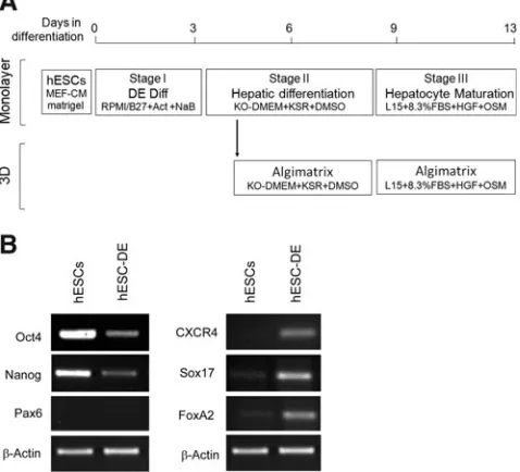
The hESC line H1 was routinely cultured and propagated in Matrigel-coated plates with a mouse embryonic fibroblast-conditioned medium supplemented with basic fibroblast growth factor as previously described.15hESCs were differ-entiated into hepatocytes in monolayer culture using the protocol previously established in our laboratory (Fig. 1A).7 Briefly, differentiation was initiated by culturing for 24 h with 100 ng/mL Activin A and 1 mM sodium butyrate (NaB), followed by 2 days with 100 ng/mL Activin A and 0.5 mM NaB. The cells were then cultured for 5 days in a stage II medium containing 20% KO serum replacement and 0.1% dimethyl sulfoxide (DMSO), followed by 4–5 days in stage III medium (L15 medium containing 8.3% fetal bovine serum, 8.3% tryptose phosphate broth, 10mM hydrocortisone 21-hemisuccinate, 1mM insulin and 2 mM glutamine, 10 ng/ mL hepatocyte growth factor, and 20 ng/mL Oncostatin M). For 3D differentiation, hESCs were differentiated in the same medium as in the monolayer cultures until the second day of stage II when they were inoculated into the Algimatrix plate (Invitrogen 12684-023) (Fig. 1A). Before inoculation, cells were treated with 10mM ROCK inhibitor Y-27632 (ROCKi) for 2 h before they were detached from the culture dish with accutase. Viable cells were transferred into a 24-well Algi-matrix plate by centrifugation at 200g for 3 min at either 0.5·106 or 1·106cells per well in a medium also supple-mented with ROCKi, which was withdrawn after 48 h.
Analysis of the spheroids
Cell spheroids were released from the scaffold by 5-min incubation with 1 mL dissolving buffer (Invitrogen A11340-01) and were then harvested by centrifugation at 200gfor 4 min. Spheroids were stained by 90 s of incubation with propidium iodide (PI) resuspended at 40mg/mL in phosphate-buffered saline (PBS), and images were captured by a Nikon E2000 phase-contrast fluorescent microscope. Dimensional analysis of the spheroids was performed using images taken under a phase-contrast microscope and using the medium axis of the spheroid. To determine the viable cell number, spheroids were dissociated into single cells by 8–10-min incubation with trypsin–ethylenediaminetetraacetic acid at 37C, during which time the spheroids were gently resuspended twice using a pipette. Single cells were then stained with trypan blue and counted with a hemocytometer.
RNA extraction and reverse transcription–polymerase chain reaction
[image:2.612.312.551.64.281.2]Total RNA was extracted using TRI reagent solution (Sigma T9424) following the manufacturer’s instructions. Remaining traces of DNAs were removed by DNase I treatment. First-strand cDNA was synthesized from 2mg
FIG. 1. Differentiation of human embryonic stem cells
total RNA using SuperScript II reverse transcriptase (In-vitrogen) with oligo-dT primer (In(In-vitrogen) in a 20-mL vol-ume. All primer sequences and polymerase chain reaction conditions used are listed in Table 1.
CYP3A4 activity assay
Cells cultured in both monolayer and Algimatrix scaffold conditions were treated with either 10mM rifampicin or 0.1% DMSO (vehicle only) for 48 h. The spheroids were then re-leased from the scaffold as described above into a new cul-ture dish. Subsequently, the cells in both spheroids and monolayer culture were subjected to the P450-Glo CYP3A4 assay using a Luciferin-IPA kit (Promega V9001) according to the manufacturer’s instructions. Briefly, cells were incu-bated for 1 h at 37C with a phenol-free DMEM supple-mented with 10% fetal bovine serum containing 3mM Luciferin-IPA after washed with PBS. A medium containing IPA substrate alone was used as background control. About 50mL of the resulting medium was transferred to a 96-well plate containing an equal volume of Luciferin Detection Reagent and incubated at room temperature for 30 min to initiate the luminescent reaction. The remaining cells were enzymatically dissociated and quantified via cell count. Ac-tivity was measured on a Perkin Elmer Victor 2 luminometer. Values were corrected to account for background. For each treatment, three independent experiments were performed to allow statistical analysis.
BrdU incorporation assay
hESC-DE or HepG2 cells were inoculated into an Algima-trix scaffold with 0.5 million cells per well. After 24 h, BrdU was added into the medium to a final concentration of 10mM and incubated for 48 h. The spheroids were then released from the scaffold, dissociated as described above, and placed onto coverslips. After 24 h culture without BrdU, cells were then fixed by 10-min incubation with 4% paraformaldehyde, wa-shed with PBS, and treated with 2M HCl for 1 h at 37C. Cells were then washed and blocked for 1 h in PBS containing 10% goat serum, incubated with mouse anti-BrdU antibody (DSHB, 1:1000 dilution) for 1 h, followed by 30-min incubation with goat anti-mouse IgG Alexa Fluor 568 secondary antibody (Invitrogen; 1:400 dilution) at room temperature. Images were taken using a Leica SP5 fluorescent microscope.
Urea secretion assay
Urea secretion was assessed by a colorimetric assay (DIUR-500 BioAssay Systems) according to the manufactur-er’s instructions. Briefly, upon a quick thaw of the frozen supernatants, 50mL water (blank), 50mL standard (5 mg/dL), or 50mL samples were transferred into triplicate wells of a clear-bottom 96-well plate. Then, 200mL working reagent was added, and samples were mixed by gentle tapping. These were then incubated for 50 min at room temperature. A spectrophotometer plate reader was used to read absor-bance at 430 nm, and urea secretion was calculated following the manufacturer’s instructions.
Results
ROCK inhibitor promotes the survival and spheroid formation of hESC-DE cells in Algimatrix scaffold
Since monolayer culture condition can efficiently differenti-ate hESCs into DE cells, an essential early stage in the gen-eration of hepatocytes,7,13 and 3D culture provides a better cell–cell contact, which enhances hepatocyte differentiation and function,14it is plausible to hypothesize that a combination of the two conditions would greatly enhance the generation of functional hepatocytes from hESCs. To test this hypothesis, we initiated DE differentiation in hESCs using monolayer culture previously established in our laboratory7(Fig. 1A) and then incorporated the resulting DE cells into 3D culture. We have previously shown that using this monolayer culture, a majority of the cells were differentiated into DE cells by the end of the stage I differentiation.7However, due to considerable cell death that occurs during the 24-h NaB (1 mM) treatment, it was dif-ficult to obtain a sufficient number of cells for the 3D culture at end of the stage I. Therefore, we continued the differentiation on monolayer into the stage II for 1 day before subjecting them into the 3D culture in a bid to increase the number of viable cells (Fig. 1A). These cells are very similar to those found at the end of stage I, as they express high levels of the DE markers, Sox17 and FoxA2, but significantly reduced expression of the plu-ripotent markers, Oct4 and Nanog, with no expression of the neural marker Pax6 (Fig. 1B). These cells will henceforth be referred to as hESC-DE cells.
To incorporate cells into 3D culture, hESC-DE cells were first disassociated into single cells from the culture plate and
Table1. Primer Sequences for the Reverse Transcription–Polymerase Chain Reaction
Name Forward primer 5¢-3¢ Reverse primer 5¢-3¢ Expected size Ta (C)
Albumin CCTTTGGCACAATGAAGTGGGTAACC CAGCAGTCAGCCATTTCACCATAGG 354 bp 55
APOF GGAAGCGATCAAACCTACCA ATCAGCCTGACAACCAGCTT 347 bp 55
B-Actin TCACCACCACGGCCGAGCG TCTCCTTCTGCATCCTGTCG 350 bp 61
CXCR4 CACCGCATCTGGAGAACCA GCCCATTTCCTCGGTGTAGTT 79 bp 55
Cyp3A4 CCTTACATATACACACCCTTTG GGTTGAAGAAGTCCTCCTAAGCT 169 bp 50
Cyp7A1 GTGCCAATCCTCTTGAGTTCC ACTCGGTAGCAGAAAGAATACATC 397 bp 57
FoxA2 AGATGGAAGGGCACGAGC CAGGCCGGCGTTCATGTT 88 bp 56
GAPDH TCTGCTCCTCCTGTTCGACA AAAAGCAGCCCTGGTGACC 120 bp 55
Nanog AGCCTCTACTCTTCCTACCACC TCCAAAGCAGCCTCCAAGTC 278 bp 60
Oct4 CTTGCTGCAGAAGTGGGTGGAGGAA CTGCAGTGTGGGTTTCGGGCA 167 bp 60
Pax6 AACAGACACAGCCCTCACAAACA CGGGAACTTGAACTGGAACTGAC 275 bp 60
Sox17 CCAGAATCCAGACCTGCACAA CTCTGCCTCCTCCACGAA 100 bp 57
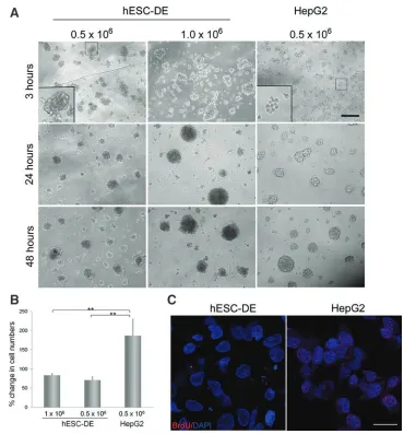
then inoculated into a 24-well Algimatrix 3D culture plate at 0.5–1·106 cells per well. HepG2 cells were also inoculated into the plate in the same way as hESC-DE cells. Interest-ingly, spheroids were observed in HepG2 cells after 5 days in the 3D culture, but none were visible in hESC-DE cultures (Fig. 2A). PI staining revealed considerable cell death in the hESC-DE cultures (Fig 2B-upper panel). As the HepG2 cells grew well in the 3D culture plate, we surmised that the scaffold itself does not induce any detrimental effect on the cells that leads to their death. As such, we reasoned that dissociation of the hESC-DE cells might be the cause of the high levels of cell death observed postinoculation. It has been reported previously that dissociation of hESCs into single cells increases ROCK-dependent hyperactivation of actin– myosin contraction, which induces apoptosis,16,17 and that treatment of hESCs with the ROCK inhibitor Y-27632 (ROCKi) significantly reduces apoptosis after dissociation.18 To test whether use of ROCK inhibitor could reduce hESC-DE cell death after dissociation, the cells were pretreated with 10mM ROCKi for 2 h before disassociation, and the treatment was continued for further 48 h after inoculation.
With this treatment, spheroids were clearly observed 24 h after inoculation and were well formed after 9 days with no signs of detectable apoptosis (Fig. 2B). By contrast, ROCKi treatment did not show a significant effect on their ability to form spheroids in HepG2 cells. These results therefore sug-gest that hESC-DE cells are different from HepG2 cells and require the inhibition of the ROCK signaling pathway to survive and to form 3D structures in Algimatrix scaffold.
hESC-DE cells form spheroid in the 3D culture via cell aggregation
[image:4.612.61.399.317.733.2]Since different growth behaviors were observed with hESC-DE cells and HepG2 cells upon their inoculation into the Algimatrix scaffold, we next examined how spheroids are formed in these two cell types, by monitoring the cells immediately after the inoculation. To our surprise, compact 3D aggregates were already visible in hESC-DE cells 3 h after inoculation, with larger aggregates formed when more cells were inoculated, whereas HepG2 cells still appeared as sin-gle cells or loose-contacted cell clusters at the time (Fig. 3A,
FIG. 2. Effect of ROCK inhibitor
(ROCKi) on cell survival and spheroid formation of hESC-DE cells in Algimatrix scaffold.(A)
Both hESC-DE and HepG2 cells were inoculated into 24-well Algimatrix at 5·105cells/well.
Images were taken 5 days after inoculation, showing no spheroids formed in hESC-DE cultures, but evidence of spheroids in HepG2 cells.(B)hESC-DE cells were treated with or without ROCKi before and 48h after inoculation of 1·106cells/well into the
Algimatrix 3D culture. Cells were collected 9 days after the
inoculation and stained with propidium iodide (PI) to detect any cell death. ROCKi treatment dramatically reduced the cell death and enhanced the spheroid formation. Scale bar=100mm. Color images available online at
upper panel). By 24 h in the 3D scaffold, hESC-DE aggregates appeared as spheroids, and similar spheroids were also ap-parent in HepG2 cells (Fig. 3A, middle panel). The size of spheroids in the hESC-DE cells did not change significantly, whereas the spheroids in the HepG2 cells gradually in-creased in size during the first 48 h in 3D culture (Fig. 3A, lower panel). These data suggest that hESC-DE cells may form spheroids predominantly via cell aggregation, which can occur within a relatively short time period and may be promoted by a ROCK inhibitor, whereas HepG2 cells may form spheroids through cell aggregation as well as cell pro-liferation.
To validate this observation, we compared the total viable cell numbers seeded at inoculation with those after 9 days of 3D culture in both cell types. As expected, the number of viable HepG2 cells in the 3D culture notably increased dur-ing this time period, although not to the same extent as in the 2D culture, whereas the number of hESC-DE cells showed a decrease in numbers, most likely due to cell death (Fig. 3B). To further confirm the proliferative properties of the two cell types, we incubated the cells with BrdU for 48 h after 3D inoculation, and then stained the cells with an anti-BrdU antibody. HepG2 cells showed positive signals with the BrdU antibody, whereas positive staining was not detected
in the nuclei of hESC-DE cells. Therefore, these results con-firm that hESC-DE cells do not proliferate after inoculation into the Algimatrix scaffold.
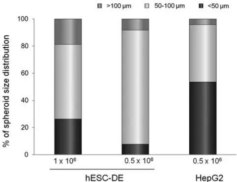
Cell density affects the size of spheroids in the Algimatrix scaffold
Spheroid size has been reported to affect hepatocyte via-bility and function in the 3D culture.14Since the cell spher-oids in culture do not have a vascular network, cells constituting the center of the spheroids become hypoxic and have a reduced uptake of oxygen from the cell medium if they are larger than 100mm in size. On the other hand, if the spheroid size is too small, they produce significantly less albumin as a result of poor maturation.19 To study factors
[image:5.612.184.554.54.451.2]that affect the spheroid size, we categorized the spheroids produced by our 3D culture into three groups according to their size: small with spheroids of <50mm in diameter, me-dium between 50 and 100mm in diameter, and larger over 100mm in diameter, and examined the effect of initial inoc-ulation seeding density on the spheroid size. We measured and categorized *100 spheroids from each inoculation condition 9 days after the cells were inoculated. The results show that in hESC-DE cells, a majority (84%) of spheroids
FIG. 3. Spheroid formation
and cell proliferation in Algimatrix 3D cultures.(A)
Both hESC-DE and HepG2 cells aggregate to form spheroids during the first day after inoculation into the Algimatrix scaffold with hESC-DE cells forming aggregates quicker than the HepG2 cells and higher seeding density producing larger spheroids. In addition, spheroids formed by hESC-DE cells exhibit no significant difference in size between 24– 48 h, while spheroids of HepG2 cells show clear increase during this time period. Scale bar=100mm.(B)
Changes in cell number after 9 days in 3D culture. Cell counts are presented as percentage of the initial seeded cell numbers. Standard error bars were calculated from six independent experiments. **p<0.001 (Student’st-tests).
produced from the lower seeding density of 5·105cells/well fell into the medium-size bracket, with <10% of spheroids falling into the larger- or smaller-size group. However, at higher seeding density of 1·106cells/well, almost 20% and 30% of the spheroids were in the large- and smaller-size group, respectively, with <60% in the medium-size category (Fig. 4). In HepG2 cells, the lower seeding density of 5·105
cells/well produced very few spheroids (<5%) of the large size. These results suggest that the initial seeding density of hESC-DE cells has an effect on the sizes of the spheroids formed in the 3D scaffold.
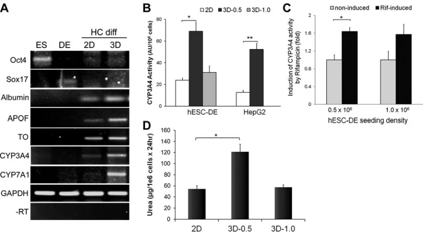
Algimatrix 3D culture promotes hepatocyte differentiation and function
Alginate scaffold has been found to promote cells from the liver of newborn rats to differentiate into functional liver tissues.20To evaluate whether the Algimatrix 3D culture has any advantage over monolayer culture in differentiating DE cells into hepatocytes, we compared the hepatic gene ex-pression profiles in the 3D culture to those in monolayer as well as to undifferentiated hESCs and the hESC-DE cells used at the 3D inoculation stage. Unsurprisingly, undiffer-entiated hESCs exhibited high levels of the pluripotent gene Oct4, but showed no expression of hepatic markers. Ex-pression of the DE marker, Sox17, was apparent in hESC-DE cells, but neitherOct4nor any hepatic genes were detectable. In contrast, hESC-DE cells that had been further differenti-ated in both 2D and 3D culture conditions expressed the hepatocyte genes, albumin, apolipoprotein F (ApoF), tryp-tophan dioxygenase (TO), and cytochrome P450 family members, CYP3A4 and CYP7A1 (Fig. 5A). Significantly, the cells differentiated in the Algimatrix 3D scaffold exhibited considerably higher levels of hepatocyte-specific markers than the cells differentiated in monolayer, particularly those that are expressed predominantly in the more mature he-patocytes, such as ApoF, TO, and CYP3A4.21
Since CYP3A4 is the most crucial P450 enzymes in the human liver, metabolizing about half of all drugs on the market today, we examined CYP3A4 activity in hESC-DE and HepG2 cells that were either cultured in 3D conditions or differentiated in monolayer to further determine the im-provement of 3D culture on the differentiation and function of hepatocytes. In comparison with the monolayer culture system, at the lower seeding density of 0.5·106cells/well, Algimatrix 3D culture significantly increased basal levels of CYP3A4 activity in both hESC-DE cells and HepG2 cells (Fig. 5B). The higher inoculation density also increased activity, but to a lesser extent, probably due to a suboptimal spheroid size. Furthermore, the cells cultured in the 3D condition also exhibited increased CYP3A4 activity in response to rifam-picin induction (Fig. 5C). To further validate the improve-ment of 3D culture on hepatocyte differentiation and function, we measured urea secretion in hESC-derived he-patocytes from both 2D and 3D cultures (Fig. 5D), which showed that Algimatrix 3D culture significantly enhanced urea secretion when hESC-DE cells were seeded at 0.5·106 cells/well. Taken together, we have shown that Algimatrix 3D culture system improves the differentiation of hESC-DE cells to hepatocytes, and as a result, enhances the expression of hepatocyte markers and certain functions.
Discussion
We have demonstrated here that DE cells derived from hESCs are able to form spheroids in an Algimatrix scaffold. In comparison with the monolayer culture, Algimatrix 3D culture enhances hepatocyte differentiation and function. These results correspond with the previous findings from hESCs.9 However, in the previous studies, the 3D culture was directly applied to the hESCs, in which hESCs were detached into small clumps and cultured in a low-adherent plate to form EBs. Since pluripotent hESCs in the EBs receive variable signals depending on their position, this method may have an increased chance of inducing the differentiation of cells to other lineages, which consequently reduces the final yield of hepatocytes. Our previous studies, as well as those of others, have shown that monolayer culture supple-mented with high-dosage Activin A can differentiate hESCs efficiently to the DE cells that are a prerequisite for the generation of hepatocytes.7,13Therefore, to increase
hepato-cyte production, we incorporated the 3D culture at a later stage in the differentiation protocol, once the hESCs were committed to DE. We differentiated hESCs for 4 days in monolayer culture with our established method and dem-onstrated that the cells were primarily differentiated into DE (Fig. 1B) before incorporating them into 3D culture.
[image:6.612.58.299.472.657.2]Here, and in accordance with others, we have demon-strated that an alginate-based 3D culture system can enhance the functionality of HepG2 cells and has no toxic effects on the cells.22,23Interestingly, we have also shown that hESC-DE cells behave differently from HepG2 cells when cultured in an Algimatrix 3D scaffold. Firstly, hESC-DE cells required the treatment of ROCKi to survive in the 3D culture after dissociation, whereas HepG2 cells do not require ROCKi to survive or to form spheroids. In addition, treatment of HepG2 cells with ROCKi showed no effect on their survival and proliferation. This phenotype of hESC-DE cells is similar to that reported in undifferentiated hESCs.24 In hESCs,
FIG. 4. Effect of seeding density on spheroids’ sizes.
apoptosis upon dissociation is thought to be caused by ROCK-dependent actomyosin hyperactivation, which is triggered by the loss of E-cadherin-dependent intercellu-lar contact.17 However, hESC-DE cells stained with anti-E-cadherin antibody exhibited no clear positive signals (data not shown), indicating that the apoptosis induced by disso-ciation in hESC-DE cells is not a downstream effect of E-cadherin. Nonetheless, one cannot exclude the possibility that this phenotype could still be related to the loss of attachment-dependent actin–myosin contraction.16 Further
experiments are required to elucidate the underlying mech-anisms. Secondly, hESC-DE cells aggregate more rapidly than the HepG2 cells. Thirdly, unlike HepG2 cells, hESC-DE cells were unable to proliferate in the Algimatrix 3D scaffold, which was demonstrated by the fact that there was no sig-nificant increase in the cell numbers after 9 days in the 3D culture, and that BrdU labeling showed no incorporation (Fig. 3). This was also different from their behavior in monolayer culture, in which hESC-DE cells were able to di-vide and incorporate BrdU. Although the exact mechanisms remain unclear, it has been demonstrated previously that the growth properties and differentiated functionality of rat he-patocytes are reciprocally modulated in culture by cell–cell contact, and that this reciprocal modulation is lost in hepa-toma cells.25,26Therefore, it is likely that increased cell–cell contact may account for the phenotype, which may also contribute to the improved differentiation and functionality. We have analyzed spheroid formation and hepatocyte function of hESC-DE cells at two different seeding densities. The two different seeding densities did not seem to have a
clear effect on the timing of spheroid formation, but did appear to impact the size of spheroids formed in the Algi-matrix scaffold. However, this effect was not a simple posi-tive correlation. Surprisingly, the higher seeding density not only resulted in a higher proportion of the larger spheroids but also increased the number of smaller spheroids. In ad-dition, our results showed that lower seeding densities that generated significantly more medium-sized spheroids had higher levels of CYP3A4 activity and a significant induction upon drug stimulation as well as increased urea secretion, indicating that the size of hESC-DE spheroids is important for the hepatocyte differentiation and function. It is not surprising that it is the medium-sized spheroids that exhibit better functionality, as the larger size reduces the nutrition/ oxygen accessibility of the cells found at the center and af-fects the survival of these cells; while the smaller spheroids do not form the proper cell–cell contacts, which may sub-sequently hinder hepatocyte differentiation and function.19 In summary, our study has demonstrated that hESC-DE cells can be incorporated into an Algimatrix scaffold 3D culture upon treatment with the ROCK inhibitor. The 3D culture enhances hESC-DE cell differentiation to HLCs and im-proves the resulting hepatocyte function, such as CYP3A4 activity.
Acknowledgments
[image:7.612.97.517.57.287.2]We thank Dr. Sam Coward and Dr. Isobel Massie for their technical help and discussion at early stage of the project and thank Dr. Fiona Lamont for their helpful discussion on the
FIG. 5. Comparison of the effect of 2D (monolayer) and 3D culture conditions on hepatocyte marker expression and
manuscript. T.S.R. was supported by a studentship from the University of Malaya/Ministry of Higher Education, Ma-laysia. This work is supported by the Genesis Research Trust.
Disclosure Statement
No competing financial interests exist.
References
1. Irion, S., Nostro, M.C., Kattman, S.J., and Keller, G.M. Di-rected differentiation of pluripotent stem cells: from devel-opmental biology to therapeutic applications. Cold Spring Harb Symp Quant Biol73,101, 2008.
2. Stutchfield, B.M., Forbes, S.J., and Wigmore, S.J. Prospects for stem cell transplantation in the treatment of hepatic disease. Liver Transpl16,827, 2010.
3. Selden, C., and Hodgson, H. Cellular therapies for liver re-placement. Transpl Immunol12,273, 2004.
4. Behbahan, I.S., Duan, Y., Lam, A., Khoobyari, S., Ma, X., Ahuja, T.P., and Zern, M.A. New approaches in the differen-tiation of human embryonic stem cells and induced pluripotent stem cells toward hepatocytes. Stem Cell Rev7,748, 2011. 5. Guguen-Guillouzo, C., Corlu, A., and Guillouzo, A. Stem
cell-derived hepatocytes and their use in toxicology. Tox-icology270,3, 2010.
6. Cai, J., Zhao, Y., Liu, Y., Ye, F., Song, Z., Qin, H., Meng, S., Chen, Y., Zhou, R., Song, X.,et al.Directed differentiation of human embryonic stem cells into functional hepatic cells. Hepatology45,1229, 2007.
7. Hay, D.C., Zhao, D., Fletcher, J., Hewitt, Z.A., McLean, D., Urruticoechea-Uriguen, A., Black, J.R., Elcombe, C., Ross, J.A., Wolf, R.,et al.Efficient differentiation of hepatocytes from human embryonic stem cells exhibiting markers reca-pitulating liver developmentin vivo. Stem Cells26,894, 2008. 8. Green, M.D., Chen, A., Nostro, M.C., d’Souza, S.L., Schaniel, C., Lemischka, I.R., Gouon-Evans, V., Keller, G., and Snoeck, H.W. Generation of anterior foregut endoderm from human embryonic and induced pluripotent stem cells. Nat Bio-technol29,267, 2011.
9. Baharvand, H., Hashemi, S.M., Kazemi, A.S., and Farrokhi, A. Differentiation of human embryonic stem cells into he-patocytes in 2D and 3D culture systemsin vitro. Int J Dev Biol50,645, 2006.
10. Elaut, G., Henkens, T., Papeleu, P., Snykers, S., Vinken, M., Vanhaecke, T., and Rogiers, V. Molecular mechanisms un-derlying the dedifferentiation process of isolated hepato-cytes and their cultures. Curr Drug Metab7,629, 2006. 11. Bone, H.K., Nelson, A.S., Goldring, C.E., Tosh, D., and
Welham, M.J. A novel chemically directed route for the generation of definitive endoderm from human embryonic stem cells based on inhibition of GSK-3. J Cell Sci124,1992, 2011.
12. Lavon, N., Yanuka, O., and Benvenisty, N. Differentiation and isolation of hepatic-like cells from human embryonic stem cells. Differentiation72,230, 2004.
13. D’Amour, K.A., Agulnick, A.D., Eliazer, S., Kelly, O.G., Kroon, E., and Baetge, E.E. Efficient differentiation of human embryonic stem cells to definitive endoderm. Nat Biotechnol
23,1534, 2005.
14. Glicklis, R., Shapiro, L., Agbaria, R., Merchuk, J.C., and Cohen, S. Hepatocyte behavior within three-dimensional porous alginate scaffolds. Biotechnol Bioeng67,344, 2000. 15. Gerrard, L., Rodgers, L., and Cui, W. Differentiation of
hu-man embryonic stem cells to neural lineages in adherent
culture by blocking bone morphogenetic protein signaling. Stem Cells23,1234, 2005.
16. Chen, G., Hou, Z., Gulbranson, D.R., and Thomson, J.A. Actin-myosin contractility is responsible for the reduced viability of dissociated human embryonic stem cells. Cell Stem Cell7,240, 2010.
17. Ohgushi, M., Matsumura, M., Eiraku, M., Murakami, K., Aramaki, T., Nishiyama, A., Muguruma, K., Nakano, T., Suga, H., Ueno, M.,et al.Molecular pathway and cell state responsible for dissociation-induced apoptosis in human pluripotent stem cells. Cell Stem Cell7,225, 2010.
18. Watanabe, K., Ueno, M., Kamiya, D., Nishiyama, A., Mat-sumura, M., Wataya, T., Takahashi, J.B., Nishikawa, S., Nishikawa, S., Muguruma, K.,et al.A ROCK inhibitor per-mits survival of dissociated human embryonic stem cells. Nat Biotechnol25,681, 2007.
19. Glicklis, R., Merchuk, J.C., and Cohen, S. Modeling mass transfer in hepatocyte spheroids via cell viability, spheroid size, and hepatocellular functions. Biotechnol Bioeng86,672, 2004.
20. Dvir-Ginzberg, M., Elkayam, T., and Cohen, S. Induced differentiation and maturation of newborn liver cells into functional hepatic tissue in macroporous alginate scaffolds. FASEB J22,1440, 2008.
21. Snykers, S., De, K.J., Rogiers, V., and Vanhaecke, T.In vitro
differentiation of embryonic and adult stem cells into he-patocytes: state of the art. Stem Cells27,577, 2009. 22. Coward, S.M., Legallais, C., David, B., Thomas, M., Foo, Y.,
Mavri-Damelin, D., Hodgson, H.J., and Selden, C. Alginate-encapsulated HepG2 cells in a fluidized bed bioreactor maintain function in human liver failure plasma. Artif Or-gans33,1117, 2009.
23. Elkayam, T., Amitay-Shaprut, S., Dvir-Ginzberg, M., Harel, T., and Cohen, S. Enhancing the drug metabolism activities of C3A—a human hepatocyte cell line—by tissue engineer-ing within alginate scaffolds. Tissue Eng12,1357, 2006. 24. Harb, N., Archer, T.K., and Sato, N. The Rho-Rock-Myosin
signaling axis determines cell-cell integrity of self-renewing pluripotent stem cells. PLoS One3,e3001, 2008.
25. Nakamura, T., Yoshimoto, K., Nakayama, Y., Tomita, Y., and Ichihara, A. Reciprocal modulation of growth and dif-ferentiated functions of mature rat hepatocytes in primary culture by cell—cell contact and cell membranes. Proc Natl Acad Sci U S A80,7229, 1983.
26. Nakamura, T., Nakayama, Y., Teramoto, H., Nawa, K., and Ichihara, A. Loss of reciprocal modulations of growth and liver function of hepatoma cells in culture by contact with cells or cell membranes. Proc Natl Acad Sci U S A81,6398, 1984.
Address correspondence to: Wei Cui, PhD Department of Surgery and Cancer Institute of Reproductive and Developmental Biology Imperial College London Hammersmith Campus Du Cane Road London W12 0NN United Kingdom
E-mail:[email protected]