Microbial Interactions in the Cystic Fibrosis Airway
Ann M. Granchelli,a*Frederick R. Adler,b,c,dRuth H. Keogh,eChristiana Kartsonaki,fDavid R. Cox,g Theodore G. Lioua,b
aThe Adult Cystic Fibrosis Center, Division of Respiratory, Critical Care and Occupational Pulmonary Medicine,
Department of Internal Medicine, University of Utah, Salt Lake City, Utah, USA
bCenter for Quantitative Biology, University of Utah, Salt Lake City, Utah, USA cDepartment of Biology, University of Utah, Salt Lake City, Utah, USA dDepartment of Mathematics, University of Utah, Salt Lake City, Utah, USA eLondon School of Hygiene and Tropical Medicine, London, United Kingdom
fNuffield Department of Population Health, University of Oxford, Oxford, United Kingdom gNuffield College, Oxford, United Kingdom
ABSTRACT Interactions in the airway ecology of cystic fibrosis may alter organism persistence and clinical outcomes. Better understanding of such interactions could guide clinical decisions. We used generalized estimating equations to fit logistic re-gression models to longitudinal 2-year patient cohorts in the Cystic Fibrosis Founda-tion Patient Registry, 2003 to 2011, in order to study associaFounda-tions between the air-way organisms present in each calendar year and their presence in the subsequent year. Models were adjusted for clinical characteristics and multiple observations per patient. Adjusted models were tested for sensitivity to cystic fibrosis-specific treat-ments. The study included 28,042 patients aged 6 years and older from 257 accred-ited U.S. care centers and affiliates. These patients had produced sputum specimens
for at least two consecutive years that were cultured for methicillin-sensitive
Staphy-lococcus aureus, methicillin-resistantS. aureus,Pseudomonas aeruginosa,Burkholderia cepacia complex, Stenotrophomonas maltophilia, Achromobacter xylosoxidans, and
Candida and Aspergillus species. We analyzed 99.8% of 538,458 sputum cultures
from the patients during the study period. Methicillin-sensitive S. aureuswas
nega-tively associated with subsequentP.aeruginosa. P.aeruginosawas negatively
associ-ated with subsequentB. cepaciacomplex,A.xylosoxidans, andS.maltophilia. B.
cepa-cia complex was negatively associated with the future presence of all bacteria
studied, as well as with that of Aspergillusspecies.P.aeruginosa,B. cepaciacomplex,
and S. maltophilia were each reciprocally and positively associated with Aspergillus
species. Independently of patient characteristics, the organisms studied interact and alter the outcomes of treatment decisions, sometimes in unexpected ways. By
inhib-iting P. aeruginosa, methicillin-sensitive S. aureus may delay lung disease
progres-sion. P.aeruginosaandB. cepaciacomplex may inhibit other organisms by
decreas-ing airway biodiversity, potentially worsendecreas-ing lung disease.
KEYWORDS Burkholderia,Pseudomonas aeruginosa,Staphylococcus aureus, airway infections, cystic fibrosis, logistic regression, microbial ecology
I
n the United States, cystic fibrosis (CF) affects roughly 30,000 people, reducing lifeexpectancy by⬎50% (1). Progressive pulmonary disease, marked by recurrent
exac-erbations, bacterial infection, and declining lung function, drives morbidity and mor-tality (2–5). Studies of the CF lung reveal a diverse microbiology. Methicillin-sensitive
Staphylococcus aureus (MSSA) and Pseudomonas aeruginosa are the two organisms most commonly isolated from the airway (1, 2). Opportunistic organisms, including
Burkholderia cepaciacomplex,Stenotrophomonas maltophilia,Achromobacter
xylosoxi-Received3 March 2018Returned for modification27 March 2018Accepted9 May 2018
Accepted manuscript posted online16 May 2018
CitationGranchelli AM, Adler FR, Keogh RH, Kartsonaki C, Cox DR, Liou TG. 2018. Microbial interactions in the cystic fibrosis airway. J Clin Microbiol 56:e00354-18.https://doi.org/10 .1128/JCM.00354-18.
EditorAlexander J. McAdam, Boston Children's Hospital
Copyright© 2018 American Society for Microbiology.All Rights Reserved.
Address correspondence to Theodore G. Liou, ted.liou@utah.edu.
*Present address: Ann M. Granchelli, Division of Pulmonary, Critical Care and Sleep Medicine, National Jewish Health, Denver, Colorado, USA.
crossm
on May 16, 2020 by guest
http://jcm.asm.org/
dans, nontuberculous mycobacteria, and fungal organisms, commonly colonize and infect patients with CF.
The presence of different organisms alters long-term outcomes for patients with CF.
MSSA appears to enhance survival, while B. cepacia complex may presage a
cata-strophic decline in health (6, 7). Acquisition of methicillin-resistantS. aureus(MRSA) or
P. aeruginosa is associated with accelerated lung disease (8–12). Published cross-sectional data from the Cystic Fibrosis Foundation Patient Registry (CFFPR) show that dominant airway infections differ with age (1). MSSA most commonly infects pediatric
patients, whileP. aeruginosainfection increases in frequency with age and commonly
dominates the bacterial community in adult patients (13). Without a clear understand-ing of the underlyunderstand-ing microbial interactions, efforts to prevent, treat, or eradicate
specific organisms, such as P. aeruginosa, may produce unexpected and undesirable
outcomes. A double-blind, randomized controlled study in 2002 showed that prophy-lactic treatment of MSSA with cephalexin in infants and young children with CF led to
earlier colonization with P. aeruginosa (14). Prophylaxis with ciprofloxacin had no
effects in preventing infection withP. aeruginosa(15). Similarly, eradication therapy for
P. aeruginosaincreased the rate of infection withS. maltophilia(15).
While antibiotic therapy may play a role, existing infections themselves appear to alter the rest of the microbiota (7, 16–19) and may thus alter the clinical disease course.
In vitro and nonhuman in vivo models show evidence of interspecies interaction
betweenP. aeruginosaand other pathogens, including MSSA andBurkholderia
ceno-cepacia. Mathematical models of disease progression explore potential airway
interac-tions between P. aeruginosa, MSSA, andBurkholderiaspecies; the results from these
models are consistent with observational data on CF (1) and illustrate the potential impact of managing these organisms (7).
In this study, we focus on eight common CF airway pathogens and show how the presence of each in a given study year is associated with infections observed in the following year. By improving our understanding of interactions between organisms, we seek to enhance understanding of the underlying mechanisms of changing airway microbial ecology, which may help us anticipate the impacts of changing practice on clinical outcomes.
(Some of the data and results of this study were reported by A. M. Granchelli in preliminary form at the 37th European Cystic Fibrosis Society Meeting, 11 to 14 June 2013, in Gothenburg, Sweden. F. R. Adler and T. G. Liou had full access to all the data in the study and take responsibility for the integrity of the data and the accuracy of the data analysis.)
MATERIALS AND METHODS
Study design and data.We analyzed data for the years 2003 to 2011 from the CFFPR, which contains
prospectively collected patient data from 257 Cystic Fibrosis Foundation-accredited care centers and affiliated programs in the United States. Data for the ongoing CFFPR study are gathered according to a defined protocol after obtaining written consent from adult patients or parental consent with assent from minors. The data include patient demographics, clinical measurements of CF disease, treatment information, the number of clinic visits, and culture results from routine quarterly and acute-illness samples. These data were monitored to confirm fidelity to medical charts (20).
We obtained approval from the University of Utah Investigational Review Board (IRB) for the performance of this study, with a waiver of informed consent and approval from the Data Use Committee of the U.S. Cystic Fibrosis Foundation for access to and use of the CFFPR. We continue to participate in data collection for the CFFPR after obtaining written informed consent, with separate approval from the IRB.
Study population and definitions.Our study cohort included all CFFPR subjects for whom data
were available for at least two consecutive years between 2003 and 2011. Since 2003, the CFFPR has recorded each culture result separately for every patient; previously, the CFFPR had reported only a single annualized result per year, thus guiding our study period selection. Patients younger than 6 years were excluded because they cannot reliably perform pulmonary function testing and usually do not produce sputum. We used sputum culture results to determine the presence of infection. The CFFPR records organisms as present or absent for each culture and records the number of cultures obtained in each given year. The presence of infection with a particular organism within a given calendar year was defined as at least one positive culture for that organism within that year. We focused on eight common infections on which the CFFPR contains sufficient data for analysis: MSSA, MRSA,B. cepaciacomplex,P. aeruginosa,S. maltophilia,A. xylosoxidans,Candidaspecies, andAspergillusspecies. We identified, and
on May 16, 2020 by guest
http://jcm.asm.org/
adjusted for, the following patient characteristics as potential confounders in statistical models: age, age at CF diagnosis, sex, CF-related diabetes, pancreatic sufficiency, weight-for-ageZ-score, percentage of predicted forced expiratory volume in 1 s (FEV1%), and acute pulmonary exacerbations (APE). Most of
these characteristics have been found previously to predict 5-year survival (6). We defined patients as diabetic in a given year if the condition was present at any time during that year. We defined patients as pancreatic sufficient in a given year if they were noted to be sufficient for all encounters and did not use pancreatic enzymes during that year. For sensitivity analyses to determine the effect of adjustment of associations for different interventions, we used treatment data on oral azithromycin, inhaled aztreonam, tobramycin, recombinant human DNase, and hypertonic saline, days per year of therapy with home intravenous (i.v.) antibiotics, hospitalization days for pulmonary exacerbation treatment, and lung transplantation.
Statistical analysis.We fitted cross-sectional univariable logistic regression models (21) with the
presence of each organism in a given year as the outcome and every other organism studied as an individual explanatory variable. Cross-sectional multivariable models were then fitted with the presence of each organism as the outcome and with all other organisms as explanatory variables with and without additional adjustment for clinical characteristics. These models were fitted separately in each calendar year, from 2003 to 2011. We fitted similar multivariable logistic models relating organisms in each year tto the presence of each organism in yeart⫹1, wheretis a particular year from 2003 to 2010. We fitted these models with and without additional adjustment for clinical characteristics. Finally, we fitted a single combined model across all observation years for each patient using generalized estimating equations with an independence working correlation matrix. This model uses multiple observations per individual across the study years. The use of the combined model increases the power of our analysis and reduces the size of confidence intervals (22, 23). It makes the assumption that the associations between organisms from one year to the next are the same across the calendar years. See Text S1 in the supplemental material for detailed methods and the individual steps to fitting the combined model. Sensitivity analyses examined the impact of adjusting for the treatments used. All analyses were performed using the R statistical system (24).
RESULTS
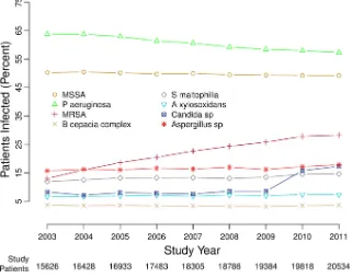
Participants.We found 28,042 patients aged 6 years or older from 257 care centers and affiliated programs accredited by the U.S. Cystic Fibrosis Foundation who were included in the CFFPR between 1 January 2003 and 31 December 2011 with at least two consecutive years of culture data (Table 1). These patients had a total of 538,458 sets of sputum culture results, of which 537,396 (99.8%) were included in our analysis (see Fig. 1). Culture results were excluded only for lack of same-patient cultures in contig-uous years. From 2003 to 2011, the median cross-sectional age of patients increased from 16.6 to 19.1 years (Table 1). The prevalence of CF-related diabetes nearly doubled over this period, from 9.62% to 17.5%. A minority of patients were pancreatic sufficient; this proportion increased between 2003 and 2011 from 6.53% to 11.2%. There were
marginal changes in the median FEV1% and the mean number of APE during the study
period. The changes in pediatric and adult groups were similar (see Tables S1 and S2 in the supplemental material).
Infection prevalence. There were changes in the percentages of patients with positive cultures for the eight most common infections recorded in the CFFPR between 2003 and 2011 (Table 1 and Fig. 1; also Tables S1 and S2 in the supplemental material).
The percentages of patients infected by MRSA andCandidaspecies more than doubled,
and the percentages of patients with S. maltophilia,A. xylosoxidans, and Aspergillus
species also increased. Between 2003 and 2011, there were small, statistically significant decreases in the percentages of patients infected by MSSA (from 50.2% to 49.2%) and
P. aeruginosa(from 63.8% to 57.3%).
Cross-sectional associations between airway infections. Figure 2 shows esti-mated odds ratios from multivariable logistic regression models fitted in each year from 2003 to 2011 with each organism as an outcome and the set of all other organisms as explanatory variables, with or without adjustment for clinical variables and the number
of cultures. The estimates were remarkably consistent across calendar years.P.
aerugi-nosainfection was negatively associated with MSSA,B. cepaciacomplex,S. maltophilia,
andA. xylosoxidansand was positively associated withAspergillusspecies infections for every study year, with or without adjustment for the presence of other organisms (Fig.
2A, D, E, F, and H, respectively).P. aeruginosaandB. cepaciacomplex were negatively
associated in every study year with or without adjustment for other organisms, and additional adjustments for clinical characteristics intensified these associations (Fig. 2B and D). After adjustment for other organisms, with or without adjustment for clinical
on May 16, 2020 by guest
http://jcm.asm.org/
TABLE 1 Selected characteristics of study patients Patient characteristic Value for the following yr: 2003 2004 2005 2006 2007 2008 2009 2010 2011 No. enrolled 15,626 16,428 16,933 17,483 18,305 18,786 19,384 19,818 20,534 No. of cultures a 42,995 (0.996) 47,516 (0.999) 51,826 (1.000) 55,922 (0.999) 61,442 (1.000) 65,319 (1.000) 68,312 (1.000) 70,294 (0.999) 74,409 (1.000) No. of cultures per patient b 2 (1–4) 2 (1–4) 3 (2–4) 3 (2–4) 3 (2–4) 3 (2–4) 3 (2–4) 3 (2–4) 3 (2–5) Age (yr) b 16.6 (11.4–24.0) 17.0 (11.7–24.5) 17.2 (11.8–24.8) 17.6 (12.0–25.3) 17.8 (12.1–25.9) 18.1 (12.2–26.4) 18.4 (12.4–27.0) 18.7 (12.5–27.4) 19.1 (12 .7–27.9) No. (%) of enrolled patients Female 7,446 (47.7) 7,811 (47.5) 8,050 (47.5) 8,319 (47.6) 8,764 (47.9) 9,060 (48.2) 9,393 (48.5) 9,618 (48.5) 9,949 (48.5) With pancreatic sufficiency 1,018 (6.53) 1,198 (7.30) 1,189 (7.03) 1,365 (7.81) 1,457 (7.96) 1,654 (8.80) 1,830 (9.44) 2,419 (12.2) 2,300 (11.2) With diabetes 1,503 (9.62) 1,744 (10.6) 1,919 (11.3) 2,410 (13.8) 2,664 (14.6) 2,845 (15.1) 3,022 (15.6) 3,270 (16.5) 3,601 (17.5) FEV 1 % b 81.0 (58.7–97.0) 81.5 (59.2–97.2) 82.0 (59.6–97.5) 82.6 (60.2–97.7) 83.0 (60.4–98.5) 83.2 (60.7–98.7) 83.5 (60.7–99.0) 83.9 (61.3–99.1) 83.7 (61 .3–99.0) No. of acute pulmonary exacerbations b 0 (0–1) 0 (0–1) 0 (0–1) 0 (0–1) 0 (0–1) 0 (0–1) 0 (0–1) 0 (0–1) 0 (0–1) Wt-for-age Z -score b ⫺ 0.52 ( ⫺ 1.23 to 0.18) ⫺ 0.47 ( ⫺ 1.20 to 0.21) ⫺ 0.45 ( ⫺ 1.17 to 0.22) ⫺ 0.40 ( ⫺ 1.12 to 0.25) ⫺ 0.38 ( ⫺ 1.10 to 0.27) ⫺ 0.33 ( ⫺ 1.06 to 0.30) ⫺ 0.29 ( ⫺ 1.04 to 0.33) ⫺ 0.27 ( ⫺ 1.04 to 0.35) ⫺ 0.25 ( ⫺ 1.03 to 0.36) No. (%) of enrolled patients with: MSSA 7,852 (50.2) 8,292 (50.5) 8,497 (50.2) 8,704 (49.8) 9,140 (49.9) 9,299 (49.5) 9,574 (49.4) 9,759 (49.2) 10,104 (49.2) P. aeruginosa 9,969 (63.8) 10,469 (63.7) 10,656 (62.9) 10,729 (61.4) 11,100 (60.6) 11,123 (59.2) 11,323 (58.4) 11,482 (57.9) 11,767 (57.3) MRSA 2,034 (13.0) 2,642 (16.1) 3,155 (18.6) 3,588 (20.5) 4,150 (22.7) 4,576 (24.4) 5,005 (25.8) 5,514 (27.8) 5,801 (28.3) Burkholderia complex 584 (3.74) 580 (3.53) 622 (3.67) 607 (3.47) 622 (3.40) 618 (3.29) 619 (3.19) 697 (3.52) 756 (3.68) S. maltophilia 1,868 (12.0) 2,064 (12.6) 2,243 (13.2) 2,320 (13.3) 2,440 (13.3) 2,457 (13.1) 2,616 (13.5) 2,881 (14.5) 3,010 (14.7) A. xylosoxidans 1,028 (6.58) 1,095 (6.67) 1,166 (6.89) 1,240 (7.09) 1,236 (6.75) 1,355 (7.21) 1,343 (6.93) 1,468 (7.41) 1,501 (7.31) Candida species 1,295 (8.29) 1,177 (7.16) 1,365 (8.06) 1,370 (7.84) 1,391 (7.60) 1,609 (8.56) 1,661 (8.57) 3,129 (15.8) 3,559 (17.3) Aspergillus species 2,458 (15.7) 2,677 (16.3) 2,724 (16.1) 2,913 (16.7) 2,996 (16.4) 3,190 (17.0) 3,141 (16.2) 3,409 (17.2) 3,678 (17.9) aThe values in parentheses are the fractions of the numbers of patients enrolled in each year in the study that had usable culture data. bValues are medians (1st and 3rd quartiles).
on May 16, 2020 by guest
http://jcm.asm.org/
[image:4.585.79.332.72.741.2]characteristics,P. aeruginosainfection was negatively associated with MRSA for each
year from 2004 through 2011 (Fig. 2C). A. xylosoxidans infections and B. cepacia
complex infections showed large negative coefficients in every study year, suggesting
that such coinfections are uncommon (Fig. 2D). In contrast,S. maltophiliawas
associ-ated with Aspergillus species infections every year with large positive coefficients,
suggesting that such coinfections are common (Fig. 2H). The odds ratios from logistic regression models examining univariable, unadjusted associations between organisms are similar to the fully adjusted results (see Fig. S1 in the supplemental material).
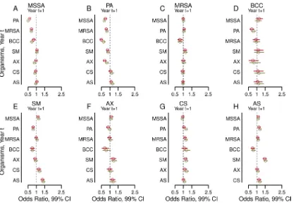
Associations between present and future airway infections.Figure 3 shows the estimated odds ratios from multivariable models for the association of microbial
infection status for each of the years 2004 through 2011 (yearst⫹1) with infections in
the previous year (wheretis a particular year from 2003 to 2010) (21). Associations were
similar for every 2-year period examined, with some variation in the degree of statistical significance for individual relationships.
For each organism, the predictor most strongly associated with its presence in year
t⫹1was the presence of the same organism in yeart(see Fig. S2 in the supplemental
material). MSSA,B. cepaciacomplex,S. maltophilia, andA. xylosoxidansin each yeart
were all negatively associated withP. aeruginosain the subsequent yeart⫹1(Fig. 3B).
CandidaorAspergillusspecies were not typically associated with subsequentP.
aerugi-nosa(Fig. 3B). Organisms other than MSSA andB. cepaciacomplex in each yeartwere
infrequently associated with MRSA in each yeart⫹1. MSSA in 3 yearst(2005, 2008, and
2010) andB. cepaciacomplex infections in 2 yearst(2005 and 2007) had potentially
significant negative statistical and clinical associations with MRSA (Fig. 3C). Adjustment for clinical variables did not substantially change the estimates or their statistical significance (Fig. 3).
When all observations are used simultaneously in the combined model, the
rela-tionships between an organism seen in yeartwith a different organism in yeart⫹1(Fig.
4; see also Tables S3 and S4 in the supplemental material) are similar to all relationships seen in multivariable logistic regression models for each of the 2-year models reported
FIG 1Percentages of patients with positive cultures for the infections studied, 2003 to 2011. Eight curves
show the changing percentages of cultures for the organisms studied, in each of the years from 2003 through 2011, for the patients in the CFFPR who were able to produce sputum samples for microbiologic cultures. The figure is similar to prior figures showing the data in somewhat different ways (59).
on May 16, 2020 by guest
http://jcm.asm.org/
[image:5.585.43.364.74.328.2]above, but with much narrower confidence intervals (Fig. 3). Adjustment for clinical characteristics produced similar results (Fig. 4; Tables S3 and S4). These models again
showed that the presence of an organism in yeartwas most strongly associated with
the same organism in yeart⫹1(see Fig. S3 in the supplemental material). Sensitivity
analyses in which we additionally adjusted for clinical treatments (one at a time) given
in yeartshowed that the associations were not substantially altered.
Missing data. Our study population included patients who had two consecutive years of culture data at least once between 2003 and 2011 (Table 1). We analyzed cohorts and patients for missing data. The proportion of patients excluded from any 2-year cohort ranged from 4.6% to 7.5% (see Table S5 in the supplemental material). There were small but statistically significant differences between study patients and patients with missing data. Excluded patients tended to be older, with a higher prevalence of diabetes and slightly decreased lung function; however, they also appeared to be healthier in other ways, with better nutritional status, fewer APE, and higher rates of pancreatic sufficiency (see Tables S6, S7, and S8 in the supplemental material).
DISCUSSION
Our study of microbiology in the human airway reveals microbial interactions that may alter therapeutic responses in individuals with CF. The organisms present currently change the odds of finding other microbes concurrently and in the future. Among the
organisms studied, the likelihood of retention is highest forP. aeruginosa, MRSA, and
especially B. cepaciacomplex, the most clinically pathogenic organisms. In contrast,
MSSA, the only organism associated with increased 5-year survival in CF (6), is the least
FIG 2Adjusted cross-sectional associations between airway infections. Forest plots show the adjusted odds ratios (circles) and 99%
confidence intervals (bars) of having a positive culture for each of the eight organisms studied within each study year, comparing the presence versus absence of a positive culture for each of the other seven organisms in the same year. The outcomes for methicillin-sensitiveStaphylococcus aureus(MSSA) (A),Pseudomonas aeruginosa(B), methicillin-resistantS. aureus(MRSA) (C), Burk-holderia cepaciacomplex (BCC) (D),Stenotrophomonas maltophilia (E),Achromobacter xylosoxidans(F),Candida species (G), and Aspergillusspecies (H) are shown. Results shown in purple are from models adjusted by the presence of the other six organisms. Results shown in turquoise are from models additionally adjusted for the following clinical characteristics: age, sex, late diagnosis of CF, best FEV1% in each year, annual number of APE, pancreatic sufficiency, diabetes status, and weight-for-ageZ-score.
on May 16, 2020 by guest
http://jcm.asm.org/
[image:6.585.41.465.72.349.2]likely to persist from one year to the next.P. aeruginosa, MRSA, andB. cepaciacomplex all reduce the likelihood of culturing MSSA in future years, thus likely reducing patient survival.
Prior studies of small groups of patients using molecular analysis methods have shown that a patient is more likely to retain current infections than to lose them (25, 26). The chronicity of specific infections (27), their interactions with human host defenses, and their clinical outcomes have been studied previously (28). Microbial species-to-species interactions among pathogens similar to those found in the CF
airway have been studied in variousin vitroand nonhumanin vivomodel systems (17,
18, 29–37), and the clinical effects of the combination ofP. aeruginosaand MSSA have
been explored (38), but interspecies microbial interactions in the CF airway have been minimally explored, and recent calls to expand this knowledge base remain outstand-ing (16, 39).
Our study answers this call quantitatively by showing precisely that current infec-tions alter the odds of finding other organisms in subsequent years. Our study uses a
⬃1,000-fold or larger group of patients than prior studies examining organism
persis-tence alone, thus providing the additional power necessary to explore interspecies interactions with confidence. We demonstrate these associations in extensive univari-able and multivariunivari-able models (adjusted for other infections and clinical variunivari-ables), and we demonstrate a lack of sensitivity to various other clinical statuses and all common treatments. The stability of the results despite the variability in clinical states and the prescribing of common treatments underscores the need to understand the potential
FIG 3Adjusted associations between airway infections in the years from 2003 to 2010 (yearst) and infections with other organisms
in subsequent years 2004 to 2011 (yearst⫹1). Forest plots show the odds ratios (circles) and 99% confidence intervals (bars) of having a positive culture in yeart⫹1for each of the eight organisms studied when each of the other organisms was present in the preceding year t (where tis a particular year between 2003 and 2010). Outcomes from years t⫹1are shown for methicillin-sensitive Staphylococcus aureus(MSSA) (A),Pseudomonas aeruginosa(B), methicillin-resistantS. aureus(MRSA) (C),Burkholderia cepaciacomplex (BCC) (D),Stenotrophomonas maltophilia(E),Achromobacter xylosoxidans(F),Candidaspecies (G), andAspergillusspecies (H). Results shown in red are from models adjusted by the presence of the remaining six organisms. Results shown in green are from models additionally adjusted for the following clinical characteristics in yeart: age, sex, late diagnosis of CF, best FEV1% in each year, annual
number of APE, pancreatic sufficiency, diabetes status, and weight-for-ageZ-score.
on May 16, 2020 by guest
http://jcm.asm.org/
[image:7.585.41.459.69.362.2]effects of persistent microbial interactions in order to avoid undesirable clinical out-comes.
The strongest association between organisms was the negative association between
MSSA in yeartandP. aeruginosain yeart⫹1, or vice versa (Fig. 3 and 4). All associations
were independent of the presence of other organisms (Fig. 3 and 4; also Table S3 in the supplemental material) and the severity of clinical characteristics (Fig. 3 and 4; also Table S4 in the supplemental material) and were insensitive to the use of multiple CF-specific therapies, including chronic and acute antibiotic treatments. MSSA may
limit the acquisition and reduce the persistence of P. aeruginosa infection in some
patients, andP. aeruginosamay supplant MSSA infection in others (1). These
observa-tions are consistent with previous findings that elimination of MSSA leads to
more-rapid infection withP. aeruginosa(14).
The two most harmful bacterial pathogens in CF, P. aeruginosa and B. cepacia
complex, are also the organisms that were associated with the greatest number of
other infections in our study (Fig. 4). The presence of eitherP. aeruginosaorB. cepacia
complex was associated with lower odds of MSSA,S. maltophilia, andA. xylosoxidansin
the future.B. cepaciacomplex was additionally associated with lower odds for
concur-rent (Fig. 2C, turquoise cluster) and subsequent (Fig. 4C) MRSA infections. By limiting
the acquisition or persistence of other infections,P. aeruginosaandB. cepaciacomplex
may decrease microbial diversity. Loss of diversity in CF airway ecology is linked with
worsening lung disease, an observation consistent with the high pathogenicity ofP.
aeruginosaandB. cepaciacomplex in CF (13, 19).
FIG 4Adjusted associations from the combined model between an organism seen in yeartwith a different organism in yeart⫹1. Each
forest plot shows the odds ratios (circles) and 99% confidence intervals (bars) from the combined model utilizing the entire 2003–2011 CFFPR data set for each of the eight organisms studied in yeart⫹1when each of the other organisms was present in the respective year t (where tis a particular year between 2003 and 2010). Outcomes from years t⫹1are shown for methicillin-sensitive Staphylococcus aureus(MSSA) (A),Pseudomonas aeruginosa(B), methicillin-resistantS. aureus(MRSA) (C),Burkholderia cepaciacomplex (BCC) (D),Stenotrophomonas maltophilia(E),Achromobacter xylosoxidans(F),Candidaspecies (G), andAspergillusspecies (H). Results shown in red are from models adjusted by the presence of the remaining six organisms. Results shown in green are from models additionally adjusted for the following clinical characteristics in yeart: age, sex, late diagnosis of CF, best FEV1% in each year, annual
number of APE, pancreatic sufficiency, diabetes status, and weight-for-ageZ-score.
on May 16, 2020 by guest
http://jcm.asm.org/
[image:8.585.42.462.68.362.2]Our study shows thatS. maltophiliaandA. xylosoxidanseach had less influence
on each other and the other six organisms in our study than didP. aeruginosaorB.
cepaciacomplex (Fig. 2, 3, and 4). The decreased impact on microbial diversity may
help explain, for example, why S. maltophilia seems less pathogenic than many
other organisms in CF (40). Our study supports previous findings that intermittent
infection with S. maltophiliadoes not substantially affect the progression of lung
disease or survival (41–43).
AspergillusandCandidaspecies are the most commonly cultured fungi in the CF airway (44). The extent and nature of interactions between fungal and bacterial
infectious agents in the CF airway is unclear. Previousin vitroand nonhumanin vivo
model-based research demonstrated inhibition of biofilm formation in both Candida
and Aspergillus species by P. aeruginosa (29, 30, 37). Our study, based on clinical
observations, suggests thatP. aeruginosaandS. maltophiliainfections are associated
with higher rather than lower odds of concurrent and subsequentAspergillusinfections
(Fig. 2H, 3H, and 4H; also Fig. S1 in the supplemental material). MRSA andS. maltophilia
were associated with slightly higher odds of subsequentCandida infection (Fig. 4G).
OnlyB. cepaciacomplex was associated with lower odds of a future fungal infection,
and only forAspergillusspecies (Fig. 4G). The discrepancies between our observations
and those from nonhuman models ofP. aeruginosa–Aspergillusinteractions (29, 30, 37)
may merely reflect differences between model system and human airway conditions but may, alternatively, indicate the presence of important differences in microbial virulence (45). Evidence of interspecies interactions may help explain why approxi-mately one-third of efforts to eradicate pathogens from the CF airway fail (46, 47): perhaps a nontargeted concurrent infection promotes the persistence of a target organism. The clinical impacts of these associations remain uncertain, but their poten-tial for altering disease course and outcomes invites further study.
Our findings suggest that microbial interactions occur in the airways of patients with CF regardless of treatments and events that may modify the presence of microbes. Potential interaction mechanisms may be considered. First, microbes produce
antimi-crobial agents (31). The strength of associations between MSSA,P. aeruginosa, andB.
cepacia complex (Fig. 4A, B, and D) is consistent with prior findings that these organisms produce novel antimicrobials (48–52). Second, organisms may compete for airway resources, such as iron (32, 53). Third, organisms may interact with human host defenses or with each other to modify interactions with yet other organisms (35, 54). Fourth, and finally, our confirmation of the consistency and strength of microbial interactions regardless of the mechanism suggests additional areas for the investiga-tion of clinical impacts. Expanded knowledge of microbial interacinvestiga-tions may explain unexpected outcomes of antimicrobial therapy (14, 15), better delineate the pathogen-esis of pulmonary exacerbations in CF that punctuate and accelerate the course of disease (55), and improve predictions of long-term outcomes critical to the well-being of patients (6, 7, 56).
Limitations. Our study has several limitations. First, there may be unrecorded treatment decisions that affect airway ecology. However, prior studies show that short-course antibiotic therapy only transiently affects a CF patient’s individual micro-biota (26, 57). Moreover, our analysis showed that adjustment for various treatments did not materially alter results. Second, we were limited to studying the eight organ-isms for which sufficient culture data are present in the CFFPR. This excluded direct study of many CF airway organisms that are infrequently present, underreported, not collected during the study period (such as nontuberculous mycobacteria), or identifi-able only by nonculture methods (58). The use of culture data is subject to variidentifi-able rates, by organism, of false-positive or false-negative results; however, similar difficulties affect the recovery of organisms by nonculture methods (58). Furthermore, results from conventional sputum cultures for aerobic organisms are the data that drive clinical decisions in treating patients with CF, are correlated with results from culture-independent methods for identifying the common aerobic infectious agents in CF
on May 16, 2020 by guest
http://jcm.asm.org/
analyzed in our study, especiallyP. aeruginosa(26, 58), and provide the basis of prior reports showing associations between organisms and survival outcomes (6, 9). Third, and last, there are potential biases from a lack of data that prevented the inclusion of some patients in the analysis. However, the proportion of patients for whom data were missing was quite low (Tables S6, S7, and S8 in the supplemental material). The patients in the CFFPR during the study period who never had sufficient data for inclusion accounted for 0.2% to 0.9% of all CFFPR patients during each year of the study period (Tables S7 and S8). There may also be data in the CFFPR that were partially available for the adjustment of our models but which we excluded from the analysis for various reasons. For example, we did not use genotype data, because it was unavailable for a large proportion of the patients studied and because it is less successful as a predictor of long-term clinical outcomes than clinical phenotype variables such as those we used previously to predict 5-year survival outcomes (6).
Conclusions.This study helps clinicians understand how current microbiology may play a role in shaping the overall subsequent microbiology of the CF airway. Mecha-nistic studies are needed in order to understand specifically how MSSA may limit
infection withP. aeruginosaand howP. aeruginosaandB. cepaciacomplex may limit
coinfecting organisms. Such understanding has the potential to influence strategic decisions in CF clinical care. While a bacterial pathogen found in an otherwise healthy host is often met with an attempt at eradication, this strategy in CF may be defeated by interspecies interactions that promote the persistence of multiple species and have unintended consequences even when the treatment seems successful. Eliminating specific infections within the diverse CF airway ecology may disrupt a delicate balance and accelerate the time to infection with more-pathogenic organisms. Determining the potential influence of each organism on the CF microbiome may help clinicians understand the extended impact of modifying a patient’s airway ecology and ulti-mately improve patient survival.
SUPPLEMENTAL MATERIAL
Supplemental material for this article may be found athttps://doi.org/10.1128/JCM
.00354-18.
SUPPLEMENTAL FILE 1,PDF file, 0.7 MB.
SUPPLEMENTAL FILE 2,PDF file, 15.7 MB.
ACKNOWLEDGMENTS
We thank the Cystic Fibrosis Foundation (CFF) for the use of Cystic Fibrosis Foun-dation Patient Registry (CFFPR) data to conduct this study. Additionally, we thank the patients, care providers, and clinic coordinators at CF centers throughout the United States for their contributions to the CFFPR. Data are available upon request through the CFF Patient Registry Comparative Effectiveness Research Committee (which may be contacted at datarequests@cff.org). Restrictions on access to data are in place to ensure privacy for all persons in the CFFPR.
A. M. Granchelli, F. R. Adler, and T. G. Liou conceived the initial idea for this paper. All authors participated in the design and execution of the analysis and the interpretation of the data. T. G. Liou obtained funding, acquired the raw data, oversaw the security and integrity of the data, and, with the assistance of Judy Jensen, obtained the necessary permissions from the University of Utah IRB and the CFF Patient Registry Comparative Effectiveness Research Committee to proceed with the study. A. M. Granchelli and T. G. Liou drafted the manuscript, but all authors participated in critical reviews and revisions of the work. Throughout the study, D. R. Cox provided pivotal advice on many statistical aspects. T. G. Liou oversaw clinically oriented aspects of the work.
This project was funded by an award from the Cystic Fibrosis Foundation, Bethesda, MD, USA (LIOU14P0), a Ben B. and Iris M. Margolis Family Foundation of Utah award, continuing funding from the Claudia Ruth Goodrich Stevens Endowment Fund, and National Heart, Lung, and Blood Institute awards T32HL105321 (to A.M.G.) and R01HL125520. R.H.K. was supported by a Medical Research Council Fellowship (MR/
on May 16, 2020 by guest
http://jcm.asm.org/
M014827/1). F.R.A. received additional support from the Army Research Office (ARO W911NF-15-1-0400), the National Science Foundation Division of Mathematical Sci-ences (RTG: NSF-DMS 1148230), and the NIH Cancer Systems Biology Consortium (U54 CA209978). The Cystic Fibrosis Foundation, through a separate prospective observa-tional clinical trial, funded the collection of data in the CFFPR that were the basis of the current study, including data from the University of Utah (CC132-16AD). The other funders had no role in data collection. The CFF reviewed the manuscript to ensure that our use of CFFPR data maintains the privacy of protected health information.
The funders of the study had no roles in study design, data analysis, data interpre-tation, or the writing of this report.
The opinions expressed in this work are solely those of the authors and do not necessarily express the views of the Cystic Fibrosis Foundation, the Margolis Foundation, the Claudia Ruth Goodrich Stevens Family, the National Heart, Lung, and Blood Institute of the National Institutes of Health, the U.S. Army Research Office, the National Science Foundation Division of Mathematical Sciences, the NIH Cancer Systems Biology Consortium, the Medical Research Council in the United Kingdom, the government of the United Kingdom, or the government of the United States.
T. G. Liou reports grant support for clinical trials from Gilead Sciences, Nivalis Therapeutics, Inc., Novartis, Proteostasis Therapeutics, Inc., Savara Pharmaceuticals, and Vertex Pharmaceuticals. In addition, T. G. Liou has a provisional patent on a novel antibiotic; is a member of the Clinical Research Review Committee of the U.S. CFF, for
which he reviews grant applications; is a member of the editorial board atChest; and
reviews papers for various journals regarding CF and statistical analyses in medicine. No pharmaceutical companies or other commercial agencies paid any of the authors to write this article.
REFERENCES
1. Cystic Fibrosis Foundation. 2016. Cystic Fibrosis Foundation Patient Registry 2015. Annual data report. Cystic Fibrosis Foundation, Bethesda, MD. 2. Lipuma JJ. 2010. The changing microbial epidemiology in cystic fibrosis.
Clin Microbiol Rev 23:299 –323.https://doi.org/10.1128/CMR.00068-09. 3. Amadori A, Antonelli A, Balteri I, Schreiber A, Bugiani M, De Rose V. 2009.
Recurrent exacerbations affect FEV1decline in adult patients with cystic
fibrosis. Respir Med 103:407– 413.https://doi.org/10.1016/j.rmed.2008 .09.024.
4. Sanders DB, Bittner RCL, Rosenfeld M, Hoffman LR, Redding GJ, Goss CH. 2010. Failure to recover to baseline pulmonary function after cystic fibrosis pulmonary exacerbation. Am J Respir Crit Care Med 182: 627– 632.https://doi.org/10.1164/rccm.200909-1421OC.
5. Waters V, Stanojevic S, Atenafu EG, Lu A, Yau Y, Tullis E, Ratjen F. 2012. Effect of pulmonary exacerbations on long-term lung function decline in cystic fibrosis. Eur Respir J 40:61– 66.https://doi.org/10.1183/09031936 .00159111.
6. Liou TG, Adler FR, Fitzsimmons SC, Cahill BC, Hibbs JR, Marshall BC. 2001. Predictive 5-year survivorship model of cystic fibrosis. Am J Epidemiol 153:345–352.https://doi.org/10.1093/aje/153.4.345.
7. Adler FR, Liou TG. 2016. The dynamics of disease progression in cystic fibrosis. PLoS One 11:e0156752. https://doi.org/10.1371/journal.pone .0156752.
8. Dasenbrook EC, Merlo CA, Diener-West M, Lechtzin N, Boyle MP. 2008. Persistent methicillin-resistant Staphylococcus aureus and rate of FEV1
decline in cystic fibrosis. Am J Respir Crit Care Med 178:814 – 821.
https://doi.org/10.1164/rccm.200802-327OC.
9. Dasenbrook EC, Checkley W, Merlo CA, Konstan MW, Lechtzin N, Boyle MP. 2010. Association between respiratory tract methicillin-resistant Staphylococcus aureus and survival in cystic fibrosis. JAMA 303: 2386 –2392.https://doi.org/10.1001/jama.2010.791.
10. Nixon GM, Armstrong DS, Carzino R, Carlin JB, Olinsky A, Robertson CF, Grimwood K. 2001. Clinical outcome after early Pseudomonas aerugi-nosa infection in cystic fibrosis. J Pediatr 138:699 –704.https://doi.org/ 10.1067/mpd.2001.112897.
11. Kosorok MR, Zeng L, West SE, Rock MJ, Splaingard ML, Laxova A, Green
CG, Collins J, Farrell PM. 2001. Acceleration of lung disease in children with cystic fibrosis after Pseudomonas aeruginosa acquisition. Pediatr Pulmonol 32:277–287.
12. Li Z, Kosorok MR, Farrell PM, Laxova A, West SEH, Green CG, Collins J, Rock MJ, Splaingard ML. 2005. Longitudinal development of mucoid Pseudomonas aeruginosa infection and lung disease progression in children with cystic fibrosis. JAMA 293:581–588.https://doi.org/10.1001/ jama.293.5.581.
13. Coburn B, Wang PW, Diaz Caballero J, Clark ST, Brahma V, Donaldson S, Zhang Y, Surendra A, Gong Y, Tullis DE, Yau YCW, Waters VJ, Hwang DM, Guttman DS. 2015. Lung microbiota across age and disease stage in cystic fibrosis. Sci Rep 5:10241.https://doi.org/10.1038/srep10241. 14. Stutman HR, Lieberman JM, Nussbaum E, Marks MI, the Antibiotic
Pro-phylaxis in Cystic Fibrosis Study Group. 2002. Antibiotic proPro-phylaxis in infants and young children with cystic fibrosis: a randomized controlled trial. J Pediatr 140:299 –305.https://doi.org/10.1067/mpd.2002.121930. 15. Onakpoya IJ, Hayward G, Heneghan CJ. 2015. Antibiotics for preventing
lower respiratory tract infections in high-risk children aged 12 years and under. Cochrane Database Syst Rev 2015(9):CD011530.https://doi.org/ 10.1002/14651858.CD011530.pub2.
16. Parkins MD, Floto RA. 2015. Emerging bacterial pathogens and changing concepts of bacterial pathogenesis in cystic fibrosis. J Cyst Fibros 14: 293–304.https://doi.org/10.1016/j.jcf.2015.03.012.
17. Beaume M, Köhler T, Fontana T, Tognon M, Renzoni A, van Delden C. 2015. Metabolic pathways of Pseudomonas aeruginosa involved in com-petition with respiratory bacterial pathogens. Front Microbiol 6:321.
https://doi.org/10.3389/fmicb.2015.00321.
18. Bragonzi A, Farulla I, Paroni M, Twomey KB, Pirone L, Lorè NI, Bianconi I, Dalmastri C, Ryan RP, Bevivino A. 2012. Modelling co-infection of the cystic fibrosis lung by Pseudomonas aeruginosa and Burkholderia ceno-cepacia reveals influences on biofilm formation and host response. PLoS One 7:e52330.https://doi.org/10.1371/journal.pone.0052330.
19. Baldan R, Cigana C, Testa F, Bianconi I, De Simone M, Pellin D, Di Serio C, Bragonzi A, Cirillo DM. 2014. Adaptation of Pseudomonas aeruginosa in cystic fibrosis airways influences virulence of Staphylococcus aureus in
on May 16, 2020 by guest
http://jcm.asm.org/
vitro and murine models of co-infection. PLoS One 9:e89614.https://doi .org/10.1371/journal.pone.0089614.
20. Knapp EA, Fink AK, Goss CH, Sewall A, Ostrenga J, Dowd C, Elbert A, Petren KM, Marshall BC. 2016. The Cystic Fibrosis Foundation Patient Registry. Design and methods of a national observational disease reg-istry. Ann Am Thorac Soc 13:1173–1179. https://doi.org/10.1513/ AnnalsATS.201511-781OC.
21. Hosmer DW, Jr, Lemeshow S, Sturdivant RX. 2013. Applied logistic regression, 3rd ed. John Wiley & Sons, Inc, Hoboken, NJ.
22. Liang K-Y, Zeger SL. 1986. Longitudinal data analysis using general-ized linear models. Biometrika 73:13–22. https://doi.org/10.1093/ biomet/73.1.13.
23. Prentice RL, Zhao LP. 1991. Estimating equations for parameters in means and covariances of multivariate discrete and continuous re-sponses. Biometrics 47:825– 839.https://doi.org/10.2307/2532642. 24. R Core Team. 2015. R: a language and environment for statistical
com-puting. R Foundation for Statistical Computing, Vienna, Austria. 25. Carmody LA, Zhao J, Kalikin LM, LeBar W, Simon RH, Venkataraman A,
Schmidt TM, Abdo Z, Schloss PD, LiPuma JJ. 2015. The daily dynamics of cystic fibrosis airway microbiota during clinical stability and at exacer-bation. Microbiome 3:12.https://doi.org/10.1186/s40168-015-0074-9. 26. Tunney MM, Klem ER, Fodor AA, Gilpin DF, Moriarty TF, McGrath SJ,
Muhlebach MS, Boucher RC, Cardwell C, Doering G, Elborn JS, Wolfgang MC. 2011. Use of culture and molecular analysis to determine the effect of antibiotic treatment on microbial community diversity and abun-dance during exacerbation in patients with cystic fibrosis. Thorax 66: 579 –584.https://doi.org/10.1136/thx.2010.137281.
27. Lee TWR, Brownlee KG, Conway SP, Denton M, Littlewood JM. 2003. Evaluation of a new definition for chronic Pseudomonas aeruginosa infection in cystic fibrosis patients. J Cyst Fibros 2:29 –34.https://doi.org/ 10.1016/S1569-1993(02)00141-8.
28. Callaghan M, McClean S. 2012. Bacterial host interactions in cystic fibrosis. Curr Opin Microbiol 15:71–77. https://doi.org/10.1016/j.mib .2011.11.001.
29. Ferreira JAG, Penner JC, Moss RB, Haagensen JAJ, Clemons KV, Spor-mann AM, Nazik H, Cohen K, Banaei N, Carolino E, Stevens DA. 2015. Inhibition of Aspergillus fumigatus and its biofilm by Pseudomonas aeruginosa is dependent on the source, phenotype and growth condi-tions of the bacterium. PLoS One 10:e0134692.https://doi.org/10.1371/ journal.pone.0134692.
30. Mowat E, Rajendran R, Williams C, McCulloch E, Jones B, Lang S, Ramage G. 2010. Pseudomonas aeruginosa and their small diffusible extracellular molecules inhibit Aspergillus fumigatus biofilm formation. FEMS Micro-biol Lett 313:96 –102.https://doi.org/10.1111/j.1574-6968.2010.02130.x. 31. Bakkal S, Robinson SM, Ordonez CL, Waltz DA, Riley MA. 2010. Role of bacteriocins in mediating interactions of bacterial isolates taken from cystic fibrosis patients. Microbiology (Reading, Engl) 156:2058 –2067.
https://doi.org/10.1099/mic.0.036848-0.
32. Weaver VB, Kolter R. 2004. Burkholderia spp. alter Pseudomonas aerugi-nosa physiology through iron sequestration. J Bacteriol 186:2376 –2384.
https://doi.org/10.1128/JB.186.8.2376-2384.2004.
33. Machan ZA, Pitt TL, White W, Watson D, Taylor GW, Cole PJ, Wilson R. 1991. Interaction between Pseudomonas aeruginosa and Staphylococ-cus aureus: description of an anti-staphylococcal substance. J Med Microbiol 34:213–217.https://doi.org/10.1099/00222615-34-4-213. 34. Pernet E, Brunet J, Guillemot L, Chignard M, Touqui L, Wu Y. 2015.
Staphylococcus aureus adenosine inhibits sPLA2-IIA-mediated host kill-ing in the airways. J Immunol 194:5312–5319.https://doi.org/10.4049/ jimmunol.1402665.
35. Bernier SP, Workentine ML, Li X, Magarvey NA, O’Toole GA, Surette MG. 2016. Cyanide toxicity to Burkholderia cenocepacia is modulated by polymicrobial communities and environmental factors. Front Microbiol 7:725.https://doi.org/10.3389/fmicb.2016.00725.
36. McAlester G, O’Gara F, Morrissey JP. 2008. Signal-mediated interactions between Pseudomonas aeruginosa and Candida albicans. J Med Micro-biol 57:563–569.https://doi.org/10.1099/jmm.0.47705-0.
37. Sass G, Nazik H, Penner J, Shah H, Ansari SR, Clemons KV, Groleau M-C, Dietl A-M, Visca P, Haas H, Déziel E, Stevens DA. 2018. Studies of Pseudomonas aeruginosa mutants indicate pyoverdine as the central factor in inhibition of Aspergillus fumigatus biofilm. J Bacteriol 200: e00345-17.https://doi.org/10.1128/JB.00345-17.
38. Hubert D, Réglier-Poupet H, Sermet-Gaudelus I, Ferroni A, Le Bourgeois M, Burgel P-R, Serreau R, Dusser D, Poyart C, Coste J. 2013. Association between Staphylococcus aureus alone or combined with Pseudomonas
aeruginosa and the clinical condition of patients with cystic fibrosis. J Cyst Fibros 12:497–503.https://doi.org/10.1016/j.jcf.2012.12.003. 39. Harrison F. 2007. Microbial ecology of the cystic fibrosis lung.
Microbi-ology (Reading, Engl) 153:917–923.https://doi.org/10.1099/mic.0.2006/ 004077-0.
40. Waters V, Atenafu EG, Lu A, Yau Y, Tullis E, Ratjen F. 2013. Chronic Stenotrophomonas maltophilia infection and mortality or lung trans-plantation in cystic fibrosis patients. J Cyst Fibros 12:482– 486.https:// doi.org/10.1016/j.jcf.2012.12.006.
41. Waters V, Atenafu EG, Salazar JG, Lu A, Yau Y, Matukas L, Tullis E, Ratjen F. 2012. Chronic Stenotrophomonas maltophilia infection and exacerba-tion outcomes in cystic fibrosis. J Cyst Fibros 11:8 –13.https://doi.org/ 10.1016/j.jcf.2011.07.008.
42. Waters V, Yau Y, Prasad S, Lu A, Atenafu E, Crandall I, Tom S, Tullis E, Ratjen F. 2011. Stenotrophomonas maltophilia in cystic fibrosis: sero-logic response and effect on lung disease. Am J Respir Crit Care Med 183:635– 640.https://doi.org/10.1164/rccm.201009-1392OC.
43. Goss CH, Otto K, Aitken ML, Rubenfeld GD. 2002. Detecting Stenotroph-omonas maltophilia does not reduce survival of patients with cystic fibrosis. Am J Respir Crit Care Med 166:356 –361.https://doi.org/10.1164/ rccm.2109078.
44. Bakare N, Rickerts V, Bargon J, Just-Nübling G. 2003. Prevalence of Aspergillus fumigatus and other fungal species in the sputum of adult patients with cystic fibrosis. Mycoses 46:19 –23.https://doi.org/10.1046/ j.1439-0507.2003.00830.x.
45. Inglis RF, Gardner A, Cornelis P, Buckling A. 2009. Spite and virulence in the bacterium Pseudomonas aeruginosa. Proc Natl Acad Sci U S A 106:5703–5707.https://doi.org/10.1073/pnas.0810850106.
46. Taccetti G, Bianchini E, Cariani L, Buzzetti R, Costantini D, Trevisan F, Zavataro L, Campana S, Italian Group for P aeruginosa Eradication in Cystic Fibrosis. 2012. Early antibiotic treatment for Pseudomonas aerugi-nosa eradication in patients with cystic fibrosis: a randomised multicen-tre study comparing two different protocols. Thorax 67:853– 859.https:// doi.org/10.1136/thoraxjnl-2011-200832.
47. Vallières E, Rendall JC, Moore JE, McCaughan J, Hoeritzauer AI, Tunney MM, Elborn JS, Downey DG. 2016. MRSA eradication of newly acquired lower respiratory tract infection in cystic fibrosis. ERJ Open Res 2:00064 –2015.https://doi.org/10.1183/23120541.00064-2015. 48. Gross H, Loper JE. 2009. Genomics of secondary metabolite production
by Pseudomonas spp. Nat Prod Rep 26:1408 –1446.https://doi.org/10 .1039/b817075b.
49. Ghequire MGK, De Mot R. 2014. Ribosomally encoded antibacterial proteins and peptides from Pseudomonas. FEMS Microbiol Rev 38: 523–568.https://doi.org/10.1111/1574-6976.12079.
50. Janek D, Zipperer A, Kulik A, Krismer B, Peschel A. 2016. High frequency and diversity of antimicrobial activities produced by nasal Staphylococ-cus strains against bacterial competitors. PLoS Pathog 12:e1005812.
https://doi.org/10.1371/journal.ppat.1005812.
51. Song L, Jenner M, Masschelein J, Jones C, Bull MJ, Harris SR, Hartkoorn RC, Vocat A, Romero-Canelon I, Coupland P, Webster G, Dunn M, Weiser R, Paisey C, Cole ST, Parkhill J, Mahenthiralingam E, Challis GL. 2017. Discovery and biosynthesis of gladiolin: a Burkholderia gladioli antibiotic with promising activity against Mycobacterium tuberculosis. J Am Chem Soc 139:7974 –7981.https://doi.org/10.1021/jacs.7b03382.
52. Mahenthiralingam E, Song L, Sass A, White J, Wilmot C, Marchbank A, Boaisha O, Paine J, Knight D, Challis GL. 2011. Enacyloxins are products of an unusual hybrid modular polyketide synthase encoded by a cryptic Burkholderia ambifaria genomic island. Chem Biol 18:665– 677.https:// doi.org/10.1016/j.chembiol.2011.01.020.
53. Hunter RC, Asfour F, Dingemans J, Osuna BL, Samad T, Malfroot A, Cornelis P, Newman DK. 2013. Ferrous iron is a significant component of bioavailable iron in cystic fibrosis airways. mBio 4(4):e00557-13.https:// doi.org/10.1128/mBio.00557-13.
54. Pernet E, Guillemot L, Burgel P-R, Martin C, Lambeau G, Sermet-Gaudelus I, Sands D, Leduc D, Morand PC, Jeammet L, Chignard M, Wu Y, Touqui L. 2014. Pseudomonas aeruginosa eradicates Staphylococcus aureus by manipulating the host immunity. Nat Commun 5:5105.https://doi.org/ 10.1038/ncomms6105.
55. Whelan FJ, Surette MG. 2015. Clinical insights into pulmonary exac-erbations in cystic fibrosis from the microbiome. What are we miss-ing? Ann Am Thorac Soc 12(Suppl 2):S207–S211.
56. Liou TG, Adler FR, Keogh RH, Li Y, Jensen JL, Walsh W, Packer K, Clark T, Carveth H, Chen J, Rogers SL, Lane C, Moore J, Sturrock A, Paine R, III, Cox DR, Hoidal JR. 2012. Sputum biomarkers and the prediction of clinical
on May 16, 2020 by guest
http://jcm.asm.org/
outcomes in patients with cystic fibrosis. PLoS One 7:e42748.https://doi .org/10.1371/journal.pone.0042748.
57. Fodor AA, Klem ER, Gilpin DF, Elborn JS, Boucher RC, Tunney MM, Wolfgang MC. 2012. The adult cystic fibrosis airway microbiota is stable over time and infection type, and highly resilient to antibiotic treatment of exacerbations. PLoS One 7:e45001.https://doi.org/10.1371/journal.pone.0045001.
58. Mahboubi MA, Carmody LA, Foster BK, Kalikin LM, VanDevanter DR,
LiPuma JJ. 2016. Culture-based and culture-independent bacteriologic analysis of cystic fibrosis respiratory specimens. J Clin Microbiol 54: 613– 619.https://doi.org/10.1128/JCM.02299-15.
59. Salsgiver EL, Fink AK, Knapp EA, LiPuma JJ, Olivier KN, Marshall BC, Saiman L. 2016. Changing epidemiology of the respiratory bacteriology of patients with cystic fibrosis. Chest 149:390 – 400.https://doi.org/10 .1378/chest.15-0676.