© Associated Asia Research Foundation (AARF)
A Monthly Double-Blind Peer Reviewed Refereed Open Access International e-Journal - Included in the International Serial Directories.
Page | 1
A REVIEW ON BRAIN COMPUTER INTERFACE
Pragyesh Kumar Agrawal Department of Physics
Atal Bihari Vajpayee Hindi Vishwavidyalaya Bhopal-462016, M.P., INDIA
ABSTRACT
A brain-computer interface (BCI) establishes a link between the human brain and the external
devices. BCIs measure the brain activity for fetching the user’s intent and subsequently provide
the control signals to the supporting hardware. This technology has varied uses ranging from
assistive devices for disabled individuals to advanced simulator control. The main use of BCI is
as an assistive technology for individuals suffering from loss of motor control caused by spinal
cord injury, amyotrophic lateral sclerosis or any other possible incidence. BCIs take advantage
of the brain’s electrochemical signals. There are billions of neurons in human brain with trillions
of interconnections known as synapses. These devices also make use of neuroplasticity which is
the brain’s ability to change physically and functionally over time. Author has discussed the
basics of BCI in this paper and has presented details regarding brain waves, control centers of
various organs in brain, invasive and non-invasive sensors. This paper also presents a summary
of the research work going on in this area.
Keywords: Brain computer interface, neuron, sensormotor rhythms, control center, electroencephalography.
International Research Journal of Mathematics, Engineering and IT
ISSN: (2349-0322) Impact Factor- 5.489, Volume 6, Issue 01, January 2019
Website- www.aarf.asia, Email : [email protected] , [email protected]
© Associated Asia Research Foundation (AARF)
A Monthly Double-Blind Peer Reviewed Refereed Open Access International e-Journal - Included in the International Serial Directories.
Page | 2 1. Introduction
Brain-computer interface (BCI) is an interface established between a brain and a device
that receives signals from the brain to command some external activity with the help of a
computer or any other electro-mechanical device. It translates neuronal information into
command signals which are capable of controlling external software or hardware. This interface
facilitates a direct communication pathway between the brain and the target object. BCI
intercepts signals from neurons and uses proper combination of hardware and software to
translate the signals into commands, thereby enabling a disabled person to perform desired tasks
or control a mechanized wheelchair or prosthetic limb through thoughts alone. At present
brain-interface devices need deliberate conscious thoughts; but efforts are going on to develop future
applications such as prosthetic control which would be able to work effortlessly. Development of
electrodes or sensors for fetching the brain signals is one of the biggest challenges in developing
BCI technology. It is also expected to make surgical methods for implanting these electrodes
minimally invasive. Traditionally the brain accepts an implanted mechanical device in BCI and
controls the device by producing desired signals. Current research in this area is focused on the
development of non-invasive BCI. Brain is practically "disconnected" from its target (such as a
limb or the facial musculature) in spinal cord injury, brainstem stroke, and a host of
neuromuscular disorders, thereby preventing mobility and movements. BCIs are often used as
assisted living devices for persons with sensory or motor impairments (Wolpaw et al, 2002;
Daly et al, 2008). This is particularly a helpful aid for individuals who suffer from severe
motor disabilities. Researchers have been working with BCIs based on electroencephalo graphy
(EEG) with the goal of helping persons with motor impairments such as spinal-cord injury
(SCI), amyotrophic lateral sclerosis or stroke survivors (Daly et al, 2008).
BCIs, in general involve four components:
(1) Sensors or electrodes for measuring brain activity
(2) Automated software for converting brain activity into commands
(3) Interface to control an output device in real-time
© Associated Asia Research Foundation (AARF)
A Monthly Double-Blind Peer Reviewed Refereed Open Access International e-Journal - Included in the International Serial Directories.
Page | 3 A BCI device basically consists of sensors (often in the form of ‘electrodes’) that receive brain
signals, an amplifier to boost the weak signals, and a proper hardware that translates these
signals into commands to control devices or computer programs. The constituent components of
BCIs can be made portable and/or wearable. BCI controlled devices can offer assistive
technology for people with disabilities, internet devices for healthy people and computer games
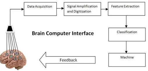
[image:3.612.181.435.172.294.2]or toys. Figure 1 presents a self explanatory schematic diagram of basic BCI system.
Figure 1: Functional block diagram of Brain computer Interface 2. Components of BCI
2.1 Brain signals
A BCI records and interprets or decodes brain activity to generate useful command
signals. Neurons (brain cells) transmit electrical signals to communicate with each other. It is
possible to intercept these signals or ‘brain activity’ with advanced electrical sensors. These
brain signals play vital role in day-to-day life of all living creatures. Movements of healthy
creatures are possible because the brain sends signals via the central nervous system to the
muscles of the body. Precise communication between the brain and muscles make all kinds of
actions of body possible. This communication between the brain and body muscles disrupts or
breaks due to medical conditions such as stroke or neuromuscular diseases and subsequently
leads to paralysis or cerebral palsy. In many such cases the brain is still able to generate the
activity signals for intended movements and a BCI can use the brain signals to control assistive
devices.
BCIs are designed to read the signals produced from the brain at different locations in the
© Associated Asia Research Foundation (AARF)
A Monthly Double-Blind Peer Reviewed Refereed Open Access International e-Journal - Included in the International Serial Directories.
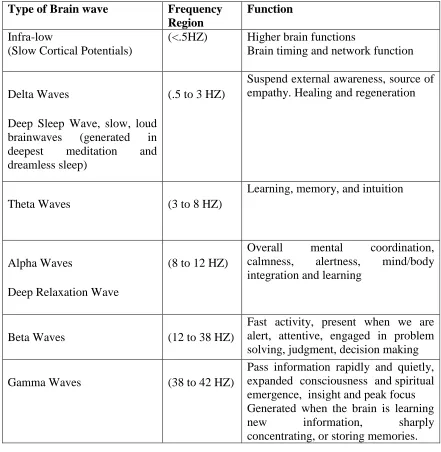
Page | 4 Brainwaves which are detected using sensors placed on the scalp are divided into bandwidths to
describe their functions, but are best thought of as a continuous spectrum of consciousness.
Human brainwaves change according to our actions and feelings. We can feel tired, retarded,
lethargic, or dreamy when lower frequency brainwaves are dominant. On the other hand, the
[image:4.612.85.526.179.631.2]higher frequencies are dominant when we feel energetic, or hyper-alert.
Table 1: Various brain waves and their functions
Type of Brain wave Frequency Region
Function
Infra-low
(Slow Cortical Potentials)
(<.5HZ) Higher brain functions
Brain timing and network function
Delta Waves
Deep Sleep Wave, slow, loud brainwaves (generated in deepest meditation and dreamless sleep)
(.5 to 3 HZ)
Suspend external awareness, source of empathy. Healing and regeneration
Theta Waves (3 to 8 HZ)
Learning, memory, and intuition
Alpha Waves
Deep Relaxation Wave
(8 to 12 HZ)
Overall mental coordination, calmness, alertness, mind/body integration and learning
Beta Waves (12 to 38 HZ)
Fast activity, present when we are alert, attentive, engaged in problem solving, judgment, decision making
Gamma Waves (38 to 42 HZ)
© Associated Asia Research Foundation (AARF)
A Monthly Double-Blind Peer Reviewed Refereed Open Access International e-Journal - Included in the International Serial Directories.
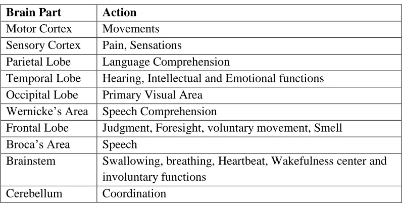
Page | 5 2.2 Brain function
Every part of body has a tiny part in the brain through which it performs its activities. In
general each part of the body has its own ‘control center’ in the brain that is responsible for its
movements. It is possible to detect the activities of various active centers with the help of various
techniques. This helps BCIs to detect the movement of body parts from the brain activity. A
special quality of the brain is that these control centers are also active when we simply think
[image:5.612.107.505.248.451.2]about making a movement without actually moving.
Table 2: Control centers in brain
Brain Part Action Motor Cortex Movements Sensory Cortex Pain, Sensations
Parietal Lobe Language Comprehension
Temporal Lobe Hearing, Intellectual and Emotional functions Occipital Lobe Primary Visual Area
Wernicke’s Area Speech Comprehension
Frontal Lobe Judgment, Foresight, voluntary movement, Smell Broca’s Area Speech
Brainstem Swallowing, breathing, Heartbeat, Wakefulness center and involuntary functions
Cerebellum Coordination
Persons who suffer from locked-in syndrome (LIS) still have fully functioning control centers in
their brains. They are able to activate distinct areas in their brain by the ways of thinking about a
particular movement, or attempting to make movements even if they are not able to move the
part of the body which is controlled by that particular area of their brain. Various brain functions,
in addition to movements of body parts, can be detected. Brain areas are involved in different
aspects of numerical calculations, understanding language and speaking. When these areas are
active a BCI can detect if a person is doing calculations in their head, or is talking. In brief we
can say that there are many distinct areas in the brain that a person can intentionally turn on
© Associated Asia Research Foundation (AARF)
A Monthly Double-Blind Peer Reviewed Refereed Open Access International e-Journal - Included in the International Serial Directories.
Page | 6 assistive technology for persons suffering from paralysis by detecting, interpreting and utilizing
mental tasks performed by them.
2.3 What can a BCI do with the brain signals?
Brain signals can be obtained by placing the electrodes exactly on brain areas can be controlled
by the person. After detecting a signal it can be converted to a command to operate a device or
software. A pre-programmed computer can use this information to perform specific tasks. This
way a BCI can be used to make a computer mouse ‘click’ every time the person counts
backwards in his/her head and select from a menu in a computer program. Such controls are
already commonly used by people suffering from paralysis with special ‘buttons’ that can be
activated by whatever type of movement they are still able to make, such as lifting their
eyebrows or with a little movement of their fingers. Thus, a BCI device can also be used as a
‘button’ to control many types of devices designed for button press control.
Usage of sensorimotor rhythms (SMRs) is also being utilized in one particular type of
BCIs. SMRs are electrical oscillations (i.e., mu [8–12 Hz] and beta [18–30 Hz]) in
electroencephalographic (EEG) activity recorded over sensorimotor cortices. Amplitudes of
these signals change with movements, imagined movements, or preparation for movements.
Needy persons can learn through a training protocol to alter the SMR amplitude. They can use
this control to move a computer cursor or operate another device (Pfurtscheller et al, 2012;
Perdikis et al, 2014; Leeb et al, 2015).
It is important to distinguish that there are various neurological as well as engineering
challenges between building BCIs for the peripheral nervous system (PNS) and central nervous
system (CNS). In particular, miniaturization of processing units, isolation of targeted structures,
replacement of failed probes, and delivery of nonmaterial, etc. are the major aspects to be looked
upon. Understanding the information transfer and processing of the nervous system are amongst
the most urgent challenges faced by the biomedical community, with a plethora of academic and
clinical applications. It includes better understanding of aging, neurodegenerative diseases and
interfaces for prosthetics and implants. For example, recent advances in chronic neural recording
© Associated Asia Research Foundation (AARF)
A Monthly Double-Blind Peer Reviewed Refereed Open Access International e-Journal - Included in the International Serial Directories.
Page | 7 paralysis and improved seizure prevention with chronic telemetry in refractory epilepsy (Cook et
al, 2013; Morrell, 2011). There are many different kinds of potential BCIs that will each serve
independent functions. However, all systems must tackle three fundamental problems: accurate
recording of information from relevant neural systems, decoding such information, and
stimulating and manipulating neuronal dynamics in appropriate as well as meaningful ways.
Further, BCIs must present real-time feedback to the user. It is essential for the BCI system to act
as per user’s intent so that the user can recognize whether he/she successfully conveyed the
desired command or not.
3. Present Status
In a research paper authors (Winda et al, 2017) have proposed system adopted Linear Predictive
Coding (LPC) coefficients as the feature of the person’s mind and Support Vector Machines to
develop the brain signal recognition system. In their work, brain signal was processed and
provided as input to the recognition system. Right-Left sign lamp based on the brain signal
recognition system could potentially replace the button or switch of the specific device in order
to make the lamp work. The system then would decide whether the signal is ‘Right’ or ‘Left’.
The decision of the Right-Left side of brain signal recognition would be sent to a processing
board in order to activate the automotive relay, which would be used to activate the sign lamp. In
another research (Jalal Karam et al, 2016) a Radial Basis Functions (RBF) Artificial Neural
Network (ANN) was constructed and a BCI was implemented using NeuroSkyS EEG biosensor
for the recognition of brain signals. The analysis was presented through the consideration of a
noisy environment to simulate a BCI in real world applications. The data were transmitted via
Bluetooth for MATLAB documentation and recognition rates in the highest 70 percent were
recorded.
An EEG Based Emotion Recognition System has been proposed in a study (Lahane et al,
2014). They have presented a model for making a system which is able to detect human emotion
through brain signals. In another study (Hiwaki et al, 2018) authors have presented a study on
noninvasive measurement of dynamic brain signals using light penetrating the brain. They
© Associated Asia Research Foundation (AARF)
A Monthly Double-Blind Peer Reviewed Refereed Open Access International e-Journal - Included in the International Serial Directories.
Page | 8 which they termed as opt encephalography (OEG), led to the detection of dynamic brain signals
that varied concurrently with the electrophysiological neural activity.
Superconducting Quantum Interference Devices (SQUIDs) are widely used to detect the
extremely weak magnetic field of brain. In order to measure brain activity in normal
environment, a group of researchers (Wang et al, 2017) have constructed and proposed
a measurement system based on highly sensitive Magneto-Impedance (MI) sensor. They have
reported the study of measuring Auditory Evoked Field (AEF) brain waves. The system was
improved in this study, and the sensor signals could be processed in real-time to
monitor brain activity. In this study, they measured the alpha rhythm and the P300 brain activity
in the frontal, the parietal and both the temporal regions using a real-time measuring and
processing system based on highly sensitive MI sensor.
4. Measurement of brain signals
It is essential to measure the brain signals in order to make proper devices for their
interpretation. Various techniques are available for measuring these signals. These techniques
offer certain advantages as well as disadvantages. Fundamentally there are three types of BCI
systems; invasive; partially invasive, and non-invasive.
Noninvasive and invasive methods both benefit from improved recording techniques.
Invasive methods being used at present do not adequately deal with the expectation for long-term
stability of their results. The brain’s complex reaction to an implant is still inadequately
understood and might hamper long term performance. Skills of the person placing noninvasive
EEG electrodes play vital role in their performance. These electrodes need periodic maintenance
to ensure sufficiently good contact with the skin. Improvement in methods for extracting key
features from EEG signals and fetching device control signals from them would also help in
improving the BCI performance.
Conventional techniques for noninvasive measurement of brain signals such as functional
magnetic resonance imaging (fMRI), near-infrared spectroscopy (NIRS), magneto
encephalography (MEG), and electroencephalography (EEG), suffer from critical limitations in
© Associated Asia Research Foundation (AARF)
A Monthly Double-Blind Peer Reviewed Refereed Open Access International e-Journal - Included in the International Serial Directories.
Page | 9 electrical sensors (electrodes) that are placed on the scalp. These electrodes can also be placed
under the scalp directly on or in the brain tissue. This requires a surgical procedure to place such
electrodes. This method provides better signals as compared to signals recorded from the scalp. It
is possible to place the electrodes on the brain without causing any damages. This has
accelerated use of implantable BCIs for paralyzed people. Near-infrared spectroscopy (NIRS)
can be used to measure brain activity by exposing the skull through near-infrared light. As such
NIRS does not require any surgery.
A variety of sensors for monitoring brain activity exists, and could in principle provide
the basis for a BCI. These include electroencephalography (EEG), electrocorticography (ECoG),
magnetoencephalography (MEG), positron emission tomography (PET), functional magnetic
resonance imaging (fMRI), and optical imaging (i.e., functional Near InfraRed (fNIR)) (Kübler
et al, 2005).
EEG is a least invasive alternative which is recorded from the scalp. These BCIs support
much higher performance than previously assumed, including two- and three dimensional cursor
movements. Extensive user training is required for the acquisition of such high levels of control.
Furthermore, EEG has low spatial resolution, which will eventually limit the amount of
information that can be extracted, and it is also susceptible to artifacts from other sources
(McFarland et al, 2008).
The second alternative uses ECoG, which is recorded from the cortical surface (Wilson et
al, 2006). It has higher spatial resolution compared to EEG (i.e., tenths of millimeters vs.
centimeters), broader bandwidth (i.e., 0–500 Hz vs. 0–50 Hz), higher signal strength (i.e., 50–
100 µV vs. 10–20 µV), and far less vulnerability to artifacts such as EMG (Ball et al, 2009) or
ambient noise. While this method is invasive, the use of these electrodes that do not penetrate the
cortex may combine excellent signal fidelity with good long-term stability.
The third and most invasive alternative uses microelectrodes to measure local activity
(i.e., action or field potentials) from multiple neurons within the brain (Taylor et al, 2002).
Cortex provides signals having higher fidelity and can support BCI systems that require lesser
training than EEG-based systems. However, clinical implementations of intracortical BCIs are
© Associated Asia Research Foundation (AARF)
A Monthly Double-Blind Peer Reviewed Refereed Open Access International e-Journal - Included in the International Serial Directories.
Page | 10 substantial technical requirements of single-neuron recordings, and by the need for continued
intensive expert oversight. For these reasons, practically all BCI demonstrations in humans to
date have been achieved with, and the examples in this book are using or meant for, EEG or
ECoG recordings.
A study reported in 2012 (Chi et al, 2012) compares wet electrodes to dry and through
hair, noncontact electrodes within a steady state visual evoked potential (SSVEP) BCI paradigm.
They have also presented the construction of a dry contact electrode, featuring fingered contact
posts and active buffering circuitry. In addition to this they have also introduced the development
of a new, noncontact, capacitive electrode that utilizes a custom integrated, high-impedance
analog front-end.
Brain-computer interface (BCI) technology is utilized in paired associative stimulation
(PAS) to evaluate movement imagery in real-time. Pas uses this information to control feedback
presented to the patient. Another group (Sabathie et al, 2016) introduced this approach and the
RecoveriX system, a hardware and software platform for PAS. They have also presented initial
results from two stroke patients who used RecoveriX, followed by future directions.
Stroke is a major cause of acquired disability which results in distal upper extremity
functional motor impairment. Ongoing research to evaluate the effectiveness of BCI-based stroke
rehabilitation for hand therapy is currently in progress. A review of the progression and future
implications of brain-computer interface therapies for restoration of distal upper extremity motor
function after stroke has been proposed (Remsik et al, 2016). Further, researchers (Almajidi et al,
2014) have presented the design and characterization of novel, differential functional NIRS
sensors, intended to record hemodynamic changes of the human motor cortex in the hand-area
during motor imagery tasks. They have reported on the spatial characterization of a portable,
multi-channel NIRS system with one module consisting of two central light emitting diodes
(LED) (770 nm and 850 nm) alongwith four symmetric pairs of photodiodes (PD). These diodes
resemble a plus symbol. The other sensor module includes four similar and differential light
paths crossing in the center of a star. Apart from the literature presenting technological
developments of BCI there are some papers where the ethical challenges of BCI have been
© Associated Asia Research Foundation (AARF)
A Monthly Double-Blind Peer Reviewed Refereed Open Access International e-Journal - Included in the International Serial Directories.
Page | 11 life saving equipments may be questionable at times (Glannon, 2014). The impulsive responses
of BCI users may cause irreparable losses to them.
4. Conclusions
Brain-computer interfaces provide a new communication-and-control option for individuals
for whom conventional methods are ineffective. Brain-Computer Interface (BCI) is a
fast-growing emergent technology in which researchers aims to build a direct channel between the
human brain and the computer. A BCI derives its outputs from brain activity which is directly
consciously controlled by the user, independently from external events, for controlling an
application. It is a collaboration in which a brain accepts and controls a mechanical device as a
natural part of its representation of the body. The BCI can lead to many applications especially
for disabled persons. A brain-computer interface (BCI) systems permit encephalic activity to
solely control computers or external devices. Accordingly, people suffering from neuromuscular
diseases can highly benefit from these technologies, since a computer could allow them to
perform multiple tasks. Further development of applications is also needed, particularly
applications of BCI technology to rehabilitation. The design of rehabilitation applications hinges
on the nature of BCI control and how it might be used to induce and guide beneficial plasticity in
the brain. As the rise in the BCI, the user would be haunting with the privacy issue; it means that
attackers would be able to hack targets brain. Undoubtedly there is great potential in BCI devices
but there are certain possible ethical implications as well. The benefits of BCIs can introduce
moral and societal challenges. It is, therefore, important to consider the impact of this technology
on legal and moral responsibilities of both user as well as producer.
References
Almajidy R. A., Kirch R. D., Christ O. & Hofmann U. G. (2014). Estimating the spatial
resolution of fNIRS sensors for BCI purposes. Proc. SPIE 8945, Design and Performance
Validation of Phantoms Used in Conjunction with Optical Measurement of Tissue VI,
© Associated Asia Research Foundation (AARF)
A Monthly Double-Blind Peer Reviewed Refereed Open Access International e-Journal - Included in the International Serial Directories.
Page | 12 Ball T., Kern M., Mutschler I., Aertsen A. & Schulze-Bonhage, A. (2009). Signal quality of
simultaneously recorded invasive and non-invasive EEG. NeuroImage, 46(3), 708–716.
DOI:10.1016/j.neuroimage.2009.02.028.
Chi Y. M., Wang Y. T. , Wang Y., Maier C., Jung T. P. & Cauwenberghs G. (2012). Dry and
Noncontact EEG Sensors for Mobile Brain–Computer Interfaces. EE Transactions on Neural
Systems and Rehabilitation Engineering, 20(2). DOI: 10.1109/TNSRE.2011.2174652.
Cook M. J., O'Brien T. J., Berkovic S. F., Murphy M., Morokoff A,. Fabinyi G., D'Souza
W., Yerra R., Archer J., Litewka L., Hosking S., Lightfoot P., Ruedebusch V., Sheffield W.
D., Snyder D., Leyde K., Himes D. (2013). Prediction of seizure likelihood with a long-term,
implanted seizure advisory system in patients with drug-resistant epilepsy: a first-in-man
study. Lancet Neurol., 12(6), 563–71.
Daly J. J. & Wolpaw J. R. (2008). Brain–computer interfaces in neurological rehabilitation.
Lancet Neurol. 7, 1032–1043.
Glannon W. (2014). Ethical issues with brain-computer interfaces. Front Syst Neurosci., 8, 136. DOI: 10.3389/fnsys.2014.00136.
Hiwaki O. & Miyaguchi H. (2018). Noninvasive measurement of dynamic brain signals using
light penetrating the brain. PLoS One, e0192095. Published online 2018 Jan
31. DOI: 10.1371/journal.pone.0192095, 13(1).
Karam J., Majeed S. A., Christofer N. Y. & Mirtskhulava L. (2016). Neural Network for
Recognition of Brain Wave Signals. Int. J. of Enhanced Research in Science, Technology &
Engineering. 5(10), 36-42.
Kübler, A., Nijboer F., Mellinger J., Vaughan T. M., Pawelzik H., Schalk G., McFarland D. J.,
Birbaumer N. & Wolpaw J. R. (2005). Patients with ALS can use sensorimotor rhythms to
operate a brain–computer interface. Neurol. 64(10), 1775–1777. DOI:10.1212/
© Associated Asia Research Foundation (AARF)
A Monthly Double-Blind Peer Reviewed Refereed Open Access International e-Journal - Included in the International Serial Directories.
Page | 13 Lahane P., Lokannavar S., Gangurde A., Bhosale P. & Chidre P. (2014). EEG Based Emotion
Recognition System. Int. J. of Computer Science and Information Technologies, 5(6),
7656-7658.
Leeb R., Tonin L., Rohm M., Desideri L., Carlson T. & Millan J. d. R. (2015). Towards
independence: a BCI telepresence robot for people with severe motor disabilities. Proc IEEE;
103, 969–982.
McFarland D. J., Krusienski D. J., Sarnacki W. A. & Wolpaw J. R. (2008). Emulation of
computer mouse control with a noninvasive brain–computer interface. J. Neural Eng., 5(2),
101–110. DOI:10.1088/1741-2560/5/2/001.
Morrell M. J. (2011). RNS System in Epilepsy Study Group: Responsive cortical stimulation for
the treatment of medically intractable partial epilepsy. Neurology, 77(13), 1295–304.
Pfurtscheller G. & McFarland D. J. (2012). BCIs that use sensorimotor rhythms. In Wolpaw JR
Wolpaw EW Brain–Computer Interfaces: Principles and Practice. New York, NY: Oxford
University Press, 227–240.
Perdikis S., Leeb R., Williamson J., Ramsay A., Tavella M., Desideri L., Hoogerwerf E. J.,
Al-Khodairy A., Murray-Smith R. & Millán J. D. (2014). Clinical evaluation of BrainTree, a
motor imagery hybrid BCI speller. J Neural Eng., 11: 036003. pmid:24737114.
Remsik A., Young B., Vermilyea R., Kiekoefer L., Abrams J., Elmore S. E., Schultz P., Nair
V., Edwards D., Williams J. & Prabhakaran V. (2016). A review of the progression and
future implications of brain-computer interface therapies for restoration of distal upper
extremity motor function after stroke. Expert Rev Med Devices. 13(5), 445–454.
DOI: 10.1080/17434440.2016.1174572.
Sabathiel N., Irimia D. C., Allison B. Z., Guger C. & Edlinger G. (2016). Paired Associative
Stimulation with Brain-Computer Interfaces: A New Paradigm for Stroke Rehabilitation.
© Associated Asia Research Foundation (AARF)
A Monthly Double-Blind Peer Reviewed Refereed Open Access International e-Journal - Included in the International Serial Directories.
Page | 14 Neuroergonomics and Operational Neuroscience. AC 2016, Lecture Notes in Computer
Science, Springer, 9743. DOI: https://doi.org/10.1007/978-3-319-39955-3_25,
Sasha Burwell S., Sample M. & Racine E. (2017). Ethical aspects of brain computer interfaces: a
scoping review. BMC Medical Ethics 18:60. DOI: 10.1186/s12910-017-0220-y.
Taylor D. M., Tillery S. I. & Schwartz A. B. (2002). Direct cortical control of 3D
neuroprosthetic devices. Science, 296, 1829–1832.
Wang K., Cai C., Yamamoto M. & Uchiyama T. (2017). Real-time brain activity measurement
and signal processing system using highly sensitive MI sensor. AIP Advances, 7(5), 056635.
https://doi.org/10.1063/1.4974528.
Wilson J., Felton E., Garell P., Schalk G. & Williams J. (2006). ECoG factors underlying
multimodal control of a brain–computer interface. IEEE Trans. Neural Syst. Rehabil. Eng.,
14, 246–250.
Winda A., Sofyan, Sthevany & Vincent R. S. (2017). Intelligent automatic right-left sign lamp
based on brain signal recognition system. IOP Conf. Series: Earth and Environmental
Science, 109, 012019.
Wolpaw J. R., Birbaumer N., McFarland D. J., Pfurtscheller G. & Vaughan T. M. (2002).
Brain–computer interfaces for communication and control. Clin. Neurophysiol. 113, 767–