S T U D Y P R O T O C O L
Open Access
Exercise and load modification versus
corticosteroid injection versus
‘
wait and
see
’
for persistent gluteus medius/minimus
tendinopathy (the LEAP trial): a protocol for
a randomised clinical trial
Rebecca Mellor
1, Alison Grimaldi
2, Henry Wajswelner
3, Paul Hodges
4, J. Haxby Abbott
5, Kim Bennell
6and Bill Vicenzino
1*Abstract
Background:Lateral hip pain is common, particularly in females aged 40–60 years. The pain can affect sleep and daily activities, and is frequently recalcitrant. The condition is often diagnosed as trochanteric bursitis, however radiological and surgical studies have revealed that the most common pathology is gluteus medius/minimus tendinopathy. Patients are usually offered three treatment options: (a) corticosteroid injection (CSI), (b) physiotherapy, or (c) reassurance and observation. Research on Achilles and patellar tendons has shown that load modification and exercise appears to be more effective than other treatments for managing tendinopathy, however, it is unclear whether a CSI, or a load modification and exercise-based physiotherapy approach is more effective in gluteal tendinopathy. This randomised controlled trial aims to compare the efficacy on pain and function of a load modification and exercise-based programme with a CSI and a‘wait and see’approach for gluteal tendinopathy. Methods:Two hundred one people with gluteal tendinopathy will be randomly allocated into one of three groups: (i) CSI; (ii) physiotherapist-administered load modification and exercise intervention; and (iii) wait and see approach. The CSI therapy will consist of one ultrasound (US) guided CSI around the affected tendons and advice on tendon care. Education about load modification will be delivered in physiotherapy clinics and the exercise programme will be both home-based and supervised. The group allocated the wait and see approach will receive basic tendon care advice and reassurance in a single session by a trial physiotherapist. Outcomes will be evaluated at baseline, 4, 8, 12, 26 and 52 weeks using validated global rating of change, pain and physical function scales, psychological measures, quality of life and physical activity levels. Hip abductor muscle strength will be measured at baseline and 8 weeks. Economic evaluation will be performed to investigate the cost-effectiveness of the active interventions compared with the wait and see approach. Analyses will be conducted on an intention-to-treat basis using logistic and linear mixed regression models and the economic evaluation will report incremental cost-utility ratios. The trial reporting will comply with CONSORT guidelines.
Discussion:This study will provide clinicians with directly applicable evidence of the relative efficacy of three common approaches to the management of gluteal tendinopathy.
Trial registration:Australia New Zealand Clinical Trials Registry ACTRN12612001126808. Date Registered: 22/10/2012.
Keywords:Gluteal tendinopathy, Greater trochanteric pain syndrome, Corticosteroid injection, Physiotherapy
* Correspondence:[email protected]
1School of Health and Rehabilitation Sciences, The University of Queensland,
Brisbane, Qld 4072, Australia
Full list of author information is available at the end of the article
Background
Gluteal tendinopathy or greater trochanteric pain syn-drome is a debilitating condition, characterised by pain situated at or around the greater trochanter of the hip, and tenderness on palpation. Although traditionally con-sidered to be trochanteric bursitis, more advanced im-aging and surgical procedures in people with lateral hip pain have revealed a primary pathology of insertional gluteus medius or minimus tendinopathy, with bursal distention generally a concomitant finding [1–3]. Mag-netic Resonance Imaging (MRI) is very effective in rec-ognizing partial and full thickness tears of the tendons of gluteus medius and minimus, tendon calcification and fatty muscle atrophy [2]. For the purposes of this paper we will refer to the condition of gluteus medius tendino-pathy, and/or gluteus minimus tendinotendino-pathy, with or without bursal distention, as gluteal tendinopathy.
The condition is more frequent in women aged 40–60 years [4], and various studies have described its preva-lence as ranging from 10 to 25 % of the general popula-tion [5, 6]. A recent study of general medical practice in the Netherlands found gluteal tendinopathy to have the highest prevalence (4.22 per 1000 person years) and inci-dence (3.29 per 1000 person years) of all presenting lower limb tendinopathies [7]. The impact of gluteal ten-dinopathy can be substantial. The pain experienced in side lying often creates significant sleep disturbance, and the pain commonly experienced with walking and stair climbing usually results in reduction of physical activity levels, which could be expected to have negative implica-tions for general health and well-being, as well as quality of life and employment status [8].
It is proposed that abnormal hip biomechanics may predispose to gluteal tendinopathy [9], and although commonly diagnosed in sedentary, overweight people [5], it is also often seen in runners, possibly due to both poor training habits and technique [10]. It has been hypothesised that gluteal tendinopathy, with or without bursitis, occurs due to compressive impingement of these structures onto the underlying greater trochanter by the iliotibial band (ITB) as the hip moves into adduc-tion [11, 12]. In an upright weight bearing posture, such as walking, weakness of the hip abductor muscles will result in lateral pelvic tilt or ipsilateral shift in single leg stance, and hip adduction, which will contribute to com-pression of the tendons between the greater trochanter and the thick fascia of the ITB.
The literature reflects an assumption that tight lateral structures are a key issue for the development of gluteal tendinopathy. Despite a lack of evidence of such tight-ness, the conservative management approach commonly suggested is stretching [13]. Compression as a primary aetiological factor is consistent with current theories for development of insertional tendinopathy [14, 15], and
failure to control compression in a treatment protocol for insertional Achilles tendinopathy has been reported to result in poor outcomes [16]. The piriformis and ilio-tibial band stretches frequently prescribed [13] involve sustaining the hip in forced adduction, which is a pos-ition of high compressive loads on the gluteal tendons [17]. We hypothesise that a specific focus on hip ab-ductor muscle function and avoidance of compressive loads on the tendons will provide a better approach to achieve effective treatment outcomes.
A range of conservative management approaches are generally recommended, including anti-inflammatory medications, rest, ice, heat, stretching, strengthening, ultrasound, shock-wave therapy (SWT), and local cor-ticosteroid injection (CSI). Surgery is usually reserved for when the condition has become refractory and con-servative measures have failed. However, optimal man-agement of gluteal tendinopathy remains unclear. Reports concerning management of this condition pre-dominantly relate to CSI and surgery. Early response to CSI in patients with lateral hip pain is reported to be very good, with 70–75 % of patients reporting a signifi-cant improvement at 1 month post injection in a rando-mised clinical trial (RCT) [13, 18]. However, at 3–4 months post injection, researchers have reported only 41–55 % positive response [13, 19]. At 12 months an RCT showed no difference between a CSI and a group that adopted a wait and see policy [19]. In the only clin-ical trial (non-randomised) to date to compare CSI to home exercise, the success rates at 1 month were 75 and 7 % respectively, but by 15 months were 48 % for CSI and 80 % for the home exercise group [13], demonstrat-ing only short term benefits of CSI. These studies of CSI highlight the typical trajectory of delayed healing after a short term improvement, which in some studies is also coupled with substantial recurrence rates [20].
stretches that increase compression), a lack of specific load modification and management strategies related to tendon healing, and a low level of supervision of the home-based exercise programme.
The contemporary non-operative approach to effect-ively managing tendinopathy involves specific exercise for the affected muscles and load management in the form of advice and practical strategies [15]. This ap-proach has yet to be rigorously tested in an RCT. We propose to test different conservative management options for gluteal tendinopathy by comparing an ex-ercise and load management approach supervised by physiotherapists, CSI and the adoption of a wait and see approach.
Our primary hypotheses are that:
HI: Both the exercise and load management programme and the CSI will be superior to a wait and see approach in terms of treatment success rates based on global rating of change and reductions in pain at 8 weeks.
H2: The exercise and load management programme will be superior to CSI in terms of treatment success rates and reductions in pain at 52 weeks.
The secondary objectives are to: compare an exercise and load management programme with CSI on out-comes including pain, function, hip abductor muscle strength, psychological measures, quality of life and physical activity levels; evaluate the effects of the two treatments compared to a wait and see approach on these outcomes; determine the cost-effectiveness of the two treatments at 52 weeks; compare the adverse events associated with both treatments.
Methods
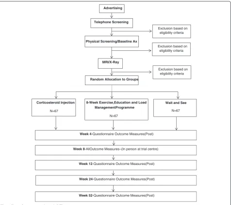
This is a pragmatic, assessor-blinded, RCT conducted in medical and physiotherapy clinics in Brisbane and Melbourne, Australia. In people with persistent gluteal tendinopathy, it compares the effect of CSI, a physio-therapy supervised load management, education and exercise programme and a wait and see approach for gluteal tendinopathy over 12 months. The trial protocol will permit its reporting in line with the CONSORT guide-lines [22].
Participants
Lateral hip pain, which is a symptom of gluteal tendi-nopathy, might also present with other pathologies, such as trochanteric bursitis, osteoarthritis, referred lumbar spine pain, and femoral stress fracture [23]. As the load modification and exercise programme in this trial has been designed specifically to address
gluteal tendinopathy, it is important that other differ-ential diagnoses for lateral hip pain are excluded.
Recruitment details
We will recruit people from the community in both Brisbane and Melbourne via advertisements in Univer-sity News, social media and local newspapers. Initial contact will be by phone or electronic media at which time a preliminary screening for suitability will occur. If the phone interview indicates potential eligibility, the volunteer will attend the trial centre for a physical exam-ination, which will assess against specific selection cri-teria. Those who meet these criteria will then undergo diagnostic imaging to confirm eligibility prior to being included in the trial.
Selection criteria
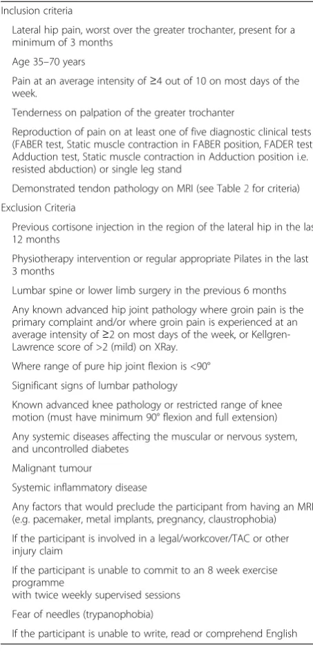
We will include participants between the ages of 35 and 70 years who have experienced lateral hip pain for at least 3 months, of an intensity of ≥4/10 on an 11-point numeric rating scale on most days of the last 3 months. Table 1 outlines the selection criteria for inclusion into the study. These criteria were based on a previous study [13].
As clinical tests to diagnose gluteal tendinopathy ap-pear to have limited validity [24], we have included a small battery of clinical tests that have been considered to be most provocative in reproducing symptoms of glu-teal tendinopathy [23]. To be eligible, the participant must experience pain on direct palpation of the gluteal tendons’insertion on the greater trochanter. They must also test positive (reproduction of trochanteric pain) to at least one of the following clinical tests: the Hip FADER (passive) test, static muscle test in the FADER position, the FABER (Patrick’s) test, passive hip adduc-tion in side lying (ADD), a static muscle contracadduc-tion in the ADD test position, and a Single Leg Stance on the affected leg for 30 s.
A.Hip FADER–With the patient supine, the hip is passively flexed to 90°, adducted and externally rotated to end of range (FADER = Flexion/Adduction/ External Rotation). The pain Numeric Rating Scale (NRS) and area of pain is recorded. This test positions the ITB over the greater trochanter and places the Gluteus Medius (GMed) and Gluteus Minimus (GMin) tendons under tension while being compressed against the greater trochanter by the overlying fascia of the ITB. The test is only recorded as positive if the pain (≥2/10) is experienced over the lateral hip. B. Hip FADER with Static muscle test(internal rotation)
90° hip flexion all portions of GMed and GMin are internal rotators [25]. This test requires the participant to activate these muscles, and therefore place further tension across their tendons, while they are in a compressed state. Again, a positive result refers to reproduction of pain at the lateral hip. As clinical features of gluteal tendinopathy include pain reproduction with elongation and compression of the involved tendons, as well as active contraction of these tendons, these two tests together may have improved diagnostic accuracy. This test is a modification of the resisted external
de-rotation test, which has been reported to have 88 % sensitivity and 97.3 % specificity [26].
C.Hip FABER–(FABER = Flexion/Abduction/External Rotation). The lateral malleolus of the test leg is placed above the patella of the opposite side, the pelvis is stabilised via the opposite anterior superior iliac spine (ASIS) and the knee is passively lowered into abduction and external rotation. This test places the anterior portions of the GMed and GMin on tensile load. A positive pain response is usually felt in the lateral hip region. Lateral hip pain with a FABER test has been shown to have a high sensitivity, specificity, positive and negative predictive value (82.9, 90, 94.4 and 72 % respectively) for differentiating the diagnosis for greater trochanteric pain syndrome from hip osteoarthritis [27].
D.Passive Hip Adduction in Side Lying (ADD)–The participant is placed in side-lying, with the under-neath hip and knee flexed 80–90°, and the upper-most leg supported by the examiner with the knee extended, in neutral rotation, and the femur in line with the trunk. The anterior superior iliac spines are aligned vertically in the frontal plane. The examiner passively moves the hip through a pure frontal plane motion into end range hip adduction with overpres-sure, while stablilising the pelvis with the other hand. This test places the lateral insertions of the gluteal tendons under compressive load, and a posi-tive response is felt over the lateral hip. This is based on Ober’s test, which has been reported as having a high specificity (95 %), but a low sensitivity (41 %) and low negative predictive value (45.2 %) [27]. E. ADD with resisted isometric abduction (ADD-R)–In
the ADD test end position, the participant is asked to push the thigh up, against the resistance of the examiner’s hand at the lateral knee. This test places tensile load on the compressed tendons, with pain elicited over the lateral hip.
[image:4.595.56.283.98.574.2]F. Single Leg Stance for 30 s (SLS)–the participant stands side-on to a wall with one finger touching the wall at shoulder height for balance, then lifts the foot closest to the wall, maintaining single leg stance for up to 30 s. The participant is asked to immediately report the development of pain by pointing to the area of pain. If the region of the greater trochanter is indicated, the timer is stopped, the test ceased and recorded as positive. This time is reported, as well as the intensity of the pain. The single leg stance test has been shown to have good sensitivity and specificity (100 and 97.3 % respectively) [26] for the diagno-sis of tendinopathy and bursitis in people with MRI-documented gluteal tendinopathy.
Table 1Inclusion and Exclusion Criteria
Inclusion criteria
Lateral hip pain, worst over the greater trochanter, present for a minimum of 3 months
Age 35–70 years
Pain at an average intensity of≥4 out of 10 on most days of the week.
Tenderness on palpation of the greater trochanter
Reproduction of pain on at least one of five diagnostic clinical tests (FABER test, Static muscle contraction in FABER position, FADER test, Adduction test, Static muscle contraction in Adduction position i.e. resisted abduction) or single leg stand
Demonstrated tendon pathology on MRI (see Table2for criteria) Exclusion Criteria
Previous cortisone injection in the region of the lateral hip in the last 12 months
Physiotherapy intervention or regular appropriate Pilates in the last 3 months
Lumbar spine or lower limb surgery in the previous 6 months
Any known advanced hip joint pathology where groin pain is the primary complaint and/or where groin pain is experienced at an average intensity of≥2 on most days of the week, or Kellgren-Lawrence score of >2 (mild) on XRay.
Where range of pure hip joint flexion is <90°
Significant signs of lumbar pathology
Known advanced knee pathology or restricted range of knee motion (must have minimum 90° flexion and full extension)
Any systemic diseases affecting the muscular or nervous system, and uncontrolled diabetes
Malignant tumour
Systemic inflammatory disease
Any factors that would preclude the participant from having an MRI (e.g. pacemaker, metal implants, pregnancy, claustrophobia)
If the participant is involved in a legal/workcover/TAC or other injury claim
If the participant is unable to commit to an 8 week exercise programme
with twice weekly supervised sessions
Fear of needles (trypanophobia)
In addition to these tests, the physical screening will also ensure that the participant has≥90°hip flexion range of movement bilaterally, knee flexion range≥90° and full knee extension bilaterally, and that the hip quadrant test [28] is clear bilaterally. If groin pain on quadrant testing is greater than 5/10 on the Pain NRS, or the difference in pain levels between sides is greater than 2/10, the par-ticipant is excluded. Additionally, the parpar-ticipant must be able to flex the trunk forward with hands reaching at least to the knees with ≤2/10 back pain, and have ad-equate hip, knee and ankle mobility to be able to per-form a squat to 60° flexion at the hips.
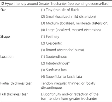
The participant will then be referred for MRI (if no contraindications e.g. cardiac pacemaker, metal im-plants etc.) and X-ray investigations at a participating radiology clinic, as a confirmed diagnosis of gluteal tendinopathy on MRI, based on a classification system from a previous study [11] will also be required for eligibility. Tendinopathy will be defined as an intra-tendinous increase in signal intensity on T2-weighted images (Table 2). Participants must have no contrain-dications to MRI (e.g. cardiac pacemaker, metal im-plants etc). An X-ray (AP and Lateral) is required to grade osteoarthritis severity using the Kellgren-Lawrence Scale [29]. Those with a score of >2 will be excluded from the study. To minimize unnecessary radiation exposure, if the patient has had previous ap-propriate X-rays within the last 6 months, they will not require a second lateral hip X-ray.
Concealed allocation
A computerised randomisation schedule stratified for site (Brisbane, Melbourne) will be prepared by an in-dependent off-site body (The Berghofer Queensland Institute of Medical Research, Clinical Trial and Bio-statistics Unit). To conceal randomisation, consecu-tively numbered, sealed, opaque envelopes will be prepared by a research assistant not involved in re-cruitment, and kept in a locked cabinet accessible only to the assistant. These envelopes will be opened at each site sequentially once the participant has been confirmed into the study by MRI, and all baseline measurements have been completed. If allocated into the CSI group, an appointment will be made for the participant to receive an US-guided injection, adminis-tered by an experienced Radiologist/Sports Physician. If allocated into the exercise and load management group or into the group that will adopt the wait and see approach, an appointment will be made for the participant to attend a trial physiotherapy clinic.
Blinding
The investigator assessing the outcome measures will be blind to group allocation, and will not be involved in any of the interventions. Participants will be informed in writing and verbally that they have an equal chance of being allocated into one of the three groups (CSI, exer-cise and load management, wait and see). They will not be made aware of the study hypotheses. Participants will be requested not to divulge any details about their treat-ment group to the investigator involved in assessing the outcome measures, and will be reminded of this prior to each encounter. It is not possible to blind the patients, physiotherapists providing the treatment, the radiologist administering the CSI, nor the physiotherapists provid-ing the basic information to the wait and see group. Statistical analysis will be conducted on a blinded intention-to-treat basis.
Interventions
[image:5.595.56.290.492.706.2]Corticosteroid injection: Participants allocated to this group will attend the clinic of the experienced radiolo-gist or sports physician who will be performing the in-jection. Seven specialist radiologists are participating in this trial, with a range of 15–40 years of experience. The injection will be performed at the same radiology clinic that the participant attended to have their MRI and XRay investigations performed. After standard prepar-ation procedures, the participant will be positioned on the table with the affected side raised. A preliminary ultrasound scan will be performed with pre procedural images taken in a longitudinal plane. The depth of injec-tion required will be measured and area of maximal ten-derness marked. A linear probe which is appropriate for Table 2MRI Image Analysis–Classification of Pathology for
definition of gluteal tendinopathy
T2 Hyperintensity around Greater Trochanter (representing oedema/fluid)
Size (1) Tiny (thin slit of fluid)
(2) Small (localized, mild distension)
(3) Medium (localized, moderate distension)
(4) Large (localized, marked distension)
Shape (1) Feathery
(2) Crescentic
(3) Round (distended bursa)
Location (1) Subtendinous
(2) Intratendinous*
(3) Subfascia lata
(4) Superficial to fascia lata
Partial thickness tear Tendon irregular, thinned or focally discontinuous
Full thickness tear Discontinuity and/or retraction of the torn tendon from greater trochanter
the participant body habitus (i.e. 12 MHz) will be cleaned using sterile wipes, and the appropriate syringe and needle selected. A further preliminary scan will be performed, and the radiologist washes their hands and gloves up. The steroid (Celestone 1 ml or Kenacort A40 1 ml (depending on availability)) and local anaesthetic (Bupivacaine 2 ml or Marcaine 1 ml) will be drawn up using sterile techniques and the participant’s skin and ultrasound probe will be cleansed with chlorhexidine and the area draped. The radiologist will place the needle through the superficial structures until it reaches the bursa following which the steroid and local anaesthetic mixture will be injected widely in different directions through the bursa. On com-pletion of the procedure, a dressing will be applied to the puncture site. A post procedural check will be performed, and a further explanation of what to expect and basic ad-vice on tendon care will be given to the participant. A weekly diary will be completed, recording levels of pain and function and any adverse effects.
Exercise, education and load management programme: Participants allocated to this group will be prescribed a home exercise programme to be performed daily, which will be limited to four to six exercises (to optimise ad-herence), and should take no longer than 15–20 min to complete. Participants will also receive more specific and detailed advice and education on tendon care. This information will be delivered in hard copy handouts, verbal explanation and DVD. They will attend the physiotherapy clinic for individual supervised exercise sessions, and be treated by an experienced and regis-tered musculoskeletal physiotherapist. Treating practi-tioners will have attended a training session outlining the study objectives and requirements, demonstrating the exercise protocol and progressions, as well as the de-tailed education material, and will be expected to be proficient in the intervention. They will also be provided with a detailed study manual for reference. The physio-therapy sessions will be once a week for the first 2 weeks, then twice a week for the next 6 weeks. The first session will be 1 h long, in order to perform detailed education and demonstration of the exercises. Successive sessions will be 30 min in duration, and include a brief interview and physical assessment to gauge the response to the ex-ercise and load management program, modify or pro-gress the home exercises, and supervise a twice weekly heavier loading exercise programme.
The exercises will include functional retraining, and tar-geted strengthening for the hip and thigh muscles, with a particular focus on the hip abductor muscles, and dy-namic control of adduction during function. Exercise diffi-culty will be gradually increased as tolerated by the participant, in order to optimise improvements in muscle strength and function without significant aggravation of the participant’s pain. Difficulty level will be monitored
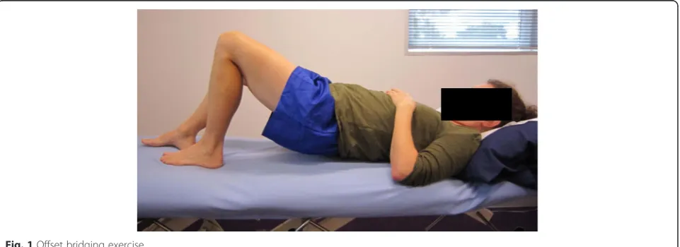
with the Borg Scale [30] where warm up will be performed at a light level (Borg 11–12), functional retraining at a somewhat hard to hard level (Borg 13–15), and the slow heavy targeted strengthening moving from somewhat hard towards the hard to very hard level (14–17), de-pending on the participants response to loading. No change in pain over the greater trochanter will be ac-ceptable during functional retraining, as this may in-dicate inadequate alignment control, and excessive compressive tendon loading. As the heavy slow strengthening exercises avoid tendon compression completely, a maximum of NRS 5/10 pain will be tol-erated as long as this eases afterwards and does not result in increased pain levels that night or the next morning. Participants’ responses to the exercises will be closely monitored, and loading levels adjusted as required to prevent any increases in pain from week to week. Table 3 outlines the clinic and home based exercise protocols, and Figs. 1 and 2 show some ex-amples of the prescribed exercises.
Adherence to the exercise programme will be moni-tored by the physiotherapists and with the partici-pants also completing a daily diary, outlining the exercises completed (number of sets and repetitions), any adverse responses and action taken. If any ad-verse event occurs during the treatment period, the participants will also be encouraged to report directly to their physiotherapist (e.g. worsening of pain, new symptoms or any injury). Any reports of adverse events will be recorded in the treatment notes and diary. The diary will be collected from the participant at the 8 week follow up assessment.
Wait and see: Participants allocated to adopt a wait and see approach will attend one session with a trial physiotherapist, where they will receive reassurance that the condition is likely to resolve over time, as well as ad-vice regarding general tendon care and self-management. They will also receive a standard information pamphlet about the condition and basic self-management. The ther-apists will take time to answer any questions about adopt-ing a wait and see approach to ensure the participant is confident that this is an appropriate and sensible approach to adopt. Participants in this group will complete a weekly diary, outlining any problems that may have been encoun-tered related to the study.
Outcome measures
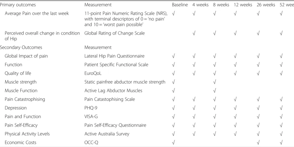
A battery of primary and secondary outcome measures will be recorded. They are summarized in Table 4.
Primary outcome measures
Table 3Exercise Dosage and Progressions
Stage Exercise Effort Speed Reps Sets Freq
Week 1- Familiarisation Low load activations Light Slow onset 10 1–2 BD
Static Abduction: Hold 5–10 s
In supine lying Light Slow onset 3–5 1 BD
In standing Hold 5–15 s
Pelvic Control during Functional Loading: daily
Bridging Light Moderate 10 1 daily
Double Leg Bridging
Functional Strengthening: Light- SWH Slow 10 1
Double leg squats
Abductor LoadingviaFrontal Plane Movement:
Light Moderate 10 each 1 daily
Sidestepping
Week 2–Early Loading & Movement Optimisation
Low load activations Maintain as per week 1
Static Abduction:
Pelvic Control during Functional Loading:
Bridging:
Double leg bridging Light Slow 10 1 daily
Single leg biased ex: SWH Slow 5 1
Offset bridging
Functional Strengthening:
Double leg squats Light Slow 10 1 daily
Single leg biased ex: SWH Slow 5 1
Offset squat
Abductor LoadingviaFrontal Plane Movement:
Light Moderate 15 each 1 daily
Sidestepping
Week 3–8–Graduated Loading Low load activations Maintain as per week 1
Static Abduction:
Pelvic Control during Functional Loading:
Bridging: Light Slow 5 1 daily
Double leg bridging
Single leg biased ex SWH–Hard 5–10 2 daily
Functional Strengthening:
Double leg squats Light Slow 5 1
Single leg biased ex SWH - Hard 5–10 2
Abductor LoadingviaFrontal Plane Movement:
daily
Sidestepping Light Moderate 10 each 1
Band Sideslides SWH- Hard 5–10 each 1–2
Week 3–8–Graduated Loading; Sliding platform with spring resistance
All supervised by Physiotherapist in Clinic
Warm up Abductor LoadingviaFrontal Plane
Movement:
Bilateral Abduction: Twice weekly
In upright Light Moderate 5 each way 1
1. The Global Rating of Change Scale (GROC) is an 11-point scale in which the participant is asked to rate their perceived overall change in condition of their hip from the time that they began the study until the present, as Worse, No Change, or Better. If they indicate worse, the patient will then be askedhow muchworse on a five-point scale from Very Much Worse to Slightly Worse, and if they are better, they will be askedhow muchbetter on a five-point scale from Slightly Better to Very Much Better [31] (Fig.3). Measuring patient perceived change using scales such as the GROC scale has previously been shown to be clinically relevant and a stable concept for interpreting meaningful im-provements from an individual perspective [32]. 2. The average pain over the previous week is measured
on an 11-point Pain Numeric Rating Scale (NRS), with terminal descriptors of 0 =‘no pain’and 10 =‘worst pain possible’. The minimum important difference (MID) for the NRS has been found to be -1.5 points for musculoskeletal disorders [33].
Secondary outcome measures
To ensure we have sufficient information regarding treat-ment responses there are a battery of secondary outcome measures: (1) Hip abductor muscle strength, (2) The VISA-G questionnaire, (3) the Patient Specific Functional Scale (PSFS), (4) the Pain Self-Efficacy Questionnaire, (5) The Pain Catastrophising Scale, (6) the Patient Health Questionnaire 9 (PHQ9),(7) the Active Australia survey, (8) EuroQoL (EQ-5D™) and (9) modified Osteoarthritis Costs and Consequences Questionnaire (OCC-Q).
[image:8.595.56.539.99.204.2]1. Hip abductor muscle strength will be measured with the participant in the supine position, with the tested leg extended and the hip at 10° abduction and 0° flexion, and the opposite leg flexed at the hip and knee. The pelvis will be stabilized with a seatbelt and towel, strapped to the plinth, and the participant will hold the side of the plinth with both hands. The centre of the dynamometer (Nicholas, Lafayett, IN47903 USA) will be positioned above the lateral malleolus of the fibula. Although such devices have
Table 3Exercise Dosage and Progressions(Continued)
Higher level loading Abductor LoadingviaFrontal Plane Movement:
Twice weekly
Bilateral Abduction:
In upright SWH-VH Slow 5–10 each way 1
In minisquat SWH-VH Slow 5–10 each way 1
Pelvic Control during Functional Loading:
Light - SWH Moderate 5–10 1–2 Twice weekly
Scooter
Repetitions (Reps); Frequency (Freq); Effort based on Borg Scale (Borg, [30]); Somewhat Hard (SWH); Very Hard (VH); Speed: Slow = 3 s each movement phase–up/ down/in/out; Moderate = 2 s each movement phase; Bi-daily (BD)
[image:8.595.59.540.549.724.2]been shown to have good to excellent reliability in different populations [34], a strap will be placed around the dynamometer and the plinth to stabilize the dynamometer and provide resistance to the abducting force, to eliminate the potential effect of examiner strength [35] (Fig.4). The distance
between the centre of the dynamometer and the most lateral aspect of the greater trochanter will be measured to calculate lever arm length. The supine position was adopted rather than side lying, as many people suffering from gluteal tendinopathy are unable to lie on the affected side due to pain. It has been found that the supine position also produces less measurement variation than the side lying position when testing hip abduction strength with a dynamometer [36]. Each participant will have one practice trial, followed by three experimental trials of hip abduction strength. To avoid pain aggravation, the participant will be asked to ramp up their force gradually, and then maintain a maximal contraction for 5 s. Peak force (Newtons) will be noted for each contraction, and the maximal value achieved over the three repetitions will be recorded. The distance between the greater trochanter and the mid-point of the dynamometer placement (proximal to the lateral malleolus) is then measured (m). Torque (Nm) is then calcu-lated by the equation T = F (N) × D (m), and then standardized to body weight (Nm/kg). Standard verbal encouragement will be provided, with a consist-ent volume and level of consist-enthusiasm. Fifteen seconds rest will be allowed between each contraction. A pain score will also be recorded for each repetition. This strength test has excellent test-retest reliability in our laboratory (ICC (95 % CI): 0.90 (0.83–0.95)).
[image:9.595.56.292.84.355.2]2. The VISA-G questionnaire is a self-reported, patient-specific tool for evaluating the severity of Fig. 2Spring resisted abduction on a sliding platform
Table 4Primary and Secondary Outcome measures
Primary outcomes Measurement Baseline 4 weeks 8 weeks 12 weeks 26 weeks 52 weeks
Average Pain over the last week 11-point Pain Numeric Rating Scale (NRS), with terminal descriptors of 0 =‘no pain’ and 10 =‘worst pain possible’
√ √ √ √ √ √
Perceived overall change in condition of Hip
Global Rating of Change Scale √ √ √ √ √
Secondary Outcomes Measurement
Global Impact of pain Lateral Hip Pain Questionnaire √ √ √ √ √ √
Function Patient Specific Functional Scale √ √ √ √ √ √
Quality of life EuroQoL √ √ √ √ √ √
Muscle strength Static painfree abductor muscle strength √ √
Muscle Function Active Lag Abductor Muscles √ √
Pain Catastrophising Pain Catastophising Scale √ √ √ √ √ √
Depression PHQ-9 √ √ √ √ √ √
Pain and Function VISA-G √ √ √ √ √ √
Pain Self-Efficacy Pain Self-Efficacy Questionnaire √ √ √ √ √ √
Physical Activity Levels Active Australia Survey √ √ √ √ √ √
Economic Costs OCC-Q √ √ √
[image:9.595.57.532.485.723.2]disability in people with gluteal tendinopathy, and is modelled on other VISA questionnaires previously developed for Achilles and patellar tendinopathies, that have been shown to be valid and reliable tools for establishing severity of tendinopathy [37,38]. This questionnaire consists of 8 items, addressing pain and function at the present time. Scores range from 0 to 100, with higher scores indicating less pain and better function. The VISA-G demonstrates good reliability and validity [39], providing an objective method for measuring changes in the severity of disability of people with gluteal tendinopathy. 3. The Patient Specific Functional Scale (PSFS) is a
self-reported, patient-specific measure, designed to assess functional change in patients presenting with musculoskeletal disorders, and has been shown to be reliable, valid, and responsive to change in a number of musculoskeletal populations [40–42]. Patients are
asked to identify three important activities they are unable to perform or are having difficulty with be-cause of their problem. They are then asked to rate the current level of difficulty associated with each activity on an 11-point scale (where 0 is‘unable to perform the activity’, and 10 is‘able to perform at the same level as before the injury or problem’). Fol-lowing intervention, the patients are then asked to rate the activities previously identified [43]. A total score is obtained by the sum of the activity scores, divided by the number of activities. Lower scores in-dicate greater functional difficulty. The MID has been found to be between 2.3 and 2.7 PSFS points in patients with musculoskeletal disorders of the lower extremity [33].
4. The Pain Self-Efficacy Questionnaire (PSEQ) [44] is a ten-item questionnaire used to assess the confidence that people with chronic pain have in Fig. 3Primary Outcome Measure 1: Global Rating of Change Scale, modified from Kamper et al [31]
[image:10.595.57.540.88.239.2] [image:10.595.60.539.501.715.2]performing activities while in pain. It covers a range of functions, including household chores, socialising, work, as well as coping with pain without medication. It takes 2 min to complete, has a high completion rate, can be used in assess-ment, treatment planning, and outcome evaluation [44], and has been shown to be a reliable and valid measure [45]. Participants are requested to rate how confidently they can presently perform the activities described on a seven-point Likert scale, where 0 = not at all confident and 6 = completely confident. The total score ranges from 0 to 60, where higher scores reflect stronger self-efficacy beliefs. 5. The Pain Catastrophising Scale is a 13-item
self-report scale to measure pain catastrophising, and has been shown to be valid and reliable [46]. Par-ticipants are asked to reflect on past painful expe-riences and to indicate the degree to which they experienced each of 13 thoughts or feelings when experiencing pain, on 5-point scales with the end points (0) not at all and (4) all the time. The Pain Catastrophising Scale produces a total score, and three subscale scores assessing rumination, magni-fication and helplessness. The total score ranges from 0 to 52, with higher scores indicating higher levels of pain catastrophization. Pain catastrophis-ing is said to relate to various levels of pain inten-sity reporting, physical disability and psychological disability in clinical and nonclinical populations. 6. The Patient Health Questionnaire 9 (PHQ-9) is a
brief self-report tool for screening, diagnosing, monitoring and measuring the severity of depres-sion. The nine item questionnaire determines the frequency of depressive symptoms over the past 2 weeks, where PHQ-9 scores of 5, 10, 15 and 20 represent mild, moderate, moderately severe and severe depression. In addition to making criteria-based diagnoses of depressive disorders, the PHQ-9 has been shown to be a valid and reliable measure of depression severity, and its brevity makes it a useful clinical and research tool [47]
7. The Active Australia Survey measures participation in leisure-time physical activity. The core questions apply to 1 week preceding completion of the survey, and consist of a short and reliable set of eight ques-tions that can be easily implemented via telephone interviewing techniques or in face-to-face interviews. The Active Australia Survey has good reliability and validity and has been used in national surveys [48]. A number of different measures of participation in physical activity during the previous week can be obtained, including how many sessions of physical activity, total time and average time spent in each activity and ultimately the proportion of people
who were doing sufficient activity to gain health benefits, or those who were sedentary.
8. The EuroQoL (EQ-5D™) is a standardised instrument for use as a measure of health-related quality of life. It is applicable to a wide range of health conditions and treatments, and provides a simple descriptive profile, where the participant indicates in tick boxes which statements about mobility, personal care, usual activ-ities, pain and anxiety/depression best describe their health status, as well as a single index value for health status. The participant is asked to grade their current level of function in each dimension into one of 3° of disability (severe, moderate or none), and each health state is ranked and transformed into a single score, called the utility. This utility score is an expression of the Quality Adjusted Life Years (QALY), and is commonly used to make evidence-based decisions in analyses of cost-effectiveness [49]. It is designed for self-completion by respondents and is cognitively simple, taking only a few minutes to complete. 9. Economic costs data will be obtained from a
modified version of the Osteoarthritis Costs and Consequences Questionnaire (OCC-Q). It is a self-administered questionnaire, which gives a broad representation of health care costs, and has been shown to be a feasible and valid method of capturing health care use and costs for patients with hip or knee pain compared with accessing administrative databases [50].
Other measures
Several other measures will be included to provide add-itional information about participants, such as demo-graphic information collected at phone interview and the initial physical screen. Participants in the CSI and wait and see groups will keep a weekly diary throughout the initial 8 weeks to record any adverse events and any co-interventions, including pain related medication use. This same information is captured via the daily diary in the physiotherapy group.
In addition to these measures the opportunity presents to test two novel condition-specific measures: (1) the Lateral Hip Pain Questionnaire (LHPQ) and (2) the hip abduction lag.
and pain beliefs (fear of physical activity, and per-manent impairment). The participant is asked to consider these aspects over the past week, and the total score ranges from 0 to 100, with higher scores indicating less pain and better function. The Sports subscale requires self-rating of pain (pain intensity while participating in sport, pain behaviour, time to pain onset) and impact on sporting participation. It is not completed if the participant does not compete in sport. Again, the time frame is considered over the past week, and the total score ranges from 0 to 100. The LHPQ is in a development phase, and will be tested alongside this main trial, and validated against information collected from other concurrent outcome measures (VISA-G) (Additional file1). 2. Active lag of hip abduction (Active -Passive
discrepancy) will be measured with the participant in the side-lying position, with the lower leg against the table in approximately 45° of hip flexion and 90° of knee flexion. The upper leg will be supported on pillows in a neutral position in the sagittal plane, with 10° abduction in the frontal plane to avoid compressive loading of the ITB over the greater trochanter. Rotation of the pelvis will be avoided, and a rolled towel will be placed under the waist angle to achieve a neutral lumbo-pelvic position. The assessor will stand behind the patient to ensure the pelvis is maintained in the starting position, and also stabilise the pelvis with a hand over the lateral iliac crest. A plurimeter (Australasian Medical & Therapeutic Instruments) will be placed on the distal femur, 5 cm proximal to the lateral joint line. The participant will be requested to actively abduct the hip to the maximal position that they are comfort-ably capable of, and this position will be recorded. The assessor will then passively abduct the hip to its end of range, stabilising the pelvis and supporting the lower leg, and this position will be recorded. A trial of this active and passive abduction movement will be performed first in order to ensure correct technique (hip in neutral flexion/extension and rotation), then this will be repeated three times. The difference between passive and active range of hip abduction is recorded as the Active Lag for each repetition, and the average of the three trials will be recorded for analysis.
Procedure
The flow of participants through the study is outlined in Fig. 5. Following baseline testing and imaging, eligible participants will be randomly allocated into one of three groups: (1) CSI, (2) exercise, education and load man-agement programme, or (3) wait and see. Participants will complete the questionnaire outcome measures at
baseline and 4, 8, 12, 26 and 52 weeks after com-mencement of the study, and physical outcome mea-sures (abductor muscle strength and lag) will be reassessed at 8 weeks. Regular contact will be main-tained with participants via phone calls and emails to ensure completion and return of questionnaires at all time points. Participants will be asked to refrain from seeking other treatments during the study period, but analgesia and anti-inflammatory drugs will be permit-ted. All medication use and co-interventions will be recorded.
Not per protocol treatments
All participants will be informed of the importance of following allocated treatments, but encouraged to record in their diary any deviations from protocol. Not per protocol treatments are for example, medications, other injections, other physiotherapy or treatment not speci-fied by the allocation.
Adverse events
Participants, physiotherapists and medical practitioners performing the treatments will report adverse effects to the research assistant who with the chief investigator will then ensure that if required the appropriate treat-ment for that adverse effect is undertaken and that the event is reported to the ethics committee. The partici-pant will be monitored over the course of resolution.
Data management
The responses to the GROC scale will be dichotomized, where “Success” will be defined as “Moderately better” to“Very Much Better”. The proportion of improved par-ticipants from each group will determine success of the intervention.
Sample size
effect of two points on the 11 point pain numerical rat-ing scale (the other primary outcome). The Clinical Trial and Biostatics Unit of the Berghofer Queensland Insti-tute of Medical Research was responsible for calculating sample size.
Statistical analysis
Statistical analyses will be conducted on a blinded intention-to-treat basis, with all participants who were initially randomised into the study included where data are available for each measurement time point.
The outcomes measured at baseline, 4, 8, 12, 26 and 52 weeks will be analysed using linear mixed and logistic regression models that will include their re-spective outcome measure scores as a covariate, par-ticipants as a random effect and treatment conditions
as fixed factors. Variables such as age, sex, body mass index (BMI) and site will be included as covariates in the analysis. Regression diagnostics will be used to check for normality of the measures and homogeneity of variance where appropriate. Treatment mean con-trasts will be defined a-priori: CSI versus load modifi-cation and exercise intervention, CSI versus wait and see approach, and load modification and exercise intervention versus wait and see approach. Alpha will be set at 0.05. Numbers needed to treat index will also be calculated.
[image:13.595.62.539.86.510.2]non-parametric methods of analysing group means will be used. Non-parametric bootstrapping will be used for calculating means and confidence intervals. Estimates of the costs and effects of each treatment group in relation to the comparator and to each other will be presented, with sampling uncertainty. The incremental differences between these groups will be reported as incremental cost-effectiveness ratios (ICER), reported from both the societal perspective (primary) and the health system per-spective (secondary), and will be presented in cost-utility scatter planes. Confidence intervals will be calculated for the ICERs. Sensitivity analyses will also be conducted to test the robustness of the models. Cost-effectiveness acceptability curves (CEACs) will also be calculated to determine the likelihood that the treatments can be con-sidered cost-effective at the commonly accepted, policy-relevant willingness-to-pay (WTP) thresholds of one, two, and three times GDP per capita.
Discussion
Despite its prevalence in the community and the associ-ated disability that ensues, optimal management of glu-teal tendinopathy has not been established. We propose that to effectively address the deficit in evidence for opti-mal management of gluteal tendinopathy, an RCT that compares commonly prescribed treatments, such as CSI, adopting a wait and see approach, and physiotherapy is required. In addition to this, current evidence suggests that management of tendinopathy needs to be targeted to the tendon and in that regard: (a) the diagnosis should involve a combination of clinical examination and MRI confirmation of tendon involvement, (b) an in-jection should be guided by imaging so as to be specific [51] and (c) any exercise be undertaken as part of a load management approach, which is now being recom-mended as the frontline treatment for managing tendi-nopathy [15].
Identification of the appropriate patient population in an RCT is critical both for the targeted applications of treatment and applicability of the study results to pa-tients in the clinic. A patient presenting with lateral hip pain might have a number of musculoskeletal conditions acting as the potential source of pain around the lateral hip area. In addition to hip joint pathology, such as osteoarthritis, inflammatory arthritis, avascular necrosis or infection, other extra-articular causes of lateral hip pain reported in the literature include femoral neck stress fractures, spinal referred pain, nerve entrapment and tumours [3]. A strength of our proposed RCT is that we will use a combination of both clinical tests and MRI diagnosis that implicate gluteal tendinopathy as our key selection criteria. This is crucial to the outcomes of the trial, as the injection and the exercise and load manage-ment programme are specifically designed to manage
and treat the condition of gluteal tendinopathy, rather than other hip conditions.
Corticosteroid injections are widely used in the man-agement of many tendinopathies [20] including gluteal tendinopathy. One of the reasons they are widely used is likely due to the remarkable improvement in pain re-ported by patients in the first 4–8 weeks. The review by Coombes et al identified that only for lateral epicondy-lalgia of the elbow has there been evidence from several high quality RCTs that show that this early good effect is followed by delayed recovery when compared to adop-tion of a wait and see approach. Also, substantially higher recurrence rates were seen with CSI compared to when a wait and see approach or manual therapy and exercise was adopted. Notwithstanding these poor long-term outcomes, the likelihood of serious adverse effects of a CSI were rare and relatively minor in nature [20], and usually only include increased or radiating pain, local skin irritation or swelling, local soft tissue atrophy, infection or depigmentation [52]. It is likely that the combination of few adverse effects and a high propor-tion of favourable responses in the short term is one of the reasons for the continued use of these injec-tions. We will minimise adverse events in our study through the use of well-trained medical practitioners who will utilise image guidance, as well as providing post-injection advice to manage load and its re-introduction in a graduated manner over the ensuing 6–8 weeks. That is, the patient will be warned that they could experience a recurrence if they overload the tendon too quickly after the injection and that there would be a temptation to do so as their pain would be much better during this period.
Exercise and load management is proposed to be the cornerstone of an effective non-operative approach to tendinopathy [15]. We have chosen to investigate an exercise and load management approach that focuses specifically on hip abductor muscle function and avoid-ance of compressive loads on the gluteal tendons. The exercise programme avoids commonly adopted/pre-scribed hip muscle stretching techniques, because they are likely to place high compressive load on the ten-dons. It also limits hip adduction in the exercises that are used to facilitate and condition the gluteal muscles. The gluteal exercises are commenced at low loads that are gradually progressed over the course of treatment. The education element of this program reinforces the attention required to avoid or minimise compressive loading of the tendons. It does this by showing the patient through multimedia and personal instruction ways in which to minimize compressive loading of the tendons during everyday activities. We propose that this will provide the best circumstances under which to test conservative management against CSI and the adoption of a wait and see approach, both in the short and long term for both recovery and recurrence rates.
In order to optimise the rigor of the RCT and to min-imise bias, a number of methodological factors have been incorporated into the design of the study. The study participants will be randomly allocated into the intervention groups via concealed allocation, as inad-equately concealed allocation has been associated with bias in RCTs [54]. Due to the nature of the interven-tions, it is not possible to blind the participants or the treating therapists to the allocated groups.
In further attempts to reduce bias, data will be ana-lysed on an intention-to-treat basis, which preserves the randomisation process and also imitates the real life situation where the possibility exists that not all partici-pants receive their prescribed treatment. Additionally, the statistical analysis will be conducted blind to treat-ment group allocation.
The RCT will utilise outcome measures that have established reliability and validity, as recommended by the CONSORT group [55]. In addition to improving measurement quality and outcomes, it also enables direct comparisons with other studies that investigate conservative management of gluteal tendinopathy and possible meta-analyses. The outcome measures are easily administered in a clinical setting, which will en-hance the relevance and application of study findings to clinicians.
Conclusion
An evidence-based, effective, appropriate conservative management strategy for gluteal tendinopathy that pro-vides both short- and long-term benefits in terms of
reduction of pain and improvement in function is needed. This RCT will implement high-quality methodologies in accordance with CONSORT guidelines. It is anticipated that findings from this study will contribute to the body of evidence-based practice available to clinicians in order to provide effective management of gluteal tendinopathy.
Ethics and consent to participate
The trial adheres to the principles of the Declaration of Helsinki with ethical approval obtained from the Human Research Ethics Committee of the University of Queensland (HREC No. 2012000930) and the Behav-ioural and Social Sciences Human Ethics Sub-Committee of the University of Melbourne (Ethics ID 1238598). All participants will provide written informed consent.
Consent to publish Not applicable.
Availability
This is a study protocol, and this trial is presently in pro-gress. Thus, no data are currently available.
Additional file
Additional file 1:Lateral Hip Pain Questionnaire. (PDF 336 kb)
Abbreviations
ADD:passive hip adduction in side lying; ADL: activities of daily living; AP: anteroposterior; ASIS: anterior superior iliac spine; Ax: assessment; BD: bidaily; BMI: body mass index; CEAC: cost effectiveness acceptability curve; CI: confidence interval; CSI: corticosteroid injection; EQ-5D™– EuroQol: European quality of life questionnaire; FABER: flexion, abduction, extenal rotation test for the hip; FADER: flexion, adduction, external rotation test for the hip; Freq: frequency; GDP: gross domestic product; GMed: gluteus medius; GMin: gluteus minimus; GROC: global rating of change; H1: hypothesis 1; H2: hypothesis 2; ICC: intraclass coefficient; ICER: incremental cost effectiveness ratio; IR: internal rotation; ITB: iliotibial band; LHPQ: lateral hip pain questionnaire; MID: minimum important difference; MRI: magnetic resonance imaging; NHMRC: National Health and Medical Research Council; NRS: numeric rating scale; OCC-Q: osteoarthritis costs and consequences questionnaire; PCS: pain catastrophizing scale; PHQ-9: patient health questionnaire 9; PSEQ: pain self efficacy questionnaire; PSFS: patient specific functional scale; QALY: quality adjusted life years; RCT: randomised clinical trial; Reps: repetitions; SLS: single leg stance; SWH: somewhat hard; SWT: shock wave therapy; TAC: transport accident commission; US: ultrasound; VH: very hard; WTP: willingness to pay.
Competing interests
AG owns a business that treats and teaches others to treat this condition, using predominantly exercise and load management, as well as exercise equipment that might be used. The remaining authors declare that they have no competing interests.
Authors’contributions
design of study, drafting and revision of manuscript. BV lead conception and design of study, drafting and revision of manuscript. All authors read and approved the final manuscript.
Acknowledgements
We would like to thank XRadiology Australia (Brisbane), CitiScan Radiology (Brisbane) and Victoria House Medical Imaging (Melbourne) for their invaluable assistance with our imaging procedures for the trial. We acknowledge the work of research assistant Philippa Nicolson.
Funding
This trial is funded by the National Health and Medical Research Council (NHMRC) Program Grant (#631717). KB is supported by a NHMRC Principal Research Fellowship (#1058440) and PH by a NHMRC Senior Principal Research Fellowship (#1002190).
Author details 1
School of Health and Rehabilitation Sciences, The University of Queensland, Brisbane, Qld 4072, Australia.2Physiotec, 23 Weller Road, Tarragindi, Qld 412, Australia.3Department of Physiotherapy & Lifecare Physiotherapy, LaTrobe University, Bundoora, VIC 3086, Australia.4NHMRC Centre of Clinical Research Excellence in Spinal Pain, Injury and Health, School of Health and
Rehabilitation Sciences, The University of Queensland, Brisbane, Qld 4072, Australia.5Centre for Musculoskeletal Outcomes Research, Dunedin School of Medicine, University of Otago, Dunedin, NZ 9054, New Zealand.6Centre for Health, Exercise and Sports Medicine, Department of Physiotherapy, University of Melbourne, Carlton, VIC 3053, Australia.
Received: 23 March 2016 Accepted: 20 April 2016
References
1. Bird PA et al. Prospective evaluation of magnetic resonance imaging and physical examination findings in patients with greater trochanteric pain syndrome. Arthritis Rheum. 2001;44(9):2138–45.
2. Kingzett-Taylor A et al. Tendinosis and tears of gluteus medius and minimus muscles as a cause of hip pain: MR imaging findings. AJR Am J Roentgenol. 1999;173(4):1123–6.
3. Kong A, Van der Vliet A, Zadow S. MRI and US of gluteal tendinopathy in greater trochanteric pain syndrome. Eur Radiol. 2007;17(7):1772–83. 4. Alvarez-Nemegyei J, Canoso JJ. Evidence-based soft tissue rheumatology: III:
trochanteric bursitis. J Clin Rheumatol. 2004;10(3):123–4.
5. Segal NA et al. Greater trochanteric pain syndrome: epidemiology and associated factors. Arch Phys Med Rehabil. 2007;88(8):988–92. 6. Williams BS, Cohen SP. Greater trochanteric pain syndrome: a review of
anatomy, diagnosis and treatment. Anesth Analg. 2009;108(5):1662–70. 7. Albers IS et al. Incidence and prevalence of lower extremity tendinopathy in a
Dutch general practice population: a cross sectional study. BMC Musculoskelet Disord. 2016;17(1):16.
8. Fearon AM, et al. Greater Trochanteric Pain Syndrome Negatively Affects Work, Physical Activity and Quality of Life: A Case Control Study. J Arthroplast. 2014;29(2):383–86.
9. Maffulli N, Wong J, Almekinders LC. Types and epidemiology of tendinopathy. Clin Sports Med. 2003;22(4):675–92.
10. Anderson K, Strickland SM, Warren R. Hip and groin injuries in athletes. Am J Sports Med. 2001;29(4):521–33.
11. Blankenbaker DG et al. Correlation of MRI findings with clinical findings of trochanteric pain syndrome. Skelet Radiol. 2008;37(10):903–9.
12. Segal NA et al. Leg-length inequality is not associated with greater trochanteric pain syndrome. Arthritis Res Ther. 2008;10(3):R62.
13. Rompe JD et al. Home training, local corticosteroid injection, or radial shock wave therapy for greater trochanter pain syndrome. Am J Sports Med. 2009; 37(10):1981–90.
14. Almekinders LC, Weinhold PS, Maffulli N. Compression etiology in tendinopathy. Clin Sports Med. 2003;22(4):703–10.
15. Cook JL, Purdam C. Is compressive load a factor in the development of tendinopathy? Br J Sports Med. 2012;46(3):163–8.
16. Jonsson P et al. New regimen for eccentric calf-muscle training in patients with chronic insertional Achilles tendinopathy: results of a pilot study. Br J Sports Med. 2008;42(9):746–9.
17. Birnbaum K et al. Anatomical and biomechanical investigations of the iliotibial tract. Surg Radiol Anat. 2004;26(6):433–46.
18. Labrosse JM et al. Effectiveness of ultrasound-guided corticosteroid injection for the treatment of gluteus medius tendinopathy. Am J Roentgenol. 2010;194(1):202–6.
19. Brinks A et al. Corticosteroid injections for greater trochanteric pain syndrome: a randomized controlled trial in primary care. Ann Fam Med. 2011;9(3):226–34.
20. Coombes BK, Bisset L, Vicenzino B. Efficacy and safety of corticosteroid injections and other injections for management of tendinopathy: a systematic review of randomised controlled trials. Lancet. 2010;376(9754): 1751–67.
21. Bisset L et al. Mobilisation with movement and exercise, corticosteroid injection, or wait and see for tennis elbow: randomised trial. BMJ. 2006; 333(7575):939.
22. Altman DG. Better reporting of randomised controlled trials: the CONSORT statement. BMJ. 1996;313(7057):570–1.
23. Woodley SJ et al. Lateral hip pain: findings from magnetic resonance imaging and clinical examination. J Orthop Sports Phys Ther. 2008;38(6): 313–28.
24. Reiman MP, Loudon JK, Goode AP. Diagnostic accuracy of clinical tests for assessment of hamstring injury: a systematic review. J Orthop Sports Phys Ther. 2013;43(4):223–31.
25. Delp SL et al. Variation of rotation moment arms with hip flexion. J Biomech. 1999;32(5):493–501.
26. Lequesne M et al. Gluteal tendinopathy in refractory greater trochanter pain syndrome: diagnostic value of two clinical tests. Arthritis Rheum. 2008;59(2): 241–6.
27. Fearon AM, et al. Greater trochanteric pain syndrome: defining the clinical syndrome. Br J Sports Med. 2012;47(10):649–53.
28. Maitland GD. Peripheral manipulation. 3rd ed. London: Butterworth-Heineman; 1991.
29. Kellgren JH, Lawrence JS. Radiological assessment of osteo-arthrosis. Ann Rheum Dis. 1957;16(4):494–502.
30. Borg GA. Psychophysical bases of perceived exertion. Med Sci Sports Exerc. 1982;14(5):377–81.
31. Kamper SJ, Maher CG, Mackay G. Global rating of change scales: a review of strengths and weaknesses and considerations for design. J Man Manip Ther. 2009;17(3):163–70.
32. ten Klooster PM et al. Patient-perceived satisfactory improvement (PPSI): interpreting meaningful change in pain from the patient’s perspective. Pain. 2006;121(1–2):151–7.
33. Abbott JH, Schmitt J. Minimum important differences for the patient-specific functional scale, 4 region-patient-specific outcome measures, and the numeric pain rating scale. J Orthop Sports Phys Ther. 2014;44(8):560–4. 34. Stark T et al. Hand-held dynamometry correlation with the gold standard
isokinetic dynamometry: a systematic review. PM R. 2011;3(5):472–9. 35. Ireland ML et al. Hip strength in females with and without patellofemoral
pain. J Orthop Sports Phys Ther. 2003;33(11):671–6.
36. Thorborg K et al. Clinical assessment of hip strength using a hand-held dynamometer is reliable. Scand J Med Sci Sports. 2010;20(3):493–501. 37. Robinson JM et al. The VISA-A questionnaire: a valid and reliable index of the
clinical severity of Achilles tendinopathy. Br J Sports Med. 2001;35(5):335–41. 38. Visentini PJ et al. The VISA score: an index of severity of symptoms in
patients with jumper’s knee (patellar tendinosis). Victorian Institute of Sport Tendon Study Group. J Sci Med Sports. 1998;1(1):22–8.
39. Fearon AM, et al. Development and validation of a VISA tendinopathy questionnaire for greater trochanteric pain syndrome, the VISA-G. Man Ther. 2015;20(6):805–13.
40. Horn KK et al. The patient-specific functional scale: psychometrics, clinimetrics, and application as a clinical outcome measure. J Orthop Sports Phys Ther. 2012;42(1):30–42.
41. Young IA et al. Reliability, construct validity, and responsiveness of the neck disability index, patient-specific functional scale, and numeric pain rating scale in patients with cervical radiculopathy. Am J Phys Med Rehabil. 2010; 89(10):831–9.
42. Abbott JH, Schmitt JS. The Patient-Specific Functional Scale was valid for group-level change comparisons and between-group discrimination. J Clin Epidemiol. 2014;67(6):681–8.
44. Nicholas MK. The pain self-efficacy questionnaire: Taking pain into account. European Journal of Pain. 2007;11(2):153–63.
45. Tonkin L. The pain self-efficacy questionnaire - commentary. Aust J Physiother. 2008;54(1):77.
46. Sullivan MJ, Bishop SR, Pivik J. The pain catastrophizing scale: development and validation. Psychol Assess. 1995;7(4):524–32.
47. Kroenke K, Spitzer RL, Williams JB. The PHQ-9: validity of a brief depression severity measure. J Gen Intern Med. 2001;16(9):606–13.
48. Timperio A, Salmon J, Bull F. Validation of adult physical activity questions for use in Australian population surveys. A.I.o.H.a. Aging, Editor. Canberra; 2002. 49. Gusi N, Olivares PR, Rajendram R. The EQ-5D Health-Related Quality of Life
Questionnaire. In: Preedy VR, Watson RR, editors. Handbook of disease burdens and quality of life measures. New York: Springer; 2010. p. 87–99. 50. Pinto D et al. Good agreement between questionnaire and administrative
databases for health care use and costs in patients with osteoarthritis. BMC Med Res Methodol. 2011;11:45.
51. Leopold SS, Battista V, Oliverio JA. Safety and efficacy of intraarticular hip injection using anatomic landmarks. Clin Orthop Relat Res. 2001;391:192–7. 52. Brinks A et al. Adverse effects of extra-articular corticosteroid injections: a
systematic review. BMC Musculoskelet Disord. 2010;11:206.
53. Smidt N et al. Corticosteroid injections, physiotherapy, or a wait-and-see policy for lateral epicondylitis: a randomised controlled trial. Lancet. 2002; 359(9307):657–62.
54. Schulz KF et al. Empirical evidence of bias. Dimensions of methodological quality associated with estimates of treatment effects in controlled trials. JAMA. 1995;273(5):408–12.
55. Altman DG et al. The revised CONSORT statement for reporting randomized trials: explanation and elaboration. Ann Intern Med. 2001;134(8):663–94.
• We accept pre-submission inquiries
• Our selector tool helps you to find the most relevant journal
• We provide round the clock customer support
• Convenient online submission
• Thorough peer review
• Inclusion in PubMed and all major indexing services
• Maximum visibility for your research
Submit your manuscript at www.biomedcentral.com/submit
![Fig. 3 Primary Outcome Measure 1: Global Rating of Change Scale, modified from Kamper et al [31]](https://thumb-us.123doks.com/thumbv2/123dok_us/211974.1019921/10.595.60.539.501.715/primary-outcome-measure-global-rating-change-modified-kamper.webp)