R E S E A R C H A R T I C L E
Open Access
Lateral Trunk Motion and Knee Pain in
Osteoarthritis of the Knee: a cross-sectional study
Martin van der Esch
1*, Martijn PM Steultjens
4, Jaap Harlaar
3, Josien C van den Noort
3, Dirk L Knol
2and
Joost Dekker
1,2Abstract
Background:Patients with osteoarthritis of the knee may change their gait in an attempt to reduce loading of the affected knee, thereby reducing pain. Especially changes in lateral trunk motion may be potentially effective, since these will affect the position of the centre of mass relative to the knee, enabling minimization of the load on the knee and thereby knee pain. The aim of the study was to test the hypothesis that a higher level of knee pain is associated with higher lateral trunk motion in patients with knee OA.
Methods:Fifty-two patients with OA of the knee were tested. Lateral trunk motion was measured during the stance phase of walking with an optoelectronic motion analysis system and a force plate. Knee pain was measured with the VAS and the WOMAC pain questionnaire. Regression analyses were performed to assess the relationship between lateral trunk motion and knee pain.
Results:It was shown that in bivariate analyses knee pain was not associated with lateral trunk motion. In
regression analyses, pain was associated with more lateral trunk motion. In addition, more lateral trunk motion was associated with younger age, being female, higher self-reported knee stiffness and higher maximum walking speed.
Conclusion:Pain is associated with lateral trunk motion. This association is weak and is influenced by age, gender, self-reported stiffness and maximum walking speed.
Keywords:Osteoarthritis, Knee, Trunk Motion, Knee Pain
Background
Osteoarthritis (OA) of the knee is characterized by changes in gait kinematics. These changes include reduced walking speed [1-3] shortening of stride length [2,4], reduced pelvic rotation [5] and increased lateral trunk motion [6-8]. The most distinct clinical sign is pain in or around the osteoarthritic knee. In an attempt to reduce pain, patients will try to reduce the load on the knee joint [9]. One of the available means is a change in gait pattern. Changes in gait kinematics (i.e. reduced walking speed, shortening of stride length, reduced pelvic rotation and increased lateral trunk motion) will influence knee loading and these gait changes are therefore related to knee pain during
walking. Alterations in gait kinematics may be second-ary, compensatory changes adopted by patients to lessen the load on the affected OA knee. Changes in lateral trunk motion may be potentially effective, seen that these will affect the position of the centre of mass rela-tive to the knee, enabling minimization of frontal plane net joint moments on the knee and thereby knee pain [7,10]. Knowledge regarding the association between lat-eral trunk motion and knee pain is scarce.
Lateral trunk motion describes the lateral motion of the trunk in the frontal plane during walking. In this study lateral trunk motion was defined as the angular deviation in the frontal plane from the global vertical axis with the axis connecting the midpoint of the trans-acromion line and the midpoint of the trans-posterior-superior-spine (Figure 1). Increased lateral trunk motion results in a greater laterally deviated trunk over the limb at midstance. This is thought to reduce the knee * Correspondence: M.vd.esch@reade.nl
1
Reade, Center for Rheumatology and Rehabilitation, Department of Rehabilitation Research, Amsterdam, the Netherlands
Full list of author information is available at the end of the article
adduction moment, and potentially the medial joint forces, which in turn may result in decreased knee joint pain [6-8]. However, the association between lateral trunk motion and knee pain was found to be rather weak (r = -0.18) [8]. An explanation for this weak association was not given. Therefore, the possible association between knee pain and lateral trunk motion in knee OA needs to be substantiated. This study aimed to test the hypothesis that a higher level of knee pain is associated with higher lateral trunk motion in patients with knee OA.
Methods
Data from earlier studies on knee varus-valgus motion during gait were used [11,12]. Sixty-three patients diag-nosed with OA of the knee were included in the study. Inclusion criteria were OA of the knee (uni- or bilateral) according to the clinical criteria of the American Col-lege of Rheumatology [13], and being between 40 and 85 years of age. Exclusion criteria were poly-arthritis, presence of rheumatoid arthritis or other systemic inflammatory arthropathies, knee surgery within the last twelve months or a history of knee arthroplastic surgery, intra-articular corticosteroid injections into either knee within the previous three months, and/or inability to understand the Dutch language. Four patients with BMI over 40 kg/m2 were excluded due to difficulty in locat-ing anatomical landmarks for proper marker placement.
Complete gait analyses data were missing from seven patients. Therefore, the data from 52 patients were used in the analysis. All patients provided written informed consent. The human research ethics committee of the VU University Medical Center in Amsterdam approved the study.
Patients visited the laboratory twice within a week. During the first visit, patients completed a question-naire, muscle strength was tested and a walking test was completed. The second visit consisted of a three-dimen-sional gait analysis.
A series of demographic variables were obtained including age, gender, height, weight, and duration of complaints.
Weight-bearing, anteroposterior radiographs of the knee joints were obtained following the Buckland-Wright protocol [14]. Radiographs of the knee were scored in a blinded fashion by an experienced radiolo-gist using the grading scales proposed by Kellgren & Lawrence (K/L) [15,16]. Additionally, Joint Space Narrowing (JSN) was assessed as the interbone distance at the narrowest points of the medial and lateral tibia femoral compartments. A 4-grade 0[1-3] scale was used: (0 = no JSN; 1 = minute JSN; 2 = definite JSN; 3 = ankylosis). The scores were dichotomized as the presence [1-3] or absence (0) of JSN. Osteophytes were assessed with a similar rating scale (0 = no osteophyte; 1 = minute osteophyte; 2 = definite osteophyte, moder-ate size; 3 = large osteophyte). The scores were dichoto-mized as the presence [1-3] or absence (0) of osteophytes.
[image:2.595.58.291.87.293.2]An Optotrak motion analysis system (model 3020, Northern Digital Inc., Waterloo, Ontario, Canada) recorded the 3D position of marker trajectories during walking at 50 Hz sampling frequency. A cluster of three markers (small infrared light emitting diodes (LEDs) were secured to each body segment. For this study, only the motions of the pelvis (cluster at the sacrum) and the trunk (cluster at the spinal processus C7) were consid-ered. These clusters were anatomically calibrated prob-ing the superior anterior and posterior iliac crest for the pelvis, and the acromion, the thoracic spinal processus 8 (Th8) and the xiphoid processus for the trunk [17]. To describe skeletal movement, body segments were con-sidered as rigid bodies (pelvis and trunk) with a local coordinate system defined to coincide with a set of ana-tomical axes [17]. An open source Matlab software pro-gram BodyMech (http://www.bodymech.nl) was used to reconstruct the anatomical axes of the pelvis and trunk orientation [18]. The anatomical axis of the trunk was defined by the line from the point halfway between the left and right superior posterior iliac crest markers to the thoracic spinal processus 8 (Th8) (see Figure 1). Lat-eral trunk angle was expressed as the anglea between
this line and a line perpendicular to the plane of the pelvis (pointing upward), projected in the frontal plane.
Ground reaction forces were recorded synchronously during walking with a sample frequency of 1000 Hz using a 51 × 46.5 cm force plate (AMTI, Watertown, Massachusetts, USA) to detect the stance phase (initial contact until toe off).
During walking all patients were instructed to walk at a self-selected speed along an 8 m walkway. In order to achieve a natural gait pattern, patients were not informed of the need to hit the force plate. The mea-surement was repeated when patients did not hit the force plate. The marker data and ground reaction forces from three accurate walking trials (i.e. hit on the force plate) were collected for both left and right knee (six in total). Trunk motion was defined by the minimum to maximum trunk angle (i.e. the range) during the stance phase on the force plate. The correlation between left and right trunk motion was high (r = 0.90; P < 0.001). Therefore, the sum score of the right and left trunk motion was used in analyses to obtain a measure for total trunk movement at the patient level (the angle 2a; see Figure 2).
Knee-pain was measured on a Visual Analogue scale (VAS) (range, 0-10). A higher score on the VAS equates to a higher level of pain. The Dutch version of the Wes-tern Ontario and McMaster Universities Osteoarthritis Index (WOMAC) was used to assess self-reported pain. The WOMAC is a disease specific measure of pain and stiffness, for individuals with OA of the knee (the WOMAC physical function was also assessed, but we
did not use that score in the present study). The WOMAC, with a possible range of 0-96, includes 5 items related to pain, 2 items related to stiffness, and 17 items related to physical function (PF). Each item is scored on a 5-point Likert scale. High scores on the WOMAC pain domain indicate a higher level of pain. Reliability and validity of the WOMAC have been estab-lished [19].
Maximum walking speed was calculated from a test that required patients to walk as fast as possible five times continuously up and down a level 20-meter corri-dor. A stopwatch was used to measure the time it took to complete the 100-meter distance, commencing from a verbal cue to start walking to culmination of the 5th pass. Maximum walking speed in m/s during this walk-ing test was used in analyses.
Muscle strength was measured isokinetically as has been described in previous studies [11,12,20,21]. The mean in Nm per kg body weight (Nm/kg) obtained from three measurements of the right and left maxi-mum voluntary contraction of quadriceps and ham-strings were used for analysis. The mean of the right and left knee were averaged to obtain a measure for total muscle strength around the knee at the patient level [22]
Statistics
[image:3.595.56.292.479.671.2]First, Pearson correlation coefficients assessed the bivari-ate association between lbivari-ateral trunk motion and knee pain. Additionally, the correlation coefficients were cal-culated between lateral trunk motion and knee pain and gender, age, stiffness (WOMAC), maximum walking speed, body mass index (BMI), duration of complaints, muscle strength and disease severity (K&L grade). Sec-ondly, regression analyses were performed to assess the association between lateral trunk motion and knee pain. Regression analyses were repeated with adjustment for gender, age, stiffness (WOMAC), maximum walking speed, body mass index (BMI), duration of complaints, muscle strength and disease severity (K&L grade). These variables were included in analyses when they were found to be significant in bivariable analyses, known as common confounders or have been mentioned in litera-ture as potentional confounder. When repeating the regression analyses each variable was entered one at a time (the enter method). The impact of the independent variables on lateral trunk motion was determined from the unstandardized and standardized regression coeffi-cient, b and B. When these independent variables chan-ged the regression coefficient of knee pain VAS or knee pain WOMAC for 10% then these variables were included in the regression model [23]. The significance level for exclusion from the final regression model was set at P< 0.05: regression coefficients were considered
to be significant at P < 0.05. All analyses were per-formed using SPSS version 17.0 software (Chicago, IL).
Results
Patient characteristics of 52 patients of the study sample are presented in Table 1. Mean ± SD lateral trunk motion was 22.7° ± 6.6° and knee pain (VAS) and knee pain (WOMAC) were 5.0 ± 2.4 and 11.29 ± 5.47, respectively.
[image:4.595.59.535.218.711.2]In bivariable analyses, lateral trunk motion did not cor-relate significantly with knee pain VAS (r = 0.24; p = 0.09), knee pain WOMAC (r = 0.19; p = 0.17), gender (r = 0.16; p = 0.26), maximum walking speed (r = 0.21; p = 0.15), BMI (r = 0.07; p = 0.60), duration of complaints (r = -0.21; p = 0.14) or muscle strength (r = 0.01; p = 0.95). Lateral trunk motion was also not correlated with K&L severity and WOMAC-pf (r = 0.14; p = 0.28, r = 0.02; p = 0.85,
Table 1 Characteristics of patients with knee osteoarthritis (N = 52)
Mean ± SD Range N(%)
Gender Female 43 (83%)
Male 9 (17%)
Age, years 60.5 ± 8.0 45 - 79
Body mass index, kg/m2 29.0 ± 3.9 22.4 - 37.4
Duration of symptoms, years 6.2 ± 8.1 1 - 47
Lateral trunk motion, degrees 22.7 ± 6.6 10.7 - 38.9
Maximum walking speed (m/s) 1.14 ± 0.28 0.51 - 1.64
WOMAC-Stiffness score 4.1 ± 1.9 0 - 8
WOMAC-Pain score 11.2 ± 5.4 0 - 20
Pain (VAS 0-10) 5.0 ± 2.5 0.3 - 9.3
Isokinetic muscle strength, (quadriceps/hamstrings) Nm/kg 0.78 ± 0.35 0.08 - 1.63
K&L grade*, no of knees Right
Grade 0 0
Grade 1 37 (71.2%)
Grade 2 8 (15.4%)
Grade 3 5 (9.6%)
Grade 4 1 (1.9%)
Missing 1 (1.9%)
Left
Grade 0 2 (3.8%)
Grade 1 33 (63.5%)
Grade 2 7 (13.3%)
Grade 3 8 (15.4%)
Grade 4 2 (3.8%)
Joint Space Narrowing Right
Medial 25 (48.0%)
Lateral 4 (7.6%)
Left
Medial 29 (55.8%)
Lateral 9 (17.3%)
Osteophytes Right
Medial 49 (94.3%)
lateral 44 (84.7%)
Left
Medial 50 (96.2%)
Lateral 44 (84.6%)
respectively). Significant correlations were found for WOMAC stiffness (r = 0.32; p = 0.02) and age (r = -0.45; p = 0.00). Pain VAS and pain WOMAC were found to be correlated (r = 0.82; p = 0.00).
In multivariable analyses, the relationship between lateral trunk motion and knee pain was evaluated in a multiple linear regression analysis. The regression model of the relationship between lateral trunk motion and knee pain VAS and the regression model between lateral trunk motion and WOMAC pain are presented in Table 2 and 3. Regression analyses demonstrated that knee pain VAS is associated with lateral trunk motion (b = 0.77, p = 0.03). However, this relationship was only significant when age, gender, stiffness and maximum walking speed were also included in the regression model. This means that younger, female patients, self-reporting stiffness and capable of walking with a higher maximal walking speed during a maximal walking speed test had greater lateral trunk motion.
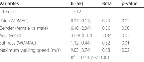
WOMAC pain was not found to be a predictor of lateral trunk motion (b = 0.27, p = 0.12), even after adjusting for the same four confounders Other variables, e.g. duration of complaints, BMI and muscle strength did not change the regression coefficient of knee pain and contributed not to the variance in lat-eral trunk motion.
Additional analyses showed that the interaction terms between knee pain and the variables age, gender, stiff-ness and maximum walking speed were not significantly associated with lateral trunk motion.
Discussion
This study showed that in bivariable analyses a higher level of pain is not associated with high lateral trunk motion during walking in patients with knee OA. How-ever, multivariable analyses showed that knee pain was
indeed weakly associated with more lateral trunk motion when age, gender, self-reported stiffness and maximum walking speed were accounted for.
[image:5.595.306.539.123.224.2]The weak relationship between lateral trunk motion and knee pain, found in this study, is in agreement with Hunt et al [8]. However, the relationship found by Hunt et al was established using bivariable analyses, whereas in our study the association was only found in multivari-able analyses. It seems that age, gender, stiffness and maximum walking speed mask the relationship between lateral trunk motion and knee pain [24]. This masking effect means that without the presence of these variables the relationship between lateral trunk motion and knee pain would be weaker, absent or inverse. These variables were chosen to control for confounding the relationship between lateral trunk motion and knee pain. The selec-tion of independent variables was based on the literature and clinical experience. The variables muscle strength, maximum walking speed, duration of complaints and BMI were selected based on the results of previous stu-dies [1,2,4-6,8-10,20-22]. Age and stiffness were selected based on clinical experience. We repeated the analysis with the former mentioned variables at the patient level with the most severe K&L disease severity knee as index knee for lateral trunk motion. Meaning that trunk motion was defined as the movement of the trunk in the direction of the knee with the highest K&L disease severity. The analysis showed similar results (data not shown). Our results are not in agreement with the study ofTanaka et al. [10]. No significant differences were found between normal, unilateral and bilateral knees in trunk motion. The difference can be explained by the difference in analyses. In our study multivariable regres-sion analyses was used and in the study of Tanaka et al. the Wilcoxon signed rank-test was conducted for differ-ences between involved and non-involved limbs. The analysis was at the knee level, whereas in our study the
Table 2 Results of the regression of lateral trunk motion on knee pain (VAS), gender, age, stiffness (WOMAC) and maximum walking speed
Variables b (SE) Beta p-value
Intercept 14.71
Pain (VAS) 0.77 (0.35) 0.31 0.03
Gender (female vs male) 6.48 (2.00) 0.39 0.00
Age (years) -0.27 (0.11) -0.32 0.02
Stiffness (WOMAC) 1.05 (0.43) 0.30 0.02
Maximum walking speed (m/s) 9.59 (3.54) 0.41 0.01 R2= 0.47 p < 0.001
(N = 52)
b = unstandardized regression coefficient SE = Standard Error
Beta = standardized regression coefficient VAS = Visual Analogue Scale
WOMAC = Western Ontario and McMaster Universities Osteoarthritis Index
Table 3 Results of the regression of lateral trunk motion on knee pain (WOMAC), gender, age, stiffness (WOMAC) and maximum walking speed
Variables b (SE) Beta p-value
Intercept 17.12
Pain (WOMAC) 0.27 (0.17) 0.23 0.12
Gender (female vs male) 6.18 (2.04) 0.36 0.00
Age (years) -0.28 (0.12) -0.34 0.02
Stiffness (WOMAC) 1.12 (0.44) 0.32 0.01
Maximum walking speed (m/s) 9.03 (3.74) 0.38 0.02 R2= 0.44 p < 0.001
(N = 52)
b = unstandardized regression coefficient SE = Standard Error
Beta = standardized regression coefficient
[image:5.595.57.290.570.673.2]analysis was at the patient level. In future studies the direction of trunk motion in relation to the most painful knee should be considered.
As age increases, it is known that joint range of motion (ROM) decreases. Joint ROM in relation to age is most evident in the spine, in particular lateral trunk flexion and axial rotation [25]. These authors also found a significant difference in lateral trunk flexion between men and women, with women exhibiting a higher mobi-lity. Although the assessment of lateral trunk motion was performed differently, our findings are in agreement with the findings reported by these authors [25].
In our study, self-reported stiffness was measured by the WOMAC questionnaire, i.e. we evaluated the patient’s perceived stiffness. Self-reported stiffness can be defined as the perception of decreased ease in mov-ing the knee joint [3,26]. In the WOMAC questionnaire, self-reported stiffness is measured by two questions; (i) how severe was your stiffness after you first rose in the morning, and (ii) how severe has your stiffness been after sitting or lying down or while resting later in the day? Our results show that a high score of self-reported stiffness was related to more lateral trunk motion. It can be speculated that patients with self-reported knee stiff-ness adjust their walking pattern by reducing flexion at their knee and thereby change their lateral trunk motion. Future studies are necessary to determine the influence of self-reported and observed knee stiffness on lateral trunk motion in patients with knee OA.
Hunt et al. (2008) reported walking speed to be corre-lated to trunk motion (r = -0.24;p = 0.05), indicating that patients who walk slower show higher lateral trunk motion. Our results showed that patients with high speed showed high lateral trunk motion. These opposing find-ings may be attributed to how walking speed was assessed. Hunt et al. assessed walking speed by taking the average of multiple gait cycles while walking at a comfor-table self-selected speed. In our study, patients were instructed to walk as fast as possible five times continu-ously up and down a level 20-meter corridor. This vari-able was chosen as an independent varivari-able in analyses for the reason that maximum walking speed during the 100 m test and walking speed during gait analysis were highly correlated (r = 0.74; p = 0.000). Although the means (SD) in m/s of our study and of Hunt el al. (2008) were comparable (1.06 (0.16) and 1.14 (0.28), respec-tively), the different assessment methods may explain the opposing results. Future studies are necessary to deter-mine the influence of different walking speeds on lateral trunk motion in patients with knee OA.
It should be noted that sagittal and transversal trunk motion would increase as lateral trunk motion increases. No studies have been found that assess the relationship between movements in all 3 dimensions in relation to
knee pain. Pelvic rotation and hip joint motion influence lateral trunk motion in knee OA patients [5], therefore, these parameters should be considered in future studies. It is also suggested that the strength of the abductor muscles of the hip is of influence on trunk motion [7]. Hip muscle strength was not assessed in our study, but could have been of influence on trunk motion, as well as knee pain [27]. In future studies it is important to measure lateral trunk motion in relation to abductor muscle strength.
Conclusions
Pain is associated with lateral trunk motion. This asso-ciation is weak and is influenced by age, gender, self-reported stiffness and maximum walking speed.
Acknowledgements
We would like to thank Karin Fiedler and Steve Webster for their help in correcting the manuscript.
Author details
1Reade, Center for Rheumatology and Rehabilitation, Department of
Rehabilitation Research, Amsterdam, the Netherlands.2VU University Medical Center, Department of Rehabilitation Medicine, Department of Psychiatry, EMGO Institute, Amsterdam, the Netherlands.3VU University Medical Center, Department of Rehabilitation Medicine, Research Institute MOVE,
Amsterdam, the Netherlands.4Glasgow Caledonian University, School of Health, Glasgow, Scotland UK.
Authors’contributions
JD conceived the study and led the co-ordination of the study. MvdE, MS, JH, JvdN, and JD assisted with protocol design. MvdE, MS and JD wrote the manuscript. DK designed the statistical analysis. MvdE, MS, JvdN and JH designed the biomechanical measures. MvdE and MS designed the biomechanical and physical impairment measures. All authors participated in the trial design, provided feedback on drafts of this paper, read, and approved the final manuscript.
Competing interests
The authors declare that they have no competing interests.
Received: 22 November 2010 Accepted: 29 June 2011 Published: 29 June 2011
References
1. Mündermann A, Dyrby CO, Hurwitz DE, Sharma L, Andriacchi TP:Potential strategies to reduce medial compartment loading in patients with knee osteoarthritis of varying severity.Arthritis Rheum2004,50:1172-1178. 2. Messier SP, DeVita P, Cowan RE, Seay J, Young HC, Marsh AP:Do older
adults with knee osteoarthritis place greater loads on the knee during gait? A preliminary study.Arch Phys Med Rehabil2005,86:703-709. 3. Zeni JA Jr, Higginson JS:Dynamic knee joint stiffness in patients with a
progressive increase in severity of knee osteoarthritis.Clin Biomech2009, 24:366-371.
4. McGibbon CA, Krebs DE:Compensatory gait mechanics in patients with unilateral knee arthritis.J Rheumatol2002,29:2410-2419.
5. Tanaka K, Miyashita K, Urabe Y, Koshida S, Takemoto Y, Ijiri T:Relationship between pelvic rotation and trunk lean motion during walking in patients with symptomatic knee osteoarthritis.J Biomech2007,40:S508. 6. Mündermann A, Asay JL, Mündermann L, Andriacchi TP:Implications of
increased medio-lateral trunk sway for ambulatory mechanics.J Biomech 2008,41:165-170.
8. Hunt MA, Birmingham TB, Bryant D, Jones I, Giffin JR, Jenkyn TR, Vandervoort AA:Lateral trunk lean explains variation in dynamic knee joint load in patients with medial compartment knee osteoarthritis. Osteoarthritis Cartilage2008,16:591-599.
9. Hurwitz DE, Ryals AR, Block JA, Sharma L, Schnitzer TJ, Andriacchi TP:Knee pain and joint loading in patients with osteoarthritis of the knee.J Orthop Res2000,18:572-579.
10. Tanaka K, Miyashita K, Urabe Y, Ijiri T, Takemoto Y, Ishii Y, Ochi M: Characteristics of trunk lean motion during walking in patients with symptomatic knee osteoarthritis.Knee2008,15:134-8.
11. Esch van der M, Steultjens M, Harlaar J, Wolterbeek N, Knol D, Dekker J: Varus-valgus motion and functional ability in patients with osteoarthritis of the knee.Ann Rheum Dis2008,67:471-477.
12. Van der Esch M, Steultjens M, Harlaar J, Wolterbeek N, Knol DL, Dekker J: Knee varus-valgus motion during gait: a measure of joint stability in patients with osteoarthritis?Osteoarthritis Cartilage2008,16:522-525. 13. Altman RD, Hochberg MC, Moskowitz RW, Schnitzer TJ:Recommendations
for the medical management of osteoarthritis of the hip and knee. American college of rheumatology subcommittee on osteoarthritis guidelines.Arthritis Rheum2000,43:1905-1915.
14. Buckland-Wright JC, Bird CF, Ritter-Hrncirik CA, Cline GA, Tonkin C, Hangartner TN,et al:X-ray technologists’reproducibility from automated measurements of the medial tibiofemoral joint space width in knee osteoarthritis for a multicenter, multinational clinical trial.J Rheumatology 2003,30:329-338.
15. Altman RD, Hochberg M, Murphy WA Jr, Wolfe F, Lequesne M:Atlas of individual features in osteoarthritis.Osteoarthritis Cartilage1995,3:3-70. 16. Kellgren JH, Lawrence JS:Radiological assessment of osteo-arthrosis.Ann
Rheum Dis1957,16:494-502.
17. Harlaar J, Doorenbosch CA:3D kinematic analysis by BodyMech: a Matlab based open source software package for research and education. Proceedings of the Ninth International Symposium on the 3D analysis of human movement, June 28-302006, Valenciennes.
18. Cappozzo A, Catani F, Croce UD, Leardini A:Position and orientation in space of bones during movement: anatomical frame definition and determination.Clin Biomech1995,10:171-178.
19. Roorda L, Jones C, Waltz M, Lankhorst G, Bouter L, van der IJken J, Willems W, Heyligers I, Voaklander D, Kelly K, Suarez-Almazor M: Satisfactory cross cultural equivalence of the Dutch WOMAC in patients with hip osteoarthritis waiting for arthroplasty.Ann Rheum Dis2004, 3:36-42.
20. Van der Esch M, Steultjens M, Knol DL, Dinant H, Dekker J:Joint laxity and the relationship between muscle strength and functional ability in patients with osteoarthritis of the knee.Arthritis Rheum2006,55:953-9. 21. Van der Esch M, Steultjens MPM, Harlaar J, Knol DL, Lems W, Dekker J:Joint
Proprioception, muscle strength and functional ability in patients with osteoarthritis of the knee.Arthritis Rheum2007,57:787-793.
22. Steultjens MPM, Dekker J, Baar van ME, Oostendorp RAB, Bijlsma JWJ: Muscle strength, pain and functional ability patients with osteoarthritis. Clin Rehab2001,15:331-341.
23. Kleinbaum DG, Kupper LL, Muller KE:Applied regression analysis and multivariable methods.Duxbury Pr2007.
24. Maassen GH, Bakker AB:Suppressor variables in path models: Definitions and interpretations.Sociological Methods & Research2001,30:241-70. 25. Doriot N, Wang X:Effects of age and gender on maximum voluntary
range of motion of the upper body joints.Ergonomics2006,49:269-281. 26. Lark SD, Buckley JG, Bennett S, Jones D, Sargeant AJ:Joint torques and
dynamic joint stiffness in elderly and young men during stepping down. Clin Biomech2003,18:848-855.
27. Bennell KL, Hunt MA, Wrigley TV, Hunter DJ, McManus FJ, Hodges PW, Li L, Hinman RS:Hip strengthening reduces symptoms but not knee load in people with medial knee osteoarthritis and varus malalignment: a randomised controlled trial.Osteoarthritis Cartilage2010,18:621-8.
Pre-publication history
The pre-publication history for this paper can be accessed here: http://www.biomedcentral.com/1471-2474/12/141/prepub
doi:10.1186/1471-2474-12-141
Cite this article as:van der Eschet al.:Lateral Trunk Motion and Knee Pain in Osteoarthritis of the Knee: a cross-sectional study.BMC Musculoskeletal Disorders201112:141.
Submit your next manuscript to BioMed Central and take full advantage of:
• Convenient online submission
• Thorough peer review
• No space constraints or color figure charges
• Immediate publication on acceptance
• Inclusion in PubMed, CAS, Scopus and Google Scholar
• Research which is freely available for redistribution