D
E
V
E
LO
P
M
E
N
T
Whether to be male or female is a critical decision in
development. Nowhere is this more important than in the germ cells, which must produce either the sperm or eggs necessary for the perpetuation of the species. How does a germ cell make this decision and how is it executed? One thing that is clear is that this process is very different in germ cells compared with other cells of the embryo. Here, we explore how sexual identity is established in the Drosophilagermline, how this affects other aspects of germ cell development and what studies in
Drosophilacan teach us about mammalian germ cells.
Introduction
Animals have evolved a fascinating array of mechanisms for conducting sexual reproduction, including those used for attracting a mate, courting a mate and getting the gametes together. These mechanisms often differ drastically between different species. However, one thing that all sexual animals have in common is the production of distinct male and female gametes that must unite to initiate development of a new individual. Thus, the study of how a germ cell is guided along a male or female path, to eventually produce either sperm or eggs, is central to our understanding of sexual reproduction and fertility.
In this review, we discuss germline sexual identity within the context of the different stages of germline sexual development and the decisions that germ cells face along the way (see Box 1 for a discussion of terms). This includes the process of germline sex determination, by which a germ cell acquires a male versus female identity, but also how sexual identity influences other events, such as the formation of male or female germline stem cells and entry into spermatogenesis or oogenesis. Although germline sex determination and sexual development have not always been studied together, it is clear that we cannot understand one without the other. An understanding of germline sex determination depends on our understanding how the developmental paths of male and female germ cells diverge. Similarly, understanding other events in male versus female germ cell development requires an understanding of the role germ cell sexual identity plays in these processes. Therefore, we first discuss work on germline sex determination that has contributed greatly to our knowledge of how this process is controlled. We then discuss our emerging understanding of germ cell sexual development and how this affects our thinking about germline sex determination. Finally, we briefly discuss how understanding these events in Drosophilacan shed light on similar events in mammalian germ cell development and human fertility. As this represents several distinct subjects, each of which is worthy of an entire review, we have, by necessity, focused on specific findings and refer readers to more-comprehensive reviews on related topics, where appropriate.
Germline sex determination in Drosophila
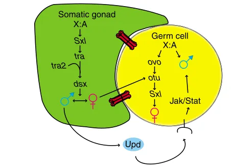
The establishment of sexual identity in the germline is thought to occur differently from in the rest of the body (the somatic cells or soma). In the soma, male versus female identity is regulated by the number of X chromosomes (or ratio of X chromosomes to autosomes), such that, in normal diploid animals, XX is female and XY is male (Fig. 1) (reviewed by Cline and Meyer, 1996). The Y chromosome is not important for sex determination in either the soma or the germline in Drosophila, but does contain genes that are required for spermatogenesis. In the soma, XX embryos activate expression of Sex-lethal (Sxl), which functions through Development 133, 2783-2791 (2006) doi:10.1242/dev.02415
The control of sexual identity in the
Drosophila
germline
Abbie Casper and Mark Van Doren*
Department of Biology, 302 Mudd Hall, Johns Hopkins University, 3400 North Charles Street, Baltimore, MD 21218, USA.
*Author for correspondence (e-mail: vandoren@jhu.edu)
Box 1. A glossary of terms
Sexual dimorphism
Any difference in the characteristics (phenotype) of an otherwise equivalent cell type, in males versus females. This includes a sex-specific pattern of gene expression, as well as any other aspects of sex-specific development.
Sexual identity
A difference in identity or developmental potential of an otherwise equivalent cell type, in males versus females. Any sexual dimorphism in a cell type, even the expression of a single sex-specific gene, can indicate that sexual identity has been initiated. However, sexual identity may also require maintenance, and a cell may receive additional sex-specific inputs that influence sexual identity. This may be particularly true in the germline, where continued interactions between the soma and germline influence all aspects of germ cell development.
Sex determination
The mechanism by which a cell, or organism, acquires its sexual identity. In some cell types, this may involve a single event that irreversibly establishes a male versus female identity. In other cell types, sex determination may be an ongoing process with multiple stages of commitment.
Sexual differentiation
How a cell or tissue uses its sexual identity to produce a sex-specific phenotype (sexual dimorphism). However, this term can be confusing for the germline because, even though the formation of male versus female germline stem cells is an interesting sexual dimorphism, germline stem cells are still considered to be in a relatively ‘undifferentiated’ state. Thus, we will only use the term sexual differentiation to refer to the processes of spermatogenesis or oogenesis.
Sexual development
D
E
V
E
LO
P
M
E
N
T
transformer (tra) and transformer 2 (tra2) to regulate the splicing of doublesex (dsx) RNA, producing a female form of this transcription factor. In XY animals, this pathway is off and a male form of DSX is produced by default. dsx regulates male versus female development in most somatic cells, including the somatic gonad, while in the nervous system, a second target for this pathway, fruitless, regulates most aspects of sex-specific behavior (Ryner et al., 1996). As we discuss below, the rules for determining sex in the germ cells are very different from those acting in the soma, and the sex of the soma also influences this decision in the germline.
Studying germline sex determination
Much of the work on germline sex determination in Drosophilahas focused on studying the relative contribution of somatic influence versus the germ cell-autonomous control of this process. Often, this has been analyzed by placing germ cells of one ‘sex’ into a soma of the opposite ‘sex’, such as through the use of genetic mosaics (see also Oliver, 2002). For example, transplanting germ cells from one embryo to another (Illmensee and Mahowald, 1974) allows one to study the fate of XY germ cells in an XX soma, and vice versa. An important conclusion from this work is that, in Drosophila, XY germ cells in an ovary do not make functional eggs, and XX germ cells in a testis do not make functional sperm (Marsh and Wieschaus, 1978; Schupbach, 1982; Steinmann-Zwicky et al., 1989; Van Deusen, 1977).
Germ cell transplantation has also revealed that most of the genes that function in Drosophilato determine the sex of the soma, such as traand dsx, have no role in germ cells; germ cells mutant for these genes produce normal eggs or sperm in wild-type hosts (Marsh and Wieschaus, 1978; Schupbach, 1982). Thus, the manipulation of these genes is another way to change the ‘sex’ of the soma without affecting the ‘sex’ of the germ cells. For example, animals that are
XX but mutant for tradevelop as males (Sturtevant, 1945), including a male somatic gonad. However, as tra is not required in the germline, this results in XX germ cells developing in a male soma, similar to the transplant experiments described above. Overexpression of tra is sufficient to feminize an XY soma (McKeown et al., 1988), creating the opposite situation.
Male germ cells in a female soma
When XY germ cells develop in a female soma [using the above methods and others; see Oliver (Oliver, 2002), for an extensive review], the result is often an ‘ovarian tumor’, where germline cysts in the ovary contain tens to hundreds of small individual germ cells instead of 15 interconnected nurse cells and an oocyte, as in a normal ovarian cyst. At times, these germ cells appear unambiguously male with characteristics of differentiating spermatocytes (Steinmann-Zwicky et al., 1989), indicating that XY germ cells can retain a male identity in a female soma. However, characteristics of oogenesis can also be observed in these experiments (Nagoshi et al., 1995; Waterbury et al., 2000) and analyses using molecular markers reveal that these germ cells have both male and female characteristics (Hinson and Nagoshi, 1999; Janzer and Steinmann-Zwicky, 2001; Waterbury et al., 2000). Furthermore, XY germ cells do not always survive in a female environment (Schüpbach, 1985; Steinmann-Zwicky et al., 1989). Thus, although the ovarian tumor phenotype is commonly observed, and is likely a result of XY germ cells maintaining aspects of male identity in a cell-autonomous manner, these germ cells are not fully male, and may also have compromised viability.
Female germ cells in a male soma
XX germ cells are likely to fail to proliferate or to die when present in a male soma. XX germ cells transplanted into a male soma are recovered at a greatly reduced frequency relative to the reciprocal transplantation (Steinmann-Zwicky, 1993; Steinmann-Zwicky et al., 1989). Similarly, in XX animals where the soma has been transformed to a male identity, many gonads contain few or no germ cells [e.g. tra mutants (Brown and King, 1961; Hinson and Nagoshi, 1999; Nöthiger et al., 1989; Sturtevant, 1945)]. The remaining germ cells form cysts that can have either male or, more rarely, female character, while many other cysts appear to be degenerating (Andrews and Oliver, 2002; Brown and King, 1961; Hinson and Nagoshi, 1999; Horabin et al., 1995; Nagoshi et al., 1995; Nöthiger et al., 1989; Oliver et al., 1993; Staab et al., 1996). Thus, XX germ cells in a male soma appear to make a more stochastic decision whether to be male, or female, and also have compromised viability.
Although the phenotype of germ cells placed in a soma of the opposite sex is variable, one conclusion is clear and consistent: they are not able to develop as either fully male or female. Thus, proper germ cell sex determination requires a combination of both somatic signals and germ cell-autonomous cues.
Somatic control over germ cell sex
Somatic control over germ cell sex determination is observed in many animals, including flies and mice. Although the somatic sex determination pathway in Drosophilais not required in the germ cells themselves for proper germ cell sex determination, it is required in the soma for proper somatic control of germ cell sex. tra, tra2 and dsxare all required in the soma, and not the germ cells, for proper germline sex determination (Nöthiger et al., 1989; Marsh and Wieschaus, 1978; Schupbach, 1982). Sxl, by contrast, is required in Somatic gonad
X:A
Sxl
tra tra2
Upd
X:A
ovo
otu
Sxl
Jak/Stat Germ cell
[image:2.612.51.283.59.223.2]dsx
D
E
V
E
LO
P
M
E
N
T
both the soma and the germline (below). The effects of mutations in tra,tra2 and dsxon the germline indicate that the pathway that controls somatic influence over germ cell sex determination is the same as that controlling other aspects of somatic sexual development and acts through dsx(Fig. 1). However, others have argued for an additional, dsx-independent mechanism for somatic control over the germline as some feminizing affects of traare still observed in the absence of dsx (Horabin et al., 1995; Waterbury et al., 2000). Although little is known about how the sex of the soma influences germline sexual identity, it has recently been shown that one such mechanism acts through the Jak/Stat (Janus kinase/signal transducer and activator of transcription) pathway (Wawersik et al., 2005).
Germ cell-autonomous control
In addition to somatic influences, germ cells also ‘know’ their own sex autonomously (Fig. 1). This is dependent on the X:A ratio in the germ cells (Schüpbach, 1985), as it is in the soma, and female germ cells require Sxlfor proper development. XX germ cells mutant for Sxlcannot make eggs but give rise to ovarian tumors (Oliver et al., 1988; Perrimon et al., 1986; Salz et al., 1987; Schüpbach, 1985; Steinmann-Zwicky et al., 1989). This resembles what is observed when XY germ cells are present in females (as discussed above), indicating that Sxlmutant XX germ cells are masculinized. Indeed, male-specific gene expression is observed in XX germ cells that lack Sxl (Staab et al., 1996; Wei et al., 1994), and Sxlfunctions to promote female germ cell development in other assays (Hinson and Nagoshi, 1999; Steinmann-Zwicky et al., 1989). However, gain of Sxl function in XY germ cells does not interfere with male development, as it does in the soma, indicating that Sxl is not sufficient to activate female germ cell development and, therefore, might not act as a ‘switch’ between male and female identity in the germline (Cline, 1983; Hager and Cline, 1997; Oliver et al., 1993). In addition, the pathway both upstream and downstream of Sxlis different in the germline than in the soma. Although germ cells depend on the X:A ratio, they count their number of X chromosomes differently. Somatic cells depend on factors such as scute/ sisterless-band maternal daughterlessto determine their X:A ratio and to activate Sxlin females, but these factors are not required in the germ cells (Granadino et al., 1993; Schüpbach, 1985; Steinmann-Zwicky, 1993). Similarly, tra is not required in the germline (Marsh and Wieschaus, 1978), so this cannot be the downstream target of Sxlin these cells. The target(s) of Sxlin the female germline remain elusive.
How is Sxlregulated in the germline if the X:A ratio is ‘read out’ differently and germ cells also respond to somatic signals? Genes that, like Sxl, give rise to ovarian tumors when mutated are candidates for acting with Sxl to promote female germ cell development. The most extensively studied of these genes, with regard to germ cell sex determination, are ovarian tumor [otu (King, 1979)] and ovo(Oliver et al., 1987). Although the role of these genes is still being resolved, one possible model for how these genes act is presented in Fig. 1. Both of these genes are normally required in female, but not in male, germ cells. ovo appears to be responsive to germ cell-autonomous cues, as ovo expression is regulated by the X:A ratio and ovois required in XX germ cells regardless of whether they are in a male or female soma (Andrews and Oliver, 2002; Bielinska et al., 2005; Nagoshi et al., 1995; Oliver et al., 1994). By contrast, otu appears to respond to somatic signals as otuexpression is activated by a female soma, and otuis required by both XX and XY germ cells when they are in a female somatic environment (Hinson and Nagoshi, 1999; Nagoshi et al., 1995; Waterbury et al., 2000). ovoencodes several
related zinc-finger transcription factors (Garfinkel et al., 1992; Garfinkel et al., 1994; Mevel-Ninio et al., 1991; Mével-Ninio et al., 1995) that directly regulate otuexpression (Andrews et al., 2000; Andrews and Oliver, 2002; Lu et al., 1998; Lu and Oliver, 2001). The molecular function of OTU is unknown, but it may involve RNA regulation (Goodrich et al., 2004). Finally, Sxl acts genetically downstream of ovo and otu, and these genes are required for the proper splicing of SxlRNA into the female (active) form (Bopp et al., 1993; Nagoshi et al., 1995; Oliver et al., 1993; Oliver and Pauli, 1998; Pauli et al., 1993).
Complications to the simplified model in Fig. 1 include that ovo can also behave genetically downstream of otuin some assays (e.g. Hinson and Nagoshi, 1999), and that both ovoand otuare required for germ cell survival in females (King and Riley, 1982; Oliver et al., 1987), unlike Sxl (Schüpbach, 1985), suggesting they have an additional role that is independent of Sxl. There are also other genes thought to act in this process, such as sans fille, a Sxl splicing factor (Salz, 1992), and stand still, which appears to regulate otu expression (Sahut-Barnola and Pauli, 1999). Thus, achieving a better understanding of how the X:A ratio is interpreted in the germline and influences sex determination, in combination with signals from the soma, remains a high priority.
The problem of germline X chromosome dosage compensation
Embryos need to adjust the amount of X chromosome gene expression to ensure that females (XX) and males (XY) have similar relative levels, a process known as X chromosome dosage compensation. However, germ cells must turn off dosage compensation, at least at some point in their development, in order to reset this system for the next generation. XX and XY germ cells would have different levels of X chromosome gene expression at this time. Some of the effects of X chromosome number in the germline may be due to general problems resulting from this difference, rather than due to specific effects on germline sex determination; it is possible that female germ cells require a 2X chromosome dose, while male germ cells cannot tolerate a 2X chromosome dose. However, it is unlikely that this is the only way in which X chromosome number affects germ cell development, as the X:A ratio clearly affects the male versus female phenotype of the germ cells and not just their survival (as discussed above). This indicates that the number of X chromosomes helps determine the sexual identity of the germline, independent of any other effects of X chromosome dose.
Germline sexual development in Drosophila
D
E
V
E
LO
P
M
E
N
T
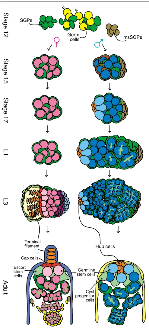
determination fully. Germ cell sexual development includes the initiation of male versus female identity in the germline. In addition, it includes the maintenance of this identity, the formation of male versus female germline stem cells, and the commitment to and completion of spermatogenesis versus oogenesis (Fig. 2). Importantly, each of these steps requires extensive interaction between the germ cells and the somatic gonad. An essential goal now is to break germline sexual development down into its component parts so that the mechanisms regulating each distinct stage can be understood.
Gonad formation
Germ cells coalesce with somatic gonadal precursors (SGPs) to form the embryonic gonad at about 12 hours after fertilization (reviewed by Van Doren, 2006). Interestingly, the somatic gonad is already sexually dimorphic at this time (see Fig. 2 and later in the review). The most posterior SGPs are known as ‘male-specific’ SGPs (msSGPs) because they contribute to only the male gonad and undergo apoptosis in females (DeFalco et al., 2003). Anterior SGPs also have a sex-specific identity and exhibit a different pattern of gene expression in males versus females (Le Bras, 2006). There is ample opportunity for soma to influence germ cell development in the embryonic gonad, as SGPs ensheath each germ cell (Jenkins et al., 2003), and gap junctions may facilitate communication between the soma and germline (Tazuke et al., 2002). In addition, the SGPs express secreted factors such as unpaired(Wawersik et al., 2005), a ligand for the JAK/STAT pathway, differently in males versus females, indicating that communication between the soma and the germline can be sex specific.
Initial germ cell sexual identity
Sexual dimorphism in the germline is also evident at the time of embryonic gonad formation. A slightly greater number of germ cells are incorporated into the male gonad relative to the female gonad (Poirie et al., 1995; Sonnenblick, 1941). As there are not thought to be differences in germ cell formation between the sexes, and germ cells are arrested in the cell cycle during migration and gonad formation (Sonnenblick, 1941; Su et al., 1998), this may be due to differences in how the germ cells respond to the somatic gonad or to differences in the somatic gonad itself (e.g. the presence of msSGPs in the male). The difference in germ cell number is then amplified after gonad formation as male germ cells begin to proliferate, while female germ cells do not (Kerkis, 1931; Steinmann-Zwicky, 1994; Wawersik et al., 2005). Last, the female germline is more sensitive
Stage 12 Stage 15 Stage 17 L1 L3
Adult
Terminal filament
Cap cells
Germline stem cells Escort
stem cells
Cyst progenitor cells
Hub cells SGPs
Germ
[image:4.612.51.298.62.605.2]cells msSGPs
D
E
V
E
LO
P
M
E
N
T
to death caused by the activation of P element transposons (hybrid dysgenesis) than is the male germline (Wei et al., 1991). As this occurs during embryonic stages, this indicates that the germline is already sexually dimorphic at this time.
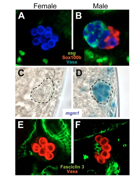
A sex-specific pattern of gene expression can also be observed in germ cells soon after gonad formation (stage 15). At this time, germ cells exhibit male-specific expression of male germline marker 1 [mgm1, Fig. 3C,D (Staab et al., 1996)], a lacZreporter that probably reflects expression of the transcription factor escargot(Streit et al., 2002), along with other male-specific genes (Wawersik et al., 2005). The fact that sex-specific germ cell phenotypes and gene expression patterns are first observed in the newly formed embryonic gonad indicates that this may be when germ cells first acquire distinct male versus female identities.
Sex-specific gene expression in embryonic germ cells is regulated by both somatic signals and germ cell-autonomous cues. XY germ cells initiate mgm1expression even in a female somatic environment, indicating that it is regulated by the germ cell genotype (Heller and Steinmann-Zwicky, 1998; Janzer and Steinmann-Zwicky, 2001). mgm1expression is also regulated by the soma, as XY germ cells cannot maintain mgm1expression in a female soma (Janzer and Steinmann-Zwicky, 2001). Thus, the initiation of mgm1 expression in the male germline may be regulated differently from its maintenance. In addition, XX germ cells can express mgm1when in a male soma (Staab et al., 1996), and there is evidence for both induction of mgm1expression by the male somatic gonad (Wawersik et al., 2005) and repression of mgm1 expression by the female somatic gonad (Heller and Steinmann-Zwicky, 1998).
How does the somatic gonad regulate initial sexual identity in the germline? One mechanism acts through the Jak/Stat pathway (Wawersik et al., 2005). A ligand for this pathway, unpaired (upd), is expressed in the embryonic somatic gonad in males, but not in females, and the Jak/Stat pathway can be activated in germ cells of either sex as long as they contact a male somatic gonad. The Jak/Stat pathway is necessary and sufficient to induce germ cell proliferation in the embryonic gonad, a male-specific behavior. It is also necessary for the maintenance of mgm1expression in male germ cells, and is sufficient to induce expression of some male-specific
genes, such as mgm1, disc proliferation abnormal and Minichromosome maintenance 5, in female germ cells (Wawersik et al., 2005). However, activation of the Jak/Stat pathway in female germ cells, at least at early stages, does not block oogenesis in adults. Thus, this pathway is only one of the factors that regulates germline sex determination, and does not necessarily lead to an irreversible commitment to male germ cell identity. It is currently unknown if other somatic signals, or genes such as Sxl,ovo and otu acting in a germ cell-autonomous fashion, contribute to specifying germ cell sexual identity at this early stage.
In summary, germ cells have a sex-specific identity at a time soon after they have joined with SGPs to form the embryonic gonad, and this may represent the point at which male versus female identity is first established in the germ cells. Even at this early stage, there is evidence that both signals from the somatic gonad and germ cell-autonomous cues are important for germline sex determination. Future work to understand how initial germ cell sexual identity is established should focus on this early timepoint.
Male versus female germline stem cells Germline stem cell formation
[image:5.612.53.471.57.210.2]D
E
V
E
LO
P
M
E
N
T
formation of the GSC niche that persists in the adult testis. As spermatogenesis begins during early larval stages (Aboïm, 1945), it is possible that the germ cells interacting with the hub are already functional GSCs in the late embryo or early (first instar) larval stage.
In the female, each of the 16 or so ovarioles in an adult ovary contains a stem cell niche, which is created largely by the somatic cap cells. This structure is formed only during ovary morphogenesis, which begins in the late (third instar) larvae (Godt and Laski, 1995; King, 1970; Sahut-Barnola et al., 1995; Spradling, 1993). Evidence suggests that the Tgfpathway, the crucial signaling pathway between the niche and GSCs in the adult ovary, is already important for GSC formation at this time (Zhu and Xie, 2003). However, recent work has shown that germ cells populating the anterior region of the female embryonic gonad are more likely to become GSCs, suggesting that some female germ cells are already predetermined to be GSCs at a much earlier stage (Asaoka and Lin, 2004). Thus, an
interesting possibility is that the anterior-most SGPs, in both the male and female embryonic gonads, may be responsible for specifying GSC identity in a subset of germ cells.
Adult GSCs and niches
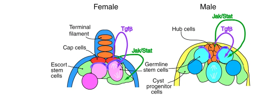
Most of our knowledge of GSCs in Drosophila comes from extensive study of the adult testis and ovary. There are some surprising similarities between male and female GSCs, including how they interact with the niche, the signaling pathways that are active in the niche, and the close interaction between GSCs and somatic stem cells in the niche (Fig. 3) (Decotto and Spradling, 2005; Gilboa and Lehmann, 2004a; Spradling et al., 2001; Wong et al., 2005; Yamashita et al., 2005). However, despite these similarities, there are also some clear differences between male and female GSCs. In both sexes, GSCs are maintained by signaling from the niche, but in the female this signal acts through the Tgfpathway (Xie and Spradling, 1998), while in the male, signals act through both the Jak/Stat (Kiger et al., 2001; Tulina and Matunis, 2001) and Tgfpathways (Kawase et al., 2004; Schulz et al., 2004; Shivdasani and Ingham, 2003). Although signaling through the Jak/Stat pathway also occurs in the female niche, it acts on the somatic (escort) stem cells; female GSCs do not require this signal (Decotto and Spradling, 2005). In addition, there are differences in gene expression in male versus female GSCs (Gönczy et al., 1992), including in mgm1expression, which is initially expressed in all male germ cells and becomes restricted to male GSCs (Staab et al., 1996). Thus, despite being exposed to similar signaling environments, male and female GSCs respond differently to these signals and maintain distinct identities. It is likely that the prior sexual identity of the germ cells is crucial for determining their differential responses to the niche environment.
Interestingly, the Jak/Stat pathway is activated in all germ cells as they enter the male embryonic gonad, but is then active only in GSCs in the adult testis. Thus, does the Jak/Stat pathway promote male germ cell identity, male GSC identity or both? One possibility is that there is no real difference between these two identities. It may be that all germ cells in a male embryo initially receive this signal, which contributes to their male identity, and allows them to proliferate and remain undifferentiated. Those germ cells that do not interact with the niche lose the signal and directly enter spermatogenesis, while those that contact the niche retain the signal and continue to act as GSCs. Similarly, it has been shown that female germ cells have the potential to enter gametogenesis at early stages, prior to ovary morphogenesis, and the Tgfpathway is important to prevent this premature differentiation (Gilboa and Lehmann, 2004b). Thus, in both sexes it is likely to be important to hold germ cells in an undifferentiated state until the stem cell niche has been formed. The same signals that regulate stem cell maintenance in the adult may act to prevent premature germ cell differentiation earlier in development.
Sex and GSCs
[image:6.612.53.279.266.560.2]How does germline sexual identity affect GSC formation and behavior? Can a male germ cell become a female GSC or vice versa? Little is currently known about these interesting questions. When male germ cells are in a female soma and exhibit the ovarian tumor phenotype, individual germline cysts are still produced in an ‘assembly line’ fashion as in normal ovarioles. This suggests that GSCs are still present and continuously produce germline cysts (Schüpbach, 1985). However, the germline is also lost over time in these gonads (Steinmann-Zwicky et al., 1989), indicating that GSC maintenance is Fig. 4. Sexual dimorphism in the embryonic gonad.(A,B) Stage 15
embryonic gonad labeled to reveal the germ cells (anti-Vasa, blue), male-specific somatic gonadal precursors (msSGPs) (anti-Sox100b, red) and esgG66Benhancer trap (anti--gal, green). msSGPs are found only
in male gonads at this stage, and anterior SGPs have a sex-specific identity as they express esgG66Bin males but not females. The esgG66B
D
E
V
E
LO
P
M
E
N
T
defective. These putative GSCs lack expression of male GSC markers, such as mgm1, and express female-specific Sxl (Janzer and Steinmann-Zwicky, 2001; Waterbury et al., 2000). Thus, these cells do not appear to have male GSC identity, and probably have a female or mixed identity.
When female germ cells develop in a male soma, many resulting adult testes have few or no germ cells (above), indicating that these germ cells do not survive or cannot populate the male GSC niche. When such gonads do contain germ cells, little is known about whether any behave as GSCs. Clearly, a more directed analysis of the GSC phenotypes in adults, and an analysis of GSC formation during development, will be required in both sexes to determine how male versus female GSCs are established and how this process is influenced by germ cell sexual identity.
Gametogenesis
Gametogenesis begins in males during the early larval stages [1-2 days after fertilization, reviewed by Fuller (Fuller, 1993)] but does not begin in females until much later [early pupae, 5+ days after fertilization, reviewed by Spradling (Spradling, 1993)]. Although spermatogenesis and oogenesis are obviously very different processes, with dramatically different end products, they are initially surprisingly similar. In both males and females, the GSC daughter that leaves the niche undergoes four mitotic divisions with incomplete cytokinesis to produce a 16-cell cyst of interconnected germ cells. The connections between germ cells are formed by ring canals, and a specialized organelle, the fusome, extends between these cells. Both male and female germ cells also express the Bag of Marbles (BAM) protein as they enter gametogenesis (Gönczy et al., 1997; McKearin and Ohlstein, 1995). Finally, the early germ cell cysts are ensheathed by somatic cells in both sexes (Decotto and Spradling, 2005; Fuller, 1993). Thus, up through the early 16-cell cyst stage, it is difficult to distinguish clearly between spermatogenesis and oogenesis.
Subsequently, the differences become obvious. In males, the germ cells of the cyst grow significantly in size and all complete meiosis to create 64 spermatids. By contrast, only one germ cell (the oocyte) commits to meiosis in females, while the other 15 become nurse cells that are easily recognizable by their polyploid nuclei. In addition, differences in ring canal and fusome character become evident at this time (Hime et al., 1996; Hinson and Nagoshi, 1999; Lin et al., 1994; Robinson et al., 1994). There are also differences in somatic cell development because, in the female, the escort cells interacting with the early cysts die and are replaced by follicle cells (Spradling, 1993, Decotto and Spradling, 2005). It is these late differences in spermatogenesis versus oogenesis, and other types of late cyst phenotypes, such as ovarian tumors, that have been used for much of the work on germline sex determination described above. As there is extensive soma/germ-cell communication during gametogenesis, defects at this stage could indeed result from sexual incompatibility between the soma and germline that is due to earlier problems in germline sex determination. However, it is also possible that these phenotypes could sometimes result from relatively late defects in gametogenesis that are separate from the events of germline sex determination. This problem highlights the need to better understand the different stages of germ cell sexual development individually, how each is affected by germ cell sexual identity and the interactions that occur between germ cells and the surrounding soma.
Conclusions
There are two main ideas on which we have focused our discussion of germline sexual identity in Drosophila, each of which represents an important area for future investigation. First,
germ cell sex determination requires a combination of somatic signals and germ cell-autonomous cues. Although some of the important players that mediate both the germ cell-autonomous and somatic effects on germline sex determination have been uncovered, much remains to be learned about how these factors interact to control germline sexual identity. The second is that germline sexual identity needs to be understood within the context of the different stages of germline sexual development. We do not yet know when the sex of the germline becomes irreversibly determined. The germline is already sexually dimorphic in the embryonic gonad, and must progress through the formation of male or female germline stem cells and through spermatogenesis or oogenesis. Extensive germline-soma interaction governs each of these stages.
Fortunately, a new window is now opening in our ability to study germ cell sexual development that will allow us to better address these ideas in the future. Considerable knowledge has been gained about the early development of the gonad in the embryo, and also about the adult ovary and testis. However, for technical reasons, it has been difficult to bridge the gap between the embryo and adult, during which most of the sexual development of the germline and somatic gonad occurs. Recently, though, researchers working from ‘both ends’ have made dramatic progress in our ability to study these stages. As described above, this includes moving forward from the embryonic gonad to understand the sexually dimorphic development of the germline and somatic gonad during late embryonic and larval stages. This also includes applying our knowledge of the adult gonad to understanding the earlier processes of testis and ovary morphogenesis, and of stem cell niche formation. Thus, we now have the tools necessary for studying the different stages of germline sexual development and for investigating how germline sexual identity is controlled at each step.
Mammalian germ cell sexual development: a view from the fly
Interestingly, the main conclusions about germline sexual development in Drosophilaare also true for the mouse. Mouse germline sex determination also requires both somatic signals and germ cell-autonomous cues, is regulated differently from in the soma and is dependent on the number of X chromosomes in the germ cells. In addition, mouse germ cell sexual development also involves several discrete stages, including the establishment of germ cell sexual identity, the formation of GSCs and entry into gametogenesis, each of which requires extensive, sex-specific interaction between the soma and germline.
D
E
V
E
LO
P
M
E
N
T
XXY germ cells, which behave initially as male but then die (Burgoyne, 1987; Hunt et al., 1998). Thus, germ cells do not need a Y chromosome to be initially ‘male-like’, but must have only one X chromosome (there are also Y chromosome genes necessary for spermatogenesis in both mouse and Drosophila). There are several explanations for the role of X chromosome number in germ cells, including the problem of germline X chromosome dose compensation discussed above. However, the data suggest that, in both flies and mice, germ cell sex determination depends on somatic signals combined with germ cell-autonomous cues that are dependent on the number of X chromosomes. This raises the intriguing possibility that the process of germ cell sex determination in these species may be highly conserved.
Understanding how somatic signals combine with germ cell-autonomous cues to control proper germ cell sexual development is also crucial for understanding human germ cell development and fertility. Human disorders such as Klinefelter’s (XXY males) and Turner’s (XO female) syndromes lead to sterility with severe defects in germ cell development and germ cell loss (Abir et al., 2001; Lanfranco et al., 2004). According to the hypothesis that X chromosome number influences germ cell sexual identity, these chromosome constitutions would lead to an ‘incompatibility’ between the sex of the soma and the sex of the germline. In individuals with Klinefelter’s syndrome, for example, the soma is male because of the presence of a Y chromosome, while the germ cells have two X chromosomes and might therefore be female, leading to germ cell loss similar to that observed when female germ cells are present in a male soma in the mouse or fly. In addition, many other patients are seen with similar severe germ cell loss phenotypes (sertoli cell only syndrome and premature ovarian failure) that are of unknown origin. A better knowledge of the mechanisms that control germ cell sexual identity and of germ cell sexual development is needed to understand the defects that occur in such individuals. The similarity between germ cell development in Drosophila and mammals indicates that Drosophilarepresents a powerful system for elucidating these mechanisms and the genes that control them.
We thank Brian Oliver, members of the Van Doren Laboratory and the anonymous reviewers for critical evaluation of this manuscript. We also thank Stephanie Le Bras and Matt Wawersik for providing images used in Fig. 4. Work from the Van Doren laboratory cited in this review has been supported by NIH grants GM63023 and HD46619 and the Pew Charitable Trust.
References
Abir, R., Fisch, B., Nahum, R., Orvieto, R., Nitke, S. and Ben Rafael, Z.(2001). Turner’s syndrome and fertility: current status and possible putative prospects.
Hum. Reprod. Update7, 603-610.
Aboïm, A. N.(1945). Développement embryonnaire et post-embryonnaire des gonades normales et agamétiques de Drosophila melanogaster. Rev. Suisse Zool.
52, 53-154.
Andrews, J. and Oliver, B.(2002). Sex determination signals control ovo-B transcription in Drosophila melanogaster germ cells. Genetics160, 537-545. Andrews, J., Garcia-Estefania, D., Delon, I., Lu, J., Mevel-Ninio, M., Spierer,
A., Payre, F., Pauli, D. and Oliver, B.(2000). OVO transcription factors function antagonistically in the Drosophila female germline. Development127, 881-892.
Asaoka, M. and Lin, H.(2004). Germline stem cells in the Drosophila ovary descend from pole cells in the anterior region of the embryonic gonad.
Development131, 5079-5089.
Bielinska, B., Lu, J., Sturgill, D. and Oliver, B.(2005). Core promoter sequences contribute to ovo-B regulation in the Drosophila melanogaster germline.
Genetics169, 161-172.
Bopp, D., Horabin, J. I., Lersch, R. A., Cline, T. W. and Schedl, P.(1993). Expression of the Sex-lethalgene is controlled at multiple levels during Drosophila oogenesis. Development118, 797-812.
Brown, E. H. and King, R. C.(1961). Studies on the expression of the transformer gene of Drosophila melanogaster. Genetics46, 143-156.
Burgoyne, P. S.(1987). The role of the mammalian Y chromosome in spermatogenesis. Development101, S133-S141.
Campos-Ortega, J. A. and Hartenstein, V.(1985). The Embryonic Development ofDrosophila melanogaster. Heidelberg: Springer-Verlag.
Cline, T. W.(1983). Functioning of the genes daughterless (da) and Sex-lethal (Sxl)
in Drosophilagerm cells. Genetics104, s16-s17.
Cline, T. W. and Meyer, B. J.(1996). Vive la difference: males vs females in flies vs worms. Annu. Rev. Genet.30, 637-702.
Decotto, E. and Spradling, A. C.(2005). The Drosophila ovarian and testis stem cell niches: similar somatic stem cells and signals. Dev. Cell9, 501-510. DeFalco, T. J., Verney, G., Jenkins, A. B., McCaffery, J. M., Russell, S. and Van
Doren, M.(2003). Sex-specific apoptosis regulates sexual dimorphism in the Drosophila embryonic gonad. Dev. Cell5, 205-216.
Fuller, M.(1993). Spermatogenesis. In The Development of Drosophila melanogaster, Vol. I (ed. M. Bate and A. Martinez Arias), pp. 71-147. Cold Spring Harbor: Cold Spring Harbor Press.
Garfinkel, M. D., Lohe, A. R. and Mahowald, A. P.(1992). Molecular genetics of the Drosophila melanogaster ovo locus, a gene required for sex determination of germline cells. Genetics130, 791-803.
Garfinkel, M. D., Wang, J., Liang, Y. and Mahowald, A. P.(1994). Multiple products from the shavenbaby-ovo gene region of Drosophila melanogaster: relationship to genetic complexity. Mol. Cell. Biol. 14, 6809-6818. Gilboa, L. and Lehmann, R.(2004a). How different is Venus from Mars? The
genetics of germ-line stem cells in Drosophila females and males. Development
131, 4895-4905.
Gilboa, L. and Lehmann, R.(2004b). Repression of primordial germ cell differentiation parallels germ line stem cell maintenance. Curr. Biol.14, 981-986. Godt, D. and Laski, F. A.(1995). Mechanisms of cell rearrangement and cell
recruitment in Drosophila ovary morphogenesis and the requirement of bric a brac. Development121, 173-187.
Gönczy, P., Viswanathan, S. and DiNardo, S.(1992). Probing spermatogenesis in Drosophilawith P-element enhancer detectors. Development114, 89-98. Gönczy, P., Matunis, E. and DiNardo, S.(1997). bag-of-marbles and benign
gonial cell neoplasm act in the germline to restrict proliferation during Drosophila spermatogenesis. Development124, 4361-4371.
Goodrich, J. S., Clouse, K. N. and Schupbach, T.(2004). Hrb27C, Sqd and Otu cooperatively regulate gurken RNA localization and mediate nurse cell chromosome dispersion in Drosophila oogenesis. Development131, 1949-1958. Granadino, B., Santamaria, P. and Sánchez, L.(1993). Sex determination in the
germ line of Drosophila melanogaster:activation of the gene Sex-lethal.
Development118, 813-816.
Hager, J. H. and Cline, T. W.(1997). Induction of female Sex-lethal RNA splicing in male germ cells: implications for Drosophila germline sex determination.
Development124, 5033-5048.
Hardy, R. W., Tokuyasu, K. T., Lindsley, D. L. and Garavito, M.(1979). The germinal proliferation center in the testis of Drosophila melanogaster. J. Ultrastruct. Res. 69, 180-190.
Heller, A. and Steinmann-Zwicky, M.(1998). In Drosophila, female gonadal cells repress male-specific gene expression in XX germ cells. Mech. Dev.73, 203-209. Hime, G. R., Brill, J. A. and Fuller, M. T.(1996). Assembly of ring canals in the
male germ line from structural components of the contractile ring. J. Cell Sci.
109, 2779-2788.
Hinson, S. and Nagoshi, R. N.(1999). Regulatory and functional interactions between the somatic sex regulatory gene transformer and the germline genes ovo and ovarian tumor. Development126, 861-871.
Horabin, J. I., Bopp, D., Waterbury, J. and Schedl, P.(1995). Selection and maintenance of sexual identity in the Drosophila germline. Genetics141, 1521-1535.
Hunt, P. A., Worthman, C., Levinson, H., Stallings, J., LeMaire, R., Mroz, K., Park, C. and Handel, M. A.(1998). Germ cell loss in the XXY male mouse: altered X-chromosome dosage affects prenatal development. Mol. Reprod. Dev.
49, 101-111.
Illmensee, K. and Mahowald, A.(1974). Transplantation of posterior polar plasm in Drosophila. Induction of germ cells at the anterior pole of the egg.
Proc. Nat. Acad. Sci. USA71, 1016-1020.
Janzer, B. and Steinmann-Zwicky, M.(2001). Cell-autonomous and somatic signals control sex-specific gene expression in XY germ cells of Drosophila.
Mech. Dev.100, 3-13.
Jenkins, A. B., McCaffery, J. M. and Van Doren, M.(2003). Drosophila E-cadherin is essential for proper germ cell-soma interaction during gonad morphogenesis. Development130, 4417-4426.
Kawase, E., Wong, M. D., Ding, B. C. and Xie, T.(2004). Gbb/Bmp signaling is essential for maintaining germline stem cells and for repressing bam transcription in the Drosophila testis. Development131, 1365-1375.
Kerkis, J.(1931). The growth of the gonads in Drosophila melanogaster. Genetics
16, 212-244.
Kiger, A. A., Jones, D. L., Schulz, C., Rogers, M. B. and Fuller, M. T.(2001). Stem cell self-renewal specified by JAK-STAT activation in response to a support cell cue. Science294, 2542-2545.
King, R. C.(1970). Ovarian Development in Drosophila melanogaster. New York: Academic Press.
D
E
V
E
LO
P
M
E
N
T
mutant of Drosophila melanogaster meigen (Diptera: Drosophilidae). Int. J. Insect Morphol. Embryol.8, 297-309.
King, R. C. and Riley, S. F.(1982). Ovarian pathologies generated by various alleles of the otulocus in Drosophila melanogaster. Dev. Genet.3, 69-89. Lanfranco, F., Kamischke, A., Zitzmann, M. and Nieschlag, E.(2004).
Klinefelter’s syndrome. Lancet364, 273-283.
Le Bras, S. and Van Doren, M.(2006). Development of the male germline stem cell niche in Drosophila. Dev. Biol. doi: 10.1016/j.ydbio.2006.02.030. Lin, H., Yue, L. and Spradling, A. C.(1994). The Drosophila fusome, a
germline-specific organelle, contains membrane skeletal proteins and functions in cyst formation. Development120, 947-956.
Lu, J. and Oliver, B.(2001). Drosophila OVO regulates ovarian tumor transcription by binding unusually near the transcription start site. Development128, 1671-1686.
Lu, J., Andrews, J., Pauli, D. and Oliver, B.(1998). Drosophila OVO zinc-finger protein regulates ovo and ovarian tumor target promoters. Dev. Genes Evol.
208, 213-222.
Marsh, J. L. and Wieschaus, E.(1978). Is sex determination in germ line and soma controlled by separate genetic mechanisms? Nature272, 249-251. McKearin, D. and Ohlstein, B.(1995). A role for the DrosophilaBag-of-marbles
protein in the differentiation of cytoblasts from germline stem cells.
Development121, 2937-2947.
McKeown, M., Belote, J. M. and Boggs, R. T.(1988). Ectopic expression of the female transformer gene product leads to female differentiation of
chromosomally male Drosophila. Cell53, 887-895.
McLaren, A.(1981). The fate of germ cells in the testis of fetal Sex-reversed mice.
J. Reprod. Fertil.61, 461-467.
McLaren, A.(2003). Primordial germ cells in the mouse. Dev. Biol.262, 1-15. Mevel-Ninio, M., Terracol, R. and Kafatos, F.(1991). The ovogene of
Drosophilaencodes a zinc finger protein required for female germ line development. EMBO J.10, 2259-2266.
Mével-Ninio, M., Terracol, R., Salles, C., Vincent, A. and Payre, F.(1995). ovo, a Drosophila gene required for ovarian development, is specifically expressed in the germline and shares most of its coding sequences with shavenbaby, a gene involved in embryo patterning. Mech. Dev.49, 83-95.
Nagoshi, R., Patton, S., Bae, E. and Geyer, P.(1995). The somatic sex determines the requirement for ovarian tumorgene activity in the proliferation of the Drosophila germline. Development121, 579-587.
Nöthiger, R., Jonglez, M., Leuthold, M., Meier-Gerschwiler, P. and Weber, T. (1989). Sex determination in the germ line of Drosophila depends on genetic signals and inductive somatic factors. Development107, 505-518. Oliver, B.(2002). Genetic control of germline sexual dimorphism in Drosophila.
Int. Rev. Cytol.219, 1-60.
Oliver, B. and Pauli, D.(1998). Suppression of distinct ovo phenotypes in the Drosophila female germline by maleslesS-and Sex-lethalM1. Dev. Genet.23,
335-346.
Oliver, B., Perrimon, N. and Mahowald, A.(1987). The ovolocus is required for sex-specific germ line maintenance in Drosophila. Genes Dev.1, 913-923. Oliver, B., Perrimon, N. and Mahowald, A. P.(1988). Genetic evidence that the
sans fille locus is involved in drosophila sex determination. Genetics120, 159-171. Oliver, B., Kim, Y.-J. and Baker, B. S.(1993). Sex-lethal, master and slave: a
hierarchy of germ-line sex determination in Drosophila. Development119, 897-908.
Oliver, B., Singer, J., Laget, V., Pennetta, G. and Pauli, D.(1994). Function of Drosophila ovo+ in germ-line sex determination depends on X-chromosome number. Development120, 3185-3195.
Pauli, D., Oliver, B. and Mahowald, A. P.(1993). The role of the ovarian tumor
locus in Drosophila melanogaster germ line sex determination. Development
119, 123-134.
Perrimon, N., Mohler, D., Engstrom, L. and Mahowald, A. P.(1986). X-linked female-sterile loci in Drosophila melanogaster. Genetics113, 695-712. Poirie, M., Niederer, E. and Steinmann-Zwicky, M.(1995). A sex-specific
number of germ cells in embryonic gonads of Drosophila. Development121, 1867-1873.
Robinson, D. N., Cant, K. and Cooley, L.(1994). Morphogenesis of Drosophila ovarian ring canals. Development120, 2015-2025.
Ryner, L. C., Goodwin, S. F., Castrillon, D. H., Anand, A., Villella, A., Baker, B. S., Hall, J. C., Taylor, B. J. and Wasserman, S. A.(1996). Control of male sexual behavior and sexual orientation in Drosophila by the fruitless gene. Cell
87, 1079-1089.
Sahut-Barnola, I. and Pauli, D.(1999). The Drosophila gene stand still encodes a germline chromatin-associated protein that controls the transcription of the ovarian tumor gene. Development126, 1917-1926.
Sahut-Barnola, I., Godt, D., Laski, F. A. and Couderc, J. L.(1995). Drosophila ovary morphogenesis: analysis of terminal filament formation and identification of a gene required for this process. Dev. Biol.170, 127-135.
Salz, H. K.(1992). The genetic analysis of snf: a Drosophila sex determination gene required for activation of Sex-lethal in both the germline and the soma.
Genetics130, 547-554.
Salz, H. K., Cline, T. W. and Schedl, P.(1987). Functional changes associated with structural alterations induced by mobilization of a P element inserted in the Sex-lethal gene of Drosophila. Genetics117, 221-231.
Schulz, C., Kiger, A. A., Tazuke, S. I., Yamashita, Y. M., Pantalena-Filho, L. C., Jones, D. L., Wood, C. G. and Fuller, M. T.(2004). A misexpression screen reveals effects of bag-of-marbles and TGF beta class signaling on the Drosophila male germ-line stem cell lineage. Genetics167, 707-723.
Schupbach, T.(1982). Autosomal mutations that interfere with sex determination in somatic cells of Drosophila have no direct effect on the germline. Dev. Biol.
89, 117-127.
Schüpbach, T.(1985). Normal female germ cell differentiation requires the female X chromosome to autosome ratio and expression of sex-lethal in Drosophlia melanogaster. Genetics109, 529-548.
Shivdasani, A. A. and Ingham, P. W.(2003). Regulation of stem cell maintenance and transit amplifying cell proliferation by tgf-beta signaling in Drosophila spermatogenesis. Curr. Biol.13, 2065-2072.
Sonnenblick, B. P.(1941). Germ cell movements and sex differentiation of the gonads in the Drosophila embryo. Proc. Natl. Acad. Sci. USA26, 373-381. Spradling, A. C.(1993). Developmental genetics of oogenesis. In The
Development of Drosophila melanogaster. Vol. I (ed. M. Bate and A. Martinez Arias), pp. 1-70. Cold Spring Harbor: Cold Spring Harbor Press.
Spradling, A., Drummond-Barbosa, D. and Kai, T.(2001). Stems cells find their niche. Nature414, 98-104.
Staab, S., Heller, A. and Steinmann-Zwicky, M.(1996). Somatic sex-determining signals act on XX germ cells in Drosophila embryos. Development
122, 4065-4071.
Steinmann-Zwicky, M.(1993). Sex determination in Drosophila: sis-b, a major numerator element of the X:A ratio in the soma, does not contribute to the X:A ratio in the germ line. Development117, 763-767.
Steinmann-Zwicky, M.(1994). Sex determination of the Drosophila germ line: tra
and dsxcontrol somatic inductive signals. Development120, 707-716. Steinmann-Zwicky, M., Schmid, H. and Nöthiger, R.(1989). Cell-autonomous
and inductive signals can determine the sex of the germ line of Drosophila by regulating the gene Sxl. Cell57, 157-166.
Streit, A., Bernasconi, L., Sergeev, P., Cruz, A. and Steinmann-Zwicky, M. (2002). mgm 1, the earliest sex-specific germline marker in Drosophila, reflects expression of the gene esg in male stem cells. Int. J. Dev. Biol.46, 159-166.
Sturtevant, A. H.(1945). A gene in Drosophila Melanogaster that transforms females into males. Genetics30. 297-299.
Su, T. T., Campbell, S. D. and O’Farrell, P. H.(1998). The cell cycle program in germ cells of the Drosophilaembryo. Dev. Biol.196, 160-170.
Tazuke, S. I., Schulz, C., Gilboa, L., Fogarty, M., Mahowald, A. P., Guichet, A., Ephrussi, A., Wood, C. G., Lehmann, R. and Fuller, M. T.(2002). A germline-specific gap junction protein required for survival of differentiating early germ cells. Development129, 2529-2539.
Tulina, N. and Matunis, E.(2001). Control of stem cell self-renewal in Drosophila spermatogenesis by JAK-STAT signaling. Science294, 2546-2549.
Van Deusen, E. B.(1977). Sex determination in germ line chimeras of Drosophila melanogaster. J. Embryol. Exp. Morphol.37, 173-185.
Van Doren, M.(2006). Development of the somatic gonad and fat bodies. In
Muscle Development in Drosophila(ed. H. Sink), pp. 51-61. Georgetown: Landes Bioscience/Eurekah.com.
Waterbury, J. A., Horabin, J. I., Bopp, D. and Schedl, P.(2000). Sex
determination in the Drosophila germline is dictated by the sexual identity of the surrounding soma. Genetics155, 1741-1756.
Wawersik, M., Milutinovich, A., Casper, A. L., Matunis, E., Williams, B. and Van Doren, M.(2005). Somatic control of germline sexual development is mediated by the JAK/STAT pathway. Nature436, 563-567.
Wei, G., Oliver, B. and Mahowald, A. P.(1991). Gonadal dysgenesis reveals sexual dimorphism in the embryonic germline of Drosophila. Genetics129, 203-210. Wei, G., Oliver, B., Pauli, D. and Mahowald, A. P.(1994). Evidence for sex
transformation of germline cells in ovarian tumor mutants of Drosophila. Dev. Biol.161, 318-320.
Wong, M. D., Jin, Z. and Xie, T.(2005). Molecular mechanisms of germline stem cell regulation. Annu. Rev. Genet.39, 173-195.
Xie, T. and Spradling, A. C.(1998). decapentaplegic is essential for the maintenance and division of germline stem cells in the Drosophila ovary. Cell94, 251-260.
Yamashita, Y. M., Fuller, M. T. and Jones, D. L.(2005). Signaling in stem cell niches: lessons from the Drosophila germline. J. Cell Sci. 118, 665-672. Zhu, C. H. and Xie, T.(2003). Clonal expansion of ovarian germline stem cells


