Summary
Polycomb group (PcG) proteins are epigenetic modifiers involved in controlling gene repression. Organized within multiprotein complexes, they regulate developmental genes in multiple cell types and tissue contexts, including embryonic and adult stem cells, and are essential for cell fate transitions and proper development. Here, we summarize recent breakthroughs that have revealed the diversity of PcG complexes acting in different cell types and genomic contexts. Intriguingly, it appears that particular PcG proteins have specific functions in embryonic development, in pluripotent stem cells and in reprogramming somatic cells into a pluripotent-like state. Finally, we highlight recent results from analyzing PcG protein functions in multipotent stem cells, such as neural, hematopoietic and epidermal stem cells.
Key words: Polycomb, Stem cells, Transcription, Differentiation, Self-renewal
Introduction
Although stem cells were discovered decades ago (Till and McCulloch, 1961; Spangrude et al., 1988), their potential as model cells for studying cell differentiation, tissue homeostasis and regeneration has only recently begun to be realized. In particular, embryonic stem cells (ESCs), which are pluripotent cells capable of giving rise to all cell types of the embryo (Boiani and Schöler, 2005), provide a valuable tool for studying embryonic development in vitro.
Several transcription factors have been identified as master regulators of pluripotent and multipotent stem cells (Niwa, 2007). Increasing evidence suggests that epigenetic modifications additionally play a crucial role in regulating stem cell characteristics. Among the chromatin modifiers, Polycomb group (PcG) proteins function as gene repressors and are involved in the regulation of stem cell characteristics (Simon and Kingston, 2009). The PcG was originally described as a set of genes responsible for controlling proper body segmentation in Drosophila(Lewis, 1978). During Drosophilaembryonic development, PcG proteins repress the homeobox genes of the Hox cluster, thereby determining the proper activation of homeotic genes (Schuettengruber and Cavalli, 2009). The function of PcG proteins as repressors of developmental genes is strongly conserved in mammals (Morey and Helin, 2010). Here, we discuss the latest insights into PcG-mediated epigenetic regulation in stem cells and embryonic development.
Molecular activities of PcG complexes
In mammals, PcG proteins are found in several multiprotein complexes (Simon and Kingston, 2009), the best characterized of which are Polycomb repressive complexes 1 and 2 (PRC1 and PRC2) (Margueron and Reinberg, 2011). As epigenetic modifiers, PcG complexes promote gene repression via particular chromatin modifications and compaction (Fig. 1).
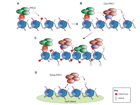
Here, we provide a brief overview of the molecular mechanisms by which PcG complexes regulate gene expression; for further details, we refer the reader to recent reviews (Lanzuolo and Orlando, 2012; Simon and Kingston, 2013). At the molecular level, PRC2 is responsible for di- and tri-methylation of lysine 27 of histone H3 (H3K27me2/me3), which act as repressive epigenetic marks (Fig. 1A) (Cao et al., 2002; Czermin et al., 2002; Kuzmichev et al., 2002; Müller et al., 2002). PRC1, by contrast, mediates the monoubiquitylation of histone H2A, which impairs transcriptional elongation (Stock et al., 2007) and is crucial for gene repression (Endoh et al., 2012) (Fig. 1B). PRC1 also represses genes through mechanisms such as chromatin compaction (Fig. 1C) (Francis et al., 2004; Endoh et al., 2012) and decreasing nucleosomal turnover (Deal et al., 2010). In addition to its function in repression, the H2AK119ub mark is essential for PRC1 displacement from chromatin, thus allowing gene activation upon differentiation stimuli (Richly et al., 2010).
PRC2 components in mammals
The PRC2 core complex of Drosophilais formed by Enhancer of zeste [E(z)], Suppressor of zeste [Su(z)] and Extra sexcombs (Esc) (Table 1). In mammals, Ezh1 and Ezh2, homologs of E(z), are histone methyltransferases responsible for the enzymatic activity of PRC2 (Margueron et al., 2008). The other core PRC2 components, which comprise a homolog of Su(z), Suz12, and a homolog of Esc, Eed, are necessary for complex assembly and for proper enzymatic activity (Cao and Zhang, 2004; Pasini et al., 2004; Ketel et al., 2005). It is still not clear how PRC2 is recruited to DNA in mammals (as discussed below). It has been suggested that the Jumonji/ARID domain-containing protein Jarid2 (Peng et al., 2009; Shen et al., 2009; Li et al., 2010; Pasini et al., 2010) and the members of the Polycomb-like family, the Pcl proteins, are responsible for PRC2 recruitment to target genes in mammals, albeit through different mechanisms (Walker et al., 2010; Ballaré et al., 2012; Brien et al., 2012; Hunkapiller et al., 2012; Musselman et al., 2012). The ARID domain of Jarid2 binds directly to DNA enriched in GC and GA dinucleotides, whereas the Tudor domain of Pcl proteins recognizes methylated H3K36, an histone mark that is associated with transcriptional elongation. This suggests that the Pcl family of proteins facilitates PcG-mediated silencing of previously active genes. Moreover, the fact that Jarid2 and the Pcl proteins are thought not to be present in the same complexes (Ballaré et al., 2012) indicates that, in mammalian cells, distinct PRC2 complexes target different genes.
Development 140, 2525-2534 (2013) doi:10.1242/dev.091553 © 2013. Published by The Company of Biologists Ltd
Polycomb complexes in stem cells and embryonic
development
Luigi Aloia1, Bruno Di Stefano1and Luciano Di Croce1,2,*
1Centre for Genomic Regulation (CRG) and UPF, Dr Aiguader 88, 08003 Barcelona, Spain. 2Institució Catalana de Recerca i Estudis Avançats (ICREA), Pg. Lluis Companys 23, 08010 Barcelona, Spain.
*Author for correspondence ([email protected])
D
E
V
E
LO
P
M
E
N
PRC1 components in mammals
The DrosophilaPRC1 core complex is formed by Polycomb (Pc), Polyhomeotic (Ph), Posterior sex combs (Psc) and Sex combs extra (Sce, also known as Ring) (Morey and Helin, 2010). In mammals, the composition of PRC1 is much more diverse and varies depending on the cellular context (Table 1) (Gao et al., 2012; Luis et al., 2012). All PRC1 complexes contain homologs of the Drosophila Ring protein. Ring1A and Ring1B (which are also known as Rnf1 and Rnf2, respectively) are E3 ubiquitin ligases (de Napoles et al., 2004; Leeb and Wutz, 2007) that decorate lysine 119 of histone H2A with a single ubiquitin group (H2AK119ub) (Wang et al., 2004a). Homologs of DrosophilaPsc, such as Mel18 (Pcgf2) or Bmi1 (Pcgf4), regulate PRC1 enzymatic activity (Brunk et al., 1991; Kanno et al., 1995).
PRC1 complexes can be divided into at least two classes according to the presence or absence of Cbx proteins, which are homologs of DrosophilaPc. Canonical PRC1 complexes contain Cbx proteins that recognize and bind H3K27me3, the mark deposited by PRC2 (Table 1). Therefore, canonical PRC1 complexes and PRC2 can act together to repress gene transcription. Non-canonical PRC1 complexes, which contain Rybp (together with additional proteins, such as L3mbtl2 or Kdm2b) rather than the Cbx proteins (Fig. 1D), have recently been described in mammals (García et al., 1999; Trojer et al., 2011; Farcas et al.,
2012; Gao et al., 2012; Hisada et al., 2012; Qin et al., 2012; Tavares et al., 2012; He et al., 2013; Wu et al., 2013) (Table 1). At the molecular level, Rybp-PRC1 and Cbx-PRC1 have been shown to regulate different target sets (Morey et al., 2013). However, this study also showed that a common subset of genes is co-regulated by both Rybp-PRC1 and Cbx-PRC1 in stem cells, indicating that the intricate interactions between these different complexes are dependent upon the developmental and cellular context.
PcG recruitment: involvement of CpG islands and DNA methylation
In addition to recruitment via Jarid2 and Pcl proteins, PRC2 occupancy has been associated with large unmethylated CpG islands (Ku et al., 2008) through a mechanism that might involve Pcl3 (Phf19) (Hunkapiller et al., 2012). DNA demethylation can be achieved via the action of the Tet proteins (Tan and Shi, 2012) and recent data indicate that Tet1 is necessary for the chromatin binding of PRC2 (Wu et al., 2011). More than 95% of PRC2 targets overlap with Tet1 targets in mouse ESCs. Tet1 depletion impairs PRC2 recruitment to most binding sites, whereas Ezh2 depletion does not affect Tet1 binding, suggesting that Tet1 contributes to PcG recruitment, promoting the demethylation of CpG islands.
UB
UB
UB
UB
UB UB Suz12
Eed
Ezh2
RF
H3K27me3
H2Aub
PRC2
A B
C
D
Cbx-PRC1
Suz12 Eed
Ezh2
RF
Psc Ring1
UB
UB
UB Cbx
Ph
Psc Ring1
Cbx
Ph Psc
Ring1
Cbx Ph
Rybp-PRC1
CpG island
Psc Ring1
Rybp Kdm2b Suz12
Eed
Ezh2
Suz12 Eed
Ezh2 RF
[image:2.612.56.509.54.395.2]Key
Fig. 1. Molecular functions of PRC1 and PRC2. (A) PRC2 decorates lysine 27 of histone H3 with a trimethyl group (H3K27me3). Specific recruiting factors (RF), such as Jarid2 and the Pcl proteins, are responsible for targeting PRC2 to genomic loci. (B,C) Cbx proteins bind to the H3K27me3 mark and recruit canonical PRC1 complexes to chromatin, leading to the deposition of the monoubiquitin mark on lysine 119 of histone H2A (H2Aub) (B) and to chromatin compaction (C). (D) Non-canonical PRC1 complexes are recruited to specific unmethylated CpG islands through the Kdm2b subunit. The deposition of the monoubiquitin moiety on histone H2A is thus independent of PRC2 activity.
D
E
V
E
LO
P
M
E
N
As mentioned above, the chromodomain of Cbx proteins recognizes the H3K27me3 mark deposited by PRC2 (Fischle et al., 2003), thus recruiting canonical PRC1 complexes and leading to co-occupancy by PRC1 and PRC2 at the same chromatin loci (Morey et al., 2012). However, H3K27me3 is not always sufficient to recruit PRC1 (Schoeftner et al., 2006; Tavares et al., 2012), and, in the case of non-canonical PRC1 variants, the absence of Cbx means that an alternative recruitment mechanism must be invoked. Recent work in mouse ESCs indicates a role for the DNA methylation state in the recruitment of PRC1 as well as PRC2. The PRC1 component Kdm2b is able to recruit PRC1 to unmethylated CpG islands independently of PRC2 (Farcas et al., 2012; He et al., 2013; Wu et al., 2013) (Fig. 1D). These data thus suggest that the methylation state of CpG islands modulates the occupancy of different Polycomb complexes. However, further studies are necessary to characterize the molecular link between DNA methylation and PcG occupancy in order to fully understand how the dynamic occupancy of Polycomb complexes is achieved in different cellular contexts.
PcG functions in mammalian embryogenesis In this Review, we focus primarily on the functions of PcG proteins in stem cells. The following section provides a brief summary of
their roles during embryogenesis, elucidated via analyses of knockout (KO) mice for various PcG components. Such studies have revealed key functions for these proteins in embryonic development, with mutant embryos typically displaying gastrulation defects. Specifically, KO embryos for the PRC2 components Suz12, Ezh2 and Eed die during early postimplantation stages (Faust et al., 1995; O’Carroll et al., 2001; Pasini et al., 2004). Unlike the early and broad developmental defects seen upon KO of core PRC2 components, Jarid2deletion has a distinct effect, causing defects in neural tube formation [at 15.5 days postcoitum (dpc)] (Takeuchi et al., 1995). By contrast, Pcl2 (Mtf2) regulates left-right asymmetry in chicken embryos (Wang et al., 2004b) but is dispensable in mouse (Wang et al., 2007). The fact that Jarid2 and Pcl2 KO mice exhibit different phenotypes, each of which is less severe than that of core PRC2 component KO, supports the idea that they are not core components of the complex but rather regulate PRC2 activity in specific contexts.
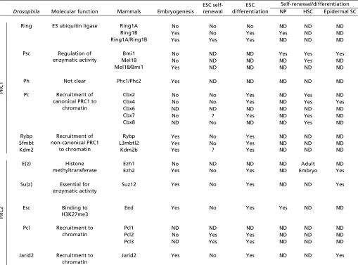
[image:3.612.52.561.70.447.2]Loss of the enzymatic subunit of PRC1, Ring1B, results in embryonic lethality, whereas Ring1A KO mice are viable (de Napoles et al., 2004). Ring1B KO causes gastrulation arrest (Voncken et al., 2003). In addition, a mouse line with a hypomorphic Ring1B allele shows posterior homeotic Table 1. Essential role of selected PRC1 and PRC2 components in embryonic development and stem cells
Drosophila Molecular function Embryogenesis
ESC self-renewal
ESC differentiation
Self-renewal/differentiation
Mammals NP HSC Epidermal SC
PRC1
Ring E3 ubiquitin ligase Ring1A No No No ND ND ND
Ring1B Yes No Yes Yes ND ND
Ring1A/Ring1B Yes Yes Yes ND ND ND
Psc Regulation of
enzymatic activity
Bmi1 No ND ND Yes Yes Yes
Mel18 No ND ND ND Yes ND
Mel18/Bmi1 Yes ND ND ND ND ND
Ph Not clear Phc1/Phc2 Yes ND ND ND ND ND
Pc Recruitment of
canonical PRC1 to chromatin
Cbx2 No No Yes ND Yes ND
Cbx4 No No Yes ND Yes Yes
Cbx6 ND ND ND ND ND ND
Cbx7 No ? Yes ND Yes ND
Cbx8 ND No ND ND Yes ND
Rybp Recruitment of
non-canonical PRC1 to chromatin
Rybp Yes No Yes ND ND ND
Sfmbt L3mbtl2 Yes No Yes ND ND ND
Kdm2 Kdm2b Yes ? Yes ND ND ND
PRC2
E(z) Histone methyltransferase
Ezh1 No ND ND ND Adult ND
Ezh2 Yes No Yes ND Embryo Yes
Su(z) Essential for
enzymatic activity
Suz12 Yes No Yes ND ND Yes
Esc Binding to
H3K27me3
Eed Yes No Yes Yes ND ND
Pcl Recruitment to
chromatin
Pcl1 ND ND ND ND ND ND
Pcl2 No Yes Yes ND ND ND
Pcl3 ND Yes Yes ND ND ND
Jarid2 Recruitment to
chromatin
Jarid2 Yes No Yes ND ND Yes
Entries indicate whether each PRC1/2 component is essential for the self-renewal or differentiation of the indicated cell types. ND, not determined; ?, controversial. ESC, embryonic stem cell; NP, neural progenitor; HSC, hematopoietic stem cell; Epidermal SC, epidermal stem cell.
D
E
V
E
LO
P
M
E
N
transformation of the axial skeleton (Suzuki et al., 2002). Non-canonical PRC1 components are essential for embryonic development: Rybp KO embryos exhibit lethality at the early postimplantation stage (Pirity et al., 2005), L3mbtl2 is essential for gastrulation (Qin et al., 2012) and Kdm2b for proper embryonic neural development (Fukuda et al., 2011). By contrast, loss of the Cbx proteins, which are responsible for recruiting the canonical PRC1 to chromatin, does not affect embryonic development. Indeed, mutants for Cbx2 or Cbx4displayed postnatal lethality (Coré et al., 1997; Liu et al., 2013). Interestingly, Cbx4 has been reported to specifically regulate the proliferation of thymic epithelial cells and the maintenance of thymic epithelium, uncovering a novel PcG function in the immune system (Liu et al., 2013). Although adult Cbx7 KO mice are viable, they show increased susceptibly to lung and liver neoplasia (Forzati et al., 2012), suggesting a role for Cbx7 as a tumor suppressor. This contrasts with previous reports indicating that Cbx7is an oncogene (Bernard et al., 2005; Scott et al., 2007); the role of this protein is therefore still a matter of debate. Genetic deletion of Mel18or Bmi1caused defects in anterior-posterior specification of the axial skeleton (van der Lugt et al., 1994; Alkema et al., 1995; Akasaka et al., 1996). Mel18/Bmi1 double-KO mice died at ~9.5 dpc (Akasaka et al., 1996) and displayed more severe developmental defects than either single KO, suggesting that Mel18 and Bmi1 have partially redundant functions as well as some independent roles, as manifested in the phenotypes of the single KOs. In the future, genetic deletion of the other putative components of PRC1, such as the mammalian homologs of DrosophilaPh and Psc, will be important to elucidate their function in development.
Roles of PcG complexes in ESCs
Accumulating data suggest that PcG proteins are essential for ESC differentiation, whereas their role in self-renewal remains controversial. We first present evidence for the role of PcG proteins in these two processes, and then discuss how the composition of PRC1 could confer specificity to complex activity.
Self-renewal
In mouse ESCs, PRC1 and PRC2 repress genes involved in differentiation (reviewed by Surface et al., 2010). In the last few years, ESC lines from several KO and knockdown mice have been generated to investigate PcG function. Genetic depletion of Eed or Ring1B, which almost completely abolished PRC1 or PRC2 activity, respectively, led to an increase in the expression of differentiation markers under basal conditions (Leeb and Wutz, 2007; Leeb et al., 2010). However, neither Eed nor Ring1B loss affects the expression of pluripotency genes or the self-renewal ability of the cells (Chamberlain et al., 2008; Endoh et al., 2008). Notably, depletion of both Ring1A and Ring1B impaired ESC self-renewal, indicating that Ring1 proteins (and hence PRC1) are essential for ESC identity (Endoh et al., 2008).
Additional PRC2 components, such as Pcl2 and Pcl3, also contribute to the ESC self-renewal network and are required for the expression of key pluripotency markers in proliferating conditions (Walker et al., 2010; Ballaré et al., 2012; Hunkapiller et al., 2012). Since such effects on the expression of pluripotency genes are not seen upon depletion of core PRC2 components, this suggests that Pcl2 and Pcl3 possess PRC2-independent functions – the molecular basis of which is not yet clear – in addition to their role in recruiting the complex to chromatin.
Interestingly, it has been reported that a set of Polycomb targets involved in metabolic processes is also expressed in mouse ESCs,
despite Polycomb being associated with repression (Brookes et al., 2012; Morey et al., 2013). Indeed, these genes exhibit elongating RNA polymerase II within the gene body. However, consecutive chromatin immunoprecipitation (re-ChIP) experiments indicate that PRC1 and the elongating RNA polymerase II are present on different alleles (Brookes et al., 2012). This suggests that the independent regulation of the two alleles contributes to the modulation of gene expression in mouse ESCs.
ESC differentiation
Although several PcG components have been characterized as positive regulators of the ESC state, they have also been clearly identified as necessary for proper ESC differentiation. Specifically, Ezh2 is required to generate mesendodermal lineages (Shen et al., 2008) and Suz12 KO ESCs fail to generate proper endodermal lineages (Pasini et al., 2004). Surprisingly, Eed KO ESCs are able to differentiate into the three germ layers and to contribute to chimera formation (Chamberlain et al., 2008), although some defects in their ability to form teratomas have been documented (Leeb et al., 2010). Other members of PRC2, such as Jarid2 and the Pcl proteins (Pcl2 and Pcl3), have also been reported to be essential for proper differentiation (Peng et al., 2009; Pasini et al., 2010; Walker et al., 2010; Ballaré et al., 2012).
The loss of the PRC1 E3 ubiquitin ligase Ring1B impaired the proper expression of differentiation markers when ESCs were grown as embryoid bodies (Leeb and Wutz, 2007). Other components of PRC1, such as the Cbx proteins (see below), Rybp and L3mbtl2, are also required for ESC differentiation (Hisada et al., 2012; Morey et al., 2012; Qin et al., 2012; Tavares et al., 2012). Interestingly, ESCs lacking either Ring1B or Eed were still able to form teratomas, but these were found to be smaller, with an increase in the ectodermal or endodermal fraction, respectively (Leeb et al., 2010). By contrast, ESCs with a double KO for Eed and Ring1B, which almost completely abolished the activity of both PRC1 and PRC2, were not able to form teratomas, indicating that depletion of both complexes blocks differentiation, and further confirming that they have at least partially independent functions.
Variation in PRC1 composition in ESC self-renewal and differentiation
How can PRC1 promote both self-renewal and differentiation? Recent evidence suggests that a switch in the Cbx protein composition of the canonical PRC1 occurs when self-renewing ESCs begin to differentiate. Cbx7 is the main component of canonical PRC1 (which is present in self-renewing ESCs), whereas Cbx2 and Cxb4 are found in PRC1 variants in differentiating ESCs (Morey et al., 2012; O’Loghlen et al., 2012). The role of Cbx7 in self-renewal is controversial: O’Loghlen and colleagues reported that depletion of Cbx7 impairs ESC self-renewal, whereas Morey and colleagues found no role for Cxb7 in this process. Both studies also addressed the role of Cbx in differentiation, finding that Cbx7-depleted ESCs gave rise to teratomas with an increased ectodermal fraction, in line with the negative regulation mediated by Cbx7-PRC1 of several ectodermal genes in ESCs.
Cbx2 and Cbx4 were found to be upregulated upon differentiation, concomitant with the downregulation of Cbx7 (Morey et al., 2012). Thus, Cbx2 and Cbx4 appear to replace Cbx7 in differentiating cells, thereby targeting PRC1 to a different set of genes, such as pluripotency regulators and specific mesodermal/endodermal markers. At the molecular level, Cbx2 and Cbx4 have non-overlapping functions, repressing distinct subsets of genes in differentiated ESCs (Morey et al., 2012).
D
E
V
E
LO
P
M
E
N
Indeed, Cbx2 and Cbx4 are required for proper ESC differentiation, with teratomas derived from Cbx2- and Cbx4-depleted ESCs displaying an aberrant increase in the number of endodermal and mesodermal cells compared with control cells, in agreement with their genome binding profiles (Morey et al., 2012).
Notably, in ESCs, Cbx7 and Ring1B occupy the Cbx2and Cbx4 promoters, indicating that Cbx7-PRC1 is responsible for the repression of these genes in self-renewing cells, whereas Cbx2/4-PRC1 appears to repress Cbx7 in differentiated ESCs (Morey et al., 2012). Therefore, an autoregulatory loop is likely to control the Cbx composition of PRC1 and to drive the transition of ESCs from self-renewal to a differentiated state.
PcG complexes regulate bivalent genes in mouse ESCs
Bivalent domains, which are regions decorated with both active (H3K4me3) and repressive (H3K27me3) marks, have been identified in mouse ESCs (Azuara et al., 2006; Bernstein et al., 2006). As mentioned above, the repressive H3K27me3 mark is deposited by PRC2 and can in turn recruit PRC1. In parallel, the active H3K4me3 mark is deposited by the trithorax/MLL complex (Schuettengruber et al., 2011). Such regions are not unique to ESCs but have also been identified in other multipotent stem cells (as discussed below) (Mohn et al., 2008; Cui et al., 2009).
In addition to the presence of an active H3K4me3 mark, bivalent genes are characterized by the presence of poised RNA polymerase II (Fig. 2A) (Brookes et al., 2012). Despite this, they remain transcriptionally silent. Current models propose that bivalency allows a rapid transition from repression to activation of developmental genes upon differentiation stimuli (Azuara et al., 2006; Bernstein et al., 2006). Thus, a fine-tuned regulation of bivalent domains is essential for proper development, as they regulate the precise course of gene expression during pluripotency and differentiation (Jia et al., 2012).
Upon ESC differentiation, bivalent domains need to be resolved into activated or repressed genomic loci according to the
differentiation process (Fig. 2B) (Mikkelsen et al., 2007; Alder et al., 2010). Mohn and collaborators have reported that new bivalent domains are acquired in neural progenitors derived from ESCs, indicating that epigenetic regulation mediated by PcG complexes is highly dynamic and cell type specific (Mohn et al., 2008). Similarly, analysis of the differentiation of ESCs into cardiomyocytes indicates a dynamic reorganization of bivalent domains (Paige et al., 2012). Recently, Marks and colleagues have reported that the number of bivalent domains decreases from ~3000 in ESCs grown in serum-containing medium to fewer than 1000 in ESCs grown in 2i-containing medium (Marks et al., 2012). This suggests that the establishment of at least some bivalent domains is dependent on cell culture conditions, thus questioning their relevance under physiological conditions. Nevertheless, evidence demonstrating that bivalent domains regulate developmental genes has recently been reported in zebrafish embryos, indicating that the observations from ESCs are likely to be relevant in vivo (Vastenhouw et al., 2010). In summary, the establishment of bivalent domains appears to be a conserved mechanism to regulate the key stem cell features of self-renewal and differentiation. Mechanistically, developmental PcG targets are kept silent, yet can be rapidly activated when the cell receives appropriate cues. Thus, the key function of the bivalent domains in pluripotent and multipotent stem cells could explain, at least in part, the dual role exerted by PcG complexes in self-renewal and differentiation (as discussed further below).
PcG proteins promote somatic cell reprogramming Somatic cells can be reverted into a pluripotent-like state using several techniques, such as somatic cell nuclear transfer (SCNT) and cell fusion (Jaenisch and Young, 2008). Pereira and colleagues provided the first evidence that PcG genes play a crucial role in reprogramming mediated by cell fusion (Pereira et al., 2010). They showed that mouse ESCs depleted of individual members of PRC1 or PRC2 (e.g. Eed, Suz12, Ezh12, Ring1A and Ring1B) fail to properly reprogram human B lymphocytes.
UB UB
UB UB
A B
UB
Suz12 RF
H3K27me3 H3K4me3 UB H2Aub
Polycomb
R
R
RF
RF RF RF
RFFF
Polycomb UUUUUBBBBBBBB
S S
S
Su Su Suz
Suzuzuz1z1z1212222
Polycomb
MLL
Stem cell Committed cell
Differentiation
Bivalent promoter
Silent promoter
Active promoter Poised
Pooos
Poised Poised Po Pooisedoisedoise
Pois Poi
Poi RNRNRNRRNNNNNNNNNNANNA PNNA PolAA PA PAA PA PA PA PAPPolPolPPPoolololllIIIIIII
MLL
Elongatinggatatingatingatinnnggggggggg
Elonga
Elonga
Elongagagaata RNRRRRRNRRRNA PA PA PPPPPol II
[image:5.612.52.535.482.682.2]Key
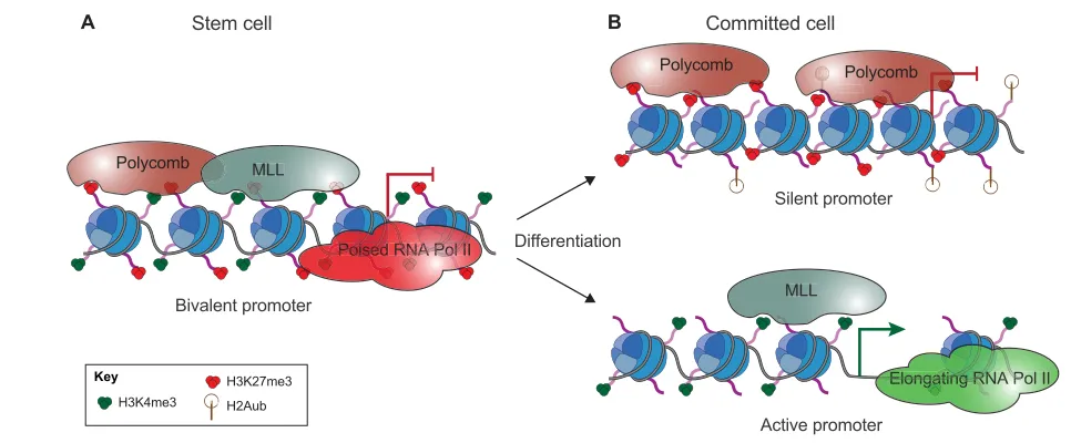
Fig. 2. Role of bivalent promoters in cell fate transition. (A) Bivalent domains are characterized by the presence of both active (H3K4me3) and repressive (H3K27me3) marks deposited by the MLL and Polycomb complexes, respectively. In stem cells, bivalent domains are found on multiple silent developmental genes. (B) The presence of H3K4me3 and poised RNA polymerase II allows rapid transcriptional activation upon differentiation stimuli: bivalent domains are resolved into actively transcribed genes (characterized by the presence of H3K4me3 and loss of H3K27me3) and silent genes
(characterized by the presence of H3K27me3 and loss of H3K4me3) according to gene function and cell type.
D
E
V
E
LO
P
M
E
N
The discovery that somatic cells can be reprogrammed into induced pluripotent stem cells (iPSCs) using a combination of four transcription factors (the so-called Yamanaka factors) opened new avenues for the use of reprogrammed cells in in vitro disease modeling and cell therapy (Takahashi and Yamanaka, 2006; Robinton and Daley, 2012). Functionally, iPSCs recapitulate all the features of pluripotent ESCs, including the ability to differentiate into the desired cell type under appropriate culture conditions. However, there are technical limitations in the generation of iPSCs, primarily in terms of the low efficiency (~0.1%) and long time required to obtain reprogrammed clones (Stadtfeld et al., 2008). Several reports suggest that these limitations are likely to be related to the difficulties in overcoming epigenetic barriers. For instance, depletion of a PRC2 component, such as Jarid2, Pcl2 or the novel component esPRC2p48, impaired the reprogramming of fibroblasts into iPSCs (Zhang et al., 2011). By contrast, overexpression of PRC2 components facilitated the reprogramming process. Similarly, Onder and colleagues reported that the loss of the PRC1 components BMI1 and RING1B, and of the core PRC2 components EZH2, EED and SUZ12, significantly decreased human iPSC generation (Onder et al., 2012). Moreover, Bugamin and colleagues recently proposed that Ezh2 is not only able to increase iPSC efficiency but can also be used as part of a new reprogramming cocktail of transcription factors, which includes Lin28, Sall4, Nanog, Klf4 and c-Myc (Buganim et al., 2012). This new combination of factors is able to generate iPSC-like cells in culture, although the cells show incomplete reactivation of the endogenous pluripotency program and are therefore not fully reprogrammed or stable.
All of these observations clearly indicate that PcG proteins modulate the reprogramming of differentiated cells into pluripotent cells. The mechanisms underlying this process are still under investigation, and, in particular, how PcG proteins affect reprogramming has not been addressed in detail.
PcG complex activities in tissue stem cells
The roles of PcG complexes have been most extensively analyzed in ESCs, but key functions have also been identified in various tissue stem cells. Below, we summarize the known roles of PcG proteins in the nervous and hematopoietic systems and in skin.
PcG proteins regulate self-renewal and the neurogenic-astrogenic switch in neural progenitors
Neural progenitors are self-renewing, multipotent cells that are able to give rise to neurons and glial cells. In the developing neocortex, neurons and astrocytes are derived from common neural progenitors. An initial neurogenic phase is followed by an astrogenic phase (Hirabayashi and Gotoh, 2005). This switch from a neurogenic to an astrogenic fate is crucial for proper cortical development. In vitro, neural progenitors that closely resemble those found in vivocan be efficiently derived from ESCs (Conti et al., 2005; Bibel et al., 2007). The presence of bivalent chromatin domains in neural progenitors derived from ESCs suggests that PcG proteins also have a function in neural progenitors (Mohn et al., 2008), and several functional analyses have established PcG complexes as crucial regulators of neural progenitor features such as proliferation, self-renewal and differentiation in vitroas well as in vivo.
In culture, neural progenitors depleted of the PRC1 enzymatic subunit Ring1B exhibited proliferation and self-renewal defects as well as premature neuronal (but not glial) differentiation in basal conditions (Román-Trufero et al., 2009). Interestingly, the PRC1
component Bmi1 has been reported to control the proliferation of neural progenitors by repressing p21 (Cdkn1a) (Fasano et al., 2007; Román-Trufero et al., 2009) and the Ink4/Arf cell cycle inhibitory proteins p16 (Cdkn2a) and p19 (Cdkn2d) (Molofsky et al., 2003; Bruggeman et al., 2005).
The essential role of Polycomb genes in the regulation of neural progenitors in vivo was demonstrated by Hirabayashi and colleagues (Hirabayashi et al., 2009). In the developing cortex, Polycomb complexes negatively regulate Ngn1 and Ngn2 expression during the astrogenic phase. Since Ngn1 and Ngn2 sustain the neurogenic phase by sequestering pro-astrogenic factors (Sun et al., 2001), the silencing of these genes by Polycomb-mediated repression allows for the proper onset of the astrogenic phase. Thus, cultured neural progenitors depleted for Eed or Ring1B maintained aberrant Ngn1 and Ngn2 at late developmental stages and were not able to differentiate into astrocytes, and mice in which Ring1B was conditionally depleted in the central nervous system exhibited improper termination of neurogenesis and an impaired onset of astrogenesis (Hirabayashi et al., 2009).
PcG proteins in hematopoietic stem cell maintenance
The hematopoietic stem cell (HSC) lineage is one of the best-studied models of stem cell self-renewal and differentiation. Long-term reconstituting HSCs (LT-HSCs) reside as rare cells in the bone marrow and sit atop a hierarchy of progenitors that become progressively restricted to several or single lineages. These progenitors are able to generate short-term repopulating HSCs (st-HSCs), which, in turn, yield blood precursors devoted to unilineage differentiation and the production of mature blood cells, including red blood cells, megakaryocytes, myeloid cells (monocytes/macrophages and neutrophils) and lymphocytes (Kondo et al., 2003; Wang and Wagers, 2011).
Several reports have elucidated the role of PcG proteins in HSC maintenance. As mentioned above for neural progenitors, the PRC1 component Bmi1 has been reported to inhibit the Ink4/Arflocus, which encodes the cell cycle inhibitors p16 and p19, and this also applies in HSCs (Park et al., 2003). Moreover, Bmi1 is also implicated in the repression of developmental genes such as Ebf1 and Pax5. Depletion of Bmi1 causes aberrant expression of Ebf1 and Pax5, which results in premature lymphoid lineage specification (Oguro et al., 2010). Interestingly, a switch of the Cbx composition in PRC1 regulates the transition from self-renewal to the differentiated state of mouse HSCs (Klauke et al., 2013), in line with the observations made in ESCs (Morey et al., 2012). The data reported suggest that Cbx7 is required for self-renewal of mouse HSCs, whereas Cbx2/4/8 are essential for their differentiation. In a separate study (van den Boom et al., 2013), the absence of CBX2 was found to strongly impair human HSC maintenance, with a decreasing level of proliferation and an increasing level of apoptosis. At the molecular level, CBX2 represses the expression of the pro-senescence factor P21 in this context (van den Boom et al., 2013). This apparent discrepancy in its activity seems to be due to species-specific functions of Cbx2. Indeed, depletion of Cbx2 in mouse does not affect HSC self-renewal (van den Boom et al., 2013).
Tight regulation of the expression of PRC2 components is also crucial for proper HSC identity. Several studies have highlighted the role of Ezh1 and Ezh2 in embryonic and adult HSCs. Loss of Ezh2 severely impaired fetal HSC self-renewal without affecting the function of adult stem cells present in the bone marrow, except that lymphopoiesis was somewhat impaired (Su et al., 2003; Su et al., 2005; Kamminga et al., 2006; Mochizuki-Kashio et al., 2011).
D
E
V
E
LO
P
M
E
N
By contrast, Ezh1 deficiency severely reduced the adult HSC fraction, impairing HSC self-renewal and quiescence (Mochizuki-Kashio et al., 2011). Hidalgo and colleagues have recently reported that Ezh1 is able to keep adult HSCs in a slow-cycling state by repressing proliferation, as well as protecting adult HSCs from senescence and premature differentiation (Hidalgo et al., 2012). At the molecular level, Ezh1 represses master senescence regulators such as the Ink4/Arflocus and Bmp2. Thus, Ezh2 is essential in fetal HSCs, whereas Ezh1 is required in the adult. This suggests that a functional switch between Ezh2 and Ezh1 regulates the specificity of PRC2 in the embryo and in the adult.
PcG promotes self-renewal in epidermal stem cells
The skin epidermis is a stratified epithelium that acts as a barrier to protect the organism against external stresses and microorganisms (Beck and Blanpain, 2012). Different stem cells contained in various epidermal niches (such as the interfollicular epidermis, hair follicles, and sebaceous glands) regulate the maintenance and repair of the epidermis. Here we present a brief overview of the role of PcG proteins in skin (for details, see Frye and Benitah, 2012). Loss of Ezh2 impairs proliferation and induces premature differentiation of the basal layer of the epidermis (Ezhkova et al., 2009; Ezhkova et al., 2011). At the molecular level, Ezh2 acts by repressing the Ink4/Arf and epidermal differentiation complex(EDC) loci, the latter of which encodes differentiation genes required for skin maturation. In addition, loss of the PRC2 component Jarid2 similarly results in increased differentiation and decreased proliferation (Mejetta et al., 2011). However, the effects of Ezh2 KO in the epidermis are predominant during embryogenesis and less severe in adults, whereas the Jarid2 epidermal KO showed postnatal defects. Supporting a role for PRC2 in the epidermis, Lien and colleagues have recently identified a low number of bivalent domains in hair follicle stem cells, indicating that PcG proteins are involved in the cell fate transition of these cells (Lien et al., 2011).
Like PRC2, PRC1 also plays an important role in the epidermis. Indeed, a recent study demonstrated that the PRC1 component Cbx4 is required for the maintenance of basal epidermal cells, preventing senescence and premature differentiation, through direct regulation of p16(Luis et al., 2011). Interestingly, Cbx4 is also likely to prevent differentiation in a PRC1-independent manner: the inhibition of differentiation mediated by Cbx4 requires its E3-SUMO ligase activity but not the recognition of H3K27me3 mediated by its chromodomain. Together, these data suggest an important role for PcG complexes in promoting the self-renewal and maintenance of epidermal stem cells by impairing their premature differentiation.
Conclusions
The use of mouse ESCs has provided a great opportunity for investigating developmental PcG functions in vitro. ESC differentiation largely recapitulates differentiation during embryonic development, with each step corresponding to a specific developmental stage. Interestingly, studies in differentiating ESCs suggest that PcG complexes set up lineage commitment from pluripotent to differentiated cells (Surface et al., 2010). This is due to the dynamic activities of PcG complexes, which regulate specific sets of genes at different developmental stages (Mohn et al., 2008). PcG complexes are involved in cell fate transitions, not only in pluripotent ESCs but also in several embryonic and adult multipotent stem cell types (Richly et al., 2011). Indeed, a key PcG function in pluripotent and multipotent stem cells is to establish
bivalent domains that allow rapid activation of the gene upon differentiation stimuli. Although the relevance of bivalent domains is still debated, several studies indicate that bivalency is an evolutionarily conserved mechanism of regulating cell fate transitions (Alder et al., 2010; Vastenhouw et al., 2010). Apart from their key role in cell fate transitions, PcG proteins are essential for the maintenance of several stem cell types. It is clear that the loss of the PRC1 components Ring1A/B and of several PRC2 components impairs ESC self-renewal. In various multipotent stem cells, PcG proteins maintain self-renewal by repressing differentiation and senescence players, such as those encoded at the Ink4/Arflocus.
Notably, at the molecular level, the roles of the PcG proteins are unique for each stem cell type and also vary within the same cell type depending on the developmental stage and the environmental stimuli. One possible explanation is that the composition of PcG proteins within the PcG complexes determines the specificity of their function. Indeed, increasing evidence from recent studies indicates that the composition of PcG complexes, particularly PRC1, is variable and dynamic in different cell types and at different developmental stages. In addition, exchanging components can have profound effects on complex function. Thus, in ESCs and HSCs, exchanging the Cbx protein in PRC1 is involved in the switch from a self-renewing to a differentiating state, whereas in HSCs the Ezh component in PRC2 differs between embryonic and adult stages, concomitant with differential activities of the complex. Whether these principles apply to other complex components or in other cell types has yet to be seen, but these examples demonstrate that a more detailed characterization of PcG complex composition will contribute greatly to our knowledge of their specific functions in different developmental and cell type contexts.
Importantly, in recent years stem cells have emerged as a possible tool for tissue regeneration upon lesions induced by trauma or disease. Further knowledge of the epigenetic networks underlying stem cell biology will increase the therapeutic use of stem cells in regeneration. Moreover, the discovery of iPSC reprogramming has opened new opportunities for regenerative medicine (Robinton and Daley, 2012), although the process remains slow and inefficient. Given that PcG complexes are essential for proper iPSC generation, more detailed information about the role of PcG genes in reprogramming could increase the efficiency and the quality of the iPSC generation process, thereby improving the therapeutic potential of iPSCs.
Finally, increasing knowledge about stem cell biology, and the roles of epigenetic modifiers therein, will aid the fight against cancer. Data indicate that a stem cell population within the cancer (cancer stem cells) is responsible for tumor initiation and for resistance to therapy. Several PcG genes are dysregulated in cancer, implying that PcG complexes are likely to play a crucial role in cancer generation and in the maintenance of cancer stem cells (Piunti and Pasini, 2011). Thus, understanding Polycomb gene functions will be crucial for elucidating the molecular mechanisms that regulate embryonic development and stem cell function in tissue homeostasis, regeneration and cancer.
Acknowledgements
We are indebted to V. A. Raker for help in preparing the manuscript and to L. Morey, P. Vizan and members of the L.D.C. laboratory for discussions.
Funding
B.D.S. was supported by a La Caixa International PhD fellowship. This work
was supported by grants from the Spanish Ministerio de Educación y Ciencia;
D
E
V
E
LO
P
M
E
N
Agència de Gestió d’Ajuts Universitaris i de Recerca (AGAUR); the Association for International Cancer Research (AICR); and the European Commissions 7th Framework Program 4DCellFate (to L.D.C.).
Competing interests statement
The authors declare no competing financial interests.
References
Akasaka, T., Kanno, M., Balling, R., Mieza, M. A., Taniguchi, M. and Koseki, H.(1996). A role for mel-18, a Polycomb group-related vertebrate gene, during the anteroposterior specification of the axial skeleton. Development122, 1513-1522.
Alder, O., Lavial, F., Helness, A., Brookes, E., Pinho, S., Chandrashekran, A., Arnaud, P., Pombo, A., O’Neill, L. and Azuara, V.(2010). Ring1B and Suv39h1 delineate distinct chromatin states at bivalent genes during early mouse lineage commitment. Development137, 2483-2492.
Alkema, M. J., van der Lugt, N. M., Bobeldijk, R. C., Berns, A. and van Lohuizen, M.(1995). Transformation of axial skeleton due to overexpression of bmi-1 in transgenic mice. Nature374, 724-727.
Azuara, V., Perry, P., Sauer, S., Spivakov, M., Jørgensen, H. F., John, R. M., Gouti, M., Casanova, M., Warnes, G., Merkenschlager, M. et al.(2006). Chromatin signatures of pluripotent cell lines. Nat. Cell Biol.8, 532-538.
Ballaré, C., Lange, M., Lapinaite, A., Martin, G. M., Morey, L., Pascual, G., Liefke, R., Simon, B., Shi, Y., Gozani, O. et al.(2012). Phf19 links methylated Lys36 of histone H3 to regulation of Polycomb activity. Nat. Struct. Mol. Biol.19, 1257-1265.
Beck, B. and Blanpain, C.(2012). Mechanisms regulating epidermal stem cells. EMBO J.31, 2067-2075.
Bernard, D., Martinez-Leal, J. F., Rizzo, S., Martinez, D., Hudson, D., Visakorpi, T., Peters, G., Carnero, A., Beach, D. and Gil, J.(2005). CBX7 controls the growth of normal and tumor-derived prostate cells by repressing the Ink4a/Arf locus. Oncogene24, 5543-5551.
Bernstein, B. E., Mikkelsen, T. S., Xie, X., Kamal, M., Huebert, D. J., Cuff, J., Fry, B., Meissner, A., Wernig, M., Plath, K. et al.(2006). A bivalent chromatin structure marks key developmental genes in embryonic stem cells. Cell125, 315-326.
Bibel, M., Richter, J., Lacroix, E. and Barde, Y. A.(2007). Generation of a defined and uniform population of CNS progenitors and neurons from mouse embryonic stem cells. Nat. Protoc.2, 1034-1043.
Boiani, M. and Schöler, H. R.(2005). Regulatory networks in embryo-derived pluripotent stem cells. Nat. Rev. Mol. Cell Biol.6, 872-884.
Brien, G. L., Gambero, G., O’Connell, D. J., Jerman, E., Turner, S. A., Egan, C. M., Dunne, E. J., Jurgens, M. C., Wynne, K., Piao, L. et al.(2012). Polycomb PHF19 binds H3K36me3 and recruits PRC2 and demethylase NO66 to embryonic stem cell genes during differentiation. Nat. Struct. Mol. Biol.19, 1273-1281.
Brookes, E., de Santiago, I., Hebenstreit, D., Morris, K. J., Carroll, T., Xie, S. Q., Stock, J. K., Heidemann, M., Eick, D., Nozaki, N. et al.(2012). Polycomb associates genome-wide with a specific RNA polymerase II variant, and regulates metabolic genes in ESCs. Cell Stem Cell10, 157-170.
Bruggeman, S. W., Valk-Lingbeek, M. E., van der Stoop, P. P., Jacobs, J. J., Kieboom, K., Tanger, E., Hulsman, D., Leung, C., Arsenijevic, Y., Marino, S. et al.(2005). Ink4a and Arf differentially affect cell proliferation and neural stem cell self-renewal in Bmi1-deficient mice. Genes Dev.19, 1438-1443.
Brunk, B. P., Martin, E. C. and Adler, P. N.(1991). Drosophila genes Posterior Sex Combs and Suppressor two of zeste encode proteins with homology to the murine bmi-1 oncogene. Nature353, 351-353.
Buganim, Y., Faddah, D. A., Cheng, A. W., Itskovich, E., Markoulaki, S., Ganz, K., Klemm, S. L., van Oudenaarden, A. and Jaenisch, R.(2012). Single-cell expression analyses during cellular reprogramming reveal an early stochastic and a late hierarchic phase. Cell150, 1209-1222.
Cao, R. and Zhang, Y.(2004). SUZ12 is required for both the histone methyltransferase activity and the silencing function of the EED-EZH2 complex. Mol. Cell15, 57-67.
Cao, R., Wang, L., Wang, H., Xia, L., Erdjument-Bromage, H., Tempst, P., Jones, R. S. and Zhang, Y.(2002). Role of histone H3 lysine 27 methylation in Polycomb-group silencing. Science298, 1039-1043.
Chamberlain, S. J., Yee, D. and Magnuson, T.(2008). Polycomb repressive complex 2 is dispensable for maintenance of embryonic stem cell pluripotency. Stem Cells26, 1496-1505.
Conti, L., Pollard, S. M., Gorba, T., Reitano, E., Toselli, M., Biella, G., Sun, Y., Sanzone, S., Ying, Q. L., Cattaneo, E. et al.(2005). Niche-independent symmetrical self-renewal of a mammalian tissue stem cell. PLoS Biol.3, e283.
Coré, N., Bel, S., Gaunt, S. J., Aurrand-Lions, M., Pearce, J., Fisher, A. and Djabali, M.(1997). Altered cellular proliferation and mesoderm patterning in Polycomb-M33-deficient mice. Development124, 721-729.
Cui, K., Zang, C., Roh, T. Y., Schones, D. E., Childs, R. W., Peng, W. and Zhao, K.(2009). Chromatin signatures in multipotent human hematopoietic stem
cells indicate the fate of bivalent genes during differentiation. Cell Stem Cell4, 80-93.
Czermin, B., Melfi, R., McCabe, D., Seitz, V., Imhof, A. and Pirrotta, V.(2002). Drosophila enhancer of Zeste/ESC complexes have a histone H3
methyltransferase activity that marks chromosomal Polycomb sites. Cell111, 185-196.
de Napoles, M., Mermoud, J. E., Wakao, R., Tang, Y. A., Endoh, M., Appanah, R., Nesterova, T. B., Silva, J., Otte, A. P., Vidal, M. et al.(2004). Polycomb group proteins Ring1A/B link ubiquitylation of histone H2A to heritable gene silencing and X inactivation. Dev. Cell7, 663-676.
Deal, R. B., Henikoff, J. G. and Henikoff, S.(2010). Genome-wide kinetics of nucleosome turnover determined by metabolic labeling of histones. Science
328, 1161-1164.
Endoh, M., Endo, T. A., Endoh, T., Fujimura, Y., Ohara, O., Toyoda, T., Otte, A. P., Okano, M., Brockdorff, N., Vidal, M. et al.(2008). Polycomb group proteins Ring1A/B are functionally linked to the core transcriptional regulatory circuitry to maintain ES cell identity. Development135, 1513-1524.
Endoh, M., Endo, T. A., Endoh, T., Isono, K., Sharif, J., Ohara, O., Toyoda, T., Ito, T., Eskeland, R., Bickmore, W. A. et al.(2012). Histone H2A mono-ubiquitination is a crucial step to mediate PRC1-dependent repression of developmental genes to maintain ES cell identity. PLoS Genet.8, e1002774.
Ezhkova, E., Pasolli, H. A., Parker, J. S., Stokes, N., Su, I. H., Hannon, G., Tarakhovsky, A. and Fuchs, E.(2009). Ezh2 orchestrates gene expression for the stepwise differentiation of tissue-specific stem cells. Cell136, 1122-1135.
Ezhkova, E., Lien, W. H., Stokes, N., Pasolli, H. A., Silva, J. M. and Fuchs, E.
(2011). EZH1 and EZH2 cogovern histone H3K27 trimethylation and are essential for hair follicle homeostasis and wound repair. Genes Dev.25, 485-498.
Farcas, A. M., Blackledge, N. P., Sudbery, I., Long, H. K., McGouran, J. F., Rose, N. R., Lee, S., Sims, D., Cerase, A., Sheahan, T. W. et al.(2012). KDM2B links the Polycomb Repressive Complex 1 (PRC1) to recognition of CpG islands. eLife1, e00205.
Fasano, C. A., Dimos, J. T., Ivanova, N. B., Lowry, N., Lemischka, I. R. and Temple, S.(2007). shRNA knockdown of Bmi-1 reveals a critical role for p21-Rb pathway in NSC self-renewal during development. Cell Stem Cell1, 87-99.
Faust, C., Schumacher, A., Holdener, B. and Magnuson, T.(1995). The eed mutation disrupts anterior mesoderm production in mice. Development121, 273-285.
Fischle, W., Wang, Y., Jacobs, S. A., Kim, Y., Allis, C. D. and Khorasanizadeh, S.(2003). Molecular basis for the discrimination of repressive methyl-lysine marks in histone H3 by Polycomb and HP1 chromodomains. Genes Dev.17, 1870-1881.
Forzati, F., Federico, A., Pallante, P., Abbate, A., Esposito, F., Malapelle, U., Sepe, R., Palma, G., Troncone, G., Scarfò, M. et al.(2012). CBX7 is a tumor suppressor in mice and humans. J. Clin. Invest.122, 612-623.
Francis, N. J., Kingston, R. E. and Woodcock, C. L.(2004). Chromatin compaction by a polycomb group protein complex. Science306, 1574-1577.
Frye, M. and Benitah, S. A.(2012). Chromatin regulators in mammalian epidermis. Semin. Cell Dev. Biol.23, 897-905.
Fukuda, T., Tokunaga, A., Sakamoto, R. and Yoshida, N.(2011).
Fbxl10/Kdm2b deficiency accelerates neural progenitor cell death and leads to exencephaly. Mol. Cell. Neurosci.46, 614-624.
Gao, Z., Zhang, J., Bonasio, R., Strino, F., Sawai, A., Parisi, F., Kluger, Y. and Reinberg, D.(2012). PCGF homologs, CBX proteins, and RYBP define functionally distinct PRC1 family complexes. Mol. Cell45, 344-356.
García, E., Marcos-Gutiérrez, C., del Mar Lorente, M., Moreno, J. C. and Vidal, M.(1999). RYBP, a new repressor protein that interacts with components of the mammalian Polycomb complex, and with the transcription factor YY1. EMBO J.18, 3404-3418.
He, J., Shen, L., Wan, M., Taranova, O., Wu, H. and Zhang, Y.(2013). Kdm2b maintains murine embryonic stem cell status by recruiting PRC1 complex to CpG islands of developmental genes. Nat. Cell Biol.15, 373-384.
Hidalgo, I., Herrera-Merchan, A., Ligos, J. M., Carramolino, L., Nuñez, J., Martinez, F., Dominguez, O., Torres, M. and Gonzalez, S.(2012). Ezh1 is required for hematopoietic stem cell maintenance and prevents senescence-like cell cycle arrest. Cell Stem Cell11, 649-662.
Hirabayashi, Y. and Gotoh, Y.(2005). Stage-dependent fate determination of neural precursor cells in mouse forebrain. Neurosci. Res.51, 331-336.
Hirabayashi, Y., Suzki, N., Tsuboi, M., Endo, T. A., Toyoda, T., Shinga, J., Koseki, H., Vidal, M. and Gotoh, Y.(2009). Polycomb limits the neurogenic competence of neural precursor cells to promote astrogenic fate transition. Neuron63, 600-613.
Hisada, K., Sánchez, C., Endo, T. A., Endoh, M., Román-Trufero, M., Sharif, J., Koseki, H. and Vidal, M.(2012). RYBP represses endogenous retroviruses and preimplantation- and germ line-specific genes in mouse embryonic stem cells. Mol. Cell. Biol.32, 1139-1149.
Hunkapiller, J., Shen, Y., Diaz, A., Cagney, G., McCleary, D., Ramalho-Santos, M., Krogan, N., Ren, B., Song, J. S. and Reiter, J. F.(2012). Polycomb-like 3 promotes polycomb repressive complex 2 binding to CpG islands and
embryonic stem cell self-renewal. PLoS Genet.8, e1002576.
D
E
V
E
LO
P
M
E
N
Jaenisch, R. and Young, R.(2008). Stem cells, the molecular circuitry of pluripotency and nuclear reprogramming. Cell132, 567-582.
Jia, J., Zheng, X., Hu, G., Cui, K., Zhang, J., Zhang, A., Jiang, H., Lu, B., Yates, J., 3rd, Liu, C. et al.(2012). Regulation of pluripotency and self- renewal of ESCs through epigenetic-threshold modulation and mRNA pruning. Cell151, 576-589.
Kamminga, L. M., Bystrykh, L. V., de Boer, A., Houwer, S., Douma, J., Weersing, E., Dontje, B. and de Haan, G.(2006). The Polycomb group gene Ezh2 prevents hematopoietic stem cell exhaustion. Blood107, 2170-2179.
Kanno, M., Hasegawa, M., Ishida, A., Isono, K. and Taniguchi, M.(1995). mel-18, a Polycomb group-related mammalian gene, encodes a transcriptional negative regulator with tumor suppressive activity. EMBO J.14, 5672-5678.
Ketel, C. S., Andersen, E. F., Vargas, M. L., Suh, J., Strome, S. and Simon, J. A.
(2005). Subunit contributions to histone methyltransferase activities of fly and worm polycomb group complexes. Mol. Cell. Biol.25, 6857-6868.
Klauke, K., Radulović, V., Broekhuis, M., Weersing, E., Zwart, E., Olthof, S., Ritsema, M., Bruggeman, S., Wu, X., Helin, K. et al.(2013). Polycomb Cbx family members mediate the balance between haematopoietic stem cell self-renewal and differentiation. Nat. Cell Biol.15, 353-362.
Kondo, M., Wagers, A. J., Manz, M. G., Prohaska, S. S., Scherer, D. C., Beilhack, G. F., Shizuru, J. A. and Weissman, I. L.(2003). Biology of hematopoietic stem cells and progenitors: implications for clinical application. Annu. Rev. Immunol.21, 759-806.
Ku, M., Koche, R. P., Rheinbay, E., Mendenhall, E. M., Endoh, M., Mikkelsen, T. S., Presser, A., Nusbaum, C., Xie, X., Chi, A. S. et al.(2008). Genomewide analysis of PRC1 and PRC2 occupancy identifies two classes of bivalent domains. PLoS Genet.4, e1000242.
Kuzmichev, A., Nishioka, K., Erdjument-Bromage, H., Tempst, P. and Reinberg, D.(2002). Histone methyltransferase activity associated with a human multiprotein complex containing the Enhancer of Zeste protein. Genes Dev.16, 2893-2905.
Lanzuolo, C. and Orlando, V.(2012). Memories from the polycomb group proteins. Annu. Rev. Genet.46, 561-589.
Leeb, M. and Wutz, A.(2007). Ring1B is crucial for the regulation of developmental control genes and PRC1 proteins but not X inactivation in embryonic cells. J. Cell Biol.178, 219-229.
Leeb, M., Pasini, D., Novatchkova, M., Jaritz, M., Helin, K. and Wutz, A.
(2010). Polycomb complexes act redundantly to repress genomic repeats and genes. Genes Dev.24, 265-276.
Lewis, E. B.(1978). A gene complex controlling segmentation in Drosophila. Nature276, 565-570.
Li, G., Margueron, R., Ku, M., Chambon, P., Bernstein, B. E. and Reinberg, D.
(2010). Jarid2 and PRC2, partners in regulating gene expression. Genes Dev.24, 368-380.
Lien, W. H., Guo, X., Polak, L., Lawton, L. N., Young, R. A., Zheng, D. and Fuchs, E.(2011). Genome-wide maps of histone modifications unwind in vivo chromatin states of the hair follicle lineage. Cell Stem Cell9, 219-232.
Liu, B., Liu, Y. F., Du, Y. R., Mardaryev, A. N., Yang, W., Chen, H., Xu, Z. M., Xu, C. Q., Zhang, X. R., Botchkarev, V. A. et al.(2013). Cbx4 regulates the proliferation of thymic epithelial cells and thymus function. Development140, 780-788.
Luis, N. M., Morey, L., Mejetta, S., Pascual, G., Janich, P., Kuebler, B., Cozutto, L., Roma, G., Nascimento, E., Frye, M. et al.(2011). Regulation of human epidermal stem cell proliferation and senescence requires polycomb-dependent and -inpolycomb-dependent functions of Cbx4. Cell Stem Cell9, 233-246.
Luis, N. M., Morey, L., Di Croce, L. and Benitah, S. A.(2012). Polycomb in stem cells: PRC1 branches out. Cell Stem Cell11, 16-21.
Margueron, R. and Reinberg, D.(2011). The Polycomb complex PRC2 and its mark in life. Nature469, 343-349.
Margueron, R., Li, G., Sarma, K., Blais, A., Zavadil, J., Woodcock, C. L., Dynlacht, B. D. and Reinberg, D.(2008). Ezh1 and Ezh2 maintain repressive chromatin through different mechanisms. Mol. Cell32, 503-518.
Marks, H., Kalkan, T., Menafra, R., Denissov, S., Jones, K., Hofemeister, H., Nichols, J., Kranz, A., Stewart, A. F., Smith, A. et al.(2012). The
transcriptional and epigenomic foundations of ground state pluripotency. Cell
149, 590-604.
Mejetta, S., Morey, L., Pascual, G., Kuebler, B., Mysliwiec, M. R., Lee, Y., Shiekhattar, R., Di Croce, L. and Benitah, S. A.(2011). Jarid2 regulates mouse epidermal stem cell activation and differentiation. EMBO J.30, 3635-3646.
Mikkelsen, T. S., Ku, M., Jaffe, D. B., Issac, B., Lieberman, E., Giannoukos, G., Alvarez, P., Brockman, W., Kim, T. K., Koche, R. P. et al.(2007). Genome-wide maps of chromatin state in pluripotent and lineage-committed cells. Nature448, 553-560.
Mochizuki-Kashio, M., Mishima, Y., Miyagi, S., Negishi, M., Saraya, A., Konuma, T., Shinga, J., Koseki, H. and Iwama, A.(2011). Dependency on the polycomb gene Ezh2 distinguishes fetal from adult hematopoietic stem cells. Blood118, 6553-6561.
Mohn, F., Weber, M., Rebhan, M., Roloff, T. C., Richter, J., Stadler, M. B., Bibel, M. and Schübeler, D.(2008). Lineage-specific polycomb targets and
de novo DNA methylation define restriction and potential of neuronal progenitors. Mol. Cell30, 755-766.
Molofsky, A. V., Pardal, R., Iwashita, T., Park, I. K., Clarke, M. F. and Morrison, S. J.(2003). Bmi-1 dependence distinguishes neural stem cell self-renewal from progenitor proliferation. Nature425, 962-967.
Morey, L. and Helin, K.(2010). Polycomb group protein-mediated repression of transcription. Trends Biochem. Sci.35, 323-332.
Morey, L., Pascual, G., Cozzuto, L., Roma, G., Wutz, A., Benitah, S. A. and Di Croce, L.(2012). Nonoverlapping functions of the Polycomb group Cbx family of proteins in embryonic stem cells. Cell Stem Cell10, 47-62.
Morey, L., Aloia, L., Cozzuto, L., Benitah, S. A. and Di Croce, L.(2013). RYBP and Cbx7 define specific biological functions of polycomb complexes in mouse embryonic stem cells. Cell Rep.3, 60-69.
Müller, J., Hart, C. M., Francis, N. J., Vargas, M. L., Sengupta, A., Wild, B., Miller, E. L., O’Connor, M. B., Kingston, R. E. and Simon, J. A.(2002). Histone methyltransferase activity of a Drosophila Polycomb group repressor complex. Cell111, 197-208.
Musselman, C. A., Avvakumov, N., Watanabe, R., Abraham, C. G., Lalonde, M. E., Hong, Z., Allen, C., Roy, S., Nunez, J. K., Nickoloff, J. et al.(2012). Molecular basis for H3K36me3 recognition by the Tudor domain of PHF1. Nat. Struct. Mol. Biol. 19, 1266-1272.
Niwa, H.(2007). How is pluripotency determined and maintained? Development
134, 635-646.
O’Carroll, D., Erhardt, S., Pagani, M., Barton, S. C., Surani, M. A. and Jenuwein, T.(2001). The polycomb-group gene Ezh2 is required for early mouse development. Mol. Cell. Biol.21, 4330-4336.
O’Loghlen, A., Muñoz-Cabello, A. M., Gaspar-Maia, A., Wu, H. A., Banito, A., Kunowska, N., Racek, T., Pemberton, H. N., Beolchi, P., Lavial, F. et al.
(2012). MicroRNA regulation of Cbx7 mediates a switch of Polycomb orthologs during ESC differentiation. Cell Stem Cell10, 33-46.
Oguro, H., Yuan, J., Ichikawa, H., Ikawa, T., Yamazaki, S., Kawamoto, H., Nakauchi, H. and Iwama, A.(2010). Poised lineage specification in multipotential hematopoietic stem and progenitor cells by the polycomb protein Bmi1. Cell Stem Cell6, 279-286.
Onder, T. T., Kara, N., Cherry, A., Sinha, A. U., Zhu, N., Bernt, K. M., Cahan, P., Marcarci, B. O., Unternaehrer, J., Gupta, P. B. et al.(2012). Chromatin-modifying enzymes as modulators of reprogramming. Nature483, 598-602.
Paige, S. L., Thomas, S., Stoick-Cooper, C. L., Wang, H., Maves, L., Sandstrom, R., Pabon, L., Reinecke, H., Pratt, G., Keller, G. et al.(2012). A temporal chromatin signature in human embryonic stem cells identifies regulators of cardiac development. Cell151, 221-232.
Park, I. K., Qian, D., Kiel, M., Becker, M. W., Pihalja, M., Weissman, I. L., Morrison, S. J. and Clarke, M. F.(2003). Bmi-1 is required for maintenance of adult self-renewing haematopoietic stem cells. Nature423, 302-305.
Pasini, D., Bracken, A. P., Jensen, M. R., Lazzerini Denchi, E. and Helin, K.
(2004). Suz12 is essential for mouse development and for EZH2 histone methyltransferase activity. EMBO J.23, 4061-4071.
Pasini, D., Cloos, P. A., Walfridsson, J., Olsson, L., Bukowski, J. P., Johansen, J. V., Bak, M., Tommerup, N., Rappsilber, J. and Helin, K.(2010). JARID2 regulates binding of the Polycomb repressive complex 2 to target genes in ES cells. Nature464, 306-310.
Peng, J. C., Valouev, A., Swigut, T., Zhang, J., Zhao, Y., Sidow, A. and Wysocka, J.(2009). Jarid2/Jumonji coordinates control of PRC2 enzymatic activity and target gene occupancy in pluripotent cells. Cell139, 1290-1302.
Pereira, C. F., Piccolo, F. M., Tsubouchi, T., Sauer, S., Ryan, N. K., Bruno, L., Landeira, D., Santos, J., Banito, A., Gil, J. et al.(2010). ESCs require PRC2 to direct the successful reprogramming of differentiated cells toward pluripotency. Cell Stem Cell6, 547-556.
Pirity, M. K., Locker, J. and Schreiber-Agus, N.(2005). Rybp/DEDAF is required for early postimplantation and for central nervous system development. Mol. Cell. Biol.25, 7193-7202.
Piunti, A. and Pasini, D.(2011). Epigenetic factors in cancer development: polycomb group proteins. Future Oncol.7, 57-75.
Qin, J., Whyte, W. A., Anderssen, E., Apostolou, E., Chen, H. H., Akbarian, S., Bronson, R. T., Hochedlinger, K., Ramaswamy, S., Young, R. A. et al.(2012). The polycomb group protein L3mbtl2 assembles an atypical PRC1-family complex that is essential in pluripotent stem cells and early development. Cell Stem Cell11, 319-332.
Richly, H., Rocha-Viegas, L., Ribeiro, J. D., Demajo, S., Gundem, G., Lopez-Bigas, N., Nakagawa, T., Rospert, S., Ito, T. and Di Croce, L.(2010). Transcriptional activation of polycomb-repressed genes by ZRF1. Nature468, 1124-1128.
Richly, H., Aloia, L. and Di Croce, L.(2011). Roles of the Polycomb group proteins in stem cells and cancer. Cell Death Dis.2, e204.
Robinton, D. A. and Daley, G. Q.(2012). The promise of induced pluripotent stem cells in research and therapy. Nature481, 295-305.
Román-Trufero, M., Méndez-Gómez, H. R., Pérez, C., Hijikata, A., Fujimura, Y., Endo, T., Koseki, H., Vicario-Abejón, C. and Vidal, M.(2009).
Maintenance of undifferentiated state and self-renewal of embryonic neural stem cells by Polycomb protein Ring1B. Stem Cells27, 1559-1570.
D
E
V
E
LO
P
M
E
N
Schoeftner, S., Sengupta, A. K., Kubicek, S., Mechtler, K., Spahn, L., Koseki, H., Jenuwein, T. and Wutz, A.(2006). Recruitment of PRC1 function at the initiation of X inactivation independent of PRC2 and silencing. EMBO J.25, 3110-3122.
Schuettengruber, B. and Cavalli, G.(2009). Recruitment of polycomb group complexes and their role in the dynamic regulation of cell fate choice. Development136, 3531-3542.
Schuettengruber, B., Martinez, A. M., Iovino, N. and Cavalli, G.(2011). Trithorax group proteins: switching genes on and keeping them active. Nat. Rev. Mol. Cell Biol.12, 799-814.
Scott, C. L., Gil, J., Hernando, E., Teruya-Feldstein, J., Narita, M., Martínez, D., Visakorpi, T., Mu, D., Cordon-Cardo, C., Peters, G. et al.(2007). Role of the chromobox protein CBX7 in lymphomagenesis. Proc. Natl. Acad. Sci. USA
104, 5389-5394.
Shen, X., Liu, Y., Hsu, Y. J., Fujiwara, Y., Kim, J., Mao, X., Yuan, G. C. and Orkin, S. H.(2008). EZH1 mediates methylation on histone H3 lysine 27 and complements EZH2 in maintaining stem cell identity and executing pluripotency. Mol. Cell32, 491-502.
Shen, X., Kim, W., Fujiwara, Y., Simon, M. D., Liu, Y., Mysliwiec, M. R., Yuan, G. C., Lee, Y. and Orkin, S. H.(2009). Jumonji modulates polycomb activity and self-renewal versus differentiation of stem cells. Cell139, 1303-1314.
Simon, J. A. and Kingston, R. E.(2009). Mechanisms of polycomb gene silencing: knowns and unknowns. Nat. Rev. Mol. Cell Biol.10, 697-708.
Simon, J. A. and Kingston, R. E.(2013). Occupying chromatin: polycomb mechanisms for getting to genomic targets, stopping transcriptional traffic, and staying put. Mol. Cell49, 808-824.
Spangrude, G. J., Heimfeld, S. and Weissman, I. L.(1988). Purification and characterization of mouse hematopoietic stem cells. Science241, 58-62.
Stadtfeld, M., Maherali, N., Breault, D. T. and Hochedlinger, K.(2008). Defining molecular cornerstones during fibroblast to iPS cell reprogramming in mouse. Cell Stem Cell2, 230-240.
Stock, J. K., Giadrossi, S., Casanova, M., Brookes, E., Vidal, M., Koseki, H., Brockdorff, N., Fisher, A. G. and Pombo, A.(2007). Ring1-mediated ubiquitination of H2A restrains poised RNA polymerase II at bivalent genes in mouse ES cells. Nat. Cell Biol.9, 1428-1435.
Su, I. H., Basavaraj, A., Krutchinsky, A. N., Hobert, O., Ullrich, A., Chait, B. T. and Tarakhovsky, A.(2003). Ezh2 controls B cell development through histone H3 methylation and Igh rearrangement. Nat. Immunol.4, 124-131.
Su, I. H., Dobenecker, M. W., Dickinson, E., Oser, M., Basavaraj, A., Marqueron, R., Viale, A., Reinberg, D., Wülfing, C. and Tarakhovsky, A.
(2005). Polycomb group protein ezh2 controls actin polymerization and cell signaling. Cell121, 425-436.
Sun, Y., Nadal-Vicens, M., Misono, S., Lin, M. Z., Zubiaga, A., Hua, X., Fan, G. and Greenberg, M. E.(2001). Neurogenin promotes neurogenesis and inhibits glial differentiation by independent mechanisms. Cell104, 365-376.
Surface, L. E., Thornton, S. R. and Boyer, L. A.(2010). Polycomb group proteins set the stage for early lineage commitment. Cell Stem Cell7, 288-298.
Suzuki, M., Mizutani-Koseki, Y., Fujimura, Y., Miyagishima, H., Kaneko, T., Takada, Y., Akasaka, T., Tanzawa, H., Takihara, Y., Nakano, M. et al.(2002). Involvement of the Polycomb-group gene Ring1B in the specification of the anterior-posterior axis in mice. Development129, 4171-4183.
Takahashi, K. and Yamanaka, S.(2006). Induction of pluripotent stem cells from mouse embryonic and adult fibroblast cultures by defined factors. Cell
126, 663-676.
Takeuchi, T., Yamazaki, Y., Katoh-Fukui, Y., Tsuchiya, R., Kondo, S.,
Motoyama, J. and Higashinakagawa, T.(1995). Gene trap capture of a novel
mouse gene, jumonji, required for neural tube formation. Genes Dev.9, 1211-1222.
Tan, L. and Shi, Y. G.(2012). Tet family proteins and 5-hydroxymethylcytosine in development and disease. Development139, 1895-1902.
Tavares, L., Dimitrova, E., Oxley, D., Webster, J., Poot, R., Demmers, J., Bezstarosti, K., Taylor, S., Ura, H., Koide, H. et al.(2012). RYBP-PRC1 complexes mediate H2A ubiquitylation at polycomb target sites independently of PRC2 and H3K27me3. Cell148, 664-678.
Till, J. E. and McCulloch, E. A.(1961). A direct measurement of the radiation sensitivity of normal mouse bone marrow cells. Radiat. Res.14, 213-222.
Trojer, P., Cao, A. R., Gao, Z., Li, Y., Zhang, J., Xu, X., Li, G., Losson, R., Erdjument-Bromage, H., Tempst, P. et al.(2011). L3MBTL2 protein acts in concert with PcG protein-mediated monoubiquitination of H2A to establish a repressive chromatin structure. Mol. Cell42, 438-450.
van den Boom, V., Rozenveld-Geugien, M., Bonardi, F., Malanga, D., van Gosliga, D., Heijink, A. M., Viglietto, G., Morrone, G., Fusetti, F., Vellenga, E. et al.(2013). Nonredundant and locus-specific gene repression functions of PRC1 paralog family members in human hematopoietic stem/progenitor cells. Blood121, 2452-2461.
van der Lugt, N. M., Domen, J., Linders, K., van Roon, M., Robanus-Maandag, E., te Riele, H., van der Valk, M., Deschamps, J., Sofroniew, M., van Lohuizen, M. et al.(1994). Posterior transformation, neurological abnormalities, and severe hematopoietic defects in mice with a targeted deletion of the bmi-1 proto-oncogene. Genes Dev.8, 757-769.
Vastenhouw, N. L., Zhang, Y., Woods, I. G., Imam, F., Regev, A., Liu, X. S., Rinn, J. and Schier, A. F.(2010). Chromatin signature of embryonic pluripotency is established during genome activation. Nature464, 922-926.
Voncken, J. W., Roelen, B. A., Roefs, M., de Vries, S., Verhoeven, E., Marino, S., Deschamps, J. and van Lohuizen, M.(2003). Rnf2 (Ring1b) deficiency causes gastrulation arrest and cell cycle inhibition. Proc. Natl. Acad. Sci. USA
100, 2468-2473.
Walker, E., Chang, W. Y., Hunkapiller, J., Cagney, G., Garcha, K., Torchia, J., Krogan, N. J., Reiter, J. F. and Stanford, W. L.(2010). Polycomb-like 2 associates with PRC2 and regulates transcriptional networks during mouse embryonic stem cell self-renewal and differentiation. Cell Stem Cell6, 153-166.
Wang, L. D. and Wagers, A. J.(2011). Dynamic niches in the origination and differentiation of haematopoietic stem cells. Nat. Rev. Mol. Cell Biol.12, 643-655.
Wang, H., Wang, L., Erdjument-Bromage, H., Vidal, M., Tempst, P., Jones, R. S. and Zhang, Y.(2004a). Role of histone H2A ubiquitination in Polycomb silencing. Nature431, 873-878.
Wang, S., Yu, X., Zhang, T., Zhang, X., Zhang, Z. and Chen, Y.(2004b). Chick Pcl2 regulates the left-right asymmetry by repressing Shh expression in Hensen’s node. Development131, 4381-4391.
Wang, S., He, F., Xiong, W., Gu, S., Liu, H., Zhang, T., Yu, X. and Chen, Y.
(2007). Polycomblike-2-deficient mice exhibit normal left-right asymmetry. Dev. Dyn.236, 853-861.
Wu, H., D’Alessio, A. C., Ito, S., Xia, K., Wang, Z., Cui, K., Zhao, K., Sun, Y. E. and Zhang, Y.(2011). Dual functions of Tet1 in transcriptional regulation in mouse embryonic stem cells. Nature473, 389-393.
Wu, X., Johansen, J. V. and Helin, K.(2013). Fbxl10/Kdm2b recruits polycomb repressive complex 1 to CpG islands and regulates H2A ubiquitylation. Mol. Cell49, 1134-1146.
Zhang, Z., Jones, A., Sun, C. W., Li, C., Chang, C. W., Joo, H. Y., Dai, Q., Mysliwiec, M. R., Wu, L. C., Guo, Y. et al.(2011). PRC2 complexes with JARID2, MTF2, and esPRC2p48 in ES cells to modulate ES cell pluripotency and somatic cell reprogramming. Stem Cells29, 229-240.